Differential control of vasomotion by angiotensins in the rostral
ventrolateral medulla of hypertensive rats
Patrícia M. Ferreira
a,⁎
, Carlos H. Xavier
a, Andreia C. Alzamora
b,
Robson A.S. Santos
c, Maria J. Campagnole-Santos
caDepartamento de Ciências Fisiológicas, Instituto de Ciências Biológicas, Universidade Federal de Goiás, Goiânia, GO, Brazil bDepartamento de Ciências Biológicas, Instituto de Ciências Exatas e Biológicas, Universidade Federal de Ouro Preto, MG, Brazil
cInstituto Nacional de Ciência e Tecnologia em Nanobiofarmacêutica (INCT-Nanobiofar), Departamento de Fisiologia e Biofísica, Instituto de Ciências Biológicas, Universidade Federal de Minas Gerais, Belo Horizonte 31270-901, MG, Brazil,
a b s t r a c t
a r t i c l e
i n f o
Article history: Received 16 April 2015
Received in revised form 25 August 2015 Accepted 8 September 2015
Available online 9 September 2015
Keywords: Ang-(1–7) Ang II
Rostral ventrolateral medulla Arterial pressure
Vascular bloodflow
Spontaneously hypertensive rats
The central and peripheral renin–angiotensin systems are known for playing a key role in cardiovascular control. In the present study, we evaluated the hemodynamic effects produced by nanoinjections of angiotensin II (Ang II) or angiotensin-(1–7) [Ang-(1–7)] into the rostral ventrolateral medulla (RVLM) of adult male normotensive (Wistar—WT) and spontaneously hypertensive rats (SHR). Animals were anesthetized (urethane 1.2 g/kg) and instrumented for recording blood pressure (BP), heart rate (HR) and bloodflow (BF) in the femoral, renal or mesenteric arteries. Afterwards, rats were positioned in a stereotaxic and prepared for nanoinjections (100 nl) of saline (NaCl 0.9%), Ang-(1–7) (40 ng) or Ang II (40 ng) into the RVLM. The vascular resistance (VR) was calcu-lated byΔMAP/ΔBF ratio. In WT, Ang-(1–7) or Ang II caused equipotent pressor effects that were not accompa-nied by changes in vascular resistance. However, MAP changes were greater in SHR. This strain also showed a concomitant increase in relative vascular resistance (ΔVR/VRbaseline) of renal (0.31 ± 0.07 and 0.3 ± 0.07 vs. 0.02 ± 0.01; Ang-(1–7), Ang II and Saline, respectively) and mesenteric beds (0.3 ± 0.06 and 0.33 ± 0.04 vs. 0.05 ± 0.02; Ang-(1–7), Ang II and saline, respectively). We conclude that Ang II and Ang-(1–7) at the RVLM control the vascular resistance of renal and mesenteric beds during hypertension.
© 2015 Elsevier Ltd. All rights reserved.
1. Introduction
The rostral ventrolateral medulla (RVLM) is a major vasomotor center in the brainstem, containing sympathetic premotor neurons re-sponsible for generating and maintaining vasomotor tone and resting levels of arterial blood pressure. RVLM is under modulatory influence of the renin–angiotensin system (RAS). Several studies demonstrated that some medullary areas are activated by injections of Angiotensin II (Ang II) and Angiotensin (1–7) [Ang-(1–7)](Allen et al., 1988; Alzamora et al., 2002; Andreatta et al., 1988; Fontes et al., 1997, 1994; Li et al., 2012; Muratani et al., 1991, 1993; Silva et al., 1993). In normotensive rats, injections of Ang II or Ang-(1–7) into the RVLM increases arterial pressure and sympathetic nerve activity (Allen et al., 1988; Averill et al., 1994; Du et al., 2013; Fontes et al., 1994; Hirooka et al., 1997; Li et al., 2013, 2012; Oliveira et al., 2013; Potts et al., 2000; Silva et al., 1993; Zhou et al., 2010). While Ang II effects are mediated by its type 1 (AT1)
receptor (Averill et al., 1994; Hirooka et al., 1997), the G protein-coupled MAS receptor, mediates those evoked by Ang-(1–7) (Santos et al., 2003).
Spontaneously hypertensive rats (SHR) are a well-known model, widely used to investigate the contribution of brain RAS to the develop-ment and maintenance of primary hypertension. Several studies showed changes in the activity and in the levels of RAS components of SHR brains (Hirooka et al., 1997; Ito et al., 2000; Nakagaki et al., 2011; Phillips and de Oliveira, 2008). Increases in central angiotensinogen ex-pression precede the development of hypertension in SHR (Tamura, 1996). These animals also show increases in the diencephalic angiotensinogen levels, likely evident from the fourth week of life (Shibata et al., 1993). In addition, renin-like activity in the anterior hy-pothalamus and in the nucleus tract solitarii (nTS) is higher during the development of hypertension in SHR, when compared to its control, Wistar rats (WT) (Ruiz et al., 1990). Ang II content as well as its turnover within the hypothalamus, and Ang II immunoreactivity within the paraventricular hypothalamus (PVN) and within the supraoptic nucleus (SFO) are substantially increased in adult SHR when compared to WT (Ganten et al., 1983; Phillips and Kimura, 1986; Weyhenmeyer and Phillips, 1982). SHR also exhibit increased density of Ang II binding sites within the median preoptic nucleus (MnPO), SFO, PVN and nTS, ⁎ Corresponding author at: Departamento de Ciências Fisiológicas, Instituto de Ciências
Biológicas, Universidade Federal de Goiás, Goiânia, GO 74690-900, Brazil. E-mail address:[email protected](P.M. Ferreira).
http://dx.doi.org/10.1016/j.npep.2015.09.002 0143-4179/© 2015 Elsevier Ltd. All rights reserved.
Contents lists available atScienceDirect
Neuropeptides
and AT1 receptor mRNA within the preoptic area (Gutkind et al., 1988; Komatus et al., 1996). Furthermore, it was demonstrated that the RVLM of SHR show an increase in the ACE and AT1receptor density, besides
showing greater pressor responses to intracerebroventricular (i.c.v.) in-jection of Ang II and by nanoinin-jections of Ang II into the preoptic area, nTS, RVLM and caudal ventrolateral medulla (CVLM) (Agarwal et al., 2011; Du et al., 2013; Matsuda et al., 1987; Muratani et al., 1991; Nakagaki et al., 2011; Phillips and de Oliveira, 2008; Wright et al., 1987; Zhu et al., 1998). More recently, Agarwal et al. demonstrated that levels of ACE2and Mas receptor in the PVN and RVLM are reduced
in SHR (Agarwal et al., 2011). Besides the involvement of central RAS in the pathophysiology of hypertension, Biancardi and colleagues sug-gested that changes in peripheral levels of Ang II modify the central per-meability, which would facilitate its access to brain regions strongly involved in the control of blood pressure (Biancardi et al., 2014).
Although bloodflow assessments show that different vascular beds are involved in arterial pressure changes evoked from both CVLM (Ferreira et al., 2008) and RVLM (Dampney, 1994; Dampney and McAllen, 1988; de Paula and Machado, 2001; Dean et al., 1992; Lovick, 1987; Willette et al., 1987), the question that still remains open is which peripheral vascular bed would be controlled by Ang II and Ang-(1–7) signaling in the RVLM. Since RVLM and the peptides of RAS are important players in cardiovascular regulation, the present study shall evaluate whether nanoinjections of Ang-(1–7) and Ang II into the RVLM are able to alter regional bloodflow in hypertensive rats.
2. Materials and methods
2.1. General procedures
2.1.1. Surgeries
Experiments were performed in adult male Wistar and spontane-ously hypertensive rats (260–300 g). All experiments conformed to the regulation set forth by the Institutional Animal Welfare Committee (CETEA, UFMG), which are in accordance with the National Institutes of Health (NIH) Guidelines for the Care and Use of Laboratory Animals (NIH publication 80-23, revised in 1996). After anesthesia with ure-thane (1.2 g/kg i.p., Sigma Chemical Co) animals were submitted to sur-gical procedures as described previously (Ferreira et al., 2008). Briefly, all the animals underwent tracheotomy and catheterization of femoral artery for arterial pressure measurement. Afterwards, animals were placed in a stereotaxic head holder (David Kopf instruments, CA) with the tooth bar−11 mm below the level of the interaural line. The dorsal surface of the brainstem was exposed by a limited occipital craniotomy. A heating pad maintained a constant body temperature (35–37 °C).
2.1.2. Injections into RVLM
Injections into the RVLM were performed with a thin tip glass micro-pipette as previously described (Alzamora et al., 2006; Ferreira et al., 2008). Unilateral injections of Ang-(1–7) (40 ng; Bachem), Ang II (40 ng; Bachem) or sterile saline (vehicle—NaCl 0.9%) were made into the RVLM (2.1 mm anterior, 1.8 mm lateral to the obex, and just above pia mater in the ventral surface). For all experiments, only one site of the RVLM was tested per animal and peptides were injected in a random order.
Arterial pressure and heart rate changes produced by injections into RVLM were recorded continuously (MP150—AcqKnowledge Software/ Biopac System). A minimum interval of 20 min was waited between the pipette positioning andfirst central injection. A period of 30 min was waited among injections into RVLM.
2.1.3. Cardiac output measurements
Cardiac output (CO) was measured by the thermodilution method using a CARDIOTHERM 500 apparatus (Columbus Instrument, Colum-bus, OH). A thermistor (Fr1.5 microprobe, outer diameter of 0.64 mm) was inserted into the aortic arch through the left carotid artery for
blood temperature measurement in WT (n = 6) and SHR (n = 4). A polyethylene catheter (PE10) was placed into the right atrium via jugu-lar vein for saline injection. CO was measured by rapidly injecting 0.1 ml of cold saline (18–20 °C) with a pump (HAMILTON, Microlab 500 series) into the right atria as a thermal tracer indicator. Three to four thermodilution curves were generated (minimum of 10 min interval) in the control period to assure the reproducibility of the measurement. The CO values obtained 5 min before and at the peak of the response elicited by RVLM microinjections were used to express the baseline and peptide-evoked responses, respectively. Total peripheral resistance (TPR) was calculated from MAP and CO values (TPR = MAP/CO, mm Hg × ml−1× min−1).
2.1.4. Bloodflow measurements
In different groups of WT and SHR, femoral (n = 10 and 9), renal (n = 6 and 4) or mesenteric (n = 6 and 5) bloodflow were determined according to the method of Welch and colleagues (Welch et al., 1995) using a transit-time bloodflowmeter (model T206; Transonics, NY, USA). A midline laparotomy was performed and miniature ultrasonic transit-timeflow probe (0.5 or 0.7 mm V-series) was carefully placed around the artery (mesenteric, renal or femoral). Bloodflow was re-corded in an acquisition system (MP150—Acqknowledge Software/ Biopac System). After 20 min of stabilization period, injections into RVLM were performed as described above (see section—Injections into RVLM). Mean vascular resistance was calculated as the ratio be-tween mean arterial pressure and mean bloodflow (mm Hg/ml/min).
2.2. Histological verification of injection sites
At the end of the experiments, the animals were euthanized with an overdose of anesthetic and the brain stem was carefully removed and fixed in 10% phosphate-buffered formalin for histological examination. Serial coronal sections (40–50μm) of the medulla oblongata were per-formed and stained with neutral red. The atlas of Paxinos and Watson (Paxinos and Watson, 1986) was used as reference. Only experiments with injections confined to the ventral part of the paragigantocellular nucleus were used in this study.
2.3. Analysis
Values at the peak of the responses were considered for each nanoinjection. All values were expressed as means ± SEM. Values obtained before and after injections into RVLM were evaluated by Student's t-test. Comparisons among different groups were made by one-way ANOVA followed by Newman–Keuls. Significance was set at pb0.05.
3. Results
Tables 1 and 2show the pre injection (baseline) values of cardiovas-cular parameters measured in normotensive (WT) and hypertensive (SHR) animals, respectively, used in current experiments.
3.1. Injections of Ang II and (1–7) into RVLM change heart rate and blood pressure of normotensive and hypertensive rats
Fig. 1is an example of coronal sections depicting injection site into RVLM. Representative chart recordings (Fig. 2) show the typical re-sponses caused by injections of vehicle, Ang II and Ang-(1–7) into RVLM. In this case, the traces are from an experiment with SHR, where the increases in MAP and BF evoked by injections of Ang II and Ang-(1–7) were greater. Injections of Ang-(1–7) and Ang II into the RVLM of SHR produced a higher pressor response when compared to those evoked by the same injections in WT (ΔMAP: 17 ± 2 vs. 12 ± 1 mm Hg andΔMAP: 21 ± 2 vs. 13 ± 1 mm Hg; pb0.05, respectively).
Injections of saline into RVLM of SHR and WT induced small and
transient pressor effects (Fig. 2—BP tracing). Although the pressor effect produced by both Ang II and Ang-(1–7) was accompanied by a bi-directional and transient chronotropy (Fig. 2—HR tracing), there were no differences in HR responses between SHR and WT.
3.2. Cardiac output responses caused by nanoinjections of Ang II and Ang-(1–7) into RVLM of normotensive and hypertensive rats
Cardiac output was unaffected by injections of Ang-(1–7) and Ang II into RVLM of WT, even with the pressor effects likely caused by medul-lary injection of both angiotensins [Ang-(1–7): 111 ± 7 ml/min vs 107 ± 4 ml/min; Ang II: 112 ± 13 ml/min vs 104 ± 9 ml/min]. Con-versely, there was an increase in TPR of WT [Ang-(1–7): 0.75 ± 0.07 mm Hg/ml/min vs 0.69 ± 0.08 mm Hg/ml/min, pb0.05; Ang II:
0.79 ± 0.05 mm Hg/ml/min vs 0.67 ± 0.06 mm Hg/ml/min, pb0.05
(Fig. 3)]. Similarly, pressor effect of the peptides was not accompanied by CO responses in SHR [Ang-(1–7):120 ± 14 ml/min vs 130 ± 10 ml/min; Ang II: 139 ± 12 ml/min vs 143 ± 13 ml/min] whereas TPR was increased [Ang-(1–7): 0.95 ± 0.1 mm Hg/ml/min vs 0.77 ± 0.1 mm Hg/ml/min, pb0.05; Ang II: 0.95 ± 0.1 mm Hg/ml/min vs
0.81 ± 0.1 mm Hg/ml/min, pb0.05;Fig. 3).
3.3. Changes in vascular conductance in mesenteric, renal and femoral beds caused by nanoinjections of Ang II and Ang-(1–7) into RVLM of normoten-sive and hypertennormoten-sive rats
The pressor effect evoked by Ang-(1–7) into RVLM of normotensive rats did not significantly alter bloodflow in mesenteric or renal arteries. However, there was a significant increase in the femoral bloodflow (ΔBF = 0.24 ± 0.10 ml/min vs. 0.05 ± 0.04 ml/min, after saline). Vascu-lar resistance (VR) was unchanged in vascuVascu-lar beds studied: femoral (ΔVR = 0.11 ± 1 mm Hg/ml/min vs. 2.5 ± 3.4 mm Hg/ml/min, after saline;ΔVR/VRbaseline =−0.004 ± 0.02 vs. 0.01 ± 0.03, after saline); renal (ΔVR =−3.7 ± 5 mm Hg/ml/min vs. 1.3 ± 0.6 mm Hg/ml/min, after saline orΔVR/VRbaseline =−0.08 ± 0.12 vs. 0.04 ± 0.01, after saline) and mesenteric (ΔVR = 1.2 ± 0.7 mm Hg/ml/min vs. 0.2 ± 1 mm Hg/ml/min, after saline orΔVR/VRbaseline = 0.04 ± 0.04 vs.
−0.01 ± 0.06, after saline).
Differently, injection of Ang II into the RVLM of normotensive rats caused significant increases in the renal artery bloodflow (ΔBF = 0.20 ± 0.04 ml/min vs. 0.02 ± 0.03 ml/min, after saline) but did not change femoral (ΔBF = 0.10 ± 0.10 ml/min vs. 0.05 ± 0.04 ml/min, after saline) or mesenteric (ΔBF = 0.60 ± 0.2 ml/min vs. 0.05 ± 0.04 ml/min, after saline) artery bloodflow. Similar to Ang-(1–7), Ang II into the RVLM did not significantly alter vascular resistance in any vascular beds studied: femoral (ΔVR = 6 ± 4.5 mm Hg/ml/min vs. 2.5 ± 3.4 mm Hg/ml/min, after saline;ΔVR/VRbaseline = 0.06 ± 0.05 vs. 0.01 ± 0.03, after saline); renal (ΔVR = 3 ± 0.7 mm Hg/ml/min vs. 1.3 ± 0.6 mm Hg/ml/min, after saline orΔVR/VRbaseline = 0.1 ± 0.03 vs. 0.04 ± 0.01, after saline) or mesenteric (ΔVR = 1.4 ± 1.0 mm Hg/ml/min vs. 0.2 ± 1 mm Hg/ml/min, after saline orΔVR/ RVbaseline = 0.06 ± 0.05 vs.−0.01 ± 0.06, after saline).
As shown inFig. 4, the pressor effect of Ang-(1–7) in the RVLM of SHR was due to increase in the vascular resistance of the renal and the mesenteric arteries. No alteration in the resistance of the femoral artery was observed. In addition, there were decreases in renal bloodflow (ΔBF =−0.2 ± 0.06 ml/min vs. 0.01 ± 0.01 ml/min, saline; Fig.4) while bloodflow in femoral or mesenteric arteries was unchanged.
Similar to Ang-(1–7), the pressor effect produced by Ang II in SHR was accompanied by increases in vascular resistance of the renal (ΔVR = 16 ± 7 mm Hg/ml/min vs. 0.9 ± 0.7 mm Hg/ml/min, after sa-line orΔVR/VRbaseline = 0.3 ± 0.07 vs. 0.02 ± 0.01, after saline) and the mesenteric arteries (ΔVR = 7.8 ± 0.7 mm Hg/ml/min vs. 1.04 ± 0.3 mm Hg/ml/min, saline orΔVR/VRbaseline = 0.33 ± 0.04 vs. 0.05 ± 0.02, saline;Fig. 4), but resistance in femoral bed was not mod-ified (ΔVR = 5.1 ± 3.7 mm Hg/ml/min vs. 4.6 ± 3.1 mm Hg/ml/min, after saline orΔVR/VRbaseline = 0.04 ± 0.03 vs. 0.02 ± 0.03, after saline). Ang II also produced significant decreases in renal bloodflow
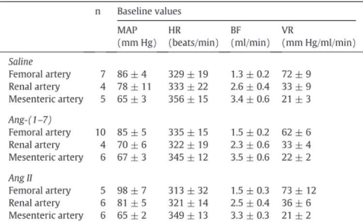
Table 1
Baseline values of mean arterial pressure (MAP, mm Hg), heart rate (HR, beats/min), mean bloodflow (BF, ml/min) and vascular resistance (VR, mm Hg/ml/min) of Wistar rats sub-mitted to microinjection into the RVLM.
n Baseline values
MAP (mm Hg)
HR (beats/min)
BF (ml/min)
VR
(mm Hg/ml/min)
Saline
Femoral artery 7 86 ± 4 329 ± 19 1.3 ± 0.2 72 ± 9
Renal artery 4 78 ± 11 333 ± 22 2.6 ± 0.4 33 ± 9
Mesenteric artery 5 65 ± 3 356 ± 15 3.4 ± 0.6 21 ± 3
Ang-(1–7)
Femoral artery 10 85 ± 5 335 ± 15 1.5 ± 0.2 62 ± 6
Renal artery 4 70 ± 6 322 ± 19 2.3 ± 0.6 33 ± 4
Mesenteric artery 6 67 ± 3 345 ± 12 3.5 ± 0.6 22 ± 2
Ang II
Femoral artery 5 98 ± 7 313 ± 32 1.5 ± 0.3 73 ± 12
Renal artery 6 81 ± 5 321 ± 14 2.5 ± 0.4 36 ± 6
Mesenteric artery 6 65 ± 2 349 ± 13 3.3 ± 0.3 21 ± 2
The values are expressed as means ± SEM; n = number of animals.
Table 2
Baseline values of mean arterial pressure (MAP, mm Hg), heart rate (HR, beats/min), mean bloodflow (BF, ml/min) and vascular resistance (VR, mm Hg/ml/min) of SHR submitted to microinjection into the RVLM.
n Baseline values
MAP (mm Hg)
HR (beats/min)
BF (ml/min)
VR
(mm Hg/ml/min)
Saline
Femoral artery 4 108 ± 15 294 ± 22 1.2 ± 0.5 120 ± 32
Renal artery 4 100 ± 7 343 ± 26 2.1 ± 0.4 58 ± 16
Mesenteric artery 5 81 ± 6 338 ± 21 3.2 ± 0.4 27 ± 3
Ang-(1–7)
Femoral artery 8 109 ± 7 303 ± 12 1.3 ± 0.3 107 ± 19
Renal artery 4 105 ± 8 346 ± 18 2.4 ± 0.3 49 ± 12
Mesenteric artery 5 82 ± 7 338 ± 22 3.1 ± 0.2 27 ± 2
Ang II
Femoral artery 9 103 ± 4 302 ± 11 1.3 ± 0.2 102 ± 17
Renal artery 4 111 ± 8 336 ± 13 2.5 ± 0.3 49 ± 10
Mesenteric artery 5 77 ± 7 340 ± 22 3.2 ± 0.4 25 ± 4
The values are expressed as means ± SEM; n = number of animals.
(ΔBF =−0.2 ± 0.06 ml/min vs. 0.01 ± 0.01 ml/min, saline), without altering femoral (ΔBF = 0.2 ± 0.07 ml/min vs. 0.04 ± 0.06 ml/min, sa-line) or mesenteric bloodflow (ΔBF =−0.1 ± 0.08 ml/min vs. -0.06 ± 0.03 ml/min, saline;Fig. 4). Therefore, in SHR, the hypertensive effect of both Ang II and Ang-(1–7) were accompanied by increase in the renal and mesenteric vascular resistance, and decrease in the renal blood flow.
4. Discussion
Our mainfindings were: i) in normotensive rats, Ang-(1–7) into RVLM increased femoral bloodflow while Ang II increased renal blood flow, without changing vascular resistance of the beds studied; and ii) in hypertensive rats, both Ang-(1–7) and Ang II increased vascular resis-tance of renal and mesenteric beds, and reduced bloodflow of renal ar-tery. Altogether, these data indicate that angiotensinergic synapses in rostral ventrolateral medulla may exert differential control of vascular resistance and bloodflow during hypertension.
Within plenty of experimental models, SHR are a widely used model of primary hypertension. SHR present significant increases in the
sympathetic outflow, which affects vasomotion (Allen, 2002; Judy et al., 1976; Li et al., 2013, 2012; Lundin et al., 1984). Several studies sug-gest that the mechanisms involved in this excessive sympathoexcitation include increases in Ang II levels in the brain, which would change the activity of sympathetic supplies and cause vasoconstriction (Berecek et al., 1984; Falcon et al., 1978; Gyurko et al., 1993; Phillips, 1983; Phillips and Kimura, 1986; Wielbo et al., 1995). RVLM is one of the main sympathetic premotor areas that exhibit increases in neuronal excitability (Chan et al., 1991) and overexpression of AT1receptors
(Gehlert et al., 1991; Gutkind et al., 1988) in SHR. In this regard, the responses we found following injection of Ang II into RVLM of SHR are consistent with several previous studies (Chan et al., 1990, 1991; Ito et al., 2002; Seyedabadi et al., 2001; Zhu et al., 1998), with the exception of contrastingfindings (Averill et al., 1994; Muratani et al., 1991, 1993; Tsuchihashi et al., 1999) likely attributed to differences in the methodol-ogy. Even so, these authors also observed potentiation of the depressor response produced by antagonism of AT1in the RVLM of SHR (Averill et al., 1994; Ito et al., 2002; Muratani et al., 1993; Zhu et al., 1998).
Two plausible hypotheses may explain the increases in the respon-siveness of RVLM of SHR to angiotensins: i) changes in neuronal activity
Fig. 2.Panel A: chart recordings exemplifying cardiovascular responses produced by injection of vehicle (100 nL), Ang-(1–7) (40 ng) or Ang II (40 ng) into the RVLM of an SHR. PAP—
pulsatile arterial pressure; BF—bloodflow; PAP—pulsatile arterial pressure; MAP—mean arterial pressure; HR—heart rate. Panel B: mean maximal changes in MAP (mm Hg) and HR (bpm) evoked by injections (100 nL) of vehicle, Ang-(1–7) (40 ng) or Ang II (40 ng) of the Wistar (n = 6) and SHR (n = 4).⁎pb b0.05 vs. saline;#pb0.05 vs. WT of same group. (ANOVA followed by Newman–Keuls).
and excitability; and ii) increases in the density and/or affinity to angio-tensin receptors. In fact, changes infiring rate of RVLM neurons seem to play a key role in the development and maintenance of hypertension (Chan et al., 1990, 1991; Ito et al., 2000; Kumagai et al., 2012; Kuo and Yang, 2000; Matsuura et al., 2002; Minson et al., 1996). These changes in the activity of RVLM neurons may be related to the modified responsivity to excitatory input (Ito et al., 2000; Lin et al., 1995; Tsuchihashi et al., 1994), besides the increases in the density of AT1
re-ceptors in the RVLM of SHR (Gehlert et al., 1991; Gutkind et al., 1988; Hu et al., 2002). The activity of RVLM neurons may be modified by supramedullary sources of angiotensinergic inputs (Ganten et al.,
1983; Phillips and Sumners, 1998) and mediated by AT1receptors, as
suggested by Ito and colleagues (Ito et al., 2002). The presence of Ang II in nerve terminals (Lind et al., 1985) and binding sites with high affi n-ity for Ang II in the RVLM provide further support to our hypotheses (Allen et al., 1988; Gehlert et al., 1991; Song et al., 1992; Speth and Kim, 1990).
receptors in the RVLM attenuated the responses evoked from para-ventricular hypothalamus (PVN). Ito and colleagues (Ito et al., 2002) showed that this PVN–RVLM angiotensinergic pathway is tonically active in SHR, since the hypotension evoked by activation of GABAAreceptors in
the PVH depends on an AT1-mediated relay in RVLM. It is noteworthy
that the hyperactive diencephalic neurons (Allen, 2002) may be the source of angiotensinergic output to RVLM during hypertension.
The present study pioneered in thefield in showing that the Ang-(1–
7) also acts differentially in medullary sympathetic premotor neurons of SHR. While the greater pressor responses produced by Ang II and Ang-(1–7) in the RVLM of SHR were unrelated to changes in cardiac output, the pressor response caused by Ang-(1–7) is increased in SHR. The liter-ature states that Ang-(1–7) produces central effects similar to those evoked by Ang II (Campagnole-Santos et al., 1989, 1990, 1992; Diz and Pirro, 1992; Du et al., 2013; Li et al., 2012; Oliveira et al., 2013; Silva and Schreihofer, 2011; Zhou et al., 2010). The pressor responses evoked by Ang-(1–7) in rostral areas (Fontes et al., 1997; Potts et al., 2000) are opposite to those caused by the same injections in the caudal ventrolateral medulla (Alzamora et al., 2002; Ferreira et al., 2008; Potts et al., 2000; Silva et al., 1993).
To the best of our knowledge, there is no report on the modification of Ang-(1–7) binding sites in medullary areas of different experimental models of hypertension, whereas the literature provides robust evidences for Ang II–AT1axis. Transgenic rats with low brain angiotensinogen
dis-play an increase in the density of AT1receptors at different brain areas –including VLM (Monti et al., 2001)–and augmented pressor responses to injections of Ang II or Ang-(1–7) into RVLM (Baltatu et al., 2001),
which is in agreement with currentfindings. These evidences suggest simultaneous changes in AT1and Mas (or other) receptors in the RVLM.
Although the cardiovascular effects triggered by Ang II and Ang-(1–7) in the VLM are similar (Fontes et al., 1997; Potts et al., 2000), mechanisms can be differential in the VLM (Alzamora et al., 2002; Oliveira et al., 2013). While the pressor effect evoked from Ang II prominently results from a sympathetically-mediated increase in vasomotion (Dampney, 1994; Muratani et al., 1991), those evoked by Ang-(1–7) may result from a sym-pathetic activation, vasopressin release and inhibition of cholinergic-sensitive beds (Oliveira et al., 2013). Furthermore, consistent evidences show that AT1and Mas receptors are the mediators of Ang II and
Ang-(1–7) evoked responses, respectively. Furthermore, consistent evidences show that AT1and Mas receptors are the mediators of Ang II and Ang-(1–
7) evoked responses, respectively (Du et al., 2013; Li et al., 2012; Nakagaki et al., 2011; Zhou et al., 2010).
SHR exhibit increases in immunoreactivity to AT1receptors in
gluta-matergic and GABAergic RVLM neurons expressing tyrosine hydroxy-lase (Hu et al., 2002). These results suggest that Ang II may be putative in the balance between excitatory and inhibitory inputs con-trolling the activity of RVLM neurons (Averill et al., 1994; Fontes et al., 1997, 1994; Hirooka et al., 1997).We confirmed that changes in central RAS indeed modify the amplitude of cardiovascular responses to block-ade and to stimulation of GABAAhypothalamic receptors (da Silva et al., 2011). Interestingly, the antagonism of AT1 receptor in rat strains with hyperactive RAS (SHR, TGR (mREN2)27, Dahl-salt) causes hypoten-sion (Fontes et al., 2000; Ito et al., 2003, 2002) whereas no change in blood pressure is found in transgenic mice where synthesis of brain
Fig. 4.Mean maximal changes in bloodflow (ml/min) and in vascular resistance (ΔVR, mm Hg/ml/min;ΔVR/baselineVR) sampled in femoral, renal and mesenteric arteries produced by injections of saline, Ang-(1–7) and Ang II into RVLM of SHR.⁎pb0.05 vs. saline (ANOVA followed by Newman–Keuls).
angiotensinogen is inhibited (Baltatu et al., 2001). Jointly, these data suggest that the modulatory role of Ang II on the peripheral sympa-thetic activity results from the balance between excitation and inhibition.
Despite the absence of data on the distribution of Ang-(1–7) recep-tors in different neurons of RVLM, it is possible that SHR show differential activity of glutamatergic and GABAergic neurons and/or non-uniform distribution of angiotensin receptors in these neurons, which may facili-tate sympathoexcitation. Cardiac control might be similarly affected, since the changes in the balance between excitatory/inhibitory inputs produced by angiotensins in the RVLM result in pressor responses that are accompanied by variable chronotropy. Baroreflex influences should be considered, despite the responses to injections into RVLM seem to op-pose the baroreflex patterns. Our results support this idea as the pressor responses produced by angiotensins in the RVLM of SHR were accompa-nied by significant tachycardia. However, we cannot neglect that possi-ble differences in the baroreflex responsiveness and in the RVLM between Wistar and SHR rats may modify the function of pathways in-nervating the heart. Since sympathetic outflow to different organs can be governed independently, changes in the cardiovascular control may result in different patterns of responses in the heart and different vascu-lar beds, in order to set specific adjustments in the perfusion and cardiac function (Dampney et al., 2002).
The RVLM contains sub-populations of neurons that change the ac-tivity of specific vascular territories (Dean et al., 1992; Lovick, 1987; McAllen and Dampney, 1990). Substantial evidences suggest a topo-graphic distribution in the RVLM premotor neurons controlling differ-ent vascular regions (Dampney, 1994; Dampney and McAllen, 1988; Dean et al., 1992; Lovick, 1987; McAllen and Dampney, 1990; McAllen and May, 1994; Willette et al., 1987). In spite of the probable overlap, Dampney and McAllen (Dampney and McAllen, 1988) observed that the increased activity of sympathetic supplies innervating skin and skele-tal muscles are predominantly evoked from medial and lateral aspects of RVLM, respectively. On the other hand, mapping studies accomplished by De Paula and Machado (de Paula and Machado, 2001) observed pressor responses accompanied by equipotent increases in the vascular resistance of renal, mesenteric and hind limb beds, which suggest a lack of topo-graphical organization with respect to these territories. We hypothesize that the different patterns of responses among vascular beds of SHR may be attributed to a differential control of regional sympathetic sup-plies. However, it is important to emphasize that other regional, vascular and autonomic mechanisms may be involved in differential responses observed in SHR (Kumagai et al., 2012; Safar et al., 2001).
In normotensive rats, the pressor responses produced by Ang-(1–7) or Ang II into RVLM were not accompanied by changes in vascular resis-tance of territories studied (femoral, renal or mesenteric), while a signif-icant increase in femoral and in renal bloodflows were found after injection of Ang-(1–7) and Ang II respectively. These suggest that these peptides in the RVLM may change the resistance of othervascular bed(s) and the pressor responses result from small changes in different territories. Our data, obtained from unilateral injections into RVLM, does not indicate differences between the two peptides, as observed for bilat-eral approaches in the CVLM (Ferreira et al., 2008). There is no doubt, however, that pressor responses produced by angiotensins in the RVLM are largely dependent on adjustments in peripheral vascular resistance, since these responses were not accompanied by significant changes in cardiac output. In conclusion, Ang-(1–7) and Ang II seem to govern the changes in vascular resistance of renal and mesenteric beds, besides re-ducing bloodflow of renal artery during essential hypertension. Whether or not the influence of these peptides in different brain areas plays a role in the pathophysiology of hypertension remains to be unraveled.
Acknowledgments
This study was supported by PRONEX (Programa de Núcleos de Excelência from FAPEMIG—Fundação de Amparo à Pesquisa do Estado
de Minas Gerais and CNPq—Conselho Nacional de Desenvolvimento Científico e Tecnológico (Proc# CBB-APQ-04758-10)). PM Ferreira was a recipient of CAPES-PICDT fellowship (Doctoral Degree) from the Fed-eral University of Goiás. We are thankful to Jose R. Silva for skillful tech-nical assistance.
References
Agarwal, D., Welsch, M.A., Keller, J.N., Francis, J., 2011.Chronic exercise modulates RAS components and improves balance between pro- and anti-inflammatory cytokines in the brain of SHR. Basic Res. Cardiol. 106, 1069–1085.
Allen, A.M., 2002.Inhibition of the hypothalamic paraventricular nucleus in spontaneous-ly hypertensive rats dramaticalspontaneous-ly reduces sympathetic vasomotor tone. Hypertension 39, 275–280.
Allen, A.M., Dampney, R.A., Mendelsohn, F.A., 1988.Angiotensin receptor binding and pressor effects in cat subretrofacial nucleus. Am. J. Phys. 255, H1011–H1017. Alzamora, A.C., Santos, R.A., Campagnole-Santos, M.J., 2002.Hypotensive effect of ANG II
and ANG-(1–7) at the caudal ventrolateral medulla involves different mechanisms. Am. J. Physiol. Regul. Integr. Comp. Physiol. 283, R1187–R1195.
Alzamora, A.C., Santos, R.A., Campagnole-Santos, M.J., 2006.Baroreflex modulation by angiotensins at the rat rostral and caudal ventrolateral medulla. Am. J. Physiol. Regul. Integr. Comp. Physiol. 290, R1027–R1034.
Andreatta, S.H., Averill, D.B., Santos, R.A., Ferrario, C.M., 1988.The ventrolateral medulla. A new site of action of the renin-angiotensin system. Hypertension 11, I163–I166. Averill, D.B., Tsuchihashi, T., Khosla, M.C., Ferrario, C.M., 1994.Losartan, nonpeptide
angio-tensin II-type 1 (AT1) receptor antagonist, attenuates pressor and sympathoexcitatory responses evoked by angiotensin II andL-glutamate in rostral ventrolateral medulla. Brain Res. 665, 245–252.
Baltatu, O., Fontes, M.A., Campagnole-Santos, M.J., Caligiorni, S., Ganten, D., Santos, R.A., Bader, M., 2001.Alterations of the renin-angiotensin system at the RVLM of transgen-ic rats with low brain angiotensinogen. Am. J. Physiol. Regul. Integr. Comp. Physiol. 280, R428–R433.
Berecek, K.H., Nagahama, S., Oparil, S., 1984.Effect of central administration of MK-422 (the diacid form of enalapril) on the development of hypertension in the spontane-ously hypertensive rat. J. Hypertens. Suppl. 2, S63–S66.
Biancardi, V.C., Son, S.J., Ahmadi, S., Filosa, J.A., Stern, J.E., 2014.Circulating angiotensin II gains access to the hypothalamus and brain stem during hypertension via breakdown of the blood–brain barrier. Hypertension 63, 572–579.
Campagnole-Santos, M.J., Diz, D.I., Ferrario, C.M., 1990.Actions of angiotensin peptides after partial denervation of the solitary tract nucleus. Hypertension 15, I34–I39. Campagnole-Santos, M.J., Diz, D.I., Santos, R.A., Khosla, M.C., Brosnihan, K.B., Ferrario, C.M.,
1989.Cardiovascular effects of angiotensin-(1–7) injected into the dorsal medulla of rats. Am. J. Phys. 257, H324–H329.
Campagnole-Santos, M.J., Heringer, S.B., Batista, E.N., Khosla, M.C., Santos, R.A., 1992. Dif-ferential baroreceptor reflex modulation by centrally infused angiotensin peptides. Am. J. Phys. 263, R89–R94.
Chan, R.K., Chan, Y.S., Wong, T.M., 1990.Cardiovascular responses to electrical stimulation of the ventrolateral medulla of the spontaneously hypertensive rat. Brain Res. 522, 99–106.
Chan, R.K., Chan, Y.S., Wong, T.M., 1991.Electrophysiological properties of neurons in the rostral ventrolateral medulla of normotensive and spontaneously hypertensive rats. Brain Res. 549, 118–126.
Dampney, R.A., 1994.Functional organization of central pathways regulating the cardio-vascular system. Physiol. Rev. 74, 323–364.
Dampney, R.A., McAllen, R.M., 1988.Differential control of sympatheticfibres supplying hindlimb skin and muscle by subretrofacial neurones in the cat. J. Physiol. 395, 41–56. Dampney, R.A., Coleman, M.J., Fontes, M.A., Hirooka, Y., Horiuchi, J., Li, Y.W., Polson, J.W., Potts, P.D., Tagawa, T., 2002.Central mechanisms underlying short- and long-term regulation of the cardiovascular system. Clin. Exp. Pharmacol. Physiol. 29, 261–268. Dean, C., Seagard, J.L., Hopp, F.A., Kampine, J.P., 1992.Differential control of sympathetic
activity to kidney and skeletal muscle by ventral medullary neurons. J. Auton. Nerv. Syst. 37, 1–10.
Diz, D.I., Pirro, N.T., 1992.Differential actions of angiotensin II and angiotensin-(1–7) on transmitter release. Hypertension 19, II41–II48.
Du, D., Chen, J., Liu, M., Zhu, M., Jing, H., Fang, J., Shen, L., Zhu, D., Yu, J., Wang, J., 2013.The effects of angiotensin II and angiotensin-(1–7) in the rostral ventrolateral medulla of rats on stress-induced hypertension. PLoS One 8, e70976.
Falcon II, J.C., Phillips, M.I., Hoffman, W.E., Brody, M.J., 1978.Effects of intraventricular angiotensin II mediated by the sympathetic nervous system. Am. J. Phys. 235, H392–H399.
Ferreira, P.M., Alzamora, A.C., Santos, R.A., Campagnole-Santos, M.J., 2008.Hemodynamic effect produced by microinjection of angiotensins at the caudal ventrolateral medulla of spontaneously hypertensive rats. Neuroscience 151, 1208–1216.
Fontes, M.A., Baltatu, O., Caligiorne, S.M., Campagnole-Santos, M.J., Ganten, D., Bader, M., Santos, R.A., 2000.Angiotensin peptides acting at rostral ventrolateral medulla contribute to hypertension of TGR(mREN2)27 rats. Physiol. Genomics 2, 137–142. Fontes, M.A., Martins Pinge, M.C., Naves, V., Campagnole-Santos, M.J., Lopes, O.U., Khosla,
M.C., Santos, R.A., 1997.Cardiovascular effects produced by microinjection of angio-tensins and angiotensin antagonists into the ventrolateral medulla of freely moving rats. Brain Res. 750, 305–310.
Ganten, D., Hermann, K., Bayer, C., Unger, T., Lang, R.E., 1983.Angiotensin synthesis in the brain and increased turnover in hypertensive rats. Science 221, 869–871. Gehlert, D.R., Gackenheimer, S.L., Schober, D.A., 1991.Autoradiographic localization of
subtypes of angiotensin II antagonist binding in the rat brain. Neuroscience 44, 501–514.
Gutkind, J.S., Kurihara, M., Castren, E., Saavedra, J.M., 1988.Increased concentration of an-giotensin II binding sites in selected brain areas of spontaneously hypertensive rats. J. Hypertens. 6, 79–84.
Gyurko, R., Wielbo, D., Phillips, M.I., 1993.Antisense inhibition of AT1 receptor mRNA and angiotensinogen mRNA in the brain of spontaneously hypertensive rats reduces hypertension of neurogenic origin. Regul. Pept. 49, 167–174.
Hirooka, Y., Potts, P.D., Dampney, R.A., 1997.Role of angiotensin II receptor subtypes in mediating the sympathoexcitatory effects of exogenous and endogenous angiotensin peptides in the rostral ventrolateral medulla of the rabbit. Brain Res. 772, 107–114. Hu, L., Zhu, D.N., Yu, Z., Wang, J.Q., Sun, Z.J., Yao, T., 2002.Expression of angiotensin II type
1 (AT(1)) receptor in the rostral ventrolateral medulla in rats. J. Appl. Physiol. (1985) 92, 2153–2161.
Ito, S., Hiratsuka, M., Komatsu, K., Tsukamoto, K., Kanmatsuse, K., Sved, A.F., 2003. Ventrolateral medulla AT1 receptors support arterial pressure in Dahl salt-sensitive rats. Hypertension 41, 744–750.
Ito, S., Komatsu, K., Tsukamoto, K., Kanmatsuse, K., Sved, A.F., 2002.Ventrolateral medulla AT1 receptors support blood pressure in hypertensive rats. Hypertension 40, 552–559.
Ito, S., Komatsu, K., Tsukamoto, K., Sved, A.F., 2000.Excitatory amino acids in the rostral ventrolateral medulla support blood pressure in spontaneously hypertensive rats. Hypertension 35, 413–417.
Judy, W.V., Watanabe, A.M., Henry, D.P., Besch Jr., H.R., Murphy, W.R., Hockel, G.M., 1976. Sympathetic nerve activity: role in regulation of blood pressure in the spontaenously hypertensive rat. Circ. Res. 38, 21–29.
Komatus, C., Shibata, K., Furukawa, T., 1996.The developmental increase of the AT1A, but not the AT1B, receptor mRNA level at the preoptic area in spontaneously hyperten-sive rats. Life Sci. 58, 1109–1121.
Kumagai, H., Oshima, N., Matsuura, T., Iigaya, K., Imai, M., Onimaru, H., Sakata, K., Osaka, M., Onami, T., Takimoto, C., Kamayachi, T., Itoh, H., Saruta, T., 2012.Importance of ros-tral ventrolateral medulla neurons in determining efferent sympathetic nerve activity and blood pressure. Hypertens. Res. 35, 132–141.
Kuo, T.B., Yang, C.C., 2000.Altered frequency characteristic of central vasomotor control in SHR. Am. J. Physiol. Heart Circ. Physiol. 278, H201–H207.
Li, P., Sun, H.J., Cui, B.P., Zhou, Y.B., Han, Y., 2013.Angiotensin-(1–7) in the rostral ventro-lateral medulla modulates enhanced cardiac sympathetic afferent reflex and sympa-thetic activation in renovascular hypertensive rats. Hypertension 61, 820–827. Li, P., Zhang, F., Zhou, Y.B., Cui, B.P., Han, Y., 2012.Superoxide anions modulate the effects
of angiotensin-(1–7) in the rostral ventrolateral medulla on cardiac sympathetic afferent reflex and sympathetic activity in rats. Neuroscience 223, 388–398. Lin, J.C., Tsao, W.L., Wang, Y., 1995.Cardiovascular effects of NMDA in the RVLM of
spontaneously hypertensive rats. Brain Res. Bull. 37, 289–294.
Lind, R.W., Swanson, L.W., Ganten, D., 1985.Organization of angiotensin II immunoreac-tive cells andfibers in the rat central nervous system. An immunohistochemical study. Neuroendocrinology 40, 2–24.
Lovick, T.A., 1987.Differential control of cardiac and vasomotor activity by neurones in nucleus paragigantocellularis lateralis in the cat. J. Physiol. 389, 23–35.
Lundin, S., Ricksten, S.E., Thoren, P., 1984.Renal sympathetic activity in spontaneously hy-pertensive rats and normotensive controls, as studied by three different methods. Acta Physiol. Scand. 120, 265–272.
Matsuda, T., Shibata, K., Abe, M., Tomonaga, M., Furukawa, T., 1987.Potentiation of pres-sor response to angiotensin II at the preoptic area in spontaneously hypertensive rat. Life Sci. 41, 749–754.
Matsuura, T., Kumagai, H., Kawai, A., Onimaru, H., Imai, M., Oshima, N., Sakata, K., Saruta, T., 2002.Rostral ventrolateral medulla neurons of neonatal Wistar-Kyoto and sponta-neously hypertensive rats. Hypertension 40, 560–565.
McAllen, R.M., Dampney, R.A., 1990.Vasomotor neurons in the rostral ventrolateral me-dulla are organized topographically with respect to type of vascular bed but not body region. Neurosci. Lett. 110, 91–96.
McAllen, R.M., May, C.N., 1994.Differential drives from rostral ventrolateral medullary neurons to three identified sympathetic outflows. Am. J. Phys. 267, R935–R944. Minson, J., Arnolda, L., Llewellyn-Smith, I., Pilowsky, P., Chalmers, J., 1996.Altered c-fos in
rostral medulla and spinal cord of spontaneously hypertensive rats. Hypertension 27, 433–441.
Monti, J., Schinke, M., Bohm, M., Ganten, D., Bader, M., Bricca, G., 2001.Glial angiotensinogen regulates brain angiotensin II receptors in transgenic rats TGR(ASrAOGEN). Am. J. Physiol. Regul. Integr. Comp. Physiol. 280, R233–R240.
Muratani, H., Averill, D.B., Ferrario, C.M., 1991.Effect of angiotensin II in ventrolateral medulla of spontaneously hypertensive rats. Am. J. Phys. 260, R977–R984. Muratani, H., Ferrario, C.M., Averill, D.B., 1993.Ventrolateral medulla in spontaneously
hypertensive rats: role of angiotensin II. Am. J. Phys. 264, R388–R395.
Nakagaki, T., Hirooka, Y., Ito, K., Kishi, T., Hoka, S., Sunagawa, K., 2011.Role of angiotensin-(1–7) in rostral ventrolateral medulla in blood pressure regulation via sympathetic nerve activity in Wistar-Kyoto and spontaneous hypertensive rats. Clin. Exp. Hypertens. 33, 223–230.
Oliveira, R.C., Campagnole-Santos, M.J., Santos, R.A., 2013. The pressor effect of angiotensin-(1–7) in the rat rostral ventrolateral medulla involves multiple peripher-al mechanisms. Clinics (Sao Paulo) 68, 245–252.
de Paula, P.M., Machado, B.H., 2001.Changes in regional vascular resistance in response to microinjection of L-glutamate into different antero-posterior coordinates of the RVLM in awake rats. Auton. Neurosci. 87, 301–309.
Paxinos, G., Watson, C., 1986.The Rat Brain in Stereotaxic Coordinates. Academic Press, New York.
Phillips, M.I., 1983.New evidence for brain angiotensin and for its role in hypertension. Fed. Proc. 42, 2667–2672.
Phillips, M.I., de Oliveira, E.M., 2008.Brain renin angiotensin in disease. J. Mol. Med. (Berl.) 86, 715–722.
Phillips, M.I., Kimura, B.K., 1986.Levels of brain angiotensin in the spontaneously hyper-tensive rat and treatment with ramiprilat. J. Hypertens. Suppl. 4, S391–S394. Phillips, M.I., Sumners, C., 1998.Angiotensin II in central nervous system physiology.
Regul. Pept. 78, 1–11.
Potts, P.D., Horiuchi, J., Coleman, M.J., Dampney, R.A., 2000.The cardiovascular effects of angiotensin-(1–7) in the rostral and caudal ventrolateral medulla of the rabbit. Brain Res. 877, 58–64.
Ruiz, P., Basso, N., Cannata, M.A., Taquini, A.C., 1990.The renin-angiotensin system in different stages of spontaneous hypertension in the rat (SHR). Clin. Exp. Hypertens. A 12, 63–81.
Safar, M., Chamiot-Clerc, P., Dagher, G., Renaud, J.F., 2001.Pulse pressure, endothelium function, and arterial stiffness in spontaneously hypertensive rats. Hypertension 38, 1416–1421.
Santos, R.A., Simoes e Silva, A.C., Maric, C., Silva, D.M., Machado, R.P., de Buhr, I., Heringer-Walther, S., Pinheiro, S.V., Lopes, M.T., Bader, M., Mendes, E.P., Lemos, V.S., Campagnole-Santos, M.J., Schultheiss, H.P., Speth, R., Walther, T., 2003. Angiotensin-(1–7) is an endogenous ligand for the G protein-coupled receptor Mas. Proc. Natl. Acad. Sci. U. S. A. 100, 8258–8263.
Seyedabadi, M., Goodchild, A.K., Pilowsky, P.M., 2001.Differential role of kinases in brain stem of hypertensive and normotensive rats. Hypertension 38, 1087–1092. Shibata, K., Komatsu, C., Misumi, Y., Furukawa, T., 1993.Developmental differences of
angiotensinogen mRNA in the preoptic area between spontaneously hypertensive and age-matched Wistar-Kyoto rats. Brain Res. Mol. Brain Res. 19, 115–120. Silva, A.Q., Schreihofer, A.M., 2011.Altered sympathetic reflexes and vascular reactivity in
rats after exposure to chronic intermittent hypoxia. J. Physiol. 589, 1463–1476. Silva, L.C., Fontes, M.A., Campagnole-Santos, M.J., Khosla, M.C., Campos Jr., R.R.,
Guertzenstein, P.G., Santos, R.A., 1993.Cardiovascular effects produced by micro-injection of angiotensin-(1–7) on vasopressor and vasodepressor sites of the ventro-lateral medulla. Brain Res. 613, 321–325.
da Silva, A.Q., Fontes, M.A., Kanagy, N.L., 2011.Chronic infusion of angiotensin receptor antagonists in the hypothalamic paraventricular nucleus prevents hypertension in a rat model of sleep apnea. Brain Res. 1368, 231–238.
Song, K., Allen, A.M., Paxinos, G., Mendelsohn, F.A., 1992.Mapping of angiotensin II recep-tor subtype heterogeneity in rat brain. J. Comp. Neurol. 316, 467–484.
Speth, R.C., Kim, K.H., 1990.Discrimination of two angiotensin II receptor subtypes with a selective agonist analogue of angiotensin II, p-aminophenylalanine6 angiotensin II. Biochem. Biophys. Res. Commun. 169, 997–1006.
Tagawa, T., Dampney, R.A., 1999.AT(1) receptors mediate excitatory inputs to rostral ventrolateral medulla pressor neurons from hypothalamus. Hypertension 34, 1301–1307.
Tamura, K., Umemura, S., Nyui, N., Yamakawa, T., Yamaguchi, S., Ishigami, T., Tanaka, S., Tanimoto, K., Takagi, N., Sekihara, H., Murakami, K., Ishii, M., 1996.Tissue-specific regulation of angiotensinogen gene expression in spontaneously hypertensive rats. Hypertension 27, 1216–1223.
Tsuchihashi, T., Abe, I., Fujishima, M., 1994.Role of metabotropic glutamate receptors in ventrolateral medulla of hypertensive rats. Hypertension 24, 648–652.
Tsuchihashi, T., Kagiyama, S., Matsumura, K., Abe, I., Fujishima, M., 1999.Effects of chronic oral treatment with imidapril and TCV-116 on the responsiveness to angiotensin II in ventrolateral medulla of SHR. J. Hypertens. 17, 917–922.
Welch, W.J., Deng, X., Snellen, H., Wilcox, C.S., 1995.Validation of miniature ultrasonic transit-timeflow probes for measurement of renal bloodflow in rats. Am. J. Phys. 268, F175–F178.
Weyhenmeyer, J.A., Phillips, M.I., 1982.Angiotensin-like immunoreactivity in the brain of the spontaneously hypertensive rat. Hypertension 4, 514–523.
Wielbo, D., Sernia, C., Gyurko, R., Phillips, M.I., 1995.Antisense inhibition of hypertension in the spontaneously hypertensive rat. Hypertension 25, 314–319.
Willette, R.N., Punnen-Grandy, S., Krieger, A.J., Sapru, H.N., 1987.Differential regulation of regional vascular resistance by the rostral and caudal ventrolateral medulla in the rat. J. Auton. Nerv. Syst. 18, 143–151.
Wright, J.W., Sullivan, M.J., Quirk, W.S., Batt, C.M., Harding, J.W., 1987.Heightened blood pressure and drinking responsiveness to intracerebroventricularly applied angioten-sins in the spontaneously hypertensive rat. Brain Res. 420, 289–294.
Zhou, L.M., Shi, Z., Gao, J., Han, Y., Yuan, N., Gao, X.Y., Zhu, G.Q., 2010.Angiotensin-(1–7) and angiotension II in the rostral ventrolateral medulla modulate the cardiac sympa-thetic afferent reflex and sympathetic activity in rats. Pflugers Arch. 459, 681–688. Zhu, D.N., Moriguchi, A., Mikami, H., Higaki, J., Ogihara, T., 1998.Central amino acids mediate