HEMODYNAMIC EFFECT PRODUCED BY MICROINJECTION OF
ANGIOTENSINS AT THE CAUDAL VENTROLATERAL MEDULLA OF
SPONTANEOUSLY HYPERTENSIVE RATS
P. M. FERREIRA,a,bA. C. ALZAMORA,cR. A. S. SANTOSa AND M. J. CAMPAGNOLE-SANTOSa*
a
Departamento de Fisiologia e Biofísica, Instituto de Ciências Biológicas, Universidade Federal de Minas Gerais, Av. Antonio Carlos, 6627-ICB, UFMG, 31270-901, Belo Horizonte, MG, Brazil
bDepartamento de Ciências Fisiológicas, Universidade Federal de Goiás, Goiânia, GO, Brazil
c
Departamento de Ciências Exatas e Biológicas, Universidade Federal de Ouro Preto, MG, Brazil
Abstract—In the present study, the effect of caudal ventro-lateral medulla (CVLM) microinjection of angiotensin-(1-7) (Ang-(1-7)) and angiotensin II (Ang II) on mean arterial pres-sure (MAP), heart rate (HR) and pulsatile vascular blood flow (VBF; Transonic System) of the femoral, renal or mesenteric arteries was evaluated in male Wistar and spontaneously hypertensive rats (SHR) anesthetized with urethane. The vas-cular resistance (VR) was calculated by the ratio between the changes in MAP and VBF. Ang-(1-7) (40 ng) and Ang II (40 ng) microinjection into the CVLM caused similar depressor ef-fects in Wistar rats and SHR. The hypotensive effect pro-duced by Ang-(1-7) into the CVLM of Wistar rats was accom-panied by a decrease in femoral (⌬VR/VRbaselineⴝⴚ0.12ⴞ 0.04 vs. 0.001ⴞ0.03; after saline) and renal (⌬ VR/VRbase-lineⴝⴚ0.10ⴞ0.02 vs.ⴚ0.003ⴞ0.02; after saline) vascular re-sistance. On the other hand, the Ang II hypotensive effect in Wistar rats produced only changes in renal vascular resis-tance (⌬VR/VRbaselineⴝⴚ0.16ⴞ0.02 vs.ⴚ0.003ⴞ0.02; after saline). In SHR, the hypotensive effect produced by Ang-(1-7) and Ang II caused decrease in renal vascular resistance (⌬VR/VRbaselineⴝⴚ0.18ⴞ0.03 andⴚ0.13ⴞ0.01, respectively, as compared with saline,⌬VR/VRbaselineⴝⴚ0.06ⴞ0.02), but did not alter the femoral or mesenteric vascular resistance. These data show that Ang II and Ang-(1-7) hypotensive effect at the CVLM involves the participation of different vascular beds. Further, the lack of involvement of the femoral vascular bed in SHR suggests that hypertension may induce alteration in the neural control of the different vascular beds, at least at the CVLM. © 2008 IBRO. Published by Elsevier Ltd. All rights reserved.
Key words: Ang-(1-7), Ang II, caudal ventrolateral medulla, arterial pressure, vascular blood flow, spontaneously hyper-tensive rats.
The caudal ventrolateral medulla (CVLM) is a key area in the neuronal circuitry controlling blood pressure. The CVLM contains inhibitory vasomotor neurons that when stimulated elicit a reduction in sympathetic nerve activity and arterial pressure (Guertzenstein, 1973; Blessing, 1988; Agarwal et al., 1989; Cravo and Morrison, 1993). Several studies have shown that VLM neurones are acti-vated by application of angiotensin (Ang) peptides, includ-ing angiotensin II (Ang II) and angiotensin-(1-7) (Ang-(1-7)) (Andreatta et al., 1988; Allen et al., 1988; Sasaki and Dampney, 1990; Muratani et al., 1991, 1993; Li et al., 1992; Silva et al., 1993; Fontes et al., 1994, 1997; Al-zamora et al., 2002). Microinjection of Ang II or Ang-(1-7) into the CVLM results in a significant decrease in arterial pressure (Silva et al., 1993; Alzamora et al., 2002), medi-ated by the interaction with different angiotensin receptor subtypes (Fontes et al., 1994; Santos et al., 1994, 2000). More recent in our laboratory, Alzamora et al. (2002) showed that the hypotensive effect produced by Ang II depends on the decrease in sympathetic vascular tonus, but the Ang-(1-7) effect involves a nitric oxide–related mechanism, suggesting that different peripheral mecha-nism and/or different regional vascular beds are triggered upon CVLM microinjection of Ang peptides.
One question that still remains open is which periph-eral vascular beds is involved in mediating Ang II and Ang-(1-7) effects at the CVLM. Studies evaluating regional blood flow have shown that the mesenteric, renal and femoral vascular beds are involved in arterial pressure changes evoked by Ang II at both the CVLM and the rostral ventrolateral medulla (RVLM) (Willette et al., 1987; Lovick, 1987; Dampney and McAllen, 1988; Dean et al., 1992; Dampney, 1994; Paula and Machado, 2001).
Many studies have in addition provided evidence that an overactivity of the brain renin-angiotensin system (RAS) may contribute to the hypertension observed in different models, such as the spontaneously hypertensive rats (SHR) (Casto and Phillips, 1985; Matsuda et al., 1987; Gutkind et al., 1988; Ruiz et al., 1990; Muratani et al., 1991, 1993; Tamura et al., 1996; Komatus et al., 1996; Zhu et al., 1998; Colombari et al., 2001; Hu et al., 2002).Hu et al. (2002) have shown an increased in angiotensin II receptor, type 1 (AT1) receptor density within the RVLM of SHR when compared with Wistar Kyoto rats (WKY).
Considering the importance the CVLM and the modu-lation produced by the peptides of the RAS at this site for the cardiovascular control, in the present study, we eval-uated the hemodynamic effect produced by Ang-(1-7) and *Corresponding author. Tel:⫹55-31-3409-2951; fax:⫹55-31-3409-2924.
E-mail address:[email protected](M. J. Campagnole-Santos). Abbreviations:Ang, angiotensin; Ang II, angiotensin II; Ang-(1-7), an-giotensin-(1-7); AT1, angiotensin II receptor, type 1; BFf, pulsatile
blood flow of the femoral artery; BFm, pulsatile blood flow of the mesenteric artery; BFr, pulsatile blood flow of the renal artery; CVLM, caudal ventrolateral medulla; HR, heart rate; MAP, mean arterial pres-sure; Mas, angiotensin-(1-7) receptor; nNOS, neuronal nitric oxide synthase; RAS, renin-angiotensin system; RVLM, rostral ventrolateral medulla; SHR, spontaneously hypertensive rats; VR, vascular resis-tance; WKY, Wistar Kyoto rats.
0306-4522/08$32.00⫹0.00 © 2008 IBRO. Published by Elsevier Ltd. All rights reserved. doi:10.1016/j.neuroscience.2007.11.042
Ang II microinjections at the CVLM in normotensive and SHRs.
EXPERIMENTAL PROCEDURES
General surgical preparation
All experiments were performed in male SHRs (SHR, 16 –18 weeks old) and age-matched Wistar rats (280 –320 g) anesthe-tized with urethane (1.2 g/kg i.p, Sigma Chemical Co.). All exper-iments conformed to the regulation set forth by the Institutional Animal Welfare Committee (CETEA, UFMG), which are in accor-dance with the National Institutes of Health (NIH) Guidelines for the Care and Use of Laboratory Animals (NIH publication 80-23, revised in 1996). Under urethane anesthesia the trachea was cannulated (PE90) and a catheter (PE10 connected to a PE50) was inserted into the abdominal aorta, through the femoral artery for arterial pressure measurement. Next, the animals were placed in a stereotaxic instrument (David Kopf Instruments, CA, USA) with the tooth bar⫺11 mm below the level of the interaural line. The dorsal surface of the brainstem was exposed by a limited occipital craniotomy and an incision of the atlanto-occipital mem-brane and meninges, as previously described (Ferreira et al., 2007; Silva et al., 1993). The animals were kept on a heating pad to maintain a constant body temperature (36 –37 °C), evaluated with a rectal thermometer. The adequate level of anesthesia was verified by the absence of a withdrawal response to nociceptive stimulation of the hindpaw. Supplemental doses of urethane (0.1 g/kg i.v.) were administered whenever necessary.
Microinjections technique
Microinjections into the CVLM were performed with a glass mi-cropipette as previously described (Ferreira et al., 2007; Alzamora et al., 2002). Unilateral microinjections of Ang-(1-7) (40 ng), Ang II (40 ng) or sterile saline (vehicle-NaCl 0,9%) were made over a 20 –30 s period into the CVLM [0.7 mm anterior, 1.8 mm lateral to the obex, and just above pia mater in the ventral surface. The volume (100 nl) of the injectate was measured by observing the movement of the fluid meniscus in the pipette barrel. Experiments were made only when the positioning of the micropipette produced a transitory depressor response (usually 10 –15 mm Hg). For all experiments, only one site of the CVLM was tested per animal.
Blood flow measurement
In different group of animals, mesenteric, renal or femoral blood flow was determined according to the method ofWelch et al. (1995)using a transit-time blood flowmeter (model T206; Tran-sonics, NY, USA). Briefly, a midline laparotomy or an incision in the inguinal region was performed and miniature ultrasonic transit-time flow probe (0.5 or 0.7 mm V-series) was carefully placed around the desired artery (mesenteric or renal or femoral artery). The volume flow, in ml/min, and the real-time pulsatile flow were recorded by a BIOPAC System (UIM100A, CA, USA). Mean flow was simultaneously calculated and displayed (Acqknowledge
Software, BIOPAC System). Calibration and the zero flow were pre-determined by the manufacturer value. Mean vascular resis-tance (VR) was calculated as the ratio between mean arterial pressure (MAP) and mean blood flow of the different vessels (mm Hg/ml/min) at each desired time.
Blood pressure measurement
The arterial cannula was connected to a strain-gauge transducer coupled to a computer-based data acquisition system (MP100A, BIOPAC System) in order to record pulsatile arterial pressure PAP. MAP and heart rate (HR) were simultaneously calculated by the software Acqknowledge(BIOPAC System) and continuously displayed.
Protocols
Experiments were performed in animals that presented at the beginning of the experiments a MAP of at least 80 mm Hg for Wistar group or 110 mm Hg for SHR group. MAP and HR changes produced by randomized microinjection of Ang-(1-7) (40 ng), Ang II (40 ng) or saline (NaCl, 0.9%-100 nl) into the CVLM were recorded continuously over a 30 min period. In all animals, a minimum interval of 20 min was given after positioning of the micropipette and the peptide microinjection. A period of 30 min was allowed to elapse between CVLM injections. MAP and HR values at the peak of the responses were considered for each substance microinjected.
Histological verification of injection sites
At the completion of each experiment, the animals were killed with excess of anesthetic and the brain stem was carefully removed and fixed in 10% phosphate-buffered formalin for histological ex-amination. Serial coronal sections (40 –50 m) of the medulla
oblongata were performed and stained with Neutral Red. Micro-injection site was identified by the disruption of the tissue pro-duced by the micropipette and referred to standard anatomical structures of the brain stem according to the atlas ofPaxinos and Watson (1986).
Drugs
Ang-(1-7) and Ang II were purchased from Bachem (Torance, CA, USA) or Peninsula Laboratories (Belmont, CA, USA) and dis-solved in sterile isotonic saline (NaCl, 0.9%).
Statistical analysis
All values were expressed as means⫾S.E.M. Comparisons among different groups were assessed by two-way ANOVA followed by the Bonferroni test. The criterion for statistical significance was set at
P⬍0.05. The statistical analysis was performed with the GraphPad Prism software (version 4.0).
RESULTS
As shown inFig. 1,the Ang-(1-7) and Ang II microinjection into the CVLM of normotensive Wistar rats (baseline MAP⫽83⫾4 mm Hg; baseline HR⫽312⫾9 beats/min;
n⫽24) produced a significant fall in MAP (⫺11⫾0.8 mm Hg; n⫽22 and ⫺12⫾1.1 mm Hg;n⫽24, respectively) in comparison to saline microinjection (⫺3⫾0.3 mm Hg;
n⫽20). The hypotensive effect produced by Ang II and Ang-(1-7) was accompanied by small (⬃2–3%), but signif-icant reduction in HR (⫺6⫾2 beats/min and⫺10⫾2 beats/ min, respectively; Fig. 1) in comparison to the alteration produced by saline (0.4⫾1 beats/min). Similarly, in SHR (baseline MAP⫽100⫾5 mm Hg; baseline HR⫽305⫾11 beats/min;n⫽14), Ang-(1-7) and Ang II microinjection pro-duced a significant reduction in MAP (⫺14⫾1.3 mm Hg;
n⫽13 and⫺14⫾1.1 mm Hg;n⫽14, respectively) in com-parison to saline (⫺3⫾0.5 mm Hg,n⫽13;Fig. 1). The MAP and HR effects in SHR were not significantly different from those produced in normotensive rats (Fig. 1).
of Wistar or SHR considering each vascular territory in separated.
Fig. 2shows recordings illustrating the changes in PAP (mm Hg) and MAP (mm Hg), pulsatile blood flow of the mesenteric artery (BFm, ml/min), femoral artery (BFf, ml/ min) and renal artery (BFr, ml/min) and HR (beats/min), induced by CVLM microinjection of Ang-(1-7) (40 ng; A, B and C) or Ang II (40 ng; D, E and F) in normotensive rats. In normotensive rats, CVLM microinjection of Ang-(1-7) did not significantly alter blood flow in the femoral, mesenteric
or renal artery (Fig. 3). These blood flow changes reflected the following alterations in VR: a significant fall in femoral VR (⌬VR⫽⫺18⫾7 mm Hg/ml/min vs. 1.5⫾3.8 mm Hg/ml/min, after saline;⌬VR/VRbaseline⫽⫺0.16⫾0.03 vs. 0.03⫾0.02, after saline;Fig. 3)and renal VR (⌬VR⫽⫺3.6⫾0.8 mm Hg/ ml/min vs. 0.02⫾0.5 mm Hg/ml/min, after saline or ⌬VR/ VRbaseline⫽⫺0.10⫾0.02 vs. ⫺0.003⫾0.02, after saline; Fig. 3). On the other hand, there was no significant alteration in the mesenteric VR (⌬VR⫽⫺1.9⫾0.8 mm Hg/ml/min vs.
⫺0.9⫾0.2 mm Hg/ml/min, after saline or⌬VR/VRbaseline⫽ ⫺0.08⫾0.04 vs. ⫺0.05⫾0.01, after saline;Fig. 3). In sum-mary, in the normotensive animals, the hypotensive effect triggered by Ang-(1-7) microinjection into the CVLM was due to a decrease in the VR of at least two territories, femoral and renal.
Similarly to Ang-(1-7), Ang II microinjection into the CVLM of normotensive rats, did not significantly change femoral or renal arteries blood flow (Fig. 3). However, a significant fall in the mesenteric artery blood flow was observed (⫺0.52⫾0.13 ml/min vs. ⫺0.01⫾0.03 ml/min, after saline;Fig. 3). Ang II microinjection into the CVLM produced fall in the renal VR (⌬VR⫽⫺7.02⫾1.2 mm Hg/ ml/min vs. 0.02⫾0.5 mm Hg/ml/min, after saline or⌬VR/ VRbaseline⫽⫺0.18⫾0.02 vs.⫺0.003⫾0.02, after saline), without altering the mesenteric VR (⫺0.9⫾1.1 mm Hg/ml/ min vs. ⫺0.9⫾0.2 mm Hg/ml/min, after saline or ⌬VR/ RVbaseline⫽⫺0.03⫾0.07 vs. ⫺0.05⫾0.01, after saline; Fig. 3). In addition, the alteration in renal artery resistance induced by CVLM injection of Ang II was significantly larger than that induced by Ang-(1-7) (Fig. 3). However, differ-ently from Ang-(1-7), Ang II microinjection did not alter the resistance in the femoral artery (⌬VR⫽⫺1.9⫾9.2 mm Hg/ ml/min vs. 1.5⫾3.8 mm Hg/ml/min, after saline or ⌬VR/ VRbaseline⫽⫺0.005⫾0.06 vs. 0.001⫾0.03, after saline; Fig. 3). Thus, in the normotensive rats the hypotensive effect of Ang II at CVLM was associated with a decrease in the renal VR, without involvement of the other territories evaluated.
In SHR, Ang-(1-7) microinjection into the CVLM did not induce significant changes in the blood flow of the femoral, renal or mesenteric arteries. A decrease in the VR of the renal
Fig. 1. Averaged changes in MAP (mm Hg) and HR (bpm) produced by microinjection of saline (100 nl), Ang-(1-7) (40 ng) or Ang II (40 ng) into the CVLM of the Wistar (n⫽24) and SHR (n⫽14). *P⬍0.05 in comparison to saline (two-way ANOVA followed by Bonferroni test).
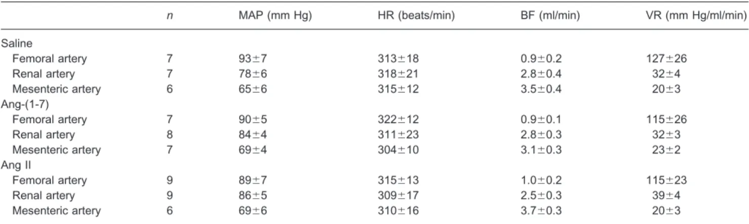
Table 1.Baseline values of MAP (mm Hg), HR (beats/min), mean blood flow (BF, ml/min) and VR (mm Hg/ml/min) of Wistar rats before CVLM microinjection
n MAP (mm Hg) HR (beats/min) BF (ml/min) VR (mm Hg/ml/min)
Saline
Femoral artery 7 93⫾7 313⫾18 0.9⫾0.2 127⫾26
Renal artery 7 78⫾6 318⫾21 2.8⫾0.4 32⫾4
Mesenteric artery 6 65⫾6 315⫾12 3.5⫾0.4 20⫾3
Ang-(1-7)
Femoral artery 7 90⫾5 322⫾12 0.9⫾0.1 115⫾26
Renal artery 8 84⫾4 311⫾23 2.8⫾0.3 32⫾3
Mesenteric artery 7 69⫾4 304⫾10 3.1⫾0.3 23⫾2
Ang II
Femoral artery 9 89⫾7 315⫾13 1.0⫾0.2 115⫾23
Renal artery 9 86⫾5 309⫾17 2.5⫾0.3 39⫾4
Mesenteric artery 6 69⫾6 310⫾16 3.7⫾0.3 20⫾3
artery (⌬VR/VRbaseline⫽⫺0.18⫾0.03 vs. ⫺0.051⫾0.014, after saline) contributed to the hypotensive effect of Ang-(1-7) at the CVLM in SHR. No significant changes were observed in the resistance of the femoral artery (⌬VR/VRbaseline⫽ ⫺0.01⫾0.07 vs. 0.06⫾0.04, after saline) or the mesenteric artery (⌬VR/VRbaseline⫽⫺0.05⫾0.04 vs.⫺0.02⫾0.01, sa-line;Fig. 3).
The depressor effect produced by Ang II in SHR was also associated with a reduction only in the renal VR (⌬VR/ VRbaseline⫽⫺0.12⫾0.01 vs. ⫺0.05⫾0.014, saline). There was no alteration of femoral VR (⌬VR/VRbaseline⫽0.13⫾
0.07 vs. 0.06⫾0.03, after saline) or mesenteric VR (⌬VR/ VRbaseline⫽⫺0.08⫾0.02 vs.⫺0.02⫾0.01, saline;Fig. 3). Ang II microinjection produced a significant alteration in femoral BF (⫺0.36⫾0.05 ml/min vs.⫺0.15⫾0.05 ml/min, saline), without altering renal BF (⫺0.1⫾0.1 ml/min vs.
⫺0.02⫾0.04 ml/min, saline) or mesenteric BF (⫺0.26⫾
0.08 ml/min vs. ⫺0.04⫾0.007 ml/min, saline; Fig. 3). Therefore, in SHR, the hypotensive effect of both Ang II and Ang-(1-7) were accompanied by decrease in the renal VR, without changes in the other territories evaluated.
Histology
Fig. 4shows on the left side a histological section of the medulla illustrating the injection site and diagram of the frontal section from the atlas of Paxinos and Watson (1986) representing the center of the microinjection of all animals in this study, which were confined to a distance of approximately 150m in the rostro-caudal level. No
differ-ences in the location of the injection sites were observed between the SHR and WKY.
DISCUSSION
In the present study using ultrasonic pulsatile blood flow measurements we showed that the depressor effect pro-duced by Ang-(1-7) at the CVLM of normotensive Wistar rats was accompanied by reduction in the femoral and renal VR, whereas the hypotensive effect produced by Ang II was accompanied only by a reduction in renal VR. These data extended our previous observations showing that the
effect of Ang II and Ang-(1-7) at the CVLM is due to a fall in peripheral resistance without significant change in car-diac output and, indicate that Ang II and Ang-(1-7) hypo-tensive effect at the CVLM involves the participation of different vascular beds. Further, in SHR only renal VR was changed after Ang II or Ang-(1-7) microinjection into the CVLM. This result suggests that hypertension may induce an alteration in the neural control of the different vascular beds, at least at the CVLM.
The involvement of different vascular beds in the sponse elicited by angiotensins at the CVLM may be re-lated to differential distribution of Ang II receptor, AT1 or the Ang-(1-7) receptor, Mas (Santos et al., 2003), into the CVLM neurons. Recently,Becker et al. (2007) showed the presence of the Ang-(1-7) receptor Mas in areas different areas of the medulla involved cardiovascular control, such as the CVLM, RVLM and the nucleus tractus solitarii, however whether AT1and Mas are expressed in different neuronal population is still under evaluation.
Although there is no report in the literature regarding the topographic organization of CVLM neurons, as de-scribed for the RVLM, studies by Cravo et al. (1991) showed that rostral CVLM neurons are involved in barore-flex modulation, while neurons located more caudally are implicated more specifically in limiting the autonomic ner-vous system, independent of the baroreflex function. Wil-lette et al. (1987) showed that microinjection of the GABA mimetic, muscimol, into the CVLM produced alterations in blood pressure accompanied by alterations in the resis-tance of mesenteric, renal and hindquarter vascular beds. In contrast, glutamate microinjection produced alteration in the mesenteric and hindquarter vascular beds, suggest-ing that different neurons of the CVLM can control the resistance of different vascular beds via changes in sym-pathetic outflow. More detailed systematic studies of mi-croinjection into the VLM will be necessary to verify whether the projections from CVLM to the RVLM also follow a topographic distribution and which is the distribu-tion of the different angiotensinergic receptors in these neurons.
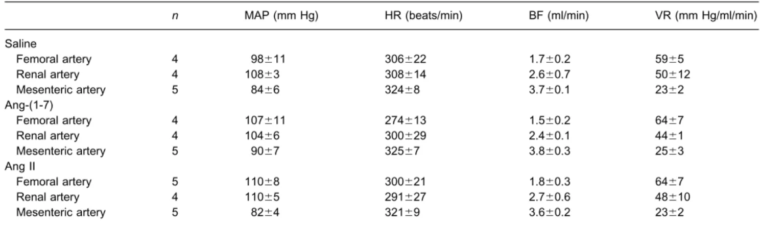
Table 2.Baseline values of MAP (mm Hg), HR (beats/min), mean blood flow (BF, ml/min) and VR (mm Hg/ml/min) of SHR submitted to microinjection into the CVLM
n MAP (mm Hg) HR (beats/min) BF (ml/min) VR (mm Hg/ml/min)
Saline
Femoral artery 4 98⫾11 306⫾22 1.7⫾0.2 59⫾5
Renal artery 4 108⫾3 308⫾14 2.6⫾0.7 50⫾12
Mesenteric artery 5 84⫾6 324⫾8 3.7⫾0.1 23⫾2
Ang-(1-7)
Femoral artery 4 107⫾11 274⫾13 1.5⫾0.2 64⫾7
Renal artery 4 104⫾6 300⫾29 2.4⫾0.1 44⫾1
Mesenteric artery 5 90⫾7 325⫾7 3.8⫾0.3 25⫾3
Ang II
Femoral artery 5 110⫾8 300⫾21 1.8⫾0.3 64⫾7
Renal artery 4 110⫾5 291⫾27 2.7⫾0.6 48⫾10
Mesenteric artery 5 82⫾4 321⫾9 3.6⫾0.2 23⫾2
Previous studies showed that the blood pressure ef-fects induced by CVLM microinjection of Ang II and Ang-(1-7) were similar, and were not accompanied by alter-ations in cardiac output. However, the hypotensive re-sponse produced by Ang II into the CVLM was abolished by the peripheral blockade of adrenergic receptors, while the depressor effect of Ang-(1-7) was only completely abolished by nitric oxide synthase inhibitors (Alzamora et al., 2002). These data suggested that the hypotensive effect provoked by Ang-(1-7) at the CVLM may involve the activation of a previously unsuspected nitrergic sym-pathetic vasodilatory pathway modulated by Ang-(1-7) in the CVLM. Whether angiotensin hypotensive effects at the CVLM are mediated by the activation of different neuronal pathways or different neuronal cell types, such
as GABAergic, catecholaminergic or glutamatergic, is still to be elucidated.
It is our hypothesis that this possible pathway that involves the release of NO in the periphery may be mod-ulated by Ang-(1-7) at the CVLM, and may be related to the vascular territory of the hindquarter of the rat. Several studies in the literature suggest the existence of a neuronal vasodilator system that uses nitric oxide as mediator in the skeletal muscle (Jones and Berne, 1963; Beck et al., 1966; Ballard et al., 1970).Davisson et al. (1994) described that the central sympathetic activation induced by the air-jet stress produces hindquarter vasodilation in conscious rats. That vasodilation could be inhibited byL-NG-nitroarginine methyl ester, a nitric oxide synthase inhibitor (Davisson et al., 1994). In another study, these authors showed that
a subpopulation of lumbar sympathetic cell bodies, post-ganglionic fibers and varicosities within the iliac and fem-oral arteries stained for neuronal nitric oxide synthase (nNOS) (Davisson et al., 1997). Additionally, these authors showed that electrical stimulation of the lumbar sympa-thetic chain produced a pronounced hind-limb vasodilation that was abolished by pretreatment with specific inhibitor of nNOS (7-NI). These data together with the result of the present study showing that Ang-(1-7), but not Ang II, alters the femoral VR and those of a previous study (Alzamora et al., 2002) showing that Ang-(1-7) effect at CVLM is completely blocked by peripheral inhibition of nitric oxide synthase suggest the possibility that a nitrergic sympa-thetic vasodilator system of central origin could be modu-lated by Ang-(1-7) at the CVLM. Interestingly, in SHR, only reductions in renal resistance were observed after Ang II or
Ang-(1-7) microinjections into the CVLM without alteration of femoral VR. The lack of change in femoral VR after CVLM microinjection of Ang-(1-7) may suggest that arterial hypertension may lead to an impairment of this vasodila-tory pathway. Future studies will be necessary to test these hypotheses.
It is interesting to observe that although different vascular beds were involved in mediating the response of Ang peptides at the CVLM, the magnitude of the hypotensive effect was not statistically different between the two peptides. Since the peptides were injected with a multibarreled micropipette in a random order, it is very likely that the site of microinjection and the spread of the injectate were the same. In addition, it is not clear why two congeners peptides exert similar blood pressure effects though distinct peripheral mechanisms. Future
Fig. 3.Alterations in mesenteric, femoral and renal blood flow (⌬BF, ml/min) and mesenteric, femoral and renal VR, calculated as⌬VR/baselineVR, produced by microinjection of saline, Ang-(1-7) and Ang II into the CVLM of Wistar and SHR. *P⬍0.05 in comparison to saline (two-way ANOVA followed by Bonferroni test);#P
studies evaluating the endogenous role of these pep-tides in different physiopathologic conditions may be helpful to address this question.
SHRs present a normal cardiac output accompanied with established hypertension, indicating that the in-creased arterial pressure was attributable exclusively to an increased peripheral resistance, however, the rise in VR is not uniform among the various organs. Comparison be-tween SHR and WKY or normotensive Wistar rats showed that, in general, SHRs have a lower blood flow to the main organs, such as kidney, skeletal muscle, skin and mesen-tery (Kimura et al., 1988; Thomas et al., 1990; Horiuchi et al., 1996; Granstam et al., 1998) and an increased blood flow in the heart and brain (Nishiyama et al., 1976; Thomas et al., 1990; Granstam et al., 1998). However, other stud-ies described a higher skeletal muscle blood flow in SHR (Nishiyama et al., 1976; Horiuchi et al., 1996). In addition, the fractional distribution of cardiac output may be different depending on the model of hypertension. In the renovas-cular Goldblatt model, an increased blood flow to heart, aorta, digestive tract, lungs and skeletal muscle and a decreased blood flow to skin were described (Bralet et al., 1973; Yates and Hiley, 1979; Teranishi and Iriuchijima, 1988). Several factors related to methodological differ-ences and/or physiopathologic mechanisms triggered while hypertension progresses can be attributable to these discrepancies. In our study, while renal VR was increased, femoral VR was decreased in SHR compared with Wistar rats. Thus, we cannot completely rule out that the lack of alteration in femoral VR after Ang-(1-7) microinjection into the CVLM of SHR could be related to the lower baseline resistance of this vascular bed. However, it is also possible that alterations of central mechanisms controlling sympa-thetic output to different vascular beds in SHR may be depending on the angiotensin levels or action at specific brain sites.
It is well known that CVLM contains GABAergic neu-rons that induce tonic inhibition of sympathoexcitatory neurons of the RVLM. Previous studies suggest that SHR may have decreased tonic GABAergic inputs into the RVLM originating directly from, or indirectly through, the CVLM, which could explain, at least in part, the increased sympathetic tonus presented by these ani-mals. SHR and WKY rats had similar responsiveness to microinjection ofL-glutamate into the RVLM (Kubo et al., 1986; Smith and Barron, 1990a,b,c; Muratani et al., 1993). In contrast, a larger depressor response to L-glutamate (Smith and Barron, 1990a,b; Muratani et al., 1993) into the CVLM, as well as to GABA (Smith and Barron, 1990c; Muratani et al., 1993) into the RVLM of SHR was observed. In addition, SHR had a lower re-sponse to GABA antagonist bicuculline into the RVLM and no response to tetrodotoxin into the CVLM. Further, tetrodotoxin injected into the CVLM attenuated the pres-sor response to bicuculline injected into the RVLM of WKY but not SHR (Smith and Barron, 1990c). In the present study we have shown that Ang II and Ang-(1-7) presented similar response in SHR when compared with Wistar rats, suggesting SHRs present alteration in giotensin endogenous levels or an up-regulation of an-giotensinergic receptors at the CVLM, which would com-pensate for the decrease in CVLM neuron activity. In-terestingly,Muratani et al. (1991, 1993) have shown an increased response to Ang II and to the non-selective angiotensin antagonist, [Sar1,Thr8]-Ang II, into the CVLM of SHR. Although the differences between our results and those ofMuratani et al. (1993) may be due to distinct baseline arterial pressure level or site of the microinjection, which in their study was closer to nucleus ambiguous, both sets of data are in keeping with the possibility of an up-regulation of angiotensin receptors in the CVLM. Further, taken together these data suggest
Fig. 4. Image of a histological section of the medulla (left side) and a diagrammatic representation of the frontal section (right side) illustrating the center of the microinjections into the CVLM (disruption of the tissue caused by the microinjection and shaded area, respectively). The diagram at
that the augmented action of endogenous Ang II in the CVLM is insufficient to suppress an apparent greater intrinsic pressor activity of RVLM neurons in SHR.
CONCLUSION
In summary, we have shown that in normotensive rats Ang II depressor effect at CVLM is mediated by a decrease in renal resistance while the effect of Ang-(1-7) involves al-teration in renal and femoral VR. In SHR, changes only in renal resistance participated in the depressor effect elicit by both peptides at the CVLM. These data suggest that the effect produced by Ang-(1-7) and Ang II in CVLM involves the participation of different neuronal populations/or differ-ent cdiffer-entral or peripheral pathways that can be partially altered in SHR.
Acknowledgments—This study was part of PM Ferreira PhD the-sis at the Post-graduation Program in Biological Sciences: Phys-iology and Pharmacology, ICB, UFMG. P. M. Ferreira was a recipient of CAPES-PICDT fellowship (doctoral degree) from the Federal University of Goiás. The financial support from FAPEMIG (Fundação de Amparo à Pesquisa do Estado de Minas Gerais) and CNPq (Conselho Nacional de Desenvolvimento Científico e Tecnológico) PRONEX grant (Programa de Núcleos de Excelên-cia) and CAPES (Coordenadoria de Apoio ao Pessoal de Nível Superior) is acknowledged. We are thankful to Jose R. Silva for skillful technical assistance.
REFERENCES
Agarwal SK, Gelsema AJ, Calaresu FR (1989) Neurons in rostral VLM are inhibited by chemical stimulation of caudal VLM in rats. Am J Physiol Regul Integr Comp Physiol 257(2):R265–R270.
Allen AM, Dampney RAL, Mendelsohn FAO (1988) Angiotensin re-ceptor binding and pressor effects in cat subretrofacial nucleus. Am J Physiol Heart Circ Physiol 255(24):H1011–H1017. Alzamora AC, Santos RAS, Campagnole-Santos MJ (2002)
Hypoten-sive effect of ANG II and Ang-(1-7) at the caudal ventrolateral medulla involves different mechanisms. Am J Physiol Regul Integr Comp Physiol 283(5):R1187–R1195.
Andreatta SH, Averill DL, Santos RAS, Ferrario CM (1988) The ven-trolateral medulla: A new site of action of the renin-angiotensin system. Hypertension 11:I163–I166.
Ballard DR, Abboud FM, Mayer HE (1970) Release of a humoral vaso-dilator substance during neurogenic vasodilatation. Am J Physiol 201:123–128.
Beck L, Pollard AA, Kaaylp SO, Weiner LM (1966) Sustained dilatation elicited by sympathetic nerve stimulation. Fed Proc 25:1596 –1606. Becker LK, Etelvino GM, Walther T, Santos RAS, Campagnole-Santos MJ (2007) Immunofluorescence localization of the receptor Mas in cardiovascular-related areas of the rat brain. Am J Physiol Heart Circ Physiol 293(3):H1416 –H1424.
Blessing WW (1988) Depressor neurons in rabbit caudal medulla act via GABA receptors in rostral medulla. Am J Physiol Heart Circ Physiol 254(4):H686 –H692.
Bralet AM, Wepierre J, Bralet J (1973) Distribution of cardiac output and nutritional blood flow in the unanesthetized rat: alterations during experimental renal hypertension. Pflugers Arch 343(3):257–266. Casto R, Phillips MI (1985) Neuropeptide action in nucleus tractus
solitarius: Angiotensin specificity and hypertensive rats. Am J Physiol Regul Integr Comp Physiol 249(3):R341–R347.
Cravo SL, Morrison SF, Reis DJ (1991) Differentiation of two cardio-vascular regions within caudal ventrolateral medulla. Am J Physiol Regul Integr Comp Physiol 261(4 Pt 2):R985–R994.
Cravo SL, Morrison SF (1993) The caudal ventrolateral medulla is a source of tonic sympathoinhibition. Brain Res 621:133–136. Colombari E, Sato MA, Cravo SL, Bergamaschi CT, Campos RRJr,
Lopes OU (2001) Role of the medulla oblongata in hypertension. Hypertension 38(3):549 –554.
Dampney RAL (1994) Functional organization of central pathways regulating the cardiovascular system. Physiol Rev 74(2):323–364. Dampney RAL, McAllen RM (1988) Differential control of sympathetic fibres supplying hindlimb skin and muscle by subretrofacial neu-rones in the cat. J Physiol 395:41–56.
Davisson RL, Johnson AK, Lewis SJ (1994) Nitrosyl factors mediate active neurogenic hindquarter vasodilation in the conscious rat. Hypertension 23:962–966.
Davisson RL, Possas OS, Murphy SP, Lewis SJ (1997) Neurogen-ically derived nitrosyl factors mediate sympathetic vasodilation in the hindlimb of the rat. Am J Physiol Heart Circ Physiol 272 (41):H2369 –H2376.
Dean C, Seagard JL, Hopp FA, Kampine JP (1992) Differential control of sympathetic activity to kidney and skeletal muscle by ventral medullary neurons. J Auton Nerv Syst 37:1–10.
Ferreira PM, Santos RAS, Campagnole-Santos MJ (2007) Angiotensin-(3–7) pressor effect at the rostral ventrolateral medulla. Regul Pept 141(1–3):168 –174.
Fontes MAP, Silva LCS, Campagnole-Santos MJ, Khosla MC, Guertzenstein PG, Santos RAS (1994) Evidence that angioten-sin-(1-7) plays a role in the central control of blood pressure at the ventrolateral medulla acting through specific receptors. Brain Res 665:175–180.
Fontes MAP, Martins Pinge MC, Naves V, Campagnole-Santos MJ, Lopes OU, Khosla MC, Santos RAS (1997) Cardiovascular effects produced by microinjection of angiotensins and angiotensin antag-onists into the ventrolateral medulla of freely moving rats. Brain Res 750:305–310.
Granstam SO, Granstam E, Fellstrom B, Lind L (1998) Regional haemo-dynamic differences between normotensive and spontaneously hy-pertensive rats: a microsphere study. Physiol Res 47(1):9 –15. Gutkind JS, Kurihara M, Castren E, Saavedra JM (1988) Increased
concentration of angiotensin II binding sites in selected brain areas of spontaneously hypertensive rats. J Hypertens 6(1):79 – 84. Guertzenstein PG (1973) Blood pressure effects obtained by drugs
ap-plied to the ventral surface of the brain stem. J Physiol 229(2): 395– 408.
Horiuchi K, He H, Tomohiro A, Aki Y, Kimura S, Tamaki T, Abe Y (1996) Lack of vasodilatory response in skeletal muscle blood vessels of aged spontaneously hypertensive rats. Heart Vessels 11(1):1–9.
Hu L, Zhu D, Yu Z, Wang JQ, Sun Z, Yao T (2002) Expression of angiotensin II type 1 (AT1) receptor in the rostral ventrolateral medulla in rats. J Appl Physiol 92:2153–2161.
Jones RD, Berne RM (1963) Vasodilatation in skeletal muscle. Am J Physiol 204:461– 466.
Kimura S, Fujioka S, Fukui K, Tamaki T, Iwao H, Abe Y (1988) Effects of an antihypertensive vasodilator, pinacidil, on regional blood flow in conscious spontaneously hypertensive rats. J Pharmacobiodyn 11(6):430 – 437.
Komatus C, Shibata K, Furukawa T (1996) The developmental increase of the AT1A, but not the AT1B, receptor mRNA level at the preoptic area in spontaneously hypertensive rats. Life Sci 58(14):1109 –1121. Kubo T, Nagura J, Kihara M, Misu Y (1986) Cardiovascular effects of l-glutamate and gaba-aminobutyric acid injected into the rostral ventrolateral medulla in normotensive and spontaneously hyper-tensive rats. Arch Int Pharmacodyn 279:150 –161.
Li Y, Polson JW, Dampney RAL (1992) Angiotensin II excites vaso-motor neurons but not respiratory neurons in the rostral and caudal ventrolateral medulla. Brain Res 577(1):161–164.
Matsuda T, Shibata K, Abe M, Tomonaga M, Furukawa T (1987) Potentiation of pressor response to angiotensin II at the preoptic area in spontaneously hypertensive rat. Life Sci 41:749 –754. Muratani H, Averill DB, Ferrario CM (1991) Effect of angiotensin II in
the ventrolateral medulla of spontaneously hypertensive rats. Am J Physiol Regul Integr Comp Physiol 260(5):R977–R984.
Muratani H, Ferrario CM, Averill DB (1993) Ventrolateral medulla of spontaneously hypertensive rats: role of angiotenisn II. Am J Physiol Regul Integr Comp Physiol 264(33):R388 –R395. Nishiyama K, Nishiyama A, Frohlich ED (1976) Regional blood flow in
normotensive and spontaneously hypertensive rats. Am J Physiol 230(3):691– 698.
Paula PM, Machado BH (2001) Changes in regional vascular resis-tance in response to microinjection of L-glutamate into different antero-posterior coordinates of the RVLM in awake rats. Auton Neurosci 87(2–3):301–309.
Paxinos G, Watson C (1986) The rat brain in stereotaxic coordinates. New York: Academic Press.
Ruiz P, Basso N, Cannata MA, Taquini AC (1990) The renin-angio-tensin system in different stages of spontaneous hypertension in the rat (SHR). Clin Exp Hypertens A 12(1):63– 81.
Santos RAS, Campagnole-Santos MJ, Baracho NCV, Fontes MAP, Silva CLS, Neves LAA, Oliveira DR, Caligiorne SM, Rodrigues ARV, Geopen C Jr, Carvalho WS, Simões e Silva AC, Khosla MC (1994) Characterization of a new angiotensin antagonist selective for angio-tensin-(1-7) evidence that the actions of angioangio-tensin-(1-7) are medi-ated by specific angiotensin receptors. Brain Res Bull 35(4):293–298. Santos RAS, Campagnole-Santos MJ, Andrade SP (2000)
angiotensin-(1-7): An update. Regul Pept 91:45– 62.
Santos RAS, Simoes e Silva AC, Maric C, Silva DM, Machado RP, Buhr I, Heringer-Walther S, Pinheiro SV, Lopes MT, Bader M, Mendes EP, Lemos VS, Campagnole-Santos MJ, Schultheiss HP, Speth R, Walther T (2003) angiotensin-(1-7) is an endogenous ligand for the g protein-coupled receptor Mas. Proc Natl Acad Sci U S A 100(14):8258 – 8263.
Sasaki S, Dampney RAL (1990) Tonic cardiovascular effects of an-giotensin II in the ventrolateral medulla. Hypertension 15:274 –283. Silva LC, Fontes MAP, Campagnole-Santos MJ, Khosla MC, Campos Jr RR, Guertzenstein PG, Santos RAS (1993) Cardiovascular
ef-fects produced by microinjection of angiotensin-(1-7) on vasopressor and vasodepressor sites of the ventrolateral medulla. Brain Res 613:321–325.
Smith JK, Barron KW (1990a) Cardiovascular effects of L-glutamate and tetrodotoxin microinjected into the rostral and caudal ventro-lateral medulla in normotensive and spontaneously hypertensive rats. Brain Res 506:1– 8.
Smith JK, Barron KW (1990b) The rostral and caudal ventrolateral medulla in young spontaneously hypertensive rats. Brain Res 506:153–158.
Smith JK, Barron KW (1990c) GABAergic responses in ventrolateral medulla in normotensive and spontaneously hypertensive rats. Am J Physiol Regul Integr Comp Physiol 258:R450 –R456. Tamura K, Umemura S, Nyui N, Yamakawa T, Yamaguchi S, Ishigami T,
Tanaka S, Tanimoto K, Takagi N, Sekihara H, Murakami K, Ishii M (1996) Tissue-specific regulation of angiotensinogen gene expression in spontaneously hypertensive rats. Hypertension 27(6):1216 –1223. Teranishi Y, Iriuchijima J (1988) Regional blood flows and resistances in conscious one-kidney, one-clip renovascular hypertensive rats. Jpn J Physiol 38(1):47–53.
Thomas GR, Walder CE, Thiemermann C, Vane JR (1990) Regional vascular resistance and haemodynamics in the spontaneously hypertensive rat: the effects of bradykinin. J Cardiovasc Pharmacol 15(2):211–217.
Welch WJ, Deng X, Snellen H, Wilcox CS (1995) Validation of minia-ture ultrasonic transit-time flow probes for measurement of renal blood flow in rats. Am J Physiol Renal Physiol 268(1 Pt 2): F175–F178.
Willette RN, Punnen-Grandy S, Krieger AJ, Sapru HN (1987) Dif-ferential regulation of regional vascular resistance by the rostral and caudal ventrolateral medulla in the rat. J Auton Nerv Syst 18(2):143–151.
Yates MS, Hiley CR (1979) Distribution of cardiac output in different models of hypertension in the conscious rat. Pflugers Arch 379(2):219 –222.
Zhu DN, Moriguchi A, Mikami H, Higaki J, Ogihara T (1998) Central amino acids mediate cardiovascular response to angiotensin II in the rat. Brain Res Bull 45(2):189 –197.