Angio te nsin and baro re fle x
co ntro l o f the circulatio n
1Neuropharmacology Laboratory, Baker Medical Research Institute, Melbourne,
Australia
2Department of Physiology, Yamanashi Medical University, Tamaho, Yamanashi, Japan
G.A. Head1, T. Saigusa2 and D.N. Mayorov1
Abstract
There is a close association between the location of angiotensin (Ang) receptors and many important brain nuclei involved in the regulation of the cardiovascular system. The present review encompasses the physiological role of Ang II in the brainstem, particularly in relation to its influence on baroreflex control of the heart and kidney. Activation of AT1 receptors in the brainstem by fourth ventricle (4V) administra-tion to conscious rabbits or local administraadministra-tion of Ang II into the rostral ventrolateral medulla (RVLM) of anesthetized rabbits acutely increases renal sympathetic nerve activity (RSNA) and RSNA barore-flex responses. Administration of the Ang antagonist Sarile into the RVLM of anesthetized rabbits blocked the effects of Ang II on the RSNA baroreflex, indicating that the RVLM is the major site of sympathoexcitatory action of Ang II given into the cerebrospinal fluid surrounding the brainstem. However, in conscious animals, blockade of endogenous Ang receptors in the brainstem by the 4V AT1 receptor antagonist losartan resulted in sympathoexcitation, suggesting an overall greater activity of endogenous Ang II within the sympathoin-hibitory pathways. However, the RSNA response to airjet stress in conscious rabbits was markedly attenuated. While we found no effect of acute central Ang on heart rate baroreflexes, chronic 4V infusion inhibited the baroreflex and chronic losartan increased baroreflex gain. Thus, brainstem Ang II acutely alters sympathetic responses to specific afferent inputs thus forming part of a potentially important mechanism for the integration of autonomic response patterns. The sympathoexcitatory AT1 receptors appear to be activated during stress,
surgery and anesthesia. Co rre spo nde nce
G.A. Head
Baker Medical Research Institute P.O . Box 6492
St. Kilda Rd. Central Melbourne, 8008 Australia
Fax: + 61-3-8532-1362
E-mail: Geoff.Head@ baker.edu.au
Presented at the IV International Symposium on Vasoactive Peptides, Belo Horizonte, MG, Brazil, O ctober 19-21, 2001.
Research supported by an Institute grant from the National Health and Medical Research Council of Australia.
Received December 6, 2001 Accepted January 22, 2002
Ke y wo rds
·Angiotensin II ·AT1 receptors
·Losartan
·Renal sympathetic activity
·Baroreflex
·Rostral ventrolateral medulla
·Blood pressure
·Rabbit
Intro ductio n
It is now becoming well recognized that angiotensin (Ang) not only plays a key role in the regulation of the cardiovascular sys-tem within the kidney and vasculature, but also in the central nervous system (1,2). There is a very close association between the loca-tion of Ang II receptors and many important brain nuclei involved in cardiovascular
More recently, brainstem vasomotor nu-clei such as the nucleus tractus solitarius (NTS), rostral ventrolateral medulla (RVLM) and caudal ventrolateral medulla (CVLM), which all contain high concentrations of Ang type 1 (AT1) receptors, also drew attention as possibly important sites of action of Ang (7,8). While these regions are key integrative centers for central cardiovascular control, they possess a blood-brain barrier which normally prevents any direct contact from circulating Ang. Presumably, Ang receptors in these nuclei are activated by an endoge-nous Ang system involving either Ang II or Ang III as neurotransmitters or neuromodu-lators (1).
These nuclei form the fundamental neu-ronal substrate of the arterial baroreflex with excitatory projections from second-order neurons in the NTS to neurons in the CVLM, and inhibitory projections from the CVLM to the sympathoexcitatory neurons in the RVLM. The pattern of AT1 receptor distri-bution thus suggests that Ang may act at these nuclei exerting both excitatory and inhibitory influence on baroreflex control of cardiovascular function. However, it would be an oversimplification to suggest that Ang exerts mostly an excitatory action in the RVLM and an inhibitory action in the CVLM because of increasing evidence that its ac-tions in the same nuclei may critically de-pend on the endogenous activity of the Ang system.
The present brief review discusses the role of Ang II in the hindbrain, particularly in relation to its influence on baroreflex control of the heart and kidney. In this re-view we will not consider the role of Ang in the area postrema on the arterial baroreflex, since it has recently been discussed in detail elsewhere (6).
Cardio vascular e ffe cts o f intrave ntricular Ang
Stimulation of central Ang II receptors
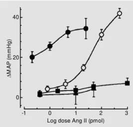
has been shown to increase arterial pressure in various species (1). A common approach used to investigate the cardiovascular role of the central Ang system has been to adminis-ter Ang peptides into the cerebral ventricles. Bickerton and Buckley (9) demonstrated in the early 1960s that Ang II given into the lateral ventricle could elicit a centrally medi-ated pressor response in the dog. Studies by Severs and colleagues (10) showed a pressor response from perfusion of the lateral ven-tricle of anesthetized cats with Ang II. Pre-venting the perfusate from leaving the lateral and third ventricles blocked the response, suggesting a site of action at or below the midbrain level. Hoffman and Phillips (11) examined the effects of intraventricular ad-ministration of Ang II to conscious rats and found marked pressor and dipsogenic re-sponses via the lateral ventricle as well as the anterior and posterior third ventricle. In this species, however, the site of action involved the anterior ventral third ventricle which correlates well with the location of the re-ceptors and is relatively close to the injection site. In conscious rabbits, however, lateral or third ventricle injections of Ang II had little effect on blood pressure with doses as large as 1000 pmol (Figure 1) (12). The rabbit also does not respond to chronic infusions of Ang into the lateral ventricle (13) nor does acute injection of Ang produce dipsogenic re-sponses (14) as it does in the rat.
about 400 times lower than those given in-travenously (Figure 1). The onset of the re-sponse was relatively quick, within 1 min, and generally lasted only 5 min, depending on the dose (12). The receptors were AT1 since the pressor response to Ang II adminis-tered into the 4V of conscious rabbits was completely blocked by low doses of the non-peptide selective antagonist losartan but not affected by the AT2 antagonist PD123319 (17).
The importance of the sympathetic ner-vous system in mediating central cardiovascu-lar effects of Ang has been recognized since the mid 1960s in all species studied although relatively few studies have measured nerve activity directly (18). A number of studies suggest that in the rat there is an important contribution to the pressor response by release of vasopressin (19). However, direct record-ings of renal sympathetic nerve activity (RSNA) do show a significant increase in RSNA con-comitant with the pressor response following Ang II injected into the third ventricle in rats (20). Tobey and colleagues (21) observed a marked increase in splanchnic nerve activity but no effect on RSNA following intraven-tricular administration of Ang II using anes-thetized, sinoaortic denervated and vagoto-mized cats, suggesting there may be differen-tial effects on sympathetic activity depending on the vascular bed.
In contrast to the rat, the pressor response observed with 4V Ang II given to conscious rabbits is predominantly due to sympathetic vasoconstriction since it is blocked by intra-venous prazosin (16). The vasoconstriction occurred in both the mesenteric and renal vascular beds but was accompanied by dila-tation of the hindlimb vascular bed and a fall in heart rate which opposed the vasocon-striction and reduced the observed pressor response. However, the hindlimb dilatation changed to vasoconstriction in rabbits after sinoaortic denervation, suggesting that it was mainly due to a baroreflex response to the rise in blood pressure (12).
D
M
A
P
(
m
m
H
g
)
40
20
0
-1 0 1 2 3
Log dose Ang II (pmol)
Effe cts o f baro re ce pto r de ne rvatio n o n the actio n o f ce ntral Ang
The profound increase in the sensitivity to central Ang II after sinoaortic denervation has been observed in rats with lateral ven-tricle administration (22). The increase was ~300 times greater than would be expected from loss of peripheral baroreflex function and was due to similar increases in sympa-thetic outflow and vasopressin-mediated va-soconstriction. In the conscious rabbit, chronic sinoaortic denervation augmented the sensitivity to Ang II given into the 4V by 900-fold. This effect was not observed in animals with depletion of spinal noradrener-gic pathways, indicating that sensitization of the brainstem noradrenergic neurons to Ang may play a key role in this phenomenon (16). This also suggests that pressor responses to central Ang II are normally suppressed by profound baroreflex inhibition. Furthermore, the Ang receptors that have been stimulated by 4V administration may be of greater physi-ological importance when the baroreflex is not functioning effectively as is the case in hypertension and heart failure (23,24).
Effe cts o f fo urth ve ntricle Ang II o n cardiac and sym pathe tic baro re fle xe s
While much has been written about the
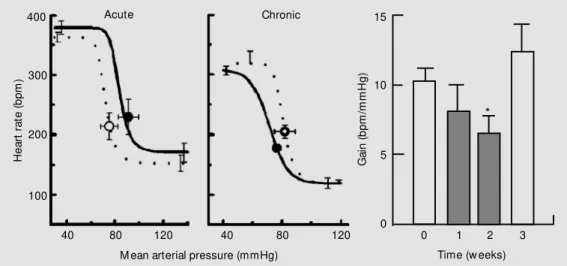
Infusion of Ang II (2.5-25 pmol/min) into the third (28) or fourth (25) ventricle acutely reset baroreflex control of heart rate to a higher pressure but did not affect the sensitivity or range of this reflex in con-scious rabbits. Importantly, in both studies intravenous infusion of the same dose of Ang II had no effect on the baroreflex. More recently, we also found similar effects of infusion of a low dose of Ang II into the lateral recess of the 4V on cardiac baroreflex in rabbits (Figure 3) (27). It is interesting to note that the resetting appears to be com-plete. However, since this occurs within 20 min, this is highly unlikely from what is known about the time course of baroreceptor resetting (29). One possible explanation for the lack of acute bradycardia may be the fact that Ang II is also exciting cardiac sympa-thetic pathways in the brainstem. The advan-tage of such a combination of actions is that Ang can modulate the level of blood pres-sure rapidly while maintaining the very short-term regulation of heart rate within the high gain area of the reflex curve.
Studies concerning the cardiovascular role of central Ang II have predominantly effect of circulating Ang on cardiac and
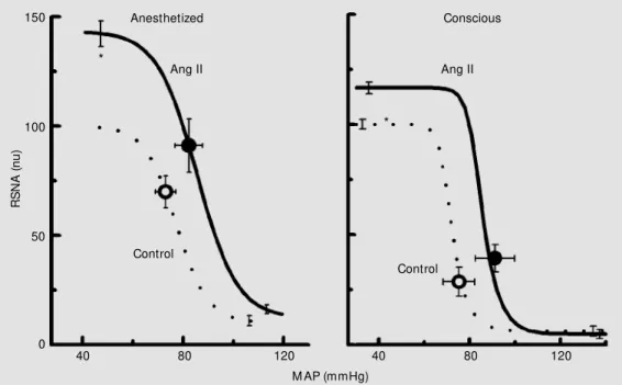
sympathetic baroreflexes, very few studies have examined the effect of intraventricular Ang on baroreflexes. In conscious rabbits Dorward and Rudd (25) found that 4V Ang II produced a marked increase in RSNA when the baroreceptors were unloaded with decreasing blood pressure. Thus, the upper plateau of the RSNA baroreflex curve was doubled and the curve shifted to the right due to the increase in blood pressure. We ob-served a similar pattern of effect of 4V Ang II on the RSNA baroreflex in urethane-anes-thetized rabbits (Figure 2) (26). Recently, we also infused Ang II into the lateral recess of the 4V in conscious rabbits (27). Although in our study Ang infusion evoked pressor responses similar to those observed by Dorward and Rudd (25), the magnitude of the RSNA increase after the baroreceptor unloading was lower (Figure 2). This differ-ence may indicate that access to the dorsal medulla and area postrema, which were much closer to the infusion site in the former study, may be critical in the excitatory action of intraventricular Ang II on the RSNA barore-flex in conscious rabbits.
Figure 2. Average mean arterial pressure (M AP) and renal sym-pathetic nerve activity (RSNA) baroreflex curves before (con-trol, dotted lines) and during in-fusion of Ang II into the fourth ventricle (solid line) from 6 ure-thane-anesthetized rabbits (left panel) and 5 conscious rabbits (right panel). Circles on curves are basal values. nu = normal-ized units. * P<0.05 for differ-ence betw een upper plateaus. Adapted w ith permission from Refs. 26,27.
150
R
S
N
A
(
n
u
)
100
50
0
Ang II Ang II
Control
Control
40 80 120 40 80 120
M AP (mmHg)
*
*
involved acute administration of agonists and antagonists. However, perhaps even more important for the cardiovascular system is the impact of chronic activation or inhibition of central Ang II receptors. It has been known since the earlier 1980s that central infusion of Ang produces hypertension and central inhibition of the renin-angiotensin system attenuates hypertension (30). Despite many such subsequent studies, relatively few have examined cardiovascular reflex mechanisms. We recently examined the effect of chronic activation of central Ang II receptors on cardiac baroreflex function in conscious nor-motensive rabbits. Animals received a 4V infusion by osmotic minipump of Ang II for 2 weeks. Assessment of the heart rate baro-reflex was performed by a single slow ramp rise and fall in blood pressure by intravenous infusion of phenylephrine and by caval bal-loon inflation, respectively. Ang II (100 ng/ h) decreased cardiac baroreflex gain by -20% after 1 week and by -37% after 2 weeks (Figure 3) (31). The intriguing feature of this study was that the effect of Ang II took the full 2 weeks to develop. Within a week of stopping the infusion, cardiac baroreflex gain had returned to control levels (Figure 3). Ringers solution or lower doses of Ang II did not modify the cardiac baroreflex func-tion. Blood pressure and heart rate were not altered by any treatment nor was their
vari-ability affected as assessed by power spec-tral analysis (31).
Effe cts o f e ndo ge no us Ang in the hindbrain o n baro re fle xe s
There has been a great deal of interest in the cardiovascular role of the endogenous Ang system within the brain. The most effec-tive method to determine the tonic activity of an endogenous Ang system has been to ap-ply a specific receptor antagonist of convert-ing enzyme inhibitor. Dorward and Rudd (25) examined the renal sympathetic and heart rate baroreflex effects of the specific, but non-subtype selective Ang II antagonist [Sar1,Ile8]-Ang II (Sarile) into the 4V of conscious rabbits. The antagonist had no effect on renal sympathetic reflexes whilst an Ang-converting enzyme inhibitor, enala-prilat, slightly enhanced maximal baroreflex sympathetic responses. By contrast, in a simi-lar conscious rabbit preparation, we found that the specific AT1 receptor antagonist lo-sartan increased resting RSNA and the upper plateau of the RSNA baroreflex, while blood pressure remained unaltered (Figure 4) (17). These results were somewhat surprising given that the receptor antagonist produced effects qualitatively similar to those of Ang itself and suggested that losartan is blocking a tonically active sympathoinhibitory action
H
e
a
rt
r
a
te
(
b
p
m
)
400
300
200
100
40 80 120 40 80 120 0 1 2 3
M ean arterial pressure (mmHg) Time (w eeks)
Acute Chronic
G
a
in
(
b
p
m
/m
m
H
g
)
15
10
5
0
*
of endogenous Ang (compare Figures 3 and 4). However, the effects of trigeminal stimu-lation were not affected by losartan, suggest-ing that Ang normally modulates specific inputs to the presympathetic neurons, possi-bly by a presynaptic action. The action of losartan was most likely a specific effect since in a later study we observed similar changes with the Ang-converting enzyme inhibitor, enalapril (32), indicating a pri-mary inhibitory role of hindbrain AT1 recep-tors in conscious normotensive rabbits. Whilst at this stage we do not know the site of action of the sympathoinhibitory Ang path-ways, possibilities include the CVLM and the NTS (see below).
In all the above studies in intact rabbits, acute administration of various Ang II recep-tor antagonists neither changed resting blood pressure or heart rate, nor affected cardiac baroreflexes (17,25,32). However, rabbits with pacing-induced heart failure have inhi-bition of cardiac baroreflex function which can be restored by the AT1 receptor antago-nist L-158,509 (33). By contrast, the cardiac baroreflex sensitivity in non-paced rabbits was not altered by AT1 receptor blockade. Thus, the participation of central Ang II in inhibiting cardiac baroreflexes does not ap-pear to occur in the normal animal but re-quires a long-term perturbation of the car-diovascular system such as heart failure.
We recently examined the effect of chronic inhibition of central Ang II receptors on cardiac baroreflex function in conscious normotensive rabbits. Animals received a 4V infusion by osmotic minipump of losar-tan for 2 weeks. Losarlosar-tan (30 µg/h) increased baroreflex gain by +24% and +58% after 1 and 2 weeks, respectively (Figure 4), but did not change resting blood pressure or heart rate (31). Similarly, chronic but not acute administration of the AT1 receptor antago-nist EXP 3174 has been shown to normalize cardiac baroreflex function in SHR (34). These data indicate that there is a long-term modulation of cardiac baroreflexes by en-dogenous Ang II which is independent of the blood pressure level. The reason for such a long time course is not clear since activation of neuronal AT1 receptors by Ang II occurs relatively rapidly, being due to a reduction in K+ current via protein kinase C and raised intracellular Ca2+ and stimulation of Ca2+ current. Possibly there is long-term regula-tion of the receptor or altered expression of AT1 receptors. Another possibility may in-volve an interaction with nitric oxide. Zucker and colleagues (35) have elegantly shown an important interaction between nitric oxide and Ang in the modulation of baroreflexes in heart failure. They suggest that the acute inhibitory effect of Ang on the baroreflex gain is opposed by the facilitatory effect of
0 1 2 3
Time (w eeks)
G
a
in
(
b
p
m
/m
m
H
g
)
15
10
5
0
R
S
N
A
(
n
u
)
150
100
50
0
40 80 120 30 60 90
M ean arterial pressure (mmHg)
Acute 400 Chronic
300
200
100
H
e
a
rt
r
a
te
(
b
p
m
)
120 0
*
nitric oxide, but chronically, as is the case with heart failure, a reduction in nitric oxide synthase amplifies the effects of Ang II.
Brainste m site s o f actio n o f fo urth ve ntricle Ang II
Intraventricular administration has been a useful tool particularly in conscious ani-mals to help unravel the role of the Ang peptides within the central nervous system but is limited by agents given in this way influencing large brain regions rather than specific nuclei. In order to determine the distribution of neurons within the medulla activated by infusion of Ang II into the 4V of conscious rabbits, we used the expression of Fos, the protein product of the immediate early gene c-fos, as a marker of neuronal activation in baroreceptor intact and barodenervated animals (36). Ang II induced a marked increase in the number of Fos-positive neurons in the NTS and in the ros-tral, intermediate, and caudal parts of the ventrolateral medulla with 30-75% of cells being double-labeled for Fos- and tyrosine hydroxylase immunoreactivity (36) (Figure 5). We found that the distribution of Fos-positive neurons closely correlated with the location of Ang II receptor-binding sites as previously determined in the rabbit.
Actio ns o f Ang II in the ro stral ve ntro late ral m e dulla o n baro re fle xe s
Of the regions that we examined using the Fos technique, it is clear that perhaps the most important is the RVLM where Ang peptides are likely to have their major sym-pathoexcitatory action (7). The RVLM is a major source of excitatory drive to the pre-ganglionic sympathetic neurons in the spinal cord and contains high Ang II receptor bind-ing in the rabbit (4) and other species includ-ing the cat and dog (37). The existence of Ang II immunoreactive fibers in the
ventro-lateral medulla and extended neural pro-cesses of the RVLM neurons close to the ventral surface (38) suggest that Ang II may have ready access to the RVLM neurons both from neural pathways and from cere-brospinal fluid. Allen and co-workers (39) demonstrated that direct microinjection of Ang II into the subretrofacial pressor region in the RVLM of the cat resulted in a pressor response. Andreatta and colleagues (40) sug-gested that the RVLM may contain a renin-angiotensin system since Ang I applied to the ventral surface of the brainstem of the cat produced an increase in blood pressure after conversion to Ang II. Sasaki and Dampney (41) found that the sites which produced the greatest increase in blood pressure in the RVLM were also the sites containing the highest concentration of Ang receptors. We initially mapped various dorsal and ventral sites with microinjections of Ang II and found that marked pressor responses were observed only when Ang II was adminis-tered into a discrete region of the RVLM corresponding to the subretrofacial nucleus. We found that injections as close as 1 mm from this region gave very much smaller pressor responses, suggesting that the Ang II-sensitive site is relatively small. Dose-response curves to Ang II in this region indicated that very low doses were required, with the half maximal dose being approxi-mately 9 fmol. This was approxiapproxi-mately 100-fold less than that required by the 4V route and consistent with the earlier findings of Sasaki and Dampney (41) and others using cats (39,40).
We also observed that local microinfusion of Ang II into the RVLM produced
facilita-N
u
m
b
e
r
o
f
F
o
s
-p
o
s
it
iv
e
n
e
u
ro
n
s
20
10
0
NTS AP CVLM IVLM RVLM
Fos TH + Fos
tion of the renal sympathetic baroreflex (Fig-ure 5) which was closely similar to that produced by 4V administration (Figure 2) (26). We found that glutamate infusions into the same region of the RVLM had effects similar to those of Ang, increasing blood pressure, resting RSNA and the upper sym-pathetic baroreflex plateau without affecting the lower plateau (Figure 6) (26). The simi-larity of Ang actions to those of glutamate suggests that it may directly excite sympa-thetic vasomotor cells in this region. Indeed, electrophysiological studies have shown that Ang appears to directly excite bulbospi-nal C1 neurons (42) by activating postsynap-tic AT1 receptors, resulting in a depolariza-tion involving the closing of K+ channels (43).
Microinjection techniques into the RVLM have been widely used to limit drug action to small brain regions, but until recently, have been confined in most cases to anesthetized preparations since this area is close to the flexion point of the cervical spinal cord and is subject to movement in the conscious animal. However, the role of Ang peptides in
the ventrolateral medulla may be very much influenced by the anesthetized animal prepa-rations. Indeed, a high degree of surgical stress is typically associated with microin-jection into the RVLM in the acute anesthe-tized preparation, and hormonal systems such as the renin-angiotensin system have been reported to become activated during anes-thesia and surgery (44). Due to their quiet nature, we have found that rabbits are suit-able animals for microinjection into the RVLM while conscious and sitting in a stan-dard rabbit box otherwise unrestrained. We therefore developed a new cannula system which permitted us to make repeated bilat-eral microinjections into the RVLM of con-scious rabbits (27). We used this new tech-nique to examine the role of Ang II in the RVLM while measuring RSNA at rest and during baroreflex responses. Bilateral mi-croinjection of Ang II (10 and 20 pmol) into histologically verified pressor regions of the RVLM did not change blood pressure or RSNA. We did observe that a higher Ang II dose of 30 pmol gradually increased blood pressure by 8 mmHg, without affecting RSNA. The time course of the increase in MAP was slow, with MAP starting to in-crease within 0.5 to 1 min and reaching a peak within 4 min of completion of the injec-tion. Bilateral microinfusion of Ang II (4 pmol/min for 20 min) did not affect resting MAP or RSNA (27). During the infusion of Ang II baroreflexes were examined but no effects were observed (Figure 6). At the same site glutamate produced increases in blood pressure and renal sympathetic activ-ity and augmentation of the RSNA barore-flex in a similar fashion to what was seen in the anesthetized animal (Figure 6, compare right and left panels). By contrast, infusion of the same dose of Ang II into the 4V cerebrospinal fluid increased blood pressure and RSNA by 22 and 34%, respectively (Figure 2), suggesting that sites other than the RVLM may be mediating these responses.
Figure 6. Average mean arterial pressure and renal sympathetic nerve activity (RSNA) curves before (control, dotted lines) and during infusion of Ang II (black lines) or glutamate (gray lines) into the rostral ventrolateral medulla in 6 urethane-anesthetized rabbits (left panel) or in 7 conscious rabbits (right panel). Error bars are average SEM calculated from analysis of variance indicating variation w ithin animals. nu = normalized units. Adapted w ith permission from M ayorov and Head (27).
R
S
N
A
(
n
u
)
150
100
50
0
40 80 120
M ean arterial pressure (mmHg)
40 80 120
Anesthetized Conscious
Glutamate Glutamate
Ang II
Ang II
Control
Cardio vascular e ffe cts o f blo cking e ndo ge no us Ang II in the ro stral ve ntro late ral m e dulla
Administration of 10 pmol of the antago-nist Sarile bilaterally into the subretrofacial region of anesthetized rabbits blocked the pressor response to locally applied Ang II and reduced the pressor responses to 4V Ang II by two thirds (Figure 7). This is perhaps the strongest evidence to suggest that the RVLM is the major site of action for Ang II given into the cerebrospinal fluid surrounding the brainstem but the remaining response suggests that other sites make a small contribution.
There has been very much interest in whether there is a contribution to maintain-ing sympathetic tone by Ang II endogenously released in the RVLM. Andreatta and co-workers (40) provided the first evidence of tonically active Ang II in the RVLM of the anesthetized cat by showing that with the same topical application approach [Sar1,Thr8]-Ang II (13 nmol) decreased blood pressure by 8 mmHg. However, we did not observe any change in blood pressure or RSNA by giving an effective dose of Sarile (10 pmol) to urethane-anesthetized rabbits. This contrasts the findings of other groups that found a marked reduction in blood pres-sure with this agent or with [Sar1,Thr8]-Ang II (41,45). A recent study suggests, however, that this effect is not related to the blockade of Ang II receptors (46). Presumably this was an effect observed only at much higher doses used in these studies (100-1000 pmol) since we observed no effect on basal blood pressure using 10 pmol, but still had a com-plete blockade of the pressor response when Ang II was administered into the RVLM. Since we did not observe any attenuation of the glutamate response, the blockage by Sarile was likely to be specific.
We found that bilateral infusion of Sarile (10 pmol + 1 pmol/min) into the RVLM did not alter the RSNA baroreflex in
urethane-anesthetized rabbits, indicating that there is little influence of endogenous Ang II on the sympathetic baroreflex. Subsequent infusion of Ang II into the 4V (10 pmol/min) had no further effect in the animals pretreated with Sarile (Figure 7). We also determined the effects of blocking Ang receptors in a con-scious rabbit preparation (27). Pretreatment with Sarile into the RVLM (100-500 pmol) did not change resting blood pressure, RSNA or renal sympathetic baroreflex parameters. The lack of an effect inhibiting Ang II recep-tors in the RVLM was unlikely to be due to leakage of the antagonist into the depressor region of the CVLM, since its bilateral mi-croinjections into the intermediate ventro-lateral medulla at or just rostral to the obex level also did not alter resting or baroreflex parameters.
The lack of effect inhibiting Ang II re-ceptors in the RVLM is consistent with our previous findings in anesthetized animals but the lack of effect of Ang II itself is somewhat surprising. One possibility is that the action of Ang II may depend very much on the state of excitatory inputs to the region. Fontes and colleagues (47) had also devel-oped a method for administration of agents into the RVLM but in conscious rats. They showed that Ang II and Ang 1-7 increased
R
S
N
A
(
n
u
)
150
100
50
0
Control
40 60 80 100 120
M ean arterial pressure (mmHg)
Sar-lle RVLM + Ang ll 4V
Vehicle RVLM + Ang ll 4V
blood pressure while the peptide antagonist Sarile produced a small decrease in blood pressure. They observed an increase in blood pressure with two different AT1 receptor an-tagonists which may be related to our obser-vations that losartan produced an increase in sympathetic activity when given 4V to con-scious rabbits (17). One possibility is that the relatively large volume of 200 nl enabled the drug to block the sympathoinhibitory action of Ang in the CVLM. The other pos-sibility is that the effect of Ang II depends on the synaptic inputs to the premotor cells. Electrophysiological studies have shown that Ang II increases input resistance of the vaso-motor cell body which should have the effect of potentiating both excitatory and inhibito-ry synaptic inputs (43). In support of this concept, Fontes and colleagues (47), who showed that the AT1 receptor antagonist CV-11974 produced an increase in blood pres-sure in normotensive rats (blocking a pre-dominant depressor role of Ang peptides), also found that this drug produced a depres-sor response in the hypertensive transgenic rat harboring the mouse Ren-2 gene (block-ing a predominant pressor response). They suggested that the role of Ang peptides de-pends on the endogenous activity of the Ang system. In support of this we have recently found that blockade of Ang II receptors in the RVLM attenuated the renal sympathoex-citatory responses to airjet stress.
Ang actio ns within the caudal ve ntro late ral m e dulla
Activation of the CVLM causes sympa-thoinhibition which is mediated by a short ascending inhibitory projection from the CVLM to the RVLM (48). Electrophysi-ological studies show that tonically active sympathoinhibitory neurons in the CVLM tonically inhibit the firing of sympathoexci-tatory barosensitive neurons in the RVLM of the rabbit (49). While the CVLM of the rabbit contains Ang II receptors (4), there
have been relatively few studies investigat-ing the role of Ang receptors in this region. Microinjection of Ang II into the region of the A1 noradrenergic cells within the CVLM of the rabbit results in a depressor response (50). The most extensive study was per-formed by Saigusa and colleagues (51) who found that Ang II infusion into the CVLM of anesthetized rabbits decreased sympathetic activity and inhibited RSNA baroreflex while blockade of CVLM Ang receptors increased the sympathetic barore-flex but did not alter resting blood pressure or RSNA.
Ang actio ns within the nucle us tractus so litarius
Co nclusio n
In recent years we have made clear progress in the understanding of the impor-tance of Ang peptides and receptors within the brainstem to cardiovascular regulation. There is a growing recognition from a vari-ety of different studies that the effects of activating Ang II receptors in the RVLM, CVLM and NTS appear to be very much dependent on the state of the animal and the activity of specific afferent information. This aspect may be a critically important mechan-ism for the adaptive ability of the central nervous system to respond with appropriate autonomic response patterns depending on the situation. The development of the micro-injecting system for conscious animals opens the way to a more complete investigation of the neurotransmitter mechanisms within the RVLMwithout the constraints of anesthesia and will permit an investigation into the role of Ang peptides in mediating the circulatory responses to emotional and behavioral stimuli. For the most part, in conscious nor-mal aninor-mals there is relatively little tonic sympathoexcitatory influence at the level of the RVLM as shown by the observations that blockade of AT1 receptor antagonists does not change blood pressure by very much and in some cases increases it. The latter finding indicates that in conscious animals the sym-pathoinhibitory influence of Ang is more evident and is less active in anesthetized
preparations. This sympathoinhibition ap-pears to be mainly mediated by an interac-tion with baroreceptor inhibitory inputs to the vasomotor neurons since the sympatho-inhibitory actions of Ang II are of greatest influence during hypotension. Studies in rats and rabbits suggest that the excitatory action of Ang II is normally under profound barore-ceptor inhibition since the sensitivity to Ang is markedly increased in barodenervated ani-mals and the sympathoexcitatory effect of Ang is most evident when baroreceptor in-put is reduced. On the other hand, barore-flexes themselves can be modulated by Ang peptides over several weeks of drug admin-istration. The precise mechanism of this long-term action remains to be elucidated. There is now much evidence to show that in situa-tions where there are increased excitatory and decreased inhibitory inputs to the RVLM brainstem, such as barodenervation, acute stress, hypertension or heart failure, the sym-pathoexcitatory Ang system within the brain-stem clearly becomes very important.
Ackno wle dgm e nts
The authors wish to acknowledge the contribution of their collaborators Jean-Luc Elghozi, Naomi Williams, Robert Bendle, Elisabeth Lambert, Yoshitaka Hirooka and Roger Dampney to the work described in this review.
Re fe re nce s
1. Phillips M I (1987). Functions of angio-tensin in the central nervous system. An-nual Review of Physiology, 49: 413-435. 2. Reid IA (1992). Interactions betw een ANG
II, sympathetic nervous system, and baro-receptor reflexes in regulation of blood pressure. American Journal of Physiolo-gy, 262: E763-E778.
3. M endelsohn FAO, Guirion R, Saavedra JM , Aguilera G & Catt KJ (1984). Autora-diographic localisation of angiotensin II receptors in rat brain. Proceedings of the
National Academy of Sciences, USA, 81: 1575-1579.
4. M endelsohn FAO, Allen AM , Clevers J, Denton DA, Tarjan E & M cKinley M J (1988). Localization of angiotensin II re-ceptor binding in rabbit brain by in vitro
autoradiography. Journal of Comparative Neurology, 270: 372-384.
5. Williams JL, Barnes KL, Brosnihan KB & Ferrario CM (1992). Area postrema - A unique regulator of cardiovascular func-tion. New s in Physiological Sciences, 7:
30-34.
6. Bishop VS & Sanderford M G (2000). An-giotensin II modulation of the arterial baro-reflex: role of the area postrema. Clinical and Experim ent al Pharm acology and Physiology, 27: 428-431.
7. Head GA (1996). Role of AT1 receptors in the central control of sympathetic vaso-motor function. Clinical and Experimental Pharmacology and Physiology, 23: S93-S98.
peptides and baroreflex control of sympa-thetic outflow : Pathw ays and mechan-isms of the medulla oblongata. Brain Re-searchBulletin, 51: 119-128.
9. Bickerton RK & Buckley JP (1961). Evi-dence for a central mechanism in angio-tensin induced hypertension. Proceedings of the Society for Experimental Biology and M edicine, 106: 834-836.
10. Severs WB, Daniels AE, Smookler HH, Kinnard WJ & Buckley JP (1966). Interre-lationship betw een angiotensin II and the sympathetic nervous system. Journal of Pharmacology and Experimental Thera-peutics, 153: 530-537.
11. Hoffman WE & Phillips M I (1976). Re-gional study of cerebral ventricle sensi-tive sites to angiotensin II. Brain Re-search, 110: 313-330.
12. Head GA & Williams NS (1992). Hemody-namic effects of central angiotensin I, II and III in conscious rabbits. American Journal of Physiology, 263: R845-R851. 13. Wright JW, Sullivan M J, Petersen EP &
Harding JW (1985). Brain angiotensin II and III binding and dipsogenicity in the rabbit. Brain Research, 358: 376-379. 14. Tarjan E, Denton DA, M cBurnie M I &
Weisinger RS (1988). Water and sodium intake of w ild and New Zealand rabbits follow ing angiotensin. Peptides, 9: 677-679.
15. Head GA, Elghozi J-L & Korner PI (1988). Baroreflex modulation of central angio-tensin II pressor responses in conscious rabbits. Journal of Hypertension, 6 (Suppl 6): S505-S507.
16. Elghozi J-L & Head GA (1990). Spinal nor-adrenergic pathw ays and the pressor re-sponses to central angiotensin II. Ameri-can Journal of Physiology, 258: H240-H246.
17. Bendle RD, M alpas SC & Head GA (1997). Role of endogenous angiotensin II on sympathetic reflexes in conscious rabbits.
American Journal of Physiology, 272: R1816-R1825.
18. Keim KL & Sigg EB (1971). Activation of central sympathetic neurons by angio-tensin II. Life Sciences, 10: 565-574. 19. Keil LC, Summy-Long J & Severs WB
(1975). Release of vasopressin by angio-tensin II. Endocrinology, 96: 1063-1065. 20. Steele M K, Gardner DG, Xie PL & Schultz
HD (1991). Interactions betw een ANP and ANG II in regulating blood pressure and sympathetic outflow . American Journal of Physiology, 260: R1145-R1151. 21. Tobey JC, Fry HK, M izejew ski CS, Fink
GD & Weaver LC (1983). Differential sym-pathetic responses initiated by
angio-tensin and sodium chloride. American Journal ofPhysiology, 245: R60-R68. 22. Barron KW, Trapani AJ, Gordon FJ &
Brody M J (1989). Baroreceptor denerva-tion profoundly enhances cardiovascular responses to central angiotensin II. Ameri-can Journal of Physiology, 257: H314-H323.
23. Head GA (1994). Cardiac baroreflexes and hypertension. Clinical and Experimental Pharmacology and Physiology, 21: 791-802.
24. Head GA (1995). Baroreflexes and cardio-vascular regulation in hypertension. Jour-nal of Cardiovascular Pharmacology, 26 (Suppl 2): S7-S16.
25. Dorw ard PK & Rudd CD (1991). Influence of the brain renin-angiotensin system on renal sympathetic and cardiac barore-flexes in conscious rabbits. American Journal ofPhysiology, 260: H770-H778. 26. Saigusa T & Head GA (1993). Renal
sym-pathetic baroreflex effects of angiotensin II infusions into the rostral ventrolateral medulla of the rabbit. Clinical and Experi-mental Pharmacology and Physiology, 20: 351-354.
27. M ayorov DN & Head GA (2001). Influence of rostral ventrolateral medulla on renal sympathetic baroreflex in conscious rab-bits. American Journal of Physiology, 280: R577-R587.
28. Reid IA & Chou L (1990). Analysis of the action of angiotensin II on the baroreflex control of heart rate in conscious rabbits.
Endocrinology, 126: 2749-2756. 29. Chapleau M W, Hajduczok G & Abboud
FM (1989). Peripheral and central mechan-isms of baroreflex resetting. Clinical and Experimental Pharmacology and Physiol-ogy, 15: 31-43.
30. Okuno T, Nagahama S, Lindheimer M D & Oparil S (1983). Attenuation of the devel-opment of spontaneous hypertension in rats by chronic central administration of captopril. Hypertension, 5: 653-662. 31. Gaudet E, Godw in SJ & Head GA (2000).
Effects of central infusion of angiotensin II and losartan on baroreflex control of heart rate in rabbits. AmericanJournal of Physiology, 278: H558-H566.
32. Gaudet E, Godw in SJ & Head GA (1998). Role of central catecholaminergic path-w ays in the actions of endogenous ANG II on sympathetic reflexes. American Jour-nalof Physiology, 275: R1174-R1184. 33. Liu JL, M urakam i H, Sanderf ord M ,
Bishop VS & Zucker IH (1999). ANG II and baroreflex function in rabbits w ith CHF and lesions of the area postrema. Ameri-can Journal of Physiology, 277:
H342-H350.
34. Bartholomeusz B & Widdop RE (1995). Effect of acute and chronic treatment w ith the angiotensin II subtype 1 receptor an-tagonist EXP 3174 on baroreflex function in conscious spontaneously hypertensive rats. Journal of Hypertension, 13: 219-225.
35. Zucker IH, Wang W, Pliquett RU, Liu JL & Patel KP (2001). The regulation of sympa-thetic outflow in heart failure. The roles of angiotensin II, nitric oxide, and exercise training. Annals of the New York Acade-my ofSciences, 940: 431-443.
36. Hirooka Y, Head GA, Potts PD, Godw in SJ, Bendle RD & Dampney RAL (1996). M edullary neurons activated by angio-tensin II in the conscious rabbit. Hyper-tension, 27: 287-296.
37. Speth RC, W am sley JK, Gehlert DR, Chernicky CL, Barnes KL & Ferrario CM (1985). Angiotensin II receptor localiza-tion in the canine CNS. Brain Research, 326: 137-143.
38. Benarroch EE, Granata AR, Ruggiero DA, Park DH & Reis DJ (1986). Neurons of C1 area mediate cardiovascular responses ini-tiated from ventral medullary surface.
American Journal of Physiology, 250: R932-R945.
39. Allen AM , Dampney RAL & M endelsohn FAO (1988). Angiotensin receptor binding and pressor effects in cat subretrofacial nucleus. American Journal of Physiology, 255: H1011-H1017.
40. Andreatta SH, Averill DB, Santos RA & Ferrario CM (1988). The ventrolateral me-dulla. A new site of action of the renin-angiotensin system. Hypertension, 11: I-163-I-166.
41. Sasaki S & Dampney RAL (1990). Tonic cardiovascular effects of angiotensin II in the ventrolateral medulla. Hypertension, 15: 274-283.
42. Chan RKW, Chan YS & Wong TM (1991). Responses of cardiovascular neurons in the rostral ventrolateral medulla of the normotensive Wistar Kyoto and sponta-neously hypertensive rats to iontophoretic application of angiotensin-II. Brain Re-search, 556: 145-150.
43. Li YW & Guyenet PG (1996). Angiotensin II decreases a resting K+ conductance in rat bulbospinal neurons of the C1 area.
Circulation Research, 78: 274-282. 44. Chernow B, Alexander HR, Smallridge RC,
45. Hirooka Y, Potts PD & Dampney RAL (1997). Role of angiotensin II receptor subtypes in mediating the sympathoexci-tatory effects of exogenous and endoge-nous angiotensin peptides in the rostral ventrolateral medulla of the rabbit. Brain Research, 772: 107-114.
46. Potts PD, Allen AM , Horiuchi J & Damp-ney RAL (2000). Does angiotensin II have a significant tonic action on cardiovascular neurons in the rostral and caudal VLM .
American Journal of Physiology, 279: R1392-R1402.
47. Fontes M AP, Baltatu O, Caligiorne SM , Campagnole-Santos M J, Ganten D, Bader M & Santos RAS (2000). Angiotensin pep-tides acting at rostral ventrolateral me-dulla contribute to hypertension of TGR (M REN2)27 rats. Physiological Genomics, 2: 137-142.
48. Chalmers J & Pilow sky P (1991). Brain-stem and bulbospinal neurotransmitter systems in the control of blood pressure.
Journal of Hypertension, 9: 675-694. 49. Li YW, Gieroba ZJ, M cAllen RM &
Bless-ing WW (1991). Neurons in rabbit caudal ventrolateral medulla inhibit bulbospinal barosensitive neurons in rostral medulla.
American Journal of Physiology, 261: R44-R51.
50. Allen AM , M endelsohn FAO, Gieroba ZJ & Blessing WW (1990). Vasopressin re-lease follow ing microinjection of angio-tensin-II into the caudal ventrolateral me-dulla oblongata in the anaesthetized rab-bit. Journal of Neuroendocrinology, 2: 867-873.
51. Saigusa T, Iriki M & Arita J (1996). Brain angiotensin II tonically modulates sympa-thetic baroreflex in rabbit ventrolateral medulla. American Journal ofPhysiology, 271: H1015-H1021.
52. Berger AJ (1979). Distribution of carotid sinus nerve afferent fibers to solitary tract nuclei of the cat using transganglionic transport of horseradish peroxidase. Neu-roscience Letters, 14: 153-158.
53. Allen AM , Chai SY, Clevers J, M cKinley M J, Paxinos G & M endelsohn FAO (1988). Localization and characterization of angio-tensin II receptor binding and angioangio-tensin converting enzyme in the human medulla oblongata. Journal of Comparative Neu-rology, 269: 249-264.
54. Paton JF & Kasparov S (2000). Sensory channel specific modulation in the nu-cleus of the solitary tract. Journal of the Autonomic Nervous System, 80: 117-129. 55. Casto R & Phillips M I (1984). Cardiovascu-lar actions of microinjections of
angioten-sin II in the brain stem of rats. American Journal of Physiology, 246: R811-R816. 56. Diz DI, Barnes KL & Ferrario CM (1984).
Hypotensive actions of microinjections of angiot ensin II int o t he dorsal m ot or nucleus of the vagus. Journal of Hyper-tension, 2: 53-56.
57. Casto R & Phillips M I (1986). Angiotensin II attenuates baroreflexes at nucleus trac-tus solitarius of rats. American Journal of Physiology, 250: R193-R198.
58. Paton JFR & Kasparov S (1999). Differen-tial effects of angiotensin II on cardiores-piratory reflexes mediated by nucleus tractus solitarii: a microinjection study in the rat. Journal of Physiology, 521: 213-225.
59. Campagnole-Santos M J, Diz DI & Ferrario CM (1988). Baroreceptor reflex modula-tion by angiotensin II at the nucleus trac-tus solitarii. Hypertension, 11: I-167-I-171. 60. Kasparov S, Butcher JW & Paton JFR (1998). Angiotensin II receptors w ithin the nucleus of the solitary tract mediate the developmental attenuation of the barore-ceptor vagal reflex in pre-w eaned rats.


![Figure 7. Average mean arterial pressure and renal sympathetic nerve activity (RSNA) baroreflex curves from 4 anesthetized rab-bits before (dotted line) and dur-ing infusion into the rostral ven-t rolaven-t eral m edulla (RVLM ) of [Sar 1 ,Ile 8 ]-Ang I](https://thumb-eu.123doks.com/thumbv2/123dok_br/15809759.650851/9.918.383.651.764.1070/average-arterial-pressure-sympathetic-activity-baroreflex-anesthetized-infusion.webp)
