Identification of Snails within the
Bulinus africanus
Group from
East Africa by Multiplex SNaPshot
Analysis of Single Nucleotide
Polymorphisms within the Cytochrome Oxidase Subunit I
JR Stothard, J Llewellyn-Hughes, CE Griffin, SJ Hubbard*, TK Kristensen**, D Rollinson/
+Wolfson Wellcome Biomedical Laboratories, Department of Zoology, The Natural History Museum, Cromwell Road, London SW7 5BD *Faculty of Agricultural and Environmental Sciences, McGill University, Québec, Canada **Danish Bilharziasis
Laboratory, Charlottenlund, Denmark
Identification of populations of Bulinus nasutus and B. globosus from East Africa is unreliable using characters of the shell. In this paper, a molecular method of identification is presented for each species based on DNA sequence variation within the mitochondrial cytochrome oxidase subunit I (COI) as detected by a novel multiplexed SNaPshotTM assay. In total, snails from 7 localities from coastal Kenya were typed using this assay and variation within shell morphology was compared to reference material from Zanzibar. Four locations were found to contain
B. nasutus and 2 locations were found to contain B. globosus. A mixed population containing both B. nasutus and
B. globosus was found at Kinango. Morphometric variation between samples was considerable and UPGMA cluster analysis failed to differentiate species. The multiplex SNaPshotTM assay is an important development for more precise methods of identification of B. africanus group snails. The assay could be further broadened for identification of other snail intermediate host species.
Key words: Bulinus - cytochrome oxidase - single nucleotide polymorphism - schistosomiasis - Schistosoma haematobium
The freshwater pulmonate snail genus Bulinus is di-vided into four species groups: B. africanus group, B. forskalii group, B. reticulatus group and the B. truncatus/ tropicus complex (Brown 1994). Despite limited morpho-logical divergence within species groups, there is consid-erable molecular divergence (Jones et al. 2001, Stothard et al. 2001). Within the B. africanus group 10 species are recognised and distributed throughout Afro-tropical re-gions and Madagascar. Several B. africanus group spe-cies are known, or suspected, to act as intermediate snail hosts for Schistosoma haematobium, a trematode para-site that causes urinary schistosomiasis.
Interactions between B. africanus group species and
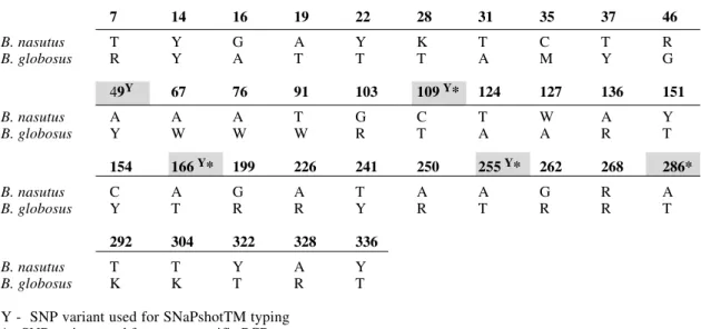
S. haematobium can be complex. Not all snail species act as intermediate hosts e.g. B. ugandae appears refractory to infection, or only certain snail species act as hosts in specific areas (Rollinson et al. 2001). Lack of clear-cut morphological characters hinders identification of natu-ral populations (Mandahl-Barth 1965). For accurate sepa-ration of these snail species it is necessary to use bio-chemical (Rollinson & Southgate 1979) or molecular DNA methods (Rollinson et al. 2001). There are many single nucleotide polymorphisms (SNPs) within the mitochon-drial cytochrome oxidase subunit I (COI) gene which may be exploited for species identification (Fig. 1). Taxon spe-cific polymerase chain reaction (PCR) primers for B.
Funded by the Wellcome Trust.
+Corresponding author. Fax + 44-0207-942-5518. E-mail:
d.rollinson@nhm.ac.uk Received 18 June 2002 Accepted 15 August 2002
globosus and B. nasutus have been designed based on genetic variation within the COI (Stothard et al. in press). Whilst taxon specific primers are highly discrimina-tory, the PCR assay is limited within the known scope of detected sequence variation; further sequence variation may lead to false negatives. SNaPshotTM is a commer-cially available product from PE Biosystems, UK for genotyping SNPs using a fluorescent based, primer ex-tension assay (Rollinson et al. 2001). Makridakis and Reichardt (2002) have taken the SNaPshotTM assay a step further by multiplexing SNaPshotTM primers of differing length coining the terminology ‘multiplex automated primer extension analysis’ (MAPA). Although this multiplex as-say requires a semi-automated DNA sequencer, the asas-say has certain key advantages; many snails can be individu-ally typed simultaneously for several key SNPs, and de-tection and precise characterisation of further DNA varia-tion is possible.
This paper reports on the development and implemen-tation of a multiplexed SNaPshotTM assay to type simul-taneously four SNPs within the COI (Fig. 2) of Bulinus
species. The assay is then used for identification of B. africanus group snails collected from 7 localities within coastal Kenya. The Kenyan shell material is compared to a selection of B. globosus and B. nasutus shells from Zan-zibar to ascertain if there is any morphological divergence.
MATERIALS AND METHODS
stan-dard methodology (Stothard et al. 1997) and the shell was retained for morphometric analysis. A collection of 79 dry shells as reported by Stothard et al. (1997) of B. africanus
group snails from Zanzibar [B. globosus Unguja (n = 18), Pemba (n = 22) and B. nasutus Unguja (n = 18), Pemba (n = 21)] was used as reference material for morphometric comparison with Kenyan material.
PCR and SNaPshotTM typing of COI - A subregion (~ 450 bp) of the COI was amplified from each snail follow-ing conditions described by Stothard and Rollinson (1997). The amplification product was then purified using a Qiagen PCR clean-up spin column and adjusted to an ap-proximate concentration of 0.2 pmol/µl.
Four SNaPshotTM primers were designed to type SNPs at positions 49, 109, 166 and 255 (Fig. 1) in the alignment of COI sequences described by Stothard and Rollinson (1997). The primer sequences were as follows:
Position 255 (29mer) 5’ – TAAAAAGAAAAATAAAYCC TAAYACTCAA
Position 166 (34mer) 5’ – ACACCTTAATTCCTGTTGG TACAGCAATAATTAT
Position 109 (38mer) 5’ – GCTCGAGTATCCACATCTATT CCHACAGTAAATATATG
Position 49 (45mer) 5’ – TAAACCTAAAATTCCAATT GAAACTATWGCATAAATTATTCCTAA
1µl of purified amplification product was added to a SNaPshotTM reaction mixture containing 0.15 pmols of each primer. The reaction mixture (10 µl) was incubated for 25 cycles of 96oC for 10 sec, 50oC for 5 sec and 60oC for 30 sec in a standard thermal cycler. The SNaPshotTM reaction mixture was purified by removal of excess fluo-rescent dye terminators by incubation with 1unit of calf intestinal alkaline phosphatase according to manu-facturer’s instructions.
1.5 µl of purified SNaPshotTM reaction was combined with 1.5 µl deionised formamide/blue dextran and the total
volume was loaded onto an ABI 377 machine using a stan-dard 5% long ranger, 6M urea sequencing gel. Electro-phoresis was conducted according to standard separa-tion condisepara-tions detailed in the PE Biosystems’ electro-phoresis manual. As the 4 SNaPshotTM primers were of differing length, upon separation by denaturing gel elec-trophoresis each SNP position could be assigned accord-ing to the order in which the now fluorescently labelled primers were separated (Fig. 2). The subsequent gel file was visually inspected and also processed with GeneScan software version 2.1 (PE Biosystems, UK).
DNA sequencing - In addition to SNaPshotTM analy-sis, the DNA sequence of the COI was determined for a total of 21 snails taken across the 7 sampling localities [Kinango (n = 7), Mbovu (n = 3), Msambweni (n = 3), Mazeras (n = 3), Nguzo (n = 1), Ramisi (n = 2) and Timboni (n = 2)] using direct DNA cycle sequencing of purified amplification product and separated on an ABI377 semi-automated DNA sequencer.
Morphometric analysis - A total of 226 snail shells were examined. Identification of Kenyan B. globosus and
B. nasutus was based upon the results of the SNaPshotTM COI profile. Digital images of the shells (aperture facing) at either x8 or x12 magnification were collected using a Leica MZ6 dissecting microscope with an attached digi-tal camera and DIC-E image capture software (World Pre-cision Instruments, UK). A 10 mm scale bar was included within each shell image for size calibration. Shell microsulpture was also noted according to descriptions given by Kristensen et al. (1987). Image analysis was con-ducted with SigmaScanPro 4.0 software package (Jandel Scientific, UK).
Nine measurements were taken for each shell: 5 linear, 3 area and 1 angular. These were, linear (mm) L: total length of shell, W: width of shell (perpendicular from ap-erture apex to opposite edge), WSp: basal width of shell
Fig. 1:a summary of thepresently known variation within the 340 bp sequenced COI region between Bulinus globosus and B. nasutus. For the original DNA alignment see Stothard and Rollinson (1997). The shaded variant positions are SNP variants utilised for SNaPshotTM [positions denoted Y (49, 109, 166, 255)] and (or) taxon specific PCR assays [positions denoted * (109, 166, 255, 286)].
7 14 16 19 22 28 31 35 37 46
B. nasutus T Y G A Y K T C T R
B. globosus R Y A T T T A M Y G
49Y 67 76 91 103 109 Y* 124 127 136 151
B. nasutus A A A T G C T W A Y
B. globosus Y W W W R T A A R T
154 166 Y* 199 226 241 250 255 Y* 262 268 286*
B. nasutus C A G A T A A G R A
B. globosus Y T R R Y R T R R T
292 304 322 328 336
B. nasutus T T Y A Y
B. globosus K K T R T
spire, LAp: length of aperture, WAp: width of aperture;
area (mm2), TA: total shell area, Asp: area of spire and AAp: area of aperture; and angular (degrees): angle sub-tended from longitudinal axis of shell rotation to maximum width point on the outer body whorl edge.
Before analysis, area measurements were transformed by taking the square root. All measurements were then standardised using the geometric mean of Log10 adjusted ratios following Clarke et al. (1999). Euclidean distances
Fig. 2: design of the multiplex SNaPshotTM assay for identification of Bulinus globosus and B. nasutus. In brief, after the gene target is amplified by PCR, a short oligonucleotide primer, or probe, is used to abut next to the variant position to be typed. The SNaPshotTM
primer can be of the same sequence as a taxon specific primer except that the 3’ terminal nucleotide is omitted. It is this nucle-otide only that is then added to the SNaPshotTM primer upon the primer extension reaction. A: snails are collected from the field; B: a subregion of the COI is amplified using standard PCR conditions; C: four SNPs are targeted within the COI which differentiate be-tween species; D: after the SNaPshotTM reaction the 4 SNaPshotTM primers, now one nucleotide longer and flourescently labelled, are separated by electrophoresis to reveal a characteristic coloured bar-code pattern typical for each species.
between each individual were calculated using the pro-gram SYN-TAX 5.0 (J Podani, Scientia Publishing, Budapest) and a dendrogram was generated using unweighted pair-group arithmetic average (UPGMA) analysis to evaluate phenetic groups.
RESULTS
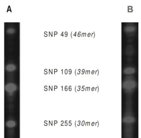
Identification and COI variation - SNaPshot TM COI reactions from all snails could be readily assigned to the expected profile for either species (Fig. 3), and no further variation within the 4 variant positions was detected. From the SNaPshot TM COI profile B. globosus was encoun-tered at Timboni and Mazeras while B. nasutus was en-countered at Mbouv, Msambweni, Nguzo and Ramisi. A mixed population of B. globosus and B. nasutus was found at Kinango. Of the 21 COI sequences obtained and in comparison to previous COI data from B. africanus group snails, 8 novel COI sequences were encountered. Three
B. globosus and 5 B. nasutus sequences have been de-posited in GenBank, accession numbers AF507035-AF507042. The majority of B. globosus from Kenya did not have a SspI restriction enzyme cutting site within the COI.
A
S N P 49 (4 6m er)
S N P 109 (3 9m er)
S N P 166 (3 5m er)
S N P 255 (3 0m er)
Fig. 3: SNaPshotTM gel profile of the Kenyan snails corresponded to the expected profiles for either Bulinus globosus or B. nasutus, no additional variation was encountered. A: profile typical of B. globosus. Note that the C/T polymorphism at position 49 within B. globosus from Zanzibar is absent within the Kenyan material. All snails possessed a C at this position; B: profile typical of B. nasutus.
Morphometric variation - The Table details the mean value with 1 standard deviation for shell measurements. A significant difference (p < 0.05) was detected between
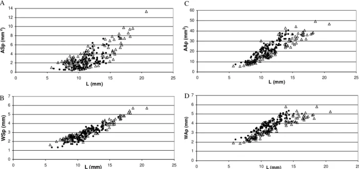
B. globosus and B. nasutus with an unpaired student t test for ASp and AAp. Generally, B. globosus had larger aperture and smaller spire areas than B. nasutus. Plotting a combination of shell variables in bivariate plots against shell length did not reveal the existence of two discrete distributions of points (Fig. 4).
UPGMA analysis of morphometric variation within Kenyan and Zanzibarian snails resulted in a dendrogram containing 8 major clusters, designated A to H (Fig. 5). The largest clusters, B and H, contained 64 and 65 snails respectively. No single cluster contained exclusively B. globosus or B. nasutus. Both species were distributed
B . glo bo s u s
B . na s u tu s
109 166 255
A S M IT1 49 A S M IT2
4 5 0 b p
B . g lo b o su s B . n a su tu s
E x tra c t D N A a n d P C R s u b re g io n o f C O I to o b ta in fra g m e n t
P e rfo rm m u ltip le x S N a P s h o t re a c tio n w ith fo u r D N A p ro b e s ta rg e tte d to s p e c ific S N P s a t p o s itio n s 4 9 , 1 0 9 , 1 6 6 & 2 5 5
S iz e fra c tio n a te e x te n s io n p ro d u c ts w ith in a n A B I 3 7 7 de n a tu rin g g e l, fo u r fra g m e n ts a re p ro d uc e d w ith d iffe rin g c o lou rs b etw e e n s p ec ies
S N P 4 9 (4 6 m e r)
S N P 1 09 (3 9 m e r)
S N P 1 66 (3 5 m e r)
S N P 2 55 (3 0m e r)
(b lue o r g re e n)
B . g lo b o su s B . n a su tu s A
B
C
D
S in g le s n a ils in a lc o h o l o b ta in e d fro m field c o lle c tio n s
quickly accomplished and as this technology requires minimal liquid handling steps, snails can be processed in microtitre plate format. This multiplex assay has great potential for regular, high-through-put DNA typing in a convenient single tube reaction. Multiplex SNaPshotTM TABLE
Shell measurements 5 linear (mm), 3 area (mm2) and 1 angular (o) with means and 1 standard deviation
Coastal Kenya Zanzibar
Bulinus globosus B. nasutus B. globosus B. nasutus
L 10.8 ± 1.9 11.7 ± 3.0 11.9 ± 1.9 11.5 ± 2.9 W 5.4 ± 1.1 5.7 ± 1.5 6.0 ± 1.1 5.5 ± 1.3 WSp 2.5 ± 0.6 3.1 ± 0.9 3.2 ± 0.6 3.2 ± 0.9 LAp 8.5 ± 1.4 8.6 ± 2.1 8.4 ± 1.3 8.1 ± 1.9 WAp 3.9 ± 0.8 3.6 ± 1.0 3.8 ± 0.7 3.3 ± 0.8 TA 60.0 ± 20.6 64.8 ± 32.5 65.1 ± 21.3 57.5 ± 29.8 AAp 23.7 ± 8.0 21.9 ± 11.0 22.9 ± 7.4 17.6 ± 8.9 ASp 1.5 ± 0.9 3.2 ± 2.1 3.6 ± 1.7 4.1 ± 2.4
28.5 ± 2.8 28.8 ± 2.7 29.9 ± 2.4 31.2 ± 3.2
0 1 2 3 4 5 6 7
0 5 10 L (mm) 15 20 25
WSp (mm)
0 2 4 6 8 10 12 14
0 5 10 15 20 25
L (mm)
ASp (mm
2)
0 10 20 30 40 50 60
0 5 10 15 20 25
L (mm)
AAp (mm
2)
0 1 2 3 4 5 6 7
0 5 10L (mm) 15 20 25
WAp (mm)
A C
B D
Fig. 4: bivariate plots of shell variation of Kenyan and Zanzibarian snails reveals no discernible groups between Bulinus globosus (•) and B. nasutus (r). A: area of spire against shell length; B: width of spire against shell length; C: area of aperture against shell length; D: width of aperture against shell length.
Fig. 5: cluster analysis of shell variation revealed 8 major groups labelled A to H. Within each group there were Bulinus globosus and B. nasutus intermingled as denoted by the adjacent table for each cluster. Whilst group B contained predominately B. globosus there was no discrete separation evidenced between species.
across clusters although the cluster B was predominately composed of Kenyan B. globosus. Following from the identification equations proposed by Kristensen et al. (1987) using presence of microsculpture and aperture band-ing, a specificity of 53% for identification of B. globosus
was found. Approximately 1 in 2 B. globosus snails would have been identified correctly within this sample.
DISCUSSION
Multiplex SNaPshotTM assay - The multiplex SNaPshotTM assay is a robust methodology for typing SNPs within the COI from individual snails. Analysis and interpretation of the resultant SNaPshotTM profile is less labour intensive than inspection of a corresponding DNA sequencing chromatogram as only 4 fragments are pro-duced (Fig. 2). Each characteristic colour bar-code is im-mediately apparent from the corresponding gel file pic-ture (Fig. 3). As such identification of many snails can be
4 0 3 0 2 0 1 0
A
B
C
D
E
F G
H
5 4 0 1 0
6 4 5 8 2 4 0
7 0 1 4 2
2 1 0 5 11 5
8 2 5 1 0 2 2 2 8 1 1 1
assays may also simultaneously screen for variation within other candidate genes e.g. ribosomal 18S and Internal Transcribed Spacer, and once sufficient DNA sequence data has been accrued for representatives of Bulinus, the assay could no doubt be extended. The methodology might also be of use for identification of problematic popu-lations of Lymnaea (Bargues et al. 1997)or Biomphalaria
(Morgan et al. 2001) or even in the detection of Schisto-soma sequences from infected snails.
Snail populations - Accurate identification of B. glo-bosus and B. nasutus populations is important. Recent work on Zanzibar has shown that B. nasutus plays no role in transmission of local S. haematobium and the distribu-tion of urinary schistosomiasis in schoolchildren is clearly linked with the distribution of B. globosus (Stothard et al. in press). A similar transmission picture might also exist in Kenyan and Tanzanian coastal regions nearby.
Both B. globosus and B. nasutus have been encoun-tered within the Kenyan material. While 6 of the 7 samples contained a single species, the sample from Kinango con-tained a mixed population of both species. This situation contrasts somewhat with Zanzibar where mixed popula-tions have yet to be encountered (Stothard et al. 1997). In Zanzibar, B. globosus appears to be associated with harder water while B. nasutus is associated with softer water but the range of water hardnesses between species does over-lap (Stothard et al. in press); potentially, mixed popula-tions could occur in nature. As water conductivity values were not recorded at the time of collection of this Kenyan material, it might be expected that water conductivity at Kinango could be within this overlapping range.
The DNA divergence within the COI between Kenyan and Zanzibarian species is confined to several further point mutations. It is worthy of note that the PCR-RFLP methodology designed by Stothard and Rollinson (1997) to differentiate B. globosus and B. nasutus in Zanzibar on the presence and absence of a SspI cutting site respec-tively (recognition sequence AATATT) does not work consistently on Kenyan B. globosus. Coastal Kenyan B. globosus has two predominant sequence types present within this selected 6 bp region: AATATT (SspI site) and GATATT (non-SspI site), while all B. nasutus have AATATA. Whilst this assay can positively identify cer-tain B. globosus by the presence of an SspI site, in this case the snails from Kinango, the absence of a SspI re-striction site cannot now be interpreted as exclusive to B. nasutus. Confident typing of the 3’ T in B. globosus and 3’ A in B. nasutus which differentiates the species, is best performed by SNaPshotTM or taxon specific PCR primers assays.
Shell morphology - It is known that the shell of B. africanus group snails from East Africa can be highly variable (Brown 1994). The problem was identified by Mandahl-Barth (1965) as long-spired and short-spired forms of either species ‘overlapped’ making separation of populations and individuals of either species problem-atic. Morphometric analysis presented here has confirmed that it is difficult to separate either species on shell char-acters alone. The bivariate plots are particularly illustra-tive of the overlapping nature of variation. Whilst the mean aperture area (AAp) of each species is significantly
different, for example aperture area is greater in B. globosus
than B. nasutus for a typical shell, this measurement is not capable of precisely differentiating species (Fig. 4).
Variation within other shell variables has been in-spected with the hope of finding clear-cut divisions be-tween species. Kristensen et al. (1987) developed a simple, powerful morphometric test which could differentiate B. africanus group species from the Lake Victoria region of Kenya. Using these equations, however, for the Kenyan and Zanzibarian material there is incongruence between molecular and morphological methods of identification. This has also been recently reported by Raahauge and Kristensen (2000) upon analysis of B. africanus group snails from Kisumu region Kenya. The presence and ab-sence of shell microsculpture has been used to differenti-ate B. nasutus and B. globosus respectively from Lake Victoria but upon inspection of further snails from differ-ent areas it seems that this character becomes less spe-cies specific. In Zanzibar shell microsculpture does not help to differentiate the species. Certain populations con-fidently typed by both genetic and biochemical methods as B. globosus do have microsulpture, as well as, aperture bands whereas others do not. Inspection of the clusters generated by UPGMA analysis of shell measurements (Fig. 5) adds further weight to the suggestion made by Stothard et al. (1997) that there may be few, if any, conchological measurement useful for reliable identifica-tion.
Conclusion - Correct identification of populations of
B. africanus group snails requires the use of biochemical or molecular DNA methods. The multiplex SNaPshotTM assay is a promising new tool for species identification. New data are required concerning the distribution and transmission status of potential intermediate hosts in East Africa in order to gain a fuller understanding of the distri-bution of urinary schistosomiasis.
REFERENCES
Bargues MD, Mangold AJ, Muñoz-Antoli C, Pointier JP, Mas-Coma S 1997. SSU rDNA characterisation of lymnaeid snails transmitting human fascioliasis in South and Central America. J Parasitol83: 1086-1092.
Brown DS 1994. Freshwater Snails of Africa and their Medical Importance, 2nd ed., Taylor & Francis Ltd., England, 609 pp.
Clarke RK, Grahame J, Mill PJ 1999. Variation and constraint in the shells of two sibling species of intertidal rough peri-winkles (Gastropoda: Littorina spp). J Zool Lond 247: 145-154.
Jones CS, Rollinson D, Mimpfoundi R, Ouma J, Kariuki HC 2001. Molecular evolution of freshwater intermediate snail hosts within the Bulinus forskalii group. Parasitology 123: S277-S292.
Kristensen TK, Frandsen F, Christensen AG 1987. Bulinus africanus-group snails in East and South East Africa, dif-ferentiated by use of biometric multivariate analysis on multivariate characters (Pulmonata: Planorbidae). Rev Zool Afric 101: 55-66.
Makridakis NM, Reichardt JKV 2002. Multiplex automated primer extension analysis: simultaneous genotyping of sev-eral polymorphisms. Biotechniques31: 1374-1380. Mandahl-Barth G 1965. The species of Bulinus, intermediate
Morgan JAT, DeJong RJ, Synder S, Mkoji GM, Loker ES 2001.
Schistosoma mansoni and Biomphalaria: past history and future trends. Parasitology123: S211-S228.
Raahauge P, Kristensen TK 2000. A comparison of Bulinus africanus group species (Planorbidae: Gastropoda) by the use of the internal transcribed spacer 1 region combined by mor-phological and anatomical characters. Acta Trop 75: 85-94. Rollinson D, Southgate VR 1979. Enzyme analyses of Bulinus
africanus group snails (Mollusca: Planorbidae) from Tan-zania. Trans R Soc Trop Med Hyg 73: 667-672.
Rollinson D, Stothard JR, Southgate VR 2001. Interactions between intermediate snail hosts of the genus Bulinus and schistosomes of the Schistosoma haematobium group. Para-sitology123: S245-S260.
Stothard JR, Rollinson D 1997. Partial DNA sequences from the mitochondrial cytochrome oxidase subunit I (COI) gene
can differentiate the intermediate snail hosts Bulinus globosus and B. nasutus (Gastropoda: Planorbidae). J Nat Hist31: 727-737.
Stothard JR, Brémond P, Andriamaro L, Sellin B, Sellin E, Rollinson D 2001. Bulinus species on Madagascar: molecu-lar evolution, genetic markers and compatibility with Schis-tosoma haematobium. Parasitology 123: S261-S275. Stothard JR, Mgeni AF, Alawi KS, Savioli L, Rollinson D 1997.
Observations on shell morphology, enzymes and random amplified polymorphic DNA (RAPD) in Bulinusafricanus
group snails (Gastropoda: Planorbidae) in Zanzibar. J Moll Stud63: 489-503.
Stothard JR, Mgeni AF, Khamis S, Seto E, Ramsan M, Hubbard SJ, Kristensen TK, Rollinson D. New insights into the trans-mission biology of urinary schistosomiasis in Zanzibar.


