online | memorias.ioc.fiocruz.br
Resurrection of
Anopheles goeldii
from synonymy with
Anopheles nuneztovari
(Diptera, Culicidae) and a new record for
Anopheles dunhami
in the Brazilian Amazon
Daniéla C Calado, Peter G Foster1, Eduardo S Bergo2, Cecília LS dos Santos/3, Allan Kardec R Galardo4, Maria Anice Mureb Sallum/+
Departamento de Epidemiologia, Faculdade de Saúde Pública, Universidade de São Paulo, Avenida Dr. Arnaldo 715, 01246-904 São Paulo, SP, Brasil 1Department of Zoology, Natural History Museum, London, United Kingdom 2Superintendência de Controle de
Endemias, Secretaria de Estado da Saúde de São Paulo, Araraquara, SP, Brasil 3Instituto Adolfo Lutz, Secretaria de Estado da Saúde de
São Paulo, São Paulo, SP, Brasil 4Instituto de Pesquisas Científicas e Tecnológicas do Estado do Amapá, Macapá, AP, Brasil
Nucleotide sequences of theinternal transcribed spacer 2 (ITS2) rDNA and partial sequences of the cytochrome c oxidase subunit I (COI) mtDNA and white gene nDNA were obtained from specimens of Anopheles nuneztovari A collected in Macapá (state of Amapá), Óbidos, Prainha and Almeirim (state of Pará), Itacoatiara and Parintins (state of Amazonas), Brazil, and compared with previously published sequences of A. nuneztovari s.l. Results of the Bayes-ian phylogenetic analyses performed using either COI or combined ITS2, COI and white gene sequences suggest that
An. nuneztovari B/C is distinct from specimens obtained in the Amazonas/Solimões River basin. Anopheles goeldii, currently in synonymy with An. nuneztovari, was described from individuals collected in Belterra (= Fordlândia) in the Tapajós River, state of Pará, Southern Amazonas River. Morphological comparisons of the characteristics of the male genitalia indicated that An. nuneztovari A and An. goeldii are similar but distinct from An. nuneztovariB/C by the apex of the aedeagus. In considering the results of the phylogenetic analyses and morphological comparisons,
An. goeldii is resurrected from synonymy with An. nuneztovari. Additionally, Anopheles dunhamiis reported for the first time in Parintins. This species can be distinguished from An. goeldiiby characters of the male genitalia and molecular data.
Key words: Anopheles - Nyssorhynchus - ITS2 - COI - white gene - Bayesian phylogeny
Anopheles nuneztovaris.l. was recorded from western Panama to northern South America (Faran 1980). This taxon is a primary vector of human Plasmodium spp. in Venezuela and Colombia (Gabaldón et al. 1975), and in the Brazilian Amazon An. nuneztovari s.l. was reported infected with Plasmodium spp. in states of Pará (Arruda et al. 1986, Póvoa et al.2001) and Amapá (Galardo et al. 2007). Additionally, specimens collected in Pará were found infected with Tucuruí, Caraipé and Arumateua arboviruses of the Anopheles A serogroup, Bunyavirus, Bunyaviridae (Travassos da Rosa et al. 1992).
Chromosomal, molecular, morphological and behav-ioral polymorphisms have been observed in An. nunezto-vari, supporting the hypothesis of a species complex (El-liot 1972, Kitzmiller et al. 1973, Panday 1977, Steiner et al.1980, Fritz et al. 1994, Hribar 1994, Linley et. al 1996, Conn et al. 1998, Tadei et al.1998, Scarpassa et al. 1999, 2000). Conn (1990) and Conn et al. (1993) described three chromosomal cytotypes in An. nuneztovari and suggested that cytotype A occurs in Brazil, cytotype B in Western
Financial support: FAPESP (Grant 05/53973-0, to MAMS), CNPq (Grant 472485/2006-7, to MAMS), NIH AI RO154139 (to Jan E Conn). + Corresponding author: masallum@usp.br
Received 5 July 2008 Accepted 19 November 2008
Venezuela, southeast of the Andes Mountains and cyto-type C in North Colombia and Western Venezuela, north-east of the Andes Mountains. When using the white gene sequence data, Mirabello and Conn (2008) found three sympatric lineages in the Brazilian Amazon.
Hribar (1994), while comparing the male genitalia of specimens of An. nuneztovari A, B and C, suggested that the aedeagus of An. nuneztovari A was similar to that of Anopheles goeldii illustrated by Rozeboom and Gabaldón (1941) (plate III, 3). An. goeldii was described from males, females, larvae and eggs collected in Bel-terra (= Fordlândia), state of Pará, Brazil (Rozeboom & Gabaldón 1941). However, based on specimens obtained in Guiana, Floch and Abonnenc (1946) synonymized
male genitalia of An. nuneztovari from Amapá did not agree with the illustrations reported in the published lit-erature from Venezuela (Cojedes, type-locality) and that they are similar to those represented in the description of
An. goeldii (plate III, 3).
In this study, we analyzed specimens collected in several localities in states of Amapá, Pará and Amazo-nas (Brazil) using sequence data from the rDNA second internal transcribed spacer 2 (ITS2), a fragment of the single copy nuclear white gene and a fragment of the cy-tochrome c oxidase subunit I (COI) of the mitochondrial genome. These markers were employed (i) to examine the taxonomic statusof An. nuneztovari A that occurs along the Solimões/Amazonas River basin and (ii) to test the hypothesis that An. goeldii is a valid species.
MATERIALS AND METHODS
Mosquito collection - Larvae were collected in ground-water habitats and reared to the adult stage to obtain adults linked with larval and pupal exuviae and male genitalia. Vouchers consisting of larval and/or pupal exuviae and dissected male genitalia mounted in Canada balsam on microscope slides are deposited in the Entomological Collection of Faculdade de Saúde Pública, Universidade de São Paulo, Brazil (FSP-USP). Details about field collections are in Table I.
DNA extraction - DNA was isolated using the pro-tocol described by Wilkerson et al. (1993), with minor modifications. Phenol:chlorophorm:isoamyl alcohol and chlorophorm:isoamyl alcohol extractions were conducted using Phase Lock Gel (Eppendorf®) following the
manu-facturer’s instructions. Template DNA from this study is retained dry at -70oC in the FSP-USP for future reference.
Amplification and sequencing - ITS2 PCR products were amplified in a 25 µL reaction mix containing: 1 X PCR buffer (20 mM Tris-HCl, 10 mM [NH4]2SO4, 10 mM KCl, 2 mM MgSO4, 0.1% Triton X -100), 0.1 mM of each dNTP (Eppendorf®), 5 picomoles of each primer and
0.5 U of Taq DNA Polymerase with ThermoPol Buffer (New England BioLabs® Inc). One µL of genomic DNA
(1/800th) was used in each PCR reaction. The PCR am-plification of the ITS2 region was carried out using the
5.8SF (5’ - ATC ACT CGG CTC GTG GAT CG -3’) and 28SR (5-’ ATG CTT AAA TTT AGG GGG TAG TC-3’) primers (Djadid et al. 2007). PCR amplification consisted of 2 min denaturation at 94oC, 40 cycles at 94oC, 60oC and
72oC for 30 sec, followed by a 10 min extension at 72oC.
PCR products were electrophoresed in 1% TBE agarose gels stained with ethidium bromide. PCR products were purified using PEG precipitation (20% polyethylene gly-col 8000/2.5 M NaCl). Two specimens were successfully directly sequenced; 18 were cloned into pGem-T Easy Vector (Promega, Madison, WI, USA). Two to four posi-tive clones per sample, yielding a total of 62 clones, were used in sequencing reactions performed with the ITS2 primer set. For analysis, the partial sequencesof the 5.8S and 28S rRNA gene regions were excluded.
COI fragments of ~ 500 base pairs were amplified using the primers C1-J-1718 (5’ - GGA GGA TTT GGA AAT TGA TTA GTT CC-3’) and C1-N-2191 (5’ - CCC GGT AAA ATT AAA ATA TAA ACT TC -3’) (Simon et al. 1994). PCR reactions were carried out in a 50-µL reaction mix containing: 1 X PCR buffer, 0.2 mM dNTP (Eppendorf®), 10 picomoles of each primer, 1 U of Taq
DNA Polymerase with Thermo Pol Buffer (New Eng-land BioLabs® Inc) and 1 µL of DNA template (1/100th).
PCR amplification protocol consisted of 2 min denatura-tion at 95ºC, 5 cycles at 94ºC for 40 sec, 37ºC for 40 sec, and 72ºC for 40 sec, 35 cycles at 94ºC for 40 sec, 48ºC for 40 sec, and 72ºC for 1 min, followed by a final exten-sion at 72oC for 7 min.
White gene fragments of ~ 600 bp were amplified using WZ2E (5’ - AAY TAY AAY CCI GCI GAY TTY TA - 3’) and WZ11X (5’ - TTI ARR AAR AAI CCI CCR AA - 3’) (Besansky & Fahey 1997). PCR amplicons were used as template in a second PCR using the inter-nal primers W1F (5’ - GAT CAA RAA GAT CTG YGA CTC GTT- 3’) and W2R (5’ - GCC ATC GAG ATG GAG GAG CTG - 3’) (designed for the study by MAM Sallum). The first PCR reactions were conducted in a 10 µL re-action mix containing: 1 X PCR buffer, 0.2 mM dNTP (Eppendorf®), 10 picomoles of each primer and 0.4 U Taq
DNA Polymerase with Thermo Pol Buffer (New England BioLabs® Inc.). One µL of genomic DNA (1/1000th) was
used per PCR reaction. For the second PCR, 1 µL of the
TABLE I
Specimen codes, localities, geographical coordinates and species studied
Specimens Locality Coordinates Species
AP20-104, AP15-115 Amapá, Macapá 0º16’17.5’’N 50º53’53.3’’W Anopheles goeldii
BRAM03-01, BRAM06-04 Amazonas, Itacotiara 3º08’48.3’’S 58º23’39.7’’W BRAM7-101, BRAM7-103 Amazonas, Itacotiara 2º54’56.1’’S 59º02’91.7’’W BRAM12-106 Amazonas, Parintins 2º38’21.7’’S 56º39’0.96’’W BRAM13-08 Amazonas, Parintins 2º38’66.2’’S 56º38’29.6’’W BRAM14-03, BRAM14-05,
BRAM15-05, BRAM16-01 Pará, Óbidos 2o32’31.9’’S 57o45’36.0’’W
BRAM22-01, BRAM22-101 Pará, Prainha 2º04’49.3’’S 53º35’27.1’’W BRAM25-01, BRAM25-07 Pará, Almeirim 1º28’34.1’’S 52º44’35.4’’W BRAM13-06, BRAM13-07,
products of the first PCR was employed, using the same protocol in 25 µL. PCR amplification protocol consisted of a cycle at 94ºC for 5 min, 40 cycles at 94ºC for 30 sec, 52oC for 30 sec, and 72ºC for 60 sec, followed by a final
extension at 72ºC for 10 min. For the second PCR, the profile was similar except for the annealing temperature for which we adopted 55oC for 30 sec.
Sequencing reactions were carried out in both direc-tions using the PCR primers. Big Dye Terminator Kit v.3.1 (PE Applied Biosystems, Warrington, England) was employed for those specimens that were electrophorezed in an ABI Prism 3100, Avant Genetic Analyzer (Applied Biosystems, Foster City, CA, USA), or ABI 3130xl Ge-netic Analyzer, or ABI 377 Sequencer. For those speci-mens processed in the MegaBACE™ DNA Analysis Systems 1000 (GE Healthcare), we used DYEnamic™ ET Terminator Kit (with Thermo Sequenase™ II DNA Polimerase). The sequences have been deposited in Gen-Bank (COI accession numbers EU848313 - EU848336; ITS2 accession numbers EU848337 - EU848400; white
gene accession numbers EU848401 - EU848416)
Sequence alignment - Nucleotide sequences were ed-ited using either Chromas Lite version 2.01 (Tecnelysi-um Pty Ltd. 2007) or BioEdit version 7.0.5.3 (Hall 1999). Sequences alignments were performed in BioEdit and adjusted by visual inspection using MacClade version 4.0 (Maddison & Maddison 2000). The ITS2 length was inferred using sequences from Fritz et al.(1994), but the nucleotide positions differ due to intragenomic and in-terspecific polymorphisms. Accuracy of nucleotide se-quence alignments (COI and white gene) was examined using amino acid sequence alignment. White gene intron position was determined based on the Anopheles albi-manus white gene sequence (U73839). Sequence simi-larities were assessed using FASTA search (http://www. ncbi.nlm.nih.gov/BLAST/).
Sequence analysis - Intraspecific sequence differ-entiation was assessed using mean uncorrected P dis-tance in Phylogenetic Analysis Using Parsimony (PAUP) (Swofford 2003). Genetic distance under the Kimura-2-parameter model implemented in PAUP was employed to examine COI sequences. ITS2 consensus sequences from clones of the same individual were generated in BioEdit program and used for Bayesian phylogenetic analysis.
Phylogenetic analysis - Modeltest version 3.7 (Pos-ada & Crandall 1998) was employed to choose a mod-el using the Akaike Information Criterion. MrBayes v.3.1.2 (Ronquist & Huelsenbeck 2003) was employed for Bayesian phylogeny. The model was chosen for each gene separately. Constant sites were removed because they have no effect on the topology. Duplicate sequences were also removed. Combined analysis (ITS2, partial COI, and partial white gene) used a model partitioned by gene, with free partition rates. To compare An. nunezto-vari A, B/C and our specimens, sequences of ITS2 and COI of An. nuneztovari from Colombia were download-ed from GenBank and designatdownload-ed as follows: Guaramito 1 (AY028083, AF368094), Guaramito 2 (AY028082, AF368089), Choco 1 (AY028121, AF368078),
Sitronel-la 1 (AY028095, AF368104), SitronelSitronel-la 2 (AY028094, AF368102), Tibu 1(AY028097, AF368106) and Tibu 2 (AY028108, AF368115). As outgroups, we used se-quences of An. dunhami.
RESULTS
ITS2 rDNA - From 20 specimens analyzed, only BRAM 13-106 and BRAM13-06 (both An. dunhami) were directly sequenced and showed 100% similar-ity. Specimens of An. dunhami had similar sequences, differing in tandem repeats regions and in a few sites that were individual-specific. The remaining samples, including two specimens of An. dunhami, showed su-perimposed chromatogram peaks due to intragenomic variation, especially in two GA repeats at positions 274-291 and 352-361.
In An. nuneztovari A, ITS2 length ranged from 358 to 373 bp, and in some individuals the difference among clones was 10 bp long. A large number of variants were observed for each specimen. Differences in fragment size across clones were mainly due to insertions/dele-tions in dinucleotide tandem repeat regions. The num-ber of these GA repeats at positions 274-291 was neither specific to any morphological form nor associated with geographical location. Rare single base polymorphisms in otherwise highly conserved regions may have been cloning artifacts. Pairwise distance between clones, the number of clones sequenced and the variants found from each individual are shown in Table II. Due to in-tragenomic polymorphisms, consensus sequences from ITS2 were generated for our specimens and these were compared to sequences of An. nuneztovari s.l. from Brazil, Colombia, Venezuela, Bolivia and Suriname, published by Fritz et al. (1994) and Sierra et al. (2004), and sequences available in GenBank [U92350, U92351, U925343 (Danoff-Burg & Conn 1997, unpublished)]. No fixed ITS2 differences were observed among cytotypes A, B and C. All polymorphisms observed represent in-traspecific variation because they were detected among the clones generated from a single individuals (Fig. 1). Sequence differences among An. nuneztovari A and An. dunhami are shown in Fig. 2.
COI mtDNA - We compared sequences of 493 bp generated for 16 individuals of An. nuneztovari A, from which 11 haplotypes were found. Under the Kimura two-parameter model, the genetic distance among the speci-mens of An. nuneztovari A varied from 0-0.02490 (the highest distances were detected among BRAM7-103 and BRAM13-08; BRAM16-1 and BRAM7-103/BRAM15-5; BRAM15-5 and BRAM13-08), whereas among An. nuneztovari B/C (Colombian specimens) distances ranged from 0-0.00407. By comparing the haplotypes of
Nuclear white gene - A 606-630 bp fragment of the
white gene flanking the fourth intron was analyzed. In-tron IV was present in all specimens and variation in this region was responsible for the length differences among sequences. Among An. nuneztovari A, four hap-lotypes were detected, three haphap-lotypes were unique and the fourth haplotype was observed in the remaining 13 individuals. Sequences generated from specimens of
An. dunhami showed poly C and T. In An. nuneztovari
A, homopolymers were observed, but the number of re-peats was lower. In these individuals, the intron region was 85-88 bp in length, whereas in An. dunhami it was 92-106 bp. Few polymorphisms were observed in exons III and IV; most were detected between An. nuneztovari
A and An. dunhami. In An. nuneztovari A, substitution occurred in four positions in the intron and three in the exon regions, in which all substitution represented syn-onymous mutations.
Phylogenetic analyses - Because there were few in-formative sites, the topologies generated from a single marker showed little resolution either under the parsi-mony criterion (not shown) or Bayesian approach. Con-sequently, Bayesian analysis was conducted using the COI data partition, as it was the most informative of the data, and combined data sets (ITS2, COI and white
gene). Alignment of the COI was 493 bp; of these only 27 (5.47%) sites were variable and 22 (4.46%) were parsi-mony informative. For Bayesian analysis, a GTR model with no among-site rate variation was used. Results of the phylogenetic analyses using COI data set showed that all specimens from Colombia are separated from specimens from the Amazonas/Solimões River basin;
however, the split leading to Colombian (An. nunezto-vari B/C) and Brazilian (An. nuneztovari A) clusters was only moderately supported (Fig. 3).
Bayesian phylogenetic analysis for the combined data (73 variable sites) was carried out using a partitioned model (COI: GTR, white: GTR, ITS2: JC) with no among-site heterogeneity but with free partition rates. Except for AF461749 (from Acre, Brazil), all Brazilian and Surinam-ese (Borokopondo) specimens clustered together in a dis-tinct clade from Colombian (Tibu, Guaramito, Sitronella and Choco), Venezuelan (Zulia and Barinas) and Bolivian (Beni) specimens. Bayesian probabilities are shown in Fig. 4. In all analyses, An. dunhami sequences clustered together in a strongly supported group.
DISCUSSION
The ITS2 has been employed for identification of
Anopheles species. However, comparisons between An. nuneztovari A from the Amazonas/Solimões River basin and published data from the literature of An. nuneztovari
A, B and C revealed no fixed difference among the Bra-zilian and Colombian/Venezuelan populations.
Sierra et al. (2004) showed that specimens of An. nuneztovari A from Suriname and Brazil differ from those of cytotypes B/C by divergences in number of GA repeats in the second microsatellite region, and a G > T transversion at positions 356-361. We found that the number of GA repeats is variable in the populations of
An. nuneztovari A from the Solimões/Amazon River ba-sin but always fewer than five. In Colombian/Venezu-elan sequences generated by Fritz et al.(1994) and Sierra et al. (2004), the number of GA repeats was always five.
TABLE II
Sequence variability of ITS2 among clones from the same individual
Specimens N of clones sequenced (variants) ITS2 length Mean uncorrected pdistance
AP20-104 4 (3) 369-373 0
AP15-105 5 (3) 361-369 0.0027-0.0081 BRAM03-01 3 (3) 361-371 0.0026-0.0080
BRAM06-04 4 (2) 361-369 0-0.0054
BRAM07-101 3 (3) 361 0.0027-0.0083
BRAM07-103 3 (3) 359-361 0.0027-0.0082
BRAM12-106 2 (2) 369-371 0
BRAM14-03 3 (3) 361-371 0.0027-0.0080
BRAM14-05 2 (2) 369 0.0081
BRAM15-05 3 (3) 361-369 0.0054-0.0081
BRAM16-01 3 (2) 361-369 0-0.0083
BRAM22-01 5 (5) 361-371 0.0027-0.0138
BRAM22-101 4 (4) 361-369 0-0.0054
BRAM25-01 5 (2) 369 0-0.0027
BRAM25-07 3 (3) 369-371 0-0.0054
BRAM13-113 4 (3) 358-360 0-0.0055
BRAM13-07 3 (3) 358-360 0.0028-0.0055
BRAM13-08 3 (2) 361-369 0-0.0054
BRAM13-06 Directly sequenced 360
-Fig. 1: partial internal transcribed spacer 2 sequence alignment of specimens of Anopheles goeldii from Brazil and Surinam, and Anopheles nuneztovari from Bolivia, Colombia and Venezuela. FBeni, FBrokopondo, FPuraquequara 1, FPuraquequara 2 and FCapanema sequences were retrieved from Fritz et al. (1994); AY028121, AY028125, AF461749, U92343, U92350 and U92351 were downloaded from GenBank.
In our analyses, the G > T transversion was both vari-able among clones obtained from a single individual and found in 12 out of 16 individuals of An. nuneztovari A. Additionally, several clones showed a polymorphic A insertion after the G > T transversion in specimens with 3 GAs. The A insertion was absent in specimens from Suriname and Brazil published by Fritz et al. (1994), but was observed by Onyabe and Conn (1999) in other Brazilian populations. These authors also showed that T (transversion) + A (insertion) is more frequent in speci-mens from states of Pará and Amazonas than in those from state of Rondônia (Brazil). In fact, T + A was fre-quent in our clones, and it is possible that the Rondônia population studied by Onyabe and Conn (1999) repre-sents a distinct lineage as suggested by Mirabello and Conn (2008). Although our results show that the ITS2 is not a good marker for separating An. nuneztovari cyto-types A, B and C, mainly due to intragenomic polymor-phisms, differences in heterogeneity can be indicative of distinct cryptic species (Onyabe & Conn 1999). Intrag-enomic polymorphisms were reported for An. nunezto-vari by Fritz et al.(1994) in individuals from Boa Vista, state of Roraima, and Puraquequara, state of Amazonas
Fig. 3: bayesian topology generated under the GTR model employing the partial cytochrome c oxidase subunit I sequences of An. dunhami, An. goeldii from Brazil and An. nuneztovari from Colombia and Ven-ezuela. Numbers on each branch represent posterior probabilities.
Fig. 4: bayesian topology generated under the GTR model employ-ing the combined internal transcribed spacer 2, partial cytochrome c oxidase subunit I and white gene sequence data of An. dunhami, An. goeldii from Brazil and Surinam, and An. nuneztovari from Bo-livia, Colombia and Venezuela. Numbers on each branch represent posterior probabilities.
(Brazil), and Suriname. Later, Onyabe and Conn (1999) observed less intragenomic heterogeneity in Colombian/ Venezuelan specimens than in Brazilian populations.
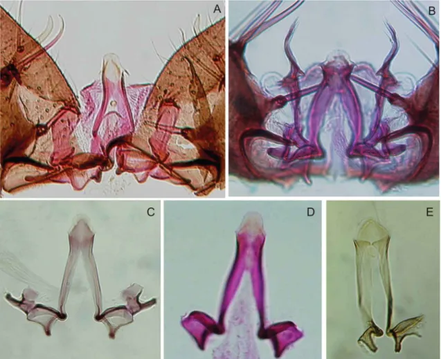
Fig. 5: details of male genitalia. A: Anopheles nuneztovari (Venezuela, image preparation #1353 WRBU); B: Anopheles goeldii (holotype); C: An. goeldii (paratype, image preparation #1356 WRBU); D: An. goeldii (Amapá, Brazil); E: Anopheles dunhami (lectotype number #58031WRBU).
from Venezuela and Colombia and thus may represent a different species.
Results of the phylogenetic analyses using the COI data set indicated that An. nuneztovari A is a different species than An. nuneztovari B/C. Similar results were obtained in the analyses using the combined data, where all Brazil-ian specimens, except for AF46174, clustered separately from Colombian and Venezuela specimens, indicating that An. nuneztovari A is not conspecific with B/C.
Morphological polymorphisms in male genitalia have been reported for An. nuneztovari s.l. and have raised questions about the taxonomic status of some morpho-logical forms, especially those from Brazil and Venezu-ela (Fig. 5A-E). In considering the description of the An. nuneztovari holotype by Savage (1986) and the illustra-tion of this species by Gabaldón (1940), we observed that the male genitalia of specimens of An. nuneztovari A collected in the Amazonas/Solimões River basin (Fig. 5A) differ from those of An. nuneztovari s.s. (Fig. 5D) in the characteristics of the apex of the aedeagus, but are similar to those of An. goeldii (Fig. 5C). By comparing genitalia from our specimens with the holotype and one paratype of An. goeldii, we observed that An. nunezto-vari A is morphologically more similar to the paratype
(Fig. 5C) than to the holotype (Fig. 5B). These differ-ences in the aedeagal apex of the holotype and paratype may represent morphological polymorphism within An. goeldii, not representative of distinct species as suggest-ed by Bergo et al. (2007). Considering the differences observed among DNA sequences and based on charac-ters observed in the male genitalia, we hereby propose the resurrection of An. goeldii from synonymy with An. nuneztovari.
positions 305-318 in U92326. We assume it is most likely a sequencing error. Other polymorphisms among our se-quences and U92326 (Lounibos et al.1998) are shown in Fig. 2 and may represent either individual variation or a cloning artifact. In considering male genitalia characters, the aedeagal leaflets are absent and the apex is some-what rounded in specimens 06, BRAM13-106 and BRAM13-07, whereas in BRAM13-113 (Bergo et al. 2007) (Fig. 1C, D) the leaflets are short, poorly sclerotized, and the apex of the aedeagus is somewhat conical. The results of this study indicate that the male genitalia of An. dunhami exhibit morphological poly-morphisms that should be taken into consideration for species identification. An. dunhami was reported in Tefé and Tabatinga (5º47’22S 65º24’32W, Amazonas, Brazil) (Lounibos et al. 1998), both localities in the Solimões River basin. This is the first record of An. dunhami from Parintins municipality along the Amazonas River. In the adult stage, An. nuneztovari and An.dunhami can be distinguished by the shape of the apical part of the aedeagus (Peyton 1993), but it remains difficult to iden-tify adult females. Morphological similarities between these species can be responsible for misidentifications and consequently mixture of information about genetics, vector competence, biting behavioral and distribution of these taxa.
ACKNOWLEDGEMENTS
To Richard C Wilkerson and J Stoffer (WRBU, USA), for the microphotos of the male genitalia of the holotype of An. goeldii, paratype of An. goeldii, lectotype of An. dunhami and non-type of An. nuneztovari, and to Henry Rupp kindly, re-viewed the English.
REFERENCES
Arruda M, Carvalho MB, Nussenzweig RS, Ferreira AW, Cochrane AH 1986. Potential vectors of malaria and their different sus-ceptibility to Plasmodium falciparum and Plasmodium vivax in Northern Brazil identified by immunoassay. Am J Trop Med Hyg 35: 873-881.
Bergo ES, Souto RNP, Galardo AKR, Nagaki SS, Calado DC, Sal-lum MAM 2007. Systematic notes on Anopheles Meigen (Dip-tera: Culicidae) species in the state of Amapá, Brazil. Mem Inst Oswaldo Cruz 102: 373-376.
Besansky NJ, Fahey GT 1997. Utility of the white gene in estimating phylogenetic relationships among mosquitoes. Mol Biol Evol 14: 442-454.
Conn JE 1990. A genetic study of the malaria vector Anopheles nuneztovari from Western Venezuela and Colombia. J Am Mosq Control Assoc 6: 400-405.
Conn JE, Mitchell SE, Cockburn AF 1998. Mitochondrial DNA analysis of the neotropical malaria vector Anopheles nuneztovari. Genome 41: 313-327.
Conn JE, Puertas YR, Seawright J 1993. A new cytotype of Ano-pheles nuneztovari from Western Venezuela and Colombia. J Am Mosq Control Assoc 9: 294-301.
Djadid ND, Gholizadeh S, Tafsiri E, Romi R, Gordeev M, Zakeri S 2007. Molecular identification of palearctic members of Ano-pheles maculipennis in Northern Iran. Malar J 17: 6.
Elliott R 1972. The influence of vector behaviour on malaria trans-mission. Am J Trop Med Hyg 21: 755-763.
Faran ME 1980. Mosquito studies (Diptera, Culicidae) XXXIV. A re-vision of the Albimanus section of the subgenus Nysorhynchus of Anopheles. Contrib Amer Entomol Inst 15: 1-215.
Floch H, Abonnenc E 1946. Sur A. nunez-tovari et A. pessoai en Guyane Francaise. Table d’identification des Nyssorhynchus guyanais. Inst Pasteur Guyane Territ Inini Publ 126: 1-5.
Fritz GN, Conn J, Cockburn AF, Seawright JA 1994. Sequence ana-lysis of the ribosomal DNA internal transcribed spacer 2 from populations of Anopheles nuneztovari (Diptera: Culicidae). Mol Biol Evol 11: 406-416.
Gabaldón A 1940. Estudios sobre anofelinos. Serie I. 1. Descripcion de Anopheles (Nyssorhynchus) nunez-tovari n. sp. y considera-ciones sobre una subdivision del grupo Nyssorhynchus (Diptera: Culicidae). Publ Div Malar 5: 3-7.
Gabaldón A 1981. Anopheles nuñez-tovari: importante vector y agente de malaria refractaria en Venezuela. Bol Dir Malaria San Amb 21: 28-38.
Gabaldón A, Martin GG, Sifontes R 1975. Necesidades en el campo de la investigacion del Programa Nacional de Eradicacion y Control de la Malaria de Venezuela. Bol Dir Malariol San Amb 15: 263-285.
Galardo AKR, Arruda M, Couto AARA, Wirtz R, Lounibos P, Zim-mermam R 2007. Malaria vector incrimination in three rural riv-erine villages in the Brazilian Amazon. Am J Trop Med Hyg 76: 461-469.
Hall TA 1999. BioEdit: a user-friendly biological sequence alignment editor and analysis program for Windows 95/98/NT. Nucl Acids Symp Ser 41: 95-98.
Hribar L 1994. Geographical variation of male genitalia of Anopheles nuneztovari Gabaldón. Mosq Syst 26: 132-144.
Kitzmiller JB, Kreutzer RD, Tallaferro E 1973. Chromosomal differ-ences in populations of Anopheles nuneztovari. Bull WHO 48: 435-445.
Lane J 1953. Neotropical Culicidae, Vol. 1, Editora da Universidade de São Paulo, São Paulo, 548 pp.
Linley JR, Lounibos LP, Conn JE, Duzak D, Nishimura N 1996. A description and morphometric comparison of eggs from eight geographic populations of the South American malaria vector Anopheles (Nyssorhynchus) nuneztovari. J Am Mosq Control Assoc 12: 275-292.
Lounibos LP, Wilkerson RC, Conn JE, Hribar LJ, Fritz GN, Danoff-Burg JA 1998. Morphological, molecular and chromosomal dis-crimination of cryptic Anopheles (Nyssorhynchus) (Diptera: Cu-licidae) from South America. J Med Entomol 35: 830-838.
Maddison WP, Maddison DR 2000.MacClade: analysis of phylogeny and character evolution,Version 4.0., Sinauer, Sunderland, MA.
Marrelli MT, Sallum MA, Marinotti O 2006. The second internal transcribed spacer of nuclear ribosomal DNA as a tool for Lat-in American anophelLat-ine taxonomy: a critical review. Mem Inst Oswaldo Cruz 101: 817-832.
Mirabello L, Conn JE 2008. Population analysis using the nuclear white gene detects Pliocene/Pleistocene lineage divergence with-in Anopheles nuneztovari with-in South America. Med Vet Entomol 22: 109-119.
Onyabe DY, Conn JE 1999. Intragenomic heterogeneity of a riboso-mal DNA spacer (ITS2) varies regionally in the neotropical ma-laria vector Anopheles nuneztovari (Diptera: Culicidae). Insect Mol Biol 8: 435-442.
Peyton EL 1993. Anopheles (Nyssorhynchus) dunhami, resurrected from synonymy with Anopheles nuneztovari and validated as a senior synonym of Anopheles trinkae (Diptera: Culicidae). Mosq Syst 25: 151-156.
Posada D, Crandall KA 1998. MODELTEST: testing the model of DNA substitution. Bioinformatics 14: 817-818.
Posso CE, González R, Cárdenas H, Gallego G, Duque MC, Suarez MF 2003. Random Amplified Polymorphic DNA Analysis of Anopheles nuneztovari (Diptera: Culicidae) from Western and Northeastern Colombia.Mem Inst Oswaldo Cruz 98: 469-476.
Póvoa MM, Wirtz RA, Lacerda RNL, Miles MA, Warhurst D 2001. Malaria Vectors in the municipality of Serra do Navio, state of Amapá, Amazon Region, Brazil. Mem Inst Oswaldo Cruz 96: 179-184.
Ronquist F, Huelsenbeck JP 2003. Mr Bayes 3: Bayesian phylogenetic inference under mixed models. Bioinformatics 19: 1572-1574.
Rozeboom LE, Gabaldón A 1941. A summary of the “tarsimaculatus” complex of Anopheles (Diptera: Culicidae). Am J Hyg 33: 88-100.
Savage HM 1986. Identification and location of the holotype and paratypes of Anopheles (Nyssorhynchus) nuneztovari Gabaldón (Diptera: Culicidae). Mosq Syst 18: 279-283.
Scarpassa VM, Tadei WP, Surez MF 1999. Population structure and genetic divergence in Anopheles nuneztovari (Diptera: Culicidae) from Brazil and Colombia. Am J Trop Med Hyg 60: 1010-1018.
Scarpassa VM, Geurgas S, Azeredo-Espin AML, Tadei WP 2000. Genetic divergence in mitochondrial DNA of Anopheles nunez-tovari (Diptera: Culicidae) from Brazil and Colombia. Genet Mol Biol 23: 71-78.
Sierra DM, Velez ID, Linton Y-M 2004. Malaria vector Anopheles (Nyssorhynchus) nuneztovari comprises one genetic species in Colombia based on homogeneity of nuclear ITS2 rDNA. J Med Entomol 41: 302-307.
Simon FF, Beckenbach A, Crespi B, Liu H, Flook P 1994. Evolution, weighting and phylogenetic utility of mitochondrial gene se-quences and a compilation of conserved polymerase chain reac-tion primers. Ann Entomol Soc Am87: 651-701.
Steiner WWM, Kitzmiller JB, Osterbur DL 1980. Gene differentia-tion in chromosome races of Anopheles nuneztovari Gabaldón. Mosquito Syst 12: 306-319.
Swofford DL 2003. PAUP*. Phylogenetic Analysis Using Parsimony (* and other methods), Version 4.01b, Sinauer Associates, Sun-derland, MA.
Tadei WP, Thatcher BD, Santos JMM, Scarpassa VM, Rodrigues IB, Rafael MS 1998. Ecologic observations on anopheline vec-tors of malaria in the Brazilian Amazon. Am J Trop Med Hyg 59: 325-335.
Technelysium Pty Ltd. 2007. Chromas lite version 2.01. Available from: http://www.technelysium.com.au/chromas_lite.html.
Travassos da Rosa JFS, Travassos da Rosa APA, Dégallier N, Vascon-celos PF da C 1992. Caracterização e relacionamento antigênico de três novos Bunyavirus no grupo Anopheles A (Bunyaviridae) dos arbovirus. Rev Saude Publ 26: 173-178.