CHARACTERIZATION AND COMPARISON OF SERRATIA MARCESCENS ISOLATED FROM EDIBLE CACTUS AND FROM SILKWORM FOR VIRULENCE POTENTIAL AND CHITOSAN SUSCEPTIBILITY
Bin Li1*, Rongrong Yu2, Baoping Liu1, Qiaomei Tang1, Guoqing Zhang1, Yanli Wang3, Guanlin Xie1*, Guochang Sun3*
1
State Key Laboratory of Rice Biology, Key Laboratory of Molecular Biology of Crop Pathogens and Insects, Ministry of
Agriculture, Institute of Biotechnology, Zhejiang University, Hangzhou 310029, China; 2Zhejiang University of Technology,
Hangzhou 310032, China; 3 State Key Laboratory Breeding Base for Zhejiang Sustainable Plant Pest and Disease Control,
Zhejiang Academy of Agricultural Sciences, Hangzhou 310021, China.
Submitted: March 17, 2010; Returned to authors for corrections: May 11, 2010; Approved: June 21, 2010.
ABSTRACT
Representative strains of Serratia marcescens from an edible cactus plant and silkworms were characterized
and a comparison based on their cellular fatty acid composition, 16S rRNA and groE gene sequence analysis
as well as silkworm virulence and chitosan susceptibility was carried out. Results from this study indicate
that there are no significant differences between the phenotypic and molecular characterization, virulence
and chitosan susceptibility of the S. marcescens strains from the cactus plant and silkworms. Silkworms
inoculated with S. marcescens from either plant or silkworm resulted in nearly 100% mortality. Chitosan
solution exhibited strong antibacterial activity against S. marcescens. This activity increased with the
increase of chitosan concentration and incubation time regardless of the strain source. Also, the results
indicate that the plant associated S. marcescens maybe plays a possible role in the contamination of humans
and animals, in particular silkworms, while chitosan showed a potential to control the contaminationcaused
by S. marcescens.
Key words:Serratia marcescens, Cactus, Silkworm, Characterization, Virulence, Chitosan susceptibility
INTRODUCTION
Various plant-associated roles have been put forward for
Serratia marcescens, including that of a plant pathogenic
bacterium (19); an herbicide degradation bacterium (24); a
plant growth promoting rhizobacterium (23); an innocuous
colonizer or endophyte of plants (25); and even a biocontrol
agent (2, 20). However, the enteric bacterium S. marcescens is
also an opportunistic human and insect, in particular silkworm,
pathogen (5, 7, 9, 15). Therefore, it is necessary to examine the
differences between S. marcescens strains from plants and that
from humans or animals, in particular silkworms.
Silkworms have been used as model animals for studying
bacterial pathogenicity in humans (10), which made it possible
for us to evaluate the virulence potential of plant associated S.
marcescens in human or animal hosts. In addition, the abilities
of S. marcescens to cause animal and human infections and
survivein the environment have been partially attributed to its
high natural resistance to antimicrobials and cleaning agents
(8). Interestingly, chitosan is been well known for its broad
antimicrobial activity (3, 11) and has shown a potential to
control bacterial septicemic disease of silkworms caused by S.
marcescens (14). Therefore, the bactericidal activity of
chitosan against plant associated S. marcescens was examined
*Corresponding Author. Mailing address: G. L.Xie, Kaixuan Road 268, Institute of Biotechnology, Zhejiang University, Hangzhou, China.; Tel: +86 571 86971412 FAX: +86 571 86971680.; E-mail: [email protected] / [email protected].; G. C. Sun, Tel.: +86-571- 86404073 FAX: +86-571- 86404225.; E-mail:
Li, B. et al. S. marcescens from cactus and silkworm
in this work.
The aim of this study is to examine and compare
phenotypic and molecular characterization as well as the
virulence potential and chitosan susceptibility between the S.
marcescens strains from an edible cactus and from silkworms.
MATERIALS AND METHODS
Strains of S. marcescens
Two representative strains of S. marcescens were used in
this study; one from a plant and the other from an animal.
Strain ZJ-C0701 of S. marcescens was isolated from the
healthy tissue of edible cactus plants (Opuntia Milpa Alta)
grown in Zhejiang province of China. Strain ZJ-S0801 of S.
marcescens was kindly provided by the College of Animal
Science, Zhejiang University, which was isolated from
diseased silkworms.
Phenotypic characterization
Classical bacteriological tests were conducted as described
by Schaad et al. (22). The bacterial strains were then grown for
24 h at 28 °C on TSBA (13) for analyses of fatty acid methyl
ester (FAME; MIDI, Inc., Newark, DE) and substrate
utilization (BIOLOG, Inc., Hayward, CA) profiles, which were
performed following manufacturers’ instructions (13). The
BIOLOG tests were carried out using substrate plates designed
for gram-negative bacteria. FAME testing was repeated a total
of three times, and BIOLOG twice.
Molecular characterization
The 16S rRNA and groE gene of the bacterial strains were
amplified and sequenced as described by Li et al. (12) and
Harada and Ishikawa (6), respectively. Phylogenetic analysis
was performed after including the consensus sequence in an
alignment of small ribosomal subunit sequences collected from
the international nucleotide sequence library GenBank.
Nucleotides of the 16S rRNA and groE gene were aligned
using CLUSTAL W. Phylogenetic and molecular evolutionary
analyses were conducted using the genetic distance-based
neighbor-joining algorithms within MEGA version 4.0
(http://www.megasoftware.net/). Bootstrap analysis for 1000
replicates was performed to estimate the confidence of tree
topology.
Virulence potential against silkworm
Cultivation of bacteria: The bacterial strains were
cultured for 48 h on nutrient agar medium at 28 °C. After
incubation, each bacterial suspension was prepared in sterilized
water, and the initial concentration of bacteria was adjusted to
approximately 108 colony forming units (CFU)/ml. All
bacterial strains involved in this study were deposited in the
culture collection of the Institute of Biotechnology, Zhejiang
University, China.
Rearing of silkworms: Hybrid strain larvae of silkworm (commercial name: Qingsong Haoyue) were reared at 25 °C in
this study. Fresh mulberry leaves (average size: 10 cm × 20
cm) were obtained from a local mulberry farm (Hangzhou,
China). Silkworm larvae were fed sufficient fresh mulberry
leaves until the fifth instar and then were used for in vivo
experiments.
The pathogenicity to silkworm: The virulence potential of the S. marcescens strains from both plant and silkworms
were examined by inoculating them in silkworms, which were
used as model animals for studying bacterial pathogenicity.
The healthy larvae of uniform size and age were inoculated by
pricking them at the third abdominal segment with sterile
needles that had been dipped into 1.0 ml of bacterial
suspension (107 CFU/ml). The experiment was carried out in a
randomized block design with three replicates of each
treatment, with twenty larvae. Control larvae were inoculated
with sterile water. Untreated healthy larvae were used as
additional controls.
Chitosan susceptibility
shells) was obtained from Sigma-Aldrich (St. Louis, MO,
USA). Stock solution of chitosan (5 mg/ml) was prepared in
1% acetic acid with pH being adjusted to 6.0 with NaOH (11).
After stirring (160 rpm) for 24 h at room temperature, the stock
solution was autoclaved at 121 °C for 20 min. Sterile deionized
water of pH 6.0 was used as a control.
Surviving cells count: Bacterial suspensions were ten-fold serially diluted and 10 l samples were inoculated on nutrient
agar medium in hexaplicate for each dilution and were
incubated for 48 h at 28 °C. After incubation, the surviving
cells on the agar were counted based on the colony forming
units and then the mean value of the cells at the lowest dilution
was calculated. Each experiment was carried out in duplicate
and was replicated twice.
Effect of chitosan concentration: Chitosan solutions of 5 ml in volume were prepared by adding chitosan stock to sterile
deionized water to give a final chitosan concentration of 0.01,
0.05 and 0.10 mg/ml. Bacterial solution was added to 5 ml of
chitosan solution to give a final bacterial concentration of 107
CFU/ml and then the mixture was incubated at 28 °C in a
rotary shaker (Hualida Company, Taicang, China) at 160 rpm.
In the control treatment chitosan stock was replaced with
sterile deionized water of pH 6.0 in order to obtain the same
pH. Two hours later, samples were collected from each cell
suspension and bacterial counting was carried out as above.
Effect of incubation time: Chitosan solutions of 5 ml in volume were prepared by adding 100 l chitosan stock to 4.90
ml sterile deionized water to give a final chitosan concentration
of 0.10 mg/ml. Bacterial strains were selected and inoculated
into chitosan solution as described above. In the control
treatment chitosan stock was replaced with sterile deionized
water of pH 6.0 in order to obtain the same pH. Antibacterial
activity of chitosan solution on the growth of S. marcescens
was determined after 0.5, 1.0, 2.0 and 4.0 h of incubation.
Statistical analysis: The software STATGRAPHICS Plus, version 4.0 (Copyright Manugistics Inc., Rockville, Md., USA)
was used to perform the statistical analysis. Levels of
significance (P<0.05) of main treatments and their interactions
were calculated by analysis of variance after testing for
normality and variance homogeneity.
RESULTS AND DISCUSSION
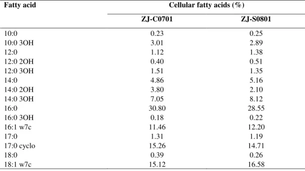
Characterization of S. marcescens
Results from this study indicated that there were no
significant differences between the phenotypic and molecular
characteristics of the S. marcescens strains from the cactus
plant and silkworms. Both two strains formed red, smooth,
convex, entire and round colonies on nutrient agar. Classical
bacteriological tests showed that they were gram-negative, rod
shaped and motile organisms. The fatty acid profiles of strain
ZJ-C0701 were very similar to those of strain ZJ-S0801 (Table
1). Comparison of the fatty acid composition of strain
ZJ-C0701 and ZJ-S0801 with species from the bacteria database
of the Microbial Identification System (Microbial ID, Inc.)
gave a similarity index value of 0.56 and 0.58 with S.
marcescens, respectively. However, it seems that the Biolog
profiles are not suitable for the characterization of this
bacterium due to the influence of pigment. The partial 16S
rRNA and groE gene sequence of strain ZJ-C0701 (EMBL
accession No. FM883708, FM946180) and strain ZJ-S0801
(EMBL accession No. FM883709, FM946181) were
determined and aligned to other known Enterobacteriaceae
sequences deposited in GenBank (6). In the phylogenetic
analysis, the two strains and S. marcescens were clustered
within a group and were well separated from either the other
Serratia species based on partial 16S gene sequences (Fig. 1)
or the other genus of Enterobacteriaceae based on partial groE
Li, B. et al. S. marcescens from cactus and silkworm
Table 1. Analysis of cellular fatty acids of strain ZJ-C0701of S. marcescens from edible cactus plant and comparison with strain ZJ-S0801 of S. marcescens from silkworm
Cellular fatty acids (%) Fatty acid
ZJ-C0701 ZJ-S0801
10:0 0.23 0.25
10:0 3OH 3.01 2.89
12:0 1.12 1.38
12:0 2OH 0.40 0.51
12:0 3OH 1.51 1.35
14:0 4.86 5.16
14:0 2OH 3.80 2.10
14:0 3OH 7.05 8.12
16:0 30.80 28.55
16:0 3OH 0.18 0.22
16:1 w7c 11.46 12.20
17:0 1.31 1.19
17:0 cyclo 15.26 14.71
18:0 0.39 0.26
18:1 w7c 15.12 16.58
Figure 1. Phylogenetic tree derived from partial 16S rRNA gene sequence analysis on strain ZJ-C0701 and ZJ-S0801 as well as reference strains of each Serratia species. The tree was generated by the neighbor-joining method based on the two-parameter
Kimura correction of evolutionary distances. Bootstrap analyses (1000 replicates) for node values from 50% are indicated.
Figure 2. Phylogenetic tree derived from partial groE gene sequences analysis on strain ZJ-C0701 and ZJ-S0801 as well as reference strains of the genus Serratia (S.), Erwinia (Er.), Enterobacter (En.) and Klebsiella (K.). The tree was generated by the
neighbor-joining method based on the two-parameter Kimura correction of evolutionary distances. Bootstrap analyses (1000
replicates) for node values from 50% are indicated.
Comparison of virulence potential
The silkworm larvae inoculated with strain ZJ-S0801
resulted in 73.3%, 86.7% and 93.3% mortality, while
inoculation with strain ZJ-C0701 resulted in 93.3%, 96.7% and
96.7% mortality after 24, 48 and 72 h of rearing, respectively.
In addition, the causal bacteria reisolated from inoculated
silkworm larvae have morphology identical to those of the
original inoculated culture of S. marcescens, which implied
that there was no significant difference in virulence between
the S. marcescens strains from the cactus plant and silkworms.
The mortality of the larvae treated with sterile water and
uninoculated control was 0% regardless of the rearing time.
Thus, these results suggest that the plant strain maybe play a
possible role in the contamination of humans and animals.
In agreement with the results of this study, vegetable plants
as a habitat for beneficial and/or human pathogenic bacteria
have received considerable attention (1, 17, 18). Indeed,
increasing numbers of foodborne illness outbreaks have been
traced to the consumption of plant-derived foods. While a
number of outbreaks caused by Escherichia coli O157:H7,
Listeria monocytogenes and Salmonella enterica have been
linked to the consumption of contaminated fruit and vegetable
produce (4, 16, 21). However, this is first report that plant
Li, B. et al. S. marcescens from cactus and silkworm
and animal health.
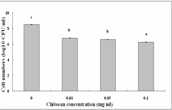
Comparison of chitosan susceptibility
Chitosan solution at different concentrations showed
effective antibacterial activity against S. marcescens strains
isolated from both the cactus plant and silkworms. In addition,
chitosan solutions up to 0.10 mg/ml showed stronger
antibacterial activity against S. marcescens compared with the
remainder treatment, which is consistent with the result of Li et
al. (11), who found that the antibacterial activity of chitosan
was influenced by its concentration in the solution. The
surviving cell numbers of strain ZJ-C0701 in chitosan solution
of 0.01 mg/ml decreased 1.74 log10 CFU/ml, while the
surviving cell numbers in chitosan solution of 0.10 mg/ml
decreased 2.24 log10 CFU/ml compared to the control (Fig. 3).
Similarly, the surviving cell numbers of strain ZJ-S0801 in
chitosan solution of 0.01 mg/ml decreased 1.92 log10 CFU/ml,
while the surviving cell numbers in chitosan solution of 0.10
mg/ml decreased 2.61 log10 CFU/ml compared to the control
(Fig. 4).
The antibacterial activity of chitosan against S. marcescens
strains was affected by the incubation time. In the absence of
chitosan, the surviving cell numbers of strain ZJ-C0701 in
sterile deionized water decreased 0.18 log10 CFU/ml after 0.5 h
of incubation compared with the starting value of 7.53 log10
CFU/ml. With the increase of incubation time, the surviving
cell numbers remained stable (Table 2). In the presence of
chitosan, the surviving cell numbers decreased significantly
compared to the starting value. The antibacterial activity of the
chitosan solution of 0.10 mg/ml increased with the incubation
time through to 4.0 h. After 0.5 h of incubation, the surviving
cell numbers in the chitosan solution decreased 1.20 log10
CFU/ml compared to the starting value and after 4.0 h of
incubation, the surviving cell numbers in the chitosan solution
decreased 2.78 log10 CFU/ml compared to the starting value
(Table 2), which shows that a certain incubation time is
required for the antibacterial activity of chitosan solution to
take effect.
Figure 3. Effect of chitosan concentration on the antibacterial activity of strainZJ-C0701 of S. marcescens. Columns with the same letters are not significantly different (P<0.05). Error bars represent the standard error of the mean (n = 6). Data are from a
Figure 4. Effect of chitosan concentration on the antibacterial activity of strain ZJ-S0801 of S. marcescens. Columns with the same letters are not significantly different (P<0.05). Error bars represent the standard error of the mean (n = 6). Data are from a
representative experiment repeated twice with similar results.
Table 2. Effect of incubation time on the antibacterial activity of chitosan solution at 0.10 mg/ml against strainZJ-C0701 of S. marcescensa
Cell numbers (log10 CFU/ml) Incubation time (h)
Control Chitosan
0.0 7.53 + 0.15a 7.53 + 0.15d
0.5 7.35 + 0.11a 6.33 + 0.10c
1.0 7.20 + 0.13a 6.21 + 0.09c
2.0 7.22 + 0.10a 5.78 + 0.10b
4.0 7.27 + 0.10a 4.75 + 0.09a
a The data were shown as means ± standard error from a representative experiment repeated twice with similar results. All the
meanswithin a column followed by the same letter are not significantly different (P<0.05, Fisher’s LSD test). Each value represents the average of six replicates.
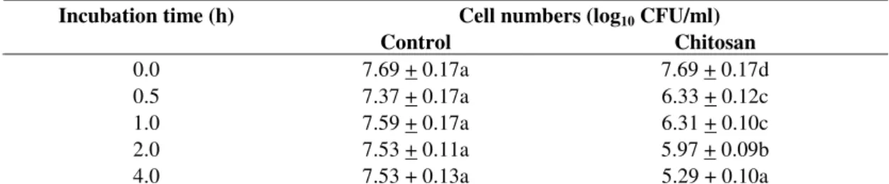
In agreement with the antibacterial effect of chitosan
solution against strain ZJ-C0701, the surviving cell numbers of
strain ZJ-S0801 remained stable in the absence of chitosan
(Table 3). In the presence of chitosan, the surviving cell
numbers decreased significantly compared to the starting value.
The antibacterial activity of the chitosan solution of 0.10
mg/ml against strain ZJ-S0801 increased with the incubation
time through to 4.0 h. The surviving cell numbers in the
chitosan solution decreased 1.36 log10 CFU/ml after 0.5 h of
incubation, and they decreased 2.40 log10 CFU/ml after 4.0 h of
incubation compared to the starting value (Table 3).
The results from this study indicate that there was no
significant difference in chitosan susceptibility between the S.
marcescens strains from the cactus plant and silkworms. In the
last three decades there has been a steady increase in
life-Li, B. et al. S. marcescens from cactus and silkworm
threatening to both animals and humans (7). As many S.
marcescens strains are also resistant to multiple antibiotics (5,
7, 15), it represents a growing problem for animal and public
health. Considering the absence of any sort of remedial
measures for S. marcescens infections, the present
investigation may prove helpful. The antibacterial activity of
chitosan may be enhanced by combination with radiation
processing in the control of S. marcescens.
Table 3. Effect of incubation time on the antibacterial activity of chitosan solution at 0.10 mg/ml against strain ZJ-S0801 of S.
marcescensa
Cell numbers (log10 CFU/ml) Incubation time (h)
Control Chitosan
0.0 7.69 + 0.17a 7.69 + 0.17d
0.5 7.37 + 0.17a 6.33 + 0.12c
1.0 7.59 + 0.17a 6.31 + 0.10c
2.0 7.53 + 0.11a 5.97 + 0.09b
4.0 7.53 + 0.13a 5.29 + 0.10a
a The data were shown as means ± standard error from a representative experiment repeated twice with similar results. All the
meanswithin a column followed by the same letter are not significantly different (P<0.05, Fisher’s LSD test). Each value represents the average of six replicates.
CONCLUSION
In summary, our results clearly demonstrated that there
was no significant difference between the phenotypic and
molecular characterization, virulence and chitosan
susceptibility of the S. marcescens strains from the cactus plant
and silkworms, which indicated that the plant strain maybe
play a possible role in the contamination of human and animals
alike. In addition, results from this study showed that a
chitosan solution has a strong antibacterial activity against
plant associated S. marcescens, which will be helpful in the
control of contaminated fruit and vegetable produce.
ACKNOWLEDGEMENTS
This project was supported by Zhejiang Provincial Natural
Science Foundation of China (Y3090150), the Fundamental
Research Funds for the Central Universities (KYJD09022),
National Natural Science Foundation of China (30600475) and
the Agricultural Ministry of China (nyhyzx200803010).
REFERENCES
1. Caggia, C.; Randazzo, C.L.; Di Salvo, M.; Romeo, F.; Giudici, P. (2004).
Occurrence of Listeria monocytogenes in green table olives. J. Food Prot. 67, 2189–2194.
2. de Queiroz, B.P.V.; de Melo, I.S. (2006). Antagonism of Serratia marcescens towards Phytophthora parasitica and its effects in promoting the growth of citrus. Braz. J. Microbiol. 37, 448–450.
3. Fang, Y.; Lou, M.M.; Li, B.; Xie, G.L.; Wang, F.; Zhang, L.; Luo, Y.C. (2010). Characterization of Burkholderia cepacia complex from cystic fibrosis patients in China and their chitosan susceptibility. World J. Microb. Biot. 26, 443–450.
4. Gorski, L.; Palumbo, J.D.; Nguyen, K.D. (2004). Strain-specific differences in the attachment of Listeria monocytogenes to alfalfa sprouts. J. Food Prot. 67, 2488–2495.
5. Guimaraes, M.A.; Tibana, A.; Nunes, M.P.; dos Santos, K.R.N. (2000). Disinfectant and antibiotic activities: A comparative analysis in Brazilian hospital bacterial isolates. Braz. J. Microbio. 31, 193–199.
6. Harada, H.; Ishikawa, H. (1997). Phylogenetical relationship based on groE genes among phenotypically related Enterobacter, Pantoea, Klebsiella, Serratia and Erwinia species. J. Gen. Appl. Microbiol. 43, 355–361.
7. Hejazi, A.; Falkiner, F.R. (1997). Serratia marcescens. J. Med. Microbiol. 46, 903–912.
8. Iranshahi, M.; Shahverdi, A.R.; Mirjani, R.; Amin, G.; Shafiee, A. (2004). Umbelliprenin from Ferula persica roots inhibits the red pigment production in Serratia marcescens. Z. Naturforsch C. 59, 506–508. 9. Iwaya, A.; Nakagawa, S.; Iwakura, N.; Taneike, I.; Kurihara, M.;
10. Kaito, C.; Akimitsu, N.; Watanab, H.; Sekimizu, K. (2002). Silkworm larvae as an animal model of bacterial infection pathogenic to humans. Microb. Pathog. 32, 183–190.
11. Li, B.; Wang, X.; Chen, R.X.; Huangfu, W.G.; Xie, G.L. (2008b). Antibacterial activity of chitosan solution against Xanthomonas pathogenic bacteria isolated from Euphorbia pulcherrima. Carbohyd. Polym. 72, 287–292.
12. Li, B.; Xie, G.L.; Zhang, J.Z.; Janssens, D.; Swings, J. (2006). Identification of the bacterial leaf spot pathogen of poinsettia in China. J. Phytopathol. 154, 711–715.
13. Li, B.; Xu, L.H.; Lou, M.M.; Li, F.; Zhang, Y.D.; Xie, G.L. (2008a). Isolation and characterization of antagonistic bacteria against bacterial leaf spot of Euphorbia pulcherrima. Lett. Appl. Microbiol. 46, 450–455. 14. Li, B.; Su, T.; Chen, X.L.; Liu, B.P.; Zhu, B.; Fang, Y.; Qiu, W.; Xie,
G.L. (2009). Effect of chitosan solution on the bacterial septicemia disease of Bombyx mori (Lepidoptera: Bombycidae) caused by Serratia marcescens. Appl. Entomol. Zool. 45, 145–152.
15. Loureiro, M.M.; de Moraes, B.A.; Quadra, M.R.R.; Pinheiro, G.S.; Asensi, M.D. (2002). Study of multi-drug resistant microorganisms isolated from blood cultures of hospitalized newborns in Rio de Janeiro city, Brazil. Braz. J. Microbiol. 33, 73–78.
16. Milillo, S.R.; Badamo, J.M.; Boor, K.J.; Wiedmann, M. (2008). Growth and persistence of Listeria monocytogenes isolates on the plant model Arabidopsis thaliana. Food Microbiol. 25, 698–704.
17. Rahme, L.G.; Ausubel, F.M.; Cao, H.; Drenkard, E.; Goumnerov, B.C.; Lau, G.W.; Mahajan-Miklos, S.; Plotnikova, J.; Tan, M.W.; Tsongalis, J.; Walendziewicz, C.L.; Tompkins, R.G. (2000). Plants and animals share functionally common bacterial virulence factors. Proc. Natl. Acad. Sci. USA 97, 8815–8821.
18. Rahme, L.G.; Stevens, E.J.; Wolfort, S.F.; Shao, J.; Tompkins, R.G.; Ausubel, F.M. (1995). Common virulence factors for bacterial
pathogenicity in plants and animals. Science 268, 1899–1902.
19. Rascoe, J.; Berg, M.; Melcher, U.; Mitchell, F.L.; Bruton, B.D.; Pair, S.D.; Fletcher, J. (2003). Identification, phylogenetic analysis, and biological characterization of Serratia marcescens strains causing cucurbit yellow vine disease. Phytopathology 93, 1233–1239.
20. Roberts, D.P.; McKenna, L.F.; Lakshman, D.K.; Meyer, S.L.F.; Kong, H.; de Souza, J.T.; Lydon, J.; Baker, C.J.; Buyer, J.S.; Chung, S. (2007). Suppression of damping-off of cucumber caused by Pythium ultimum with live cells and extracts of Serratia marcescens N4-5. Soil Biol. Biochem. 39, 2275–2288.
21. Samadpour, M.; Barbour, M.W.; Nguyen, T.; Cao, T.M.; Buck, F.; Depavia, G.A.; Mazengia, E.; Yang, P.; Alfi, D.; Lopes, M.; Stopforth, J.D. (2006). Incidence of enterohemorrhagic Escherichia coli, Escherichia coli O157, Salmonella, and Listeria monocytogenes in retail fresh ground beef, sprouts, and mushrooms. J. Food Prot. 69, 441–443. 22. Schaad, N.W.; Jones, J.B.; Chun, W. (2001). Laboratory guide for
identification of plant pathogenic bacteria. 3rd Edition. American Phytopathological Society, St. Paul, Minn.
23. Selvakumar, G.; Mohan, M.; Kundu, S.; Gupta, A.D.; Joshi, P.; Nazim, S.; Gupta, H.S. (2008). Cold tolerance and plant growth promotion potential of Serratia marcescens strain SRM (MTCC 8708) isolated from flowers of summer squash (Cucurbita pepo). Lett. Appl. Microbiol. 46, 171–175.
24. Silva, T.M.; Stets, M.I.; Mazzetto, A.M.; Andrade, F.D.; Pileggi, S.A.V.; Favero, P.R.; Cantu, M.D.; Carrilho, E.; Carneiro, P.I.B.; Pileggi, M. (2007). Degradation of 2,4-D herbicide by microorganisms isolated from Brazilian contaminated soil. Braz. J. Microbiol. 38, 522–525.