Arq Neuropsiquiatr 2006;64(2-B):394-397
Brain Research and Treatment Center. Division of Neuro l o g y. Scripps Clinic, La Jolla, CA, USA:1MD, PhD;2PhD;3MD.S u p p o rted by
the Darlene and Donald Shiley Foundation. JRC is recipient of the Clark Fellowship in Neurophysiology.
Received 11 August 2005, received in final form 6 December 2005. Accepted 4 February 2006.
Shirley M. Otis, MD. - The Brain Research and Treatment Center - Scripps Clinic, Division of Neurology - 10666 North To rrey Pines Road - La Jolla, CA 92037 USA. E-mail: [email protected]
MAGNETOENCEPHALOGRAM RECORDING
FROM SECONDARY MOTOR AREAS DURING
IMAGINED MOVEMENTS
Carlos Amo
1, José R. Criado
2, Shirley M. Otis
3ABSTRACT - This study determined whether the activity of the secondary motor cortex (M2) could be re c o rd-ed during imaginrd-ed movements (IM) of the right and left hand using magnetoencephalography (MEG). Results during IM were compared with a somatosensory trial during a passive tactile stimulation in one subject. During the somatosensory trial, dipoles were detected in somatosensory (SS) and motor primary (M1) areas, scoring 94.4-98.4% for SS, 1.6-5.6% for M1 and 0% for M2. During the IM trial, dipoles were detected in SS, M1 and M2 areas, scoring 61.1-68.8% for SS, 2.6-9.3% for M1 and 28.6-29.6% for M2. These data support the hypothesis that M2 areas are activated during imagined hand movements. This study aims for the development of a diagnosis test for patients with motor deficits by evaluating the whole somatomotor network with specific interest in M2 areas.
KEY WORDS: secondary motor cortex, somatosensory areas, motor primary areas, magnetoencephalogra-phy (MEG), motor imagined movements.
Registro magnetoencefalográfico de áreas motoras secundárias durante simulação interna do movimento
RESUMO - Este estudo determina se a atividade motora secundária cortical (M2) pode ser gravada durante simulação interna do movimento (IM) das mãos direita e esquerda utilizando-se magnetencefalografia (MEG). Os resultados da simulação dos movimentos estudados foram comparados com um ensaio soma-to-sensorial com estimulação tactil passiva em um sujeito. Durante o ensaio somasoma-to-sensorial dipolos foram detectados em áreas somato-sensoriais (SS) e motoras primarias (MI) tendo como score 94,4-98,4% para SS, 1,6-5,6% para M1 e 0% para M2. Durante o ensaio de simulação dos movimentos também foram detec-tados dipolos em SS 61,1-68,8%, M1 2,6-9,3% e M2 28,6-29,6%. Estes dados evidenciam a hipótese de que as áreas M2 são ativadas durante a simulação dos movimentos das mãos. Este estudo sugere o desenvolvi-mento de um teste diagnóstico para pacientes com deficites motores, que avalie a rede somatomotora com interesse específico nas áreas M2.
PA L AV R A S - C H AVE: áreas motoras secundárias, áreas somato-sensoriais, áreas motoras, magnetencefalo-grafia (MEG), simulação interna do movimento.
Motor Imagery or Imagined Movements (IM) have been defined as conscious mental rehearsal of a mo-tor act without perf o rming any overt movement and implies that the subject feels himself executing a
giv-en action1. Execution and internal simulation of
mo-vements seem to follow the same temporal character-istics and use the same neural re p re s e n t a t i o n s2. S t
u-dies using diff e rent neuroimaging techniques to char-acterize IM have revealed activation of a number of
cortical motor areas3-6shown also by
Magnetoence-phalograpy (MEG)7 , 8. MEG provides high spatial and
temporal re s o l u t i o n9allowing characterizing with
precision the temporal sequence of different motor a reas activated during IM. Based on a previous MEG p rotocol to evaluate motor and somatosensory
func-tion simultaneously1 0, the aim of the present study
Arq Neuropsiquiatr 2006;64(2-B) 395
the whole somatomotor network could complement the diagnosis of motor function in patients with dif-ferent degrees of motor deficits.
METHOD
A healthy male (39 years-old) gave his informed con-sent to participation in this study. MEG activity was re c o rd-ed by a Magnes 2500 Whole-Head system with 148 chan-nels (4D NeuroIm aging Technologies, Inc., San Diego CA, USA) inside a shielded magnetic room while the subject un-d e rwent two un-diff e rent conun-ditions. Prior to the stuun-dy, the subject was instructed to execute a hand movement con-sisting of a wrist extension with passive flexion. In the first condition, a somatosensory trial, the subject was inform e d to relax his hand while feeling a pre s s u re stimulus deliv-e rdeliv-ed to thdeliv-e inddeliv-ex fingdeliv-er by a pndeliv-eumatically drivdeliv-en mdeliv-echan- mechan-ical system (air pre s s u re, 15 psi). In the second condition (IM), the subject was asked to imagine moving the hand (as described above) immediately after feeling a pre s s u re stimulus.
I n t e r-stimulus interval was 2 seconds and 300 trials per each hand were performed first in the left hand and then in the right hand. EMG electrodes were placed in the wrist extensor muscles in order to exclude epochs with muscu-lar activity. Each re c o rding was acquired with a sample rate of 678 Hz. MEG data were digitized, filtered (1-40 Hz) and averaged. Simple equivalent dipole model was used
(equiv-alent current dipole (ECD)) to calculate the spatial localiza-tion of the neuronal currents. Only dipoles with adjust high-er than 90% and confidence volume smallhigh-er than 5 cm3w
e-re selected. Dipoles wee-re included in the subject MRI images with the software STA/R®11.
RESULTS
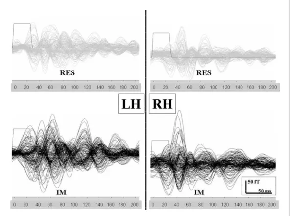
Wa v e f o rms of averaged evoked magnetic re s p o n s-es obtained from the somatosensory and IM trials a re shown in Figure 1. ECDs were calculated from e v o-ked magnetic responses. To quantify the anatomical distribution of the dipoles the following cortical re-gions were selected:
1) M1 (Primary Motor Cortex corresponding Bro d-mann’s Area 4).
2) M2 (Secondary Motor Cortex, Bro d m a n n ’s Are a s 6 and 8) including motor supplementary areas and premotor cortex.
3) S1 (Primary Somatosensory area, Bro d m a n n ’s areas 1, 2, 3a and 3b).
4) S2 (Secondary Somatosensory area, Bro d m a n n ’s Area 5 and 7).
The anatomical distribution of the dipoles (contra-lateral to the stimulus) is described in Figure 2. Soma-t o s e n s o ry (SS) and moSoma-tor primary (M1) areas were
396 Arq Neuropsiquiatr 2006;64(2-B)
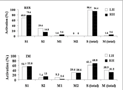
Fig 2. Dipole localization (% of to tal number of dipoles) from diff e r -ent somatosensory (S, S1, S2) and motor areas (M, M1, M2) in both h e m i s p h e res during somatosenso ry trial (upper) and imagined mo -vement (lower).
Fig 3. Dipole localization during the IM trial (white dots overlapped in a single MRI slice). Dipoles re p resent the temporal sequence of cortical activation during the first 200 ms after contralateral index finger stimulation.
activated during the somatosensory trial. From the total number of dipoles selected during the somato-s e n somato-s o ry trial, 94.4% were re c o rded in the right hemi-s p h e re (RH) and 98.4% in the left hemihemi-sphere (LH)
we-Arq Neuropsiquiatr 2006;64(2-B) 397
re re c o rded in LH and 68.8% in the RH in the SS. Con-sistent with the somatosensory trial, 2.6% (RH) and 9.3% (LH) of the dipoles were re c o rded in M1. In con-trast with the somatosensory trial, 28.6% (RH) and 29.6% (LH) of the dipoles were re c o rded in M2 (Fig 2, bottom).
The temporal sequence of the cortical areas acti-vated during the IM trial was obtained from the la-tencies of the selected dipoles. During the first 200 ms following the tactile stimulus the temporal se-quence of cortical areas activated were similar in both h e m i s p h e res. The earliest activation (16-51 ms) was p redominant in S1 (contralateral to the stimulus), fol-lowed by M2 (57-102 ms) (contralateral to the stim-ulus), and S2 (94-167 ms) (bilateral) (Fig 3). In the s o m a t o s e n s o ry trial, the temporal sequence of the c o rtical areas activated was similar in both hemisphe-res. The earliest activation (initial 100 ms) was pre d o-minant in S1 (contralateral to the stimulus), followed by S2 (100-200 ms) (bilateral).
DISCUSSION
Findings from previous studies characterizing IM
using diff e rent neuroimaging techniques3 - 8s u g g e s t
that both internal simulation and movement
execu-tion shares similar neurophysiological substrates2.
The location and latency of the MEG activity re c o r-ded in the somatosensory areas (S1 and S2) during the somatosensory and IM trials were similar. These data is consistent with previous MEG studies
charac-terizing somatosensory evoked activity9 , 1 0H o w e v e r,
our findings of S2 during IM trials could be also re l a t-ed to a potential role for S2 in the planning and
exe-cution of movements5,6.
M o re o v e r, an increase in M1 activity was found during both somatosensory and IM trials. This could be due to either somatosensory aff e rents innerv a
t-ing M11 2or the possible role of M1 during imagined
m o v e m e n t s8. M2 activity was re c o rded during IM
tri-als (50-100 ms), but not during somatosensory tritri-als. The M2 activity during IM trials is consistent with
pre-vious studies3-8.
In conclusion, this MEG study represents only an example, but shows a significant activation of M2 during IM trials (~30% of total number of dipoles re-c o rded in the trial). The temporal sequenre-ce of are-cti-
acti-vation during IM trials is as follow: S1M 2S2. Fro m
this temporal sequence we obtained latencies that correspond to each cortical area. Further research is required with a larger sample group to validate the p resent results. Findings from future studies could be used for the development of a non-invasive diag-nostic test for patients with motor deficits. This future diagnostic test could be used to follow up patients undergoing rehabilitation or pharmacological ther-apies by evaluating the whole somatomotor network with specific interest in M2 areas.
Acknowledgments– We thank Lacey Kurelowech and Patti Quint for outstanding technical support.
REFERENCES
1. J e a n n e rod M, Frak V. Mental imaging of motor activity in humans. Curr Opin Neurobiol 1999;9:735-739.
2. Crammond DJ. Motor imagery: never in your wildest dream. Tre n d s Neurosci 1997;20:54-57.
3. Roland PE, Larsen B, Lassen NA, Skinhoj E. Supplementary motor are a and other cortical areas in organization of voluntary movements in man. J Neurophysiol 1980;43:118-136.
4. Decety J, Perani D, Jeannerod M, et al. Mapping motor re p re s e n t a t i o n s with positron emission tomography. Nature 1994;371:600-602. 5. Porro CA, Francescato MP, Cettolo V, et al. Primary motor and
senso-ry cortex activation during motor performance and motor imagesenso-ry: a functional magnetic resonance imaging study. J Neurosci 1996;16:7688-7698.
6. Lotze M, Montoya P, Erb M, et al. Activation of cortical and cere b e l l a r motor areas during executed and imagined hand movements: an fMRI study. J Cogn Neurosci 1999;11:491-501.
7. Lang W, Cheyne D, Hollinger P, Gerschlager W, Lindinger G. Electric and magnetic fields of the brain accompanying internal simulation of movement. Brain Res Cogn Brain Res 1996;3:125-129.
8. Schnitzler A, Salenius S, Salmelin R, Jousmaki V, Hari R. Involvement of primary motor cortex in motor imagery: a neuromagnetic study. Neuroimage 1997;6:201-208.
9. Hämäläinen M, Hari R, Ilmoniemi RJ, Knuutila J, Lounasmaa OV. Mag-netoencephalography: theory, instrumentation, and applications to non-invasive studies of the working human brain. Rev Mod Phys 1993; 65:413-497.
10. Castillo EM, Simos PG, Wheless JW, et al. Integrating sensory and motor mapping in a comprehensive MEG protocol: clinical validity and re p l i c-ability. Neuroimage 2004;21:973-983.
11. Schwartz DP, Badier JM, Bihoué P, Bouliou A. Evaluation of a new MEG-EEG spatio-temporal localization approach using a realistic sourc e model. Brain Topography 1999;11:279-289.