www.jbp.org.br
ISSN 1806-3713
Volume 42, Number 6 November | December 2016
HIGHLIGHT
V
olume 42, Number 6
No
v
ember | Dec
ember
20
16
Exercise and
sleep apnea
Pulmonary function
and sickle cell disease
Realização:
Nos dias 02 a 05 de agosto de 2017, a cidade de Fortaleza receberá
os maiores congressos sobre doenças respiratórias e pulmonares da
atualidade, com renomados palestrantes da área médica, informações,
estudos e pesquisas internacionais.
E O MELHOR: TUDO ISSO EM UMA DAS CIDADES MAIS BONITAS
DO BRASIL.
INVISTA
NO SEU
CONHECIMENTO.
COMPAREÇA!
ISSN 1806-3713
Published once every two months J Bras Pneumol. v.42, number 6, p. 399-476 November/December 2016
Publicação Indexada em:
Latindex, LILACS, Scielo Brazil, Scopus, Index Copernicus, ISI Web of Knowledge, MEDLINE e PubMed Central (PMC)
Disponível eletronicamente nas versões português e inglês:
www.jornaldepneumologia.com.br e www.scielo.br/jbpneu
Associação Brasileira de Editores Científicos
I N T E R N A T I O N A L
EDITOR-IN-CHIEF
Rogerio Souza - Universidade de São Paulo, São Paulo - SP
EXECUTIVE EDITORS
Bruno Guedes Baldi - Universidade de São Paulo, São Paulo - SP
Caio Júlio Cesar dos Santos Fernandes - Universidade de São Paulo - São Paulo - SP Carlos Roberto Ribeiro de Carvalho - Universidade de São Paulo, São Paulo - SP Carlos Viana Poyares Jardim - Universidade de São Paulo, São Paulo - SP
ASSOCIATE EDITORS
Afrânio Lineu Kritski - Universidade Federal do Rio de Janeiro, Rio de Janeiro, RJ Andre Luis Pereira de Albuquerque - Universidade de São Paulo - São Paulo - SP Bruno Hochhegger - Universidade Federal do Rio Grande do Sul - Porto Alegre – RS Edson Marchiori - Universidade Federal Fluminense, Niterói - RJ
Fernanda Carvalho de Queiroz Mello - Universidade Federal do Rio de Janeiro - Rio de Janeiro - RJ Frederico Leon Arrabal Fernandes - Universidade de São Paulo - São Paulo - SP
Giovanni Battista Migliori - Director WHO Collaborating Centre for TB and Lung Diseases, Fondazione S. Maugeri, Care and Research Institute, Tradate - Italy
Giovanni Sotgiu - University of Sassari, Sassari - Italy Irma de Godoy - Universidade Estadual Paulista, Botucatu - SP
Marcelo Alcântara Holanda - Universidade Federal do Ceará - Fortaleza - CE Pedro Caruso - Universidade de São Paulo - São Paulo - SP
Pedro Rodrigues Genta - Universidade de São Paulo - São Paulo - SP
Renato Tetelbom Stein - Pontifícia Universidade Católica do Rio Grande do Sul, Porto Alegre - RS Ricardo de Amorim Corrêa - Universidade Federal de Minas Gerais - Belo Horizonte - MG Ricardo Mingarini Terra - Universidade de São Paulo - São Paulo - SP
Simone Dal Corso - Universidade Nove de Julho - São Paulo – SP Tomás Pulido - Instituto Nacional de Cardiología Ignacio Chávez - México Ubiratan de Paula Santos - Universidade de São Paulo, São Paulo - SP Veronica Amado - Universidade de Brasília, Brasília - DF
EDITORIAL COUNCIL
Alberto Cukier - Universidade de São Paulo, São Paulo – SP Álvaro A. Cruz - Universidade Federal da Bahia, Salvador, BA Ana C. Krieger - Weill Cornell Medical College - New York – USA
Ana Luiza Godoy Fernandes - Universidade Federal de São Paulo, São Paulo - SP Antonio Segorbe Luis - Universidade de Coimbra, Coimbra - Portugal Ascedio Jose Rodrigues - Universidade de São Paulo - São Paulo - SP Brent Winston - University of Calgary, Calgary - Canada
Carlos Alberto de Assis Viegas - Universidade de Brasília, Brasília - DF
Carlos Alberto de Castro Pereira - Universidade Federal de São Paulo, São Paulo - SP Carlos M. Luna - Hospital de Clinicas, Universidad de Buenos Aires, Buenos Aires - Argentina Carmen Silvia Valente Barbas - Universidade de São Paulo, São Paulo - SP
Celso Ricardo Fernandes de Carvalho - Universidade de São Paulo, São Paulo - SP Dany Jasinowodolinski - Universidade de São Paulo, São Paulo - SP
Denis Martinez - Universidade Federal do Rio Grande do Sul, Porto Alegre - RS Douglas Bradley - University of Toronto, Toronto, ON - Canadá
Emílio Pizzichini - Universidade Federal de Santa Catarina, Florianópolis - SC Fábio Biscegli Jatene - Universidade de São Paulo, São Paulo - SP
Frank McCormack - University of Cincinnati School of Medicine, Cincinnati, OH - USA Geraldo Lorenzi Filho - Universidade de São Paulo, São Paulo - SP
Gilberto de Castro Junior - Universidade de São Paulo, São Paulo - SP
Gustavo Javier Rodrigo - Hospital Central de las Fuerzas Armadas, Montevidéu – Uruguay Ilma Aparecida Paschoal - Universidade de Campinas, Campinas - SP
C. Isabela Silva Müller - Vancouver General Hospital, Vancouver, BC - Canadá J. Randall Curtis - University of Washington, Seattle, Wa - USA
John J. Godleski - Harvard Medical School, Boston, MA - USA José Alberto Neder - Queen’s University - Ontario, Canada
José Antonio Baddini Martinez - Universidade de São Paulo, Ribeirão Preto - SP José Dirceu Ribeiro - Universidade de Campinas, Campinas - SP
José Miguel Chatkin - Pontifícia Universidade Católica do Rio Grande do Sul, Porto Alegre - RS José Roberto de Brito Jardim - Universidade Federal de São Paulo, São Paulo - SP José Roberto Lapa e Silva - Universidade Federal do Rio de Janeiro, Rio de Janeiro - RJ Kevin Leslie - Mayo Clinic College of Medicine, Rochester, MN - USA
Luiz Eduardo Nery - Universidade Federal de São Paulo, São Paulo - SP Marc Miravitlles - University Hospital Vall d’Hebron - Barcelona, Catalonia, Spain Marisa Dolhnikoff - Universidade de São Paulo, São Paulo - SP
Marli Maria Knorst - Universidade Federal do Rio Grande do Sul, Porto Alegre - RS Mauro Musa Zamboni - Instituto Nacional do Câncer, Rio de Janeiro - RJ Nestor Muller - Vancouver General Hospital, Vancouver, BC - Canadá Noé Zamel - University of Toronto, Toronto, ON - Canadá
Oliver Augusto Nascimento - Universidade Federal de São Paulo - São Paulo - SP Paul Noble - Duke University, Durham, NC - USA
Paulo Francisco Guerreiro Cardoso - Universidade de São Paulo, São Paulo - SP Paulo Manuel Pêgo Fernandes - Universidade de São Paulo, São Paulo - SP Peter J. Barnes - National Heart and Lung Institute, Imperial College, London - UK Renato Sotto Mayor - Hospital Santa Maria, Lisboa - Portugal
Richard W. Light - Vanderbili University, Nashville, TN, USA Rik Gosselink - University Hospitals Leuven - Bélgica Robert Skomro - University of Saskatoon, Saskatoon - Canadá Rubin Tuder - University of Colorado, Denver, CO - USA
Sérgio Saldanha Menna Barreto - Universidade Federal do Rio Grande do Sul, Porto Alegre - RS Sonia Buist - Oregon Health & Science University, Portland, OR - USA
Talmadge King Jr. - University of California, San Francisco, CA - USA
ISSN 1806-3713
E
x
p
e
d
ie
n
te
BRAZILIAN THORACIC SOCIETY
Ofice: SCS Quadra 01, Bloco K, Asa Sul, salas 203/204. Edifício Denasa, CEP 70398-900, Brasília, DF, Brazil. Tel. +55 61 3245-1030/+55 0800 616218. Website: www.sbpt.org.br. E-mail: [email protected]
The Brazilian Journal of Pulmonology (ISSN 1806-3713) is published once every two months by the Brazilian Thoracic Society (BTS). The statements and opinions contained in the editorials and articles in this Journal are solely those of the authors thereof and not of the Journal’s Editor-in-Chief, peer reviewers, the BTS, its oficers, regents, members, or employees. Permission is granted to reproduce any igure, table, or other material published in the Journal provided that the source for any of these is credited.
BTS Board of Directors (2015-2016 biennium):
President: Renato Maciel - MG
Secretary-General: Paulo Henrique Ramos Feitosa - DF
Director, Professional Advocacy: Jose Eduardo Delini Cançado - SP CFO: Saulo Maia Davila Melo - SE
Scientiic Director: Miguel Abidon Aide - RJ
Director, Education and Professional Practice: Clystenes Odyr Soares Silva - SP Director, Communications: Simone Chaves Fagondes - RS
President, BTS Congress 2016: Marcus Barreto Conde - RJ
President Elect (2017/2018 biennium): Fernando Luiz Cavalcanti Lundgren - PE Chairman of the Board: Jairo Sponholz Araújo (PR)
AUDIT COMMITTEE:
Active Members: Clóvis Botelho (MT), Benedito Francisco Cabral Júnior (DF), Rafael de Castro Martins (ES)
Alternates: Maurício Meireles Góes (MG), Alina Faria França de Oliveira (PE), Paulo Cesar de Oliveira (MG)
COORDINATORS, BTS DEPARTMENTS:
Thoracic Surgery – Darcy Ribeiro Pinto Filho (RS) Sleep–disordered Breathing – Marcelo Fouad Rabahi (GO) Respiratory Endoscopy – Mauro Musa Zamboni (RJ) Pulmonary Function – John Mark Salge (SP) Imaging – Bruno Hochhegger (RS)
Lung Diseases – Ester Nei Aparecida Martins Coletta (SP) Pediatric Pulmonology – Paulo Cesar Kussek (PR)
COORDINATORS, BTS SCIENTIFIC COMMITTEES:
Asthma – Emilio Pizzichini (SC) Lung Cancer – Teresa Yae Takagaki (SP)
Pulmonary Circulation – Carlos Viana Poyares Jardim (SP) Advanced Lung Disease – Dagoberto Vanoni de Godoy (RS) Interstitial Diseases – José Antônio Baddini Martinez (SP)
Environmental and Occupational Respiratory Diseases – Ana Paula Scalia Carneiro (MG) COPD – Roberto Stirbulov (SP)
Epidemiology – Frederico Leon Arrabal Fernandes (SP) Cystic Fibrosis – Marcelo Bicalho of Fuccio (MG) Respiratory Infections and Mycoses – Mauro Gomes (SP) Pleura – Roberta Karla Barbosa de Sales (SP)
International Relations – José Roberto de Brito Jardim (SP) Smoking – Luiz Carlos Corrêa da Silva (RS)
Intensive Care – Marco Antônio Soares Reis (MG) Tuberculosis – Fernanda Carvalho de Queiroz Mello (RJ)
ADMINISTRATIVE SECRETARIAT OF THE BRAZILIAN JOURNAL OF PULMONOLOGY Address: SCS Quadra 01, Bloco K, Asa Sul, salas 203/204. Edifício Denasa, CEP 70398-900, Brasília, DF, Brazil. Tel. +55 61 3245-1030/+55 0800 616218.
Assistant Managing Editor: Luana Maria Bernardes Campos. E-mail: [email protected]
Circulation: 4.000 copies
Distribution: Free to members of the BTS and libraries Printed on acid-free paper
ISSN 1806-3713
Published once every two months J Bras Pneumol. v.42, number 6, p. 399-476 November/December 2016
C
o
n
te
n
ts
EDITORIAL
399 - Consolidating in the present, with an eye to the future Rogerio Souza
401 - The JBP and sleep medicine Pedro Rodrigues Genta
CONTINUING EDUCATION: IMAGING
402 - Clusters of small nodules with no conluence Edson Marchiori, Bruno Hochhegger, Gláucia Zanetti
CONTINUING EDUCATION: SCIENTIFIC METHODOLOGY
403 - Developing research questions that make a difference Cecilia Maria Patino, Juliana Carvalho Ferreira
ORIGINAL ARTICLE
404 - Effects of positive expiratory pressure on pulmonary clearance of aerosolized technetium-99m-labeled diethylenetriaminepentaacetic acid in healthy individuals Isabella Martins de Albuquerque, Dannuey Machado Cardoso, Paulo Ricardo Masiero, Dulciane Nunes Paiva, Vanessa Regiane Resqueti, Guilherme Augusto de Freitas Fregonezi, Sérgio Saldanha Menna-Barreto
409 - Pulmonary function in children and adolescents with sickle cell disease: have
we paid proper attention to this problem?
Ana Karine Vieira, Cristina Gonçalves Alvim, Maria Cristina Marquez Carneiro, Cássio da Cunha Ibiapina
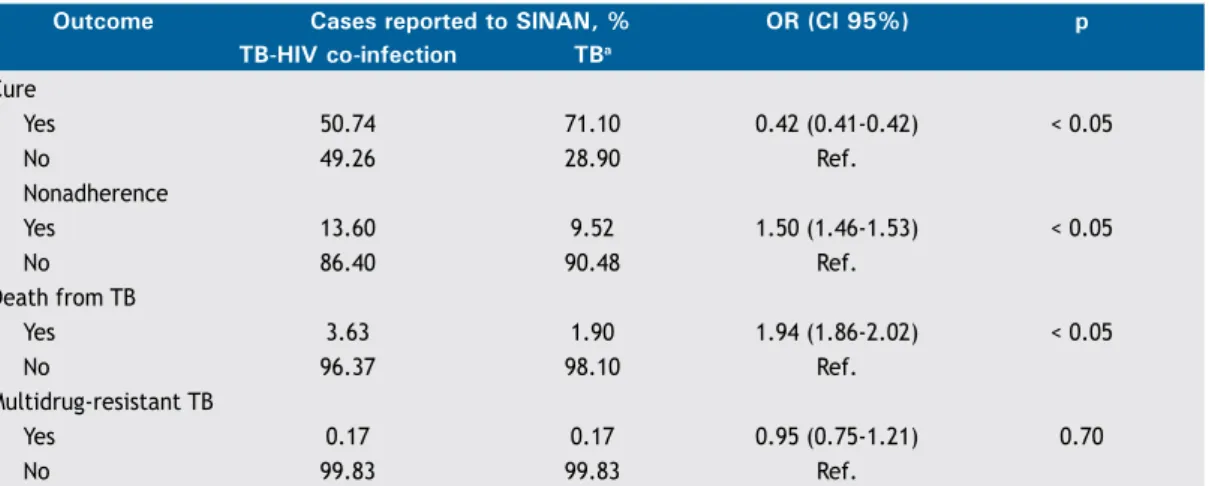
416 - Temporal analysis of reported cases of tuberculosis and of tuberculosis-HIV co-infection in Brazil between 2002 and 2012
Renato Simões Gaspar, Natália Nunes, Marina Nunes, Vandilson Pinheiro Rodrigues
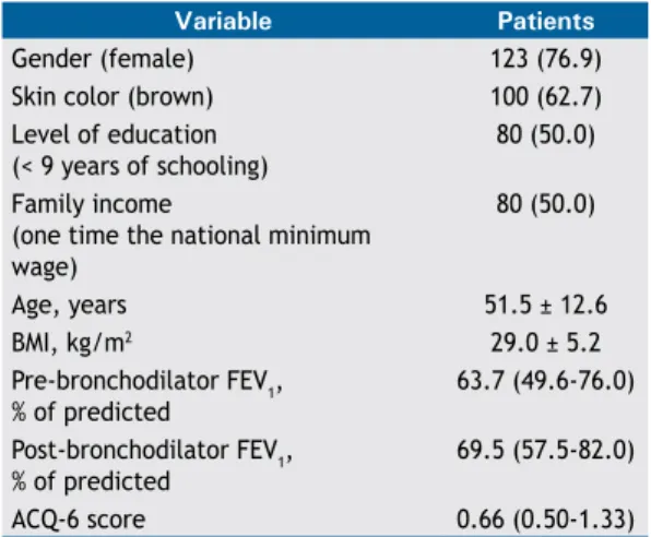
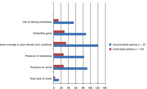
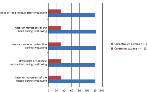
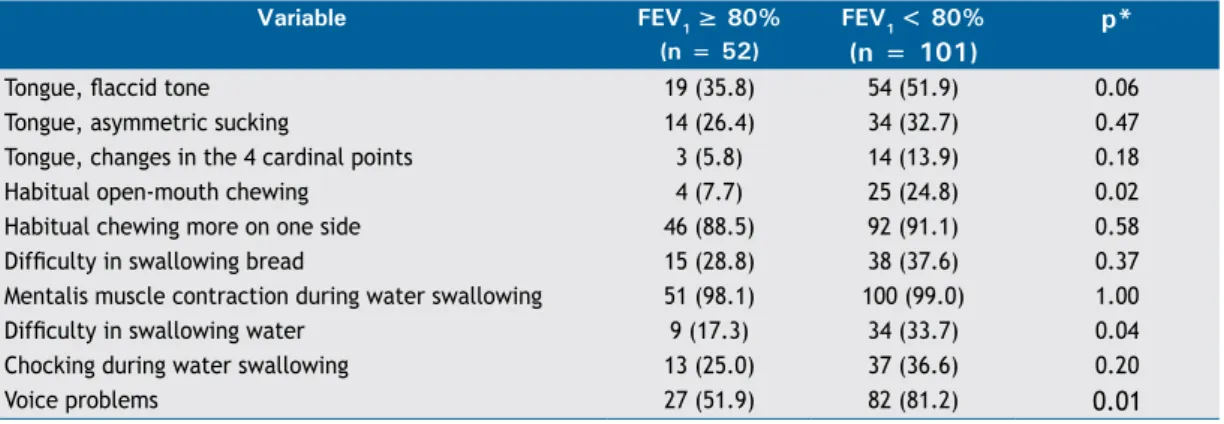
423 - Association between severe asthma and changes in the stomatognathic system Mayra Carvalho-Oliveira, Cristina Salles, Regina Terse, Argemiro D’Oliveira Júnior
429 - Perme Intensive Care Unit Mobility Score and ICU Mobility Scale: translation
into Portuguese and cross-cultural adaptation for use in Brazil
Yurika Maria Fogaça Kawaguchi, Ricardo Kenji Nawa, Thais Borgheti Figueiredo, Lourdes Martins, Ruy Camargo Pires-Neto
435 - The halo sign: HRCT indings in 85 patients
Giordano Rafael Tronco Alves, Edson Marchiori, Klaus Irion, Carlos Schuler Nin,
ISSN 1806-3713
Published once every two months J Bras Pneumol. v.42, number 6, p. 399-476 November/December 2016
C
o
n
te
n
ts
BRIEF COMMUNICATION
440 - Educational interventions to improve inhaler techniques and their impact on asthma and COPD control: a pilot effectiveness-implementation trial
Tiago Maricoto, Soia Madanelo, Luís Rodrigues, Gilberto Teixeira, Carla Valente, Lília Andrade, Alcina Saraiva
CASE SERIES
444 - An initial experience with a digital drainage system during the postoperative period of pediatric thoracic surgery
Altair da Silva Costa Jr, Thiago Bachichi, Caio Holanda, Luiz Augusto Lucas Martins De Rizzo
447 - Hard metal lung disease: a case series
Rafael Futoshi Mizutani, Mário Terra-Filho, Evelise Lima, Carolina Salim Gonçalves Freitas, Rodrigo Caruso Chate, Ronaldo Adib Kairalla, Regiani Carvalho-Oliveira,
Ubiratan Paula Santos
453 - Mouthpiece ventilation in Duchenne muscular dystrophy: a rescue strategy for noncompliant patients
Giuseppe Fiorentino, Anna Annunziata, Rosa Cauteruccio, Gianfranco Scotto di Frega, Antonio Esquinas
REVIEW ARTICLE
457 - The role of physical exercise in obstructive sleep apnea Flávio Maciel Dias de Andrade, Rodrigo Pinto Pedrosa
IMAGING IN PULMONARY MEDICINE
465 - An uncommon tomographic association: amiodarone pulmonary toxicity and adenocarcinoma
Arthur Soares Souza Jr, Gláucia Zanetti, Edson Marchiori
LETTER TO THE EDITOR
466 - Pulmonary fat embolism of neoplastic origin
Flávia Pinto, Miriam Menna Barreto, Daniela Braz Parente, Edson Marchiori
SUBJECT INDEX FOR V.41 (1-6)
468 - Índice remissivo de assuntos do volume 42 (1-6) 2016
AUTHOR INDEX FOR V.41 (1-6)
470 - Índice remissivo de autores do volume 42 (1-6) 2016
REVIEWERS FOR V.41 (1-6)
http://dx.doi.org/10.1590/S1806-37562016000600002
Consolidating in the present, with an eye to
the future
Rogerio Souza
Disciplina de Pneumologia, Instituto do Coração (InCor), Hospital das Clínicas da Faculdade de Medicina da Universidade de São Paulo, São Paulo (SP) Brasil. Editor-Chefe do Jornal Brasileiro de Pneumologia.
The scientiic literature has changed substantially over the last few decades. There has been a progressive migration to a model based on exclusive online platforms, even by traditional periodicals. In addition to reducing costs, the use of such platforms also increases the speed at which accepted articles gain exposure. In parallel with that, the appearance of an increasing number of journals has increased the portfolio of options for the publication of a given manuscript. However, the absolute majority of these new journals employ a paid submission model, which creates another type of barrier to publishing, particularly for novice researchers, whose sources of funding are more limited.
Within the context of the scenario described above but respecting the fundamental objectives of the JBP,(1)
various editorial decisions have been made, the work of the associated editors having been of fundamental importance. Their work gave greater representation to areas that previously were not well covered in the JBP. For example, among the most often cited JBP articles in the last two years, there are two articles on the physiology of the respiratory muscles,(2,3) one on the pulmonary circulation,(4) and two related to smoking. (5,6) These have been added to the already established topics covered in the JBP, such as that of obstructive lung diseases and even the study of tuberculosis. There has also been a signiicant increase in the number of JBP articles related to invasive procedures,(7,8) as well as to the ield of thoracic surgery,(9) clearly indicating an expansion of
the representativeness of the Journal. In parallel, JBP review articles have sought to illuminate subjects of more general interest to our readership, such as sleep,(10)
idiopathic pulmonary ibrosis,(11) and imaging,(12) together
with special sections on continuing education in scientiic methodology and imaging, with the goal of making the JBP an accessible tool for keeping everyone with an interest in respiratory medicine up to date.
We have been able to reduce the average time between submission and the inal decision on the manuscripts. Here, we must offer our sincere thanks to all of the reviewers who contribute to making our evaluation and publication process more agile, while it continues to be free. It was the direct participation of the reviewers that allowed such advancement. It must be acknowledged, however, that there is still much room for improvement. A number of steps in the decision-making process still need to be further streamlined. Thus, the JBP can become even more attractive to researchers as a means of broadly and rapidly bringing their results to light.
The changes described above would be illogical were they not accompanied by greater international recognition of the content published in the JBP. We are pleased to report that that is what has occurred. The JBP impact factor, as published in Thomson Reuters Journal Citation Reports has stabilized and has actually risen slightly, currently standing at 1.019. That indicates that the journal has begun to overcome the effects of the temporary suspension of 2014.(13) Supporting that impression is the fact that the two-year Scimago Jornal Rank citation index is now 1.282, corresponding to a 15% increase in relation to the previous cycle. These igures, while indicating substantial room for growth, make the JBP the leading journal in the ield of respiratory medicine in Latin America. In addition, the JBP continues to be an important journal for the dissemination of postgraduate studies conducted in Brazil, receiving a good grade in the journal ranking system of the Brazilian Ofice for the Advancement of Higher Education, a system known as Qualis.
There is a need for further consolidation of the JBP processes. There are several highly relevant research groups in our country that do not yet participate signiicantly in the publication of the JBP and that could greatly enrich the representativeness of the journal. There is also an opportunity to update several of the Brazilian Thoracic Association guidelines, which also play a fundamental role in the visibility of any scientiic journal. These opportunities will be explored in upcoming years.
The JBP can also become a platform for the discussion of pathways and alternatives in respiratory medicine. There is room to open a debate on several aspects, from the education of pulmonologists to the future of research in the area of respiratory medicine. Relevant themes for the future of respiratory medicine, which could be exposed in the JBP in the form of debates between exponents of each of the respective areas include the ideal curriculum, funding sources, the minimum structure of a postgraduate program in the pulmonology, incentives for young researchers, retention of professionals in the university environment, and professional defense.
If there is still a great need to improve the processes and solidify the continued growth of the JBP, we must not lose sight of the opportunities to give the JBP an even more comprehensive role in leading the reader to relection, not only on science, a fundamental principle of the existence of the journal, but also on health policies, vocational training, and medical education. We invite our readers to take this next step with us.
J Bras Pneumol. 2016;42(6):399-400
Consolidating in the present, with an eye to the future
REFERENCES
1. Souza R. The next 40 years. J Bras Pneumol. 2015;41(5):404. https:// doi.org/10.1590/S1806-37132015000500008
2. Caruso P, Albuquerque AL, Santana PV, Cardenas LZ, Ferreira JG, Prina E, et al. Diagnostic methods to assess inspiratory and expiratory muscle strength. J Bras Pneumol. 2015;41(2):110-23. https://doi.org/10.1590/S1806-37132015000004474
3. Marques TB, Neves Jde C, Portes LA, Salge JM, Zanoteli E, Reed UC. Air stacking: effects on pulmonary function in patients with spinal muscular atrophy and in patients with congenital muscular dystrophy. J Bras Pneumol. 2014;40(5):528-34. https://doi.org/10.1590/S1806-37132014000500009
4. Gavilanes F, Alves Jr JL, Fernandes C, Prada LF, Jardim CV, Morinaga LT, et al. Left ventricular dysfunction in patients with suspected pulmonary arterial hypertension. J Bras Pneumol. 2014;40(6):609-16. https://doi.org/10.1590/S1806-37132014000600004
5. Knorst MM, Benedetto IG, Hoffmeister MC, Gazzana MB. The electronic cigarette: the new cigarette of the 21st century? J Bras Pneumol. 2014;40(5):564-72. https://doi.org/10.1590/S1806-37132014000500013
6. Stelmach R, Fernandes FL, Carvalho-Pinto RM, Athanazio RA, Rached SZ, Prado GF, et al. Comparison between objective measures of smoking and self-reported smoking status in patients with asthma or COPD: are our patients telling us the truth? J Bras Pneumol. 2015;41(2):124-32. https://doi.org/10.1590/S1806-37132015000004526
7. Perazzo A, Gatto P, Barlascini C, Ferrari-Bravo M, Nicolini A. Can ultrasound guidance reduce the risk of pneumothorax following thoracentesis? J Bras Pneumol. 2014;40(1):6-12. https://doi. org/10.1590/S1806-37132014000100002
8. Ortakoylu MG, Iliaz S, Bahadir A, Aslan A, Iliaz R, Ozgul MA, et al. Diagnostic value of endobronchial ultrasound-guided transbronchial needle aspiration in various lung diseases J Bras Pneumol. 2015;41(5):410-4. https://doi.org/10.1590/S1806-37132015000004493
9. Marchetti-Filho MA, Leão LE, Costa-Junior Ada S. The role of intercostal nerve preservation in acute pain control after thoracotomy. J Bras Pneumol. 2014;40(2):164-70. https://doi.org/10.1590/S1806-37132014000200010
10. Beltrami FG, Nguyen XL, Pichereau C, Maury E, Fleury B, Fagondes S. Sleep in the intensive care unit. J Bras Pneumol. 2015;41(6):539-46. https://doi.org/10.1590/S1806-37562015000000056
11. Baddini-Martinez J, Baldi BG, Costa CH, Jezler S, Lima MS, Ruino R. Update on diagnosis and treatment of idiopathic pulmonary ibrosis. J Bras Pneumol. 2015;41(5):454-66. https://doi.org/10.1590/S1806-37132015000000152
12. Hochhegger B, Alves GR, Irion KL, Fritscher CC, Fritscher LG, Concatto NH, et al. PET/CT imaging in lung cancer: indications and indings. J Bras Pneumol. 2015;41(3):264-74. https://doi.org/10.1590/ S1806-37132015000004479
13. Souza R. 2015--another step along the road in a 40-year journey. J Bras Pneumol. 2015;41(1):1-2. https://doi.org/10.1590/S1806-37132015000100001
http://dx.doi.org/10.1590/S1806-37562016000600001
The JBP and sleep medicine
Pedro Rodrigues Genta1
1. Laboratório do Sono, Disciplina de Pneumologia, Instituto do Coração, Hospital das Clínicas, Faculdade de Medicina, Universidade de São Paulo, São Paulo (SP) Brasil.
Sleep medicine is a relatively new discipline that has begun to consolidate as an important area of activity for pulmonologists, in Brazil as well as in the rest of the world. The JBP has been a part of this transformation, as evidenced by the increasing number of submissions and publications related to the ield of sleep medicine. Although we are still a month away from the end of the year, the number of submissions to the JBP in 2016 has already hit a record, growing by 18% in comparison with 2013. Even more signiicant was the increase in the number of submissions related to sleep medicine, which increased from 8 in 2013 to 16 (to date) in 2016. The increasing interest in the JBP, together with an increase in the quality of the articles submitted, demonstrates the maturing of the sleep medicine community in Brazil.(1,2)
It also shows the evolution of the JBP, which has increased its visibility on various fronts. As a matter of fact, there have also been submissions on the topic from outside Brazil.(2,3)
All of this has made the JBP stand out among the Brazilian journals indexed for the Index Medicus (Medline/ PubMed) as one of the journals that publishes the most articles on the topic of sleep.
In this edition of the JBP, Andrade and Pedrosa(4) review
a current theme: the role of physical exercise in obstructive sleep apnea. Exercise has a broad range of effects. In sleep apnea, exercise can reduce the frequency of respiratory events and relieve symptoms. The treatment of sleep apnea with strategies other than the use of continuous positive airway pressure is relevant because of the dificulty that some patients have in adhering to the treatment, as well as because of the high cost of the equipment. Although the eficacy of physical exercise is limited, studies in which physical exercise is combined with other strategies, such as myofunctional speech therapy, weight loss, and positional therapy, have shown promise.
I would therefore like to thank everyone who has been contributing to the sleep medicine section of the JBP, either by submitting their scientiic production or by dedicating themselves to reviewing the manuscripts submitted. In addition, I appeal to the entire scientiic community that is interested in sleep medicine to contribute with new submissions and to volunteer to act as a JBP reviewer. The dedication of all involved will increase the international visibility of the JBP, with an emphasis on sleep medicine, which will beneit the Brazilian Thoracic Association and the scientiic community at large.
REFERENCES
1. Fonseca LB, Silveira EA, Lima NM, Rabahi MF. STOP-Bang questionnaire: translation to Portuguese and cross-cultural adaptation for use in Brazil. J Bras Pneumol. 2016:42(4):266-272. https://doi. org/10.1590/S1806-37562015000000243
2. Sunnetcioglu A, Sertogullarindan B, Ozbay B, Gunbatar H, Ekin S. Obstructive sleep apnea related to rapid-eye-movement or non-rapid-eye-movement sleep: comparison of demographic, anthropometric, and polysomnographic features. J Bras Pneumol. 2016:42(1):48-54. https://
doi.org/10.1590/S1806-37562016000000012
3. Salvaggio A, Lo Bue A, Isidoro SI, Romano S, Marrone O, Insalaco G. Gel pillow designed speciically for obstructive sleep apnea treatment with continuous positive airway pressure. J Bras Pneumol. 2016:42(5):362-366. https://doi.org/10.1590/S1806-37562016000000015
4. Andrade FM, Pedrosa RP. The role of physical exercise in obstructive sleep apnea. J Bras Pneumol. 2016:42(6):457-464.
J Bras Pneumol. 2016;42(6):401-401
ISSN 1806-3713 © 2016 Sociedade Brasileira de Pneumologia e Tisiologia
http://dx.doi.org/10.1590/S1806-37562016000000228
Clusters of small nodules with no
conluence
Edson Marchiori1,2, Bruno Hochhegger3,4, Gláucia Zanetti2,5
1. Universidade Federal Fluminense, Niterói (RJ) Brasil. 2. Universidade Federal do Rio de Janeiro, Rio de Janeiro (RJ) Brasil. 3. Santa Casa de Misericórdia de Porto Alegre, Porto Alegre (RS) Brasil.
4. Universidade Federal de Ciências da Saúde de Porto Alegre, Porto Alegre (RS) Brasil. 5. Faculdade de Medicina de Petrópolis, Petrópolis (RJ) Brasil.
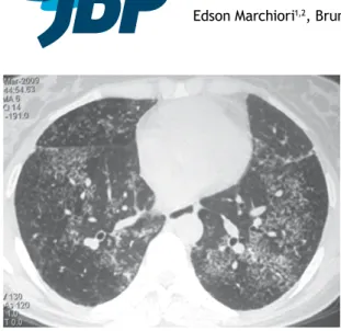
Figure 1. HRCT scan showing clusters of small nodules with no conluence in the lower lobes of both lungs.
CLINICAL HISTORY
A 36-year-old man presented with a two-month history of dry cough, fever, and weight loss (8 kg). Laboratory test results were normal. HRCT of the chest showed clusters of interstitial nodules with no conluence (Figure 1). BAL specimens were negative for tuberculosis and fungi. Open lung biopsy showed necrotic granulomas. Culture of lung tissue was positive for Mycobacterium tuberculosis.
DISCUSSION
This patient had multiple small interstitial nodules on HRCT. Small pulmonary nodules (or micronodules) are round opacities of soft tissue density and less than 1 cm in diameter. On the basis of their distribution in the lung parenchyma, they can be classiied as perilymphatic, centrilobular, or random micronodules.
A perilymphatic distribution occurs when the small nodules are located predominantly along the lung
lymphatic system (peribronchovascular interstitium, interlobular septa, and subpleural regions). A centrilobular distribution is characterized by nodules that are located within a few millimeters of the pleural surface and issures but do not touch them. A random pattern corresponds to small nodules randomly distributed in the secondary lobule and scattered uniformly throughout the lungs.
Some other patterns of distribution of small interstitial nodules have been described more recently. They include the sarcoid galaxy sign, the sarcoid cluster sign, and the nodular reversed halo sign. The reversed halo sign is deined as a round area of ground-glass attenuation surrounded by a ring of consolidation. The designation of nodular reversed halo sign is used when the walls of the dense peripheral ring are formed by nodules, instead of exhibiting the usual smooth appearance. Nodules are also frequently seen within the halo. Findings of this pattern are highly suggestive of active granulomatous disease, particularly tuberculosis or sarcoidosis.
The sarcoid galaxy sign corresponds to a large parenchymal nodule that is formed by the coalescence of small nodules and is surrounded by satellite nodules. The sarcoid cluster sign corresponds to a generally round or elongated group of small nodules with no conluence, as in the case presented here. The nodules are close to each other but do not group. These two signs were initially reported in patients with sarcoidosis, hence their names. However, they were subsequently described in patients with tuberculosis, thus requiring to be renamed. The names currently proposed for these new signs are “cluster of nodules with conluence” and “cluster of nodules without conluence”, respectively. From an anatomical and pathological standpoint, in the cases of both signs, the nodules correspond to granulomas, and the two major diagnoses that should be considered in the presence of these indings are active tuberculosis and sarcoidosis.
RECOMMENDED READING
1. Marchiori E, Zanetti G, Barreto MM, de Andrade FT, Rodrigues RS. Atypical distribution of small nodules on high resolution CT studies:
patterns and differentials. Respir Med. 2011;105(9):1263-7. http:// dx.doi.org/0.1016/j.rmed.2011.02.010
J Bras Pneumol. 2016;42(6):402-402
402
http://dx.doi.org/10.1590/S1806-37562016000000354
Developing research questions that make a
difference
Cecilia Maria Patino1,2, Juliana Carvalho Ferreira2,3
1. Department of Preventive Medicine, Keck School of Medicine, University of Southern California, Los Angeles, CA, USA.
2. Methods in Epidemiologic, Clinical and Operations Research (MECOR) Program, American Thoracic Society, New York, NY, USA, and Asociación Latinoamericana de Tórax, Montevideo, Uruguay.
3. Divisão de Pneumologia, Instituto do Coração – InCor – Hospital das Clínicas, Faculdade de Medicina, Universidade de São Paulo, São Paulo, Brasil.
BACKGROUND
A clinical research question is deined as an uncertainty about a health problem that points to the need for meaningful understanding and deliberate investigation. (1) For clinicians
interested in conducting high-quality clinical research, it is essential to recognize the fact that the research process starts with developing a question about a speciic health-related area of interest. This is important because once the research question is deined, it has an impact on every remaining component of the research process, including generating the hypothesis and deining the appropriate study design, as well as the study population, study variables, and statistical approach. However, conceiving a sound research question is not an easy task; it requires having a particular set of personal skills and utilizing structured approaches.
DEVELOPING AND WRITING A RESEARCH QUESTION
Developing a research question starts by identifying a clinical problem that is important to patients, being related to managing and ultimately improving their health. The process requires clinician scientists to be curious about and attentive to day-to-day practice outcomes, as well as to be avid readers of the scientiic literature, to participate in scientiic activities (e.g., journal clubs), and to have access to a scientiic mentor or collaborators interested in clinical research.
The research question itself should meet certain criteria, as summarized by the acronym FINGER, which stands for Feasible, Interesting, Novel, Good (for your career), Ethical, and Relevant (Chart 1).(1) We recommend going through the
FINGER criteria systematically and discussing all issues with
a mentor or colleague before writing the study protocol and conducting a study that will answer the proposed research question.
Once the research question has been deined, it should be written out in such a way that the answer can be expressed as either a number, typical of descriptive research questions (e.g., a prevalence related to disease burden, such as “What is the prevalence of asthma among favela residents in Brazil?”), or as a yes or no, typical of studies about associations between exposures and outcomes (e.g., “Is living in a
favela in Brazil associated with increased mortality among
adults with asthma?”). In addition, if the researcher has a hypothesis about the answer to the research question,(1) it
is important that it be written out using a comprehensive approach, as summarized by the acronym PICOT, which stands for Population (the population to be included in the
study), Intervention (treatment applied to participants in the treatment arm), Comparison (treatment applied to the control group), Outcome (the primary outcome variable),
and Time (follow-up time to measure the outcome).(2)
INVESTING THE TIME AND EFFORT TO COME UP WITH A HIGH-QUALITY, WELL-WRITTEN RESEARCH QUESTION IS WORTH IT!
As clinician scientists who train clinicians to become successful researchers, we cannot emphasize enough the importance of investing one’s time wisely to develop a high-quality research question. Researchers who conceive and clearly state a research question about an important health-related problem are at an advantage because they are more likely to convince key individuals to provide them with the necessary resources and support to carry out the study, as well as to increase the reporting quality of the paper to be published.(3)
Chart 1. Expanded descriptions of the recommended criteria for developing a good research question.
FINGER Criteria Feasible
Access to an adequate number of participants
Research team has technical expertise to conduct the study Affordable: costs are reasonable and funding is available Can be completed in a reasonable time period
Interesting
Results of the study will be of interest to the research community Novel
Provides new indings, extends or refutes previous indings
Good
For your career: its into your career development plan
Ethical
Risk to participants is low/acceptable, considered ethical by peers and the Institutional Review Board Relevant
To improve scientiic knowledge, inform clinicians and health policy, and to impact future research
REFERENCES
1. Hulley SB, Cummings SR, Browner WS, Grady DG, Newman TB. Designing clinical research. 4th ed. Philadelphia (PA): Lippincott Williams and Wilkins; 2013.
2. Haynes R. Forming research questions In: Haynes R, Sackett D, Guyatt GH, Tugwell P, editors. Clinical Epidemiology: How to do Clinical Practice
Research. Philadelphia, PA: Lippincott Williams & Wilkins; 2006. p. 3-14. 3. Rios LP, Ye C, Thabane L. Association between framing of the research question using the PICOT format and reporting quality of randomized controlled trials. BMC Med Res Methodol. 2010;10:11. http://dx.doi. org/10.1186/1471-2288-10-11
J Bras Pneumol. 2016;42(6):403-403 CONTINUING EDUCATION:
ISSN 1806-3713 © 2016 Sociedade Brasileira de Pneumologia e Tisiologia
http://dx.doi.org/10.1590/S1806-37562015000000320
ABSTRACT
Objective: To evaluate the effects of positive expiratory pressure (PEP) on pulmonary epithelial membrane permeability in healthy subjects. Methods: We evaluated a cohort of 30 healthy subjects (15 males and 15 females) with a mean age of 28.3 ± 5.4 years, a mean FEV1/FVC ratio of 0.89 ± 0.14, and a mean FEV1 of 98.5 ± 13.1% of predicted.
Subjects underwent technetium-99m-labeled diethylenetriaminepentaacetic acid (99m
Tc-DTPA) radioaerosol inhalation lung scintigraphy in two stages: during spontaneous breathing; and while breathing through a PEP mask at one of three PEP levels—10 cmH2O (n = 10), 15 cmH2O (n = 10), and 20 cmH2O (n = 10). The
99mTc-DTPA was
nebulized for 3 min, and its clearance was recorded by scintigraphy over a 30-min period during spontaneous breathing and over a 30-min period during breathing through a PEP mask. Results: The pulmonary clearance of 99mTc-DTPA was signiicantly shorter when
PEP was applied—at 10 cmH2O (p = 0.044), 15 cmH2O (p = 0.044), and 20 cmH2O (p =
0.004)—in comparison with that observed during spontaneous breathing. Conclusions: Our indings indicate that PEP, at the levels tested, is able to induce an increase in pulmonary epithelial membrane permeability and lung volume in healthy subjects.
Keywords: Lung/metabolism; Technetium Tc 99m pentetate/pharmacokinetics; Radiopharmaceuticals; Positive-pressure respiration.
Effects of positive expiratory pressure on
pulmonary clearance of aerosolized
technetium-99m-labeled diethylenetriaminepentaacetic acid
in healthy individuals
Isabella Martins de Albuquerque1, Dannuey Machado Cardoso2,
Paulo Ricardo Masiero3, Dulciane Nunes Paiva4, Vanessa Regiane Resqueti5,
Guilherme Augusto de Freitas Fregonezi5, Sérgio Saldanha Menna-Barreto6
Correspondence to:
Isabella Martins de Albuquerque. Avenida Roraima, 1000, Cidade Universitária, Camobi, CEP 97105-900, Santa Maria, RS, Brasil. Tel.: 55 55 3220-8234. E-mail: [email protected]
Financial support: This study received inancial support from the Fundo de Incentivo à Pesquisa do Hospital de Clínicas de Porto Alegre (FIPE-HCPA, Research Incentive Fund of the Porto Alegre Hospital de Clínicas).
INTRODUCTION
The alveolar-capillary barrier, also known as the blood-gas barrier, is excellent at maintaining the separation between alveolar air and pulmonary capillary blood, allowing rapid, eficient exchange of respiratory gases while preventing the diffusion of water-soluble particles suspended in alveolar air.(1,2) The integrity of the blood-gas barrier is extremely important to the maintenance of pulmonary homeostasis. In 1953, Frank Low published the irst high-resolution electron micrographs of the human pulmonary blood-gas barrier, showing that a structure only 0.3 µm thick separates capillary blood from alveolar gas, which suggested that the barrier
might be vulnerable to mechanical failure if the capillary pressure increased(3) or in states of high lung inlation, in which the capillary wall is under tension because of longitudinal stress in the alveolar walls.(4)
Technetium-99m-labeled diethylenetriaminepen-taacetic acid (99mTc-DTPA) radioaerosol inhalation lung
scintigraphy is a rapid, easily performed, extremely sensitive, noninvasive technique for assessing lung epithelial permeability.(5,6) When inhaled, 99mTc-DTPA
particles arrive at the alveolar epithelial surface and then diffuse from the air space into the vascular space. The clearance rate of 99mTc-DTPA is a reliable index of
alveolar epithelial permeability. Therefore, 99mTc-DTPA
aerosol-inhaled scintigraphy has been used in numerous
1. Departamento de Fisioterapia e Reabilitação, Programa de Pós-Graduação em Reabilitação Funcional, Universidade Federal de Santa Maria, Santa Maria (RS) Brasil.
2. Departamento de Educação Física e Saúde, Universidade de Santa Cruz do Sul, Santa Cruz do Sul (RS) Brasil.
3. Serviço de Medicina Nuclear, Hospital de Clínicas de Porto Alegre, Universidade Federal do Rio Grande do Sul, Porto Alegre (RS) Brasil.
4. Programa de Pós-Graduação em Promoção da Saúde, Departamento de Educação Física e Saúde, Universidade de Santa Cruz do Sul, Santa Cruz do Sul (RS) Brasil.
5. Departamento de Fisioterapia, Laboratório de Desempenho Pneumocardiovascular e Músculos Respiratórios, Universidade Federal do Rio Grande do Norte, Natal (RN) Brasil.
6. Serviço de Pneumologia, Hospital de Clínicas de Porto Alegre, Universidade Federal do Rio Grande do Sul, Porto Alegre (RS) Brasil.
Submitted: 16 December 2015.
Accepted: 19 April 2016.
Study carried out in the Serviço de Pneumologia and in the Serviço de Medicina Nuclear, Hospital de Clínicas de Porto Alegre, Universidade Federal do Rio Grande do Sul, Porto Alegre (RS) Brasil.
J Bras Pneumol. 2016;42(6):404-408
404
Albuquerque IM, Cardoso DM, Masiero PR, Paiva DN, Resqueti VR, Fregonezi GAF, Menna-Barreto SS
experiments and clinical investigations designed to assess the integrity of the respiratory epithelium.(7,8)
Positive expiratory pressure (PEP) therapy involves breathing with slightly active expiration against mild expiratory resistance (typically 10-20 cmH2O).
(9) Its
application via a PEP mask represents a reliable, safe, low-cost intervention that can increase lung volumes and intrathoracic pressure.(10,11) It has been shown to improve lung volume, promote dilation of the airways, and decrease pulmonary resistance. The purpose of PEP therapy is to increase the transpulmonary pressure gradient and improve pulmonary expansion, which consequently improves oxygenation and the response to inhaled bronchodilators.(12,13)
The effects of different intensities of PEP on the pulmonary clearance of 99mTc-DTPA in healthy subjects
have been largely unexplored in the literature and require further elucidation. Therefore, the aim of the present study was to evaluate the effects of PEP on pulmonary epithelial membrane permeability in healthy subjects.
METHODS
We studied 30 healthy subjects (15 men and 15 women). The mean age was 28.3 ± 5.4 years. Subjects with cardiovascular or neuromuscular disease were excluded, as were those with a history of smoking or respiratory disease, as well as those who were pregnant. All participants were recruited from a tertiary care hospital in the city of Porto Alegre, Brazil, and were evaluated between January and July of 2012. The study was approved by the Research Ethics Committee of the Porto Alegre Hospital de Clínicas (Protocol no. 04-418), which is accredited by the Brazilian National Commission on Research Ethics, and all subjects gave written informed consent.
Pulmonary scintigraphy with 99mTc-DTPA radioaerosol
was conducted over two 30-min periods: during spontaneous breathing; and while subjects were breathing via a PEP mask (Vital Signs/GE Healthcare, Totowa, NJ, USA), which we adapted to it each subject by using headgear, coupled to a PEP therapy device (Resistex®; Mercury Medical, Clearwater, FL, USA).
Using computer-generated randomization codes, we divided the subjects into three groups: those receiving PEP at 10 cmH2O (PEP10); those receiving PEP at 15
cmH2O (PEP15); and those receiving PEP at 20 cmH2O
(PEP20). During data acquisition, the subjects were
in a sitting position with their hands on their thighs and their arms held away from their body. For each subject, the spontaneous breathing period served as the control.
A spirometer (MasterScreen v4.31; Jaeger, Würzburg, Germany) was used in order to measure FEV1 and
FVC. The technical procedures, acceptability criteria, and reproducibility criteria, as well as standardization of measures, were in accordance with the guidelines established by the Brazilian Thoracic Association.(14)
Subjects received instruction regarding the procedures
to be performed during the spirometry assessment. The spirometer was routinely calibrated (on a daily basis) with a 3-L syringe in order to compensate for ambient temperature conditions. A minimum of three and a maximum of eight tests were conducted with a 1-min interval between them. Three reproducible maneuvers were performed, and the best curve was considered for the study. Results are expressed as absolute and percent-of-predicted values.(15)
The 99mTc-DTPA was chelated by adding 99m
Tc-pertech-netate (99mTc-O 4
−, IPEN-TEC; Institute for Energy Research and Nuclear Science, São Paulo, Brazil) to 740 MBq (20 mCi) of DTPA (Institute for Energy Research and Nuclear Science) in 5 mL of normal saline. Using instant thin layer chromatography, we determined the labeling eficiency to be above 98%. The solution was placed in the reservoir of the nebulizer (Aerogama Medical, Porto Alegre, Brazil) and was inhaled by the volunteer during 3 min of normal tidal breathing, with an oxygen low rate of 9 L/min. During nebulization, the subjects remained under supervision, which allowed us to verify the appropriate performance of the inhalation maneuvers, as well as to correct any errors in inhalation techniques. The subjects were placed in a sitting position in front of a gamma camera (Starcam 4000i; GE Medical Systems, Milwaukee, WI, USA), and images were obtained every 20 s over a 30-min period. Two regions of interest were deined—the left lung and the right lung—and were drawn manually, a time-activity curve being constructed by the same investigator. For each lung, we employed the negative slope of the curve to deine clearance, using the minimum and maximum clearance values. The 99mTc-DTPA clearance rate was
expressed as its half-time (T1/2)—i.e., the time for its activity to decrease to 50% of the peak value.
All statistical analyses were performed with the SPSS Statistics software package, version 20.0 (IBM Corp., Armonk, NY, USA). The normality of the variables was assessed with the Shapiro-Wilk test. Categorical data are presented as absolute and relative frequencies. Continuous data with normal distribution are expressed as means and standard deviations. Anthropometric variables and lung function parameters were compared among groups by one-way ANOVA (with the exception of gender, which was compared by the chi-square test). The difference in T1/2 among groups was determined with one-way analysis of covariance, with body weight, height, and body mass index (BMI) as the dependent variables. The inluence of the pressure levels on T1/2 was compared among the groups by two-way ANOVA. The level of statistical signiicance was set at 5% (p<0.05).
RESULTS
Effects of positive expiratory pressure on pulmonary clearance of aerosolized technetium-99m-labeled diethylenetriaminepentaacetic acid in healthy individuals
similar among the three study groups. Body weight, height, and BMI were higher in the PEP20 group than in
the PEP15 group (p = 0.038, p = 0.027, and p = 0.015,
respectively). The 99mTc-DTPA pulmonary clearance
rate was not found to correlate signiicantly with age (r = −0.120; p = 0.951), body weight (r = 0.115; p = 0.545), height (r = 0.085; p = 0.655), or BMI (r = 0.120; p = 0.528). In the analysis of the results related to the 99mTc-DTPA pulmonary clearance rate,
we considered the mean values for the left and right lungs together, given that the difference between the two lungs was not found to be statistically signiicant in the PEP10 group (p = 0.258), the PEP15 group (p =
0.908), or the PEP20 group (p = 0.570).
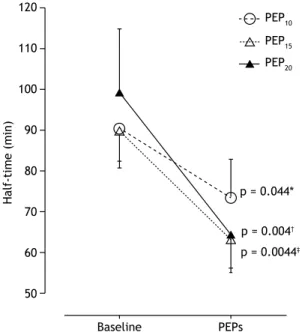
In comparison with the value obtained during sponta-neous breathing, the mean T1/2 was signiicantly lower in the PEP10 group—90.3 ± 25.4 min versus 73.3 ± 30.6 min (p = 0.044), as can be seen in Figure 1. That difference was also signiicant in the PEP15 group—89.8
± 28.9 min versus 63.1 ± 22.1 min (p = 0.044)—and in the PEP20 group—99.3 ± 49.6 min versus 64.5 ±
29.6 min (p = 0.004). Regarding the delta variation
in T1/2 values, no statistically signiicant difference was
found among the groups (p = 0.322).
DISCUSSION
To our knowledge, this was the first study to investigate the effects that different levels of PEP (10, 15, and 20 cmH2O) have on the rate of pulmonary
clearance of 99mTc-DTPA. We demonstrated that PEP
was able to induce increases in epithelial permeability, at all of the levels tested. Another important inding is that no signiicant variation in pulmonary clearance rate was observed among the three groups under the effects of any of the three PEP levels.
The effect of positive end-expiratory pressure (PEEP) on solute clearance has been described as a sigmoidal dose-response relationship dependent on the pressure level applied (5-15 cmH2O); in other words, the rate of pulmonary clearance of 99mTc-DTPA
accelerates exponentially due to the increase in lung volume caused by the administration of different PEEP
levels.(16) Contrary to our hypothesis, we did not ind any differences among the three levels of PEP. In accordance with our results, the study conducted by Bishai et al.(17) showed an increase in the pulmonary
epithelial permeability in mice at a PEEP level of 10 cmH2O. This difference in pulmonary clearance might
be because mice have smaller alveoli and are probably more sensitive to the distension of inter-epithelial junctions of the alveolar epithelium induced by the application of lower pressure levels.
Our indings differ from those reported by Paiva et al.,(18) who assessed the permeability of the alveolar
Table 1. Baseline characteristics of the study groups.a
Variable PEP group p*
PEP10 PEP15 PEP20
(n = 10) (n = 10) (n = 10)
Male gender, n (%) 6 (60) 2 (20) 7 (70) 0.061
Age, years 27.7 ± 5.1 30.4 ± 5.9 26.6 ± 5.1 0.286
Weight, kg 70.7 ± 13.6 60.4 ± 5.2 76.5 ± 11.5 0.009†
Height, cm 173 ± 7.7 165.5 ± 5.5 176.1 ± 8.5 0.017‡
BMI, kg/m2 23.4 ± 2.8 22.1 ± 1.5 24.5 ± 2.1 0.004§
FVC, % of predicted 99.5 ± 15.9 97.1 ± 17.8 99.4 ± 18.5 0.347
FEV1, % of predicted 97.8 ± 12.3 99.3 ± 12.6 98.6 ± 14.4 0.356
aValues are expressed in mean ± SD, except where otherwise indicated. PEP: positive expiratory pressure; PEP
10:
PEP at 10 cmH2O for 30 min; PEP15: PEP at 15 cmH2O for 30 min; PEP20: PEP at 20 cmH2O for 30 min; and BMI: body
mass index. *Between-group comparisons. †Signiicant difference between the PEP
15 group and the PEP20 group (p
= 0.038). ‡Signiicant difference between the PEP
15 group and the PEP20 group (p = 0.027).
§Signiicant difference between the PEP
15 group and the PEP20 group (p = 0.015).
Figure 1. Effects of positive expiratory pressure (PEP) on the pulmonary clearance of technetium-99m-labeled diethylenetriaminepentaacetic acid (99mTc-DTPA). At all PEP
levels, there was a signiicant decrease in the 99mTc-DTPA
half-time (the time for its activity to decrease to 50% of the peak value) in comparison with the baseline (spontaneous breathing) values (analysis of covariance, p < 0.05 for all). PEP10: PEP at 10 cmH2O for 30 min; PEP15: PEP at 15 cmH2O
for 30 min; and PEP20: and PEP at 20 cmH2O for 30 min.
*PEP10. †PEP
20. ‡PEP
15. 120
110
100
90
80
70
60
50
Baseline PEPs
PEP10
PEP15
PEP20
H
a
lf
-t
im
e
(
m
in
)
p = 0.044*
p = 0.004†
p = 0.0044‡
Albuquerque IM, Cardoso DM, Masiero PR, Paiva DN, Resqueti VR, Fregonezi GAF, Menna-Barreto SS
epithelial membrane in 36 healthy individuals submitted to 10 cmH2O and 20 cmH2O under continuous positive
airway pressure (CPAP). The authors showed that 20 cmH2O of CPAP induced an increase in epithelial permeability, whereas 10 cmH2O of CPAP did not.
Previous studies in humans,(19) sheep,(20,21) and dogs(22,23) showed that the application of PEEP increases lung volume and accelerates the rate of pulmonary clearance of 99mTc-DTPA. Therefore, the increased
end-expiratory lung volume produced by PEP reduces airway resistance and promotes an increase in functional residual capacity.(24) However, other factors, such as
airway/alveolar distension, prevention of alveolar collapse during expiration, and recruitment of collapsed alveoli, can also increase functional residual capacity.(25)
In keeping with the indings of the present study, Suzuki et al.(26) observed that, during the application of 20 cmH2O of PEEP, pulmonary clearance of 99mTc-DTPA
increased but returned to baseline values when PEEP was discontinued. That suggests that the increase in
the 99mTc-DTPA pulmonary clearance rate achieved
through the application of PEEP is reversible after breathing returns to atmospheric pressure levels.
The mechanisms by which pulmonary insuflation accelerates the pulmonary clearance of 99mTc-DTPA
REFERENCES
1. West JB. Comparative physiology of the pulmonary blood-gas barrier: the unique avian solution. Am J Physiol Regul Integr Comp Physiol. 2009;297(6):R1625-34. http://dx.doi.org/10.1152/ ajpregu.00459.2009
2. Maina JN, West JB. Thin and strong! The bioengineering dilemma in the structural and functional design of the blood-gas barrier. Physiol Rev. 2005;85(3):811-44. http://dx.doi.org/10.1152/ physrev.00022.2004
3. West JB. Role of the fragility of the pulmonary blood-gas barrier in the evolution of the pulmonary circulation. Am J Physiol Regul Integr Comp Physiol. 2013;304(3):R171-6. http://dx.doi.org/10.1152/ ajpregu.00444.2012
4. Budinger GR, Sznajder JI. The alveolar-epithelial barrier: a target for potential therapy. Clin Chest Med. 2006;27(4):655-69; abstract ix. http://dx.doi.org/10.1016/j.ccm.2006.06.007
5. O’Doherty MJ, Peters AM. Pulmonary technetium-99m diethylene triamine penta-acetic acid aerosol clearance as an index of lung injury. Eur J Nucl Med. 1997;24(1):81-7. http://dx.doi.org/10.1007/ BF01728316
6. Cayir D, Demirel K, Korkmaz M, Koca G. Evaluation of lung epithelial permeability in volatile substance abuse using Tc-99m DTPA aerosol scintigraphy. Ann Nucl Med. 2011;25(8):554-9. http://dx.doi. org/10.1007/s12149-011-0498-7
7. Ogi S, Gotoh E, Uchiyama M, Fukuda K, Urashima M, Fukumitsu N. Inluence of hilar deposition in the evaluation of the alveolar epithelial permeability on 99mTc-DTPA aerosol inhaled scintigraphy. Jpn J Radiol. 2009;27(1):20-4. http://dx.doi.org/10.1007/s11604-008-0288-x
8. Gumuser G, Vural K, Varol T, Parlak Y, Tuglu I, Topal G, et al. Assessment of lung toxicity caused by bleomycin and amiodarone by Tc-99m HMPAO lung scintigraphy in rats. Ann Nucl Med. 2013;27(7):592-9. http://dx.doi.org/10.1007/s12149-013-0722-8 9. Osadnik C, Stuart-Andrews C, Ellis S, Thompson B, McDonald
CF, Holland AE. Positive expiratory pressure via mask does not improve ventilation inhomogeneity more than hufing and coughing in individuals with stable chronic obstructive pulmonary disease and chronic sputum expectoration. Respiration. 2014;87(1):38-44. http:// dx.doi.org/10.1159/000348546
10. Ricksten SE, Bengtsson A, Soderberg C, Thorden M, Kvist H. Effects
of periodic positive airway pressure by mask on postoperative pulmonary function. Chest. 1986;89(6):774-81. http://dx.doi. org/10.1378/chest.89.6.774
11. Myers TR. Positive expiratory pressure and oscillatory positive expiratory pressure therapies. Respir Care. 2007;52(10):1308-26; discussion 1327.
12. Fink JB. Positive pressure techniques for airway clearance. Respir Care. 2002;47(7):786-96.
13. Alcoforado L, Brandão S, Rattes C, Brandão D, Lima V, Ferreira Lima G, et al. Evaluation of lung function and deposition of aerosolized bronchodilators carried by heliox associated with positive expiratory pressure in stable asthmatics: a randomized clinical trial. Respir Med. 2013;107(8):1178-85. http://dx.doi.org/10.1016/j.rmed.2013.03.020 14. Sociedade Brasileira de Pneumologia e Tisiologia. Diretrizes para
testes de função pulmonar. J Pneumol. 2002;28(Suppl 3):S1-238. 15. Pereira CA, Sato T, Rodrigues SC. New reference values for forced
spirometry in white adults in Brazil. J Bras Pneumol. 2007;33(4):397-406. http://dx.doi.org/10.1590/S1806-37132007000400008 16. Marks JD, Luce JM, Lazar NM, Wu JN, Lipavsky A, Murray JF. Effect
of increases in lung volume on clearance of aerosolized solute from human lungs. J Appl Physiol. 1985;59(4):1242-8.
17. Bishai JM, Mitzner W, Tankersley CG, Wagner EM. PEEP-induced changes in permeability in inbred mouse strains. Respir Physiol Neurobiol. 2007;156(3):340-4. http://dx.doi.org/10.1016/j. resp.2006.10.009
18. Paiva DN, Masiero PR, Spiro BL, Jost RT, Albuquerque IM, Cardoso DM, et al. Continuous positive airway pressure and body position alter lung clearance of the radiopharmaceutical 99mtechnetium-diethylenetriaminepentaacetic acid (99mTc-DTPA). Afr J Biotechnol. 2012;11(99):16519-24. http://dx.doi.org/10.5897/AJB12.2563 19. Rinderknecht J, Shapiro L, Krauthammer M, Taplin G, Wasserman
K, Uszler JM, et al. Accelerated clearance of small solutes from the lungs in interstitial lung disease. Am Rev Respir Dis. 1980;121(1):105-17.
20. O’Brodovich H, Coates G, Marrin, M. Effect of inspiratory resistance and PEEP on 99mTc-DTPA clearance. J Appl Physiol (1985). 1986;60(5):1461-5.
21. Cooper JA, van der Zee H, Line BR, Malik AB. Relationship of end-expiratory pressure, lung volume, and 99mTc-DTPA clearance. J remain unclear. Some authors attribute this effect to an increase in the alveolar surface area available for diffusion,(22) due to either the increase in epithelial
permeability,(27) functional alterations in the surfactant
layer,(28) or distension of the intercellular junctions of the alveolar epithelium.(29)
The present study has some limitations. First, we evaluated subjects with healthy lungs. Therefore, any conclusions drawn from this study cannot be applied to patients with lung disease. This needs to be investigated in different, clearly speciied disease groups, especially because PEP is not usually applied in patients with healthy lungs. Second, the upper airway critical closing pressure was not measured. Nevertheless, our indings offer new perspectives on the role of noninvasive ventilation, particularly in relation to PEP and its inluence on the alveolar epithelium.
Effects of positive expiratory pressure on pulmonary clearance of aerosolized technetium-99m-labeled diethylenetriaminepentaacetic acid in healthy individuals
Appl Physiol . 1987;63(4):1586-90.
22. Rizk NW, Luce JM, Hoeffel JM, Price DC, Murray JF. Site of deposition and factors affecting clearance of aerosolized solute from canine lungs, J Appl Physiol Respir Environ Exerc Physiol. 1984;56(3):723-9.
23. Oberdörster G, Utell MJ, Weber DA, Ivanovich M, Hyde RW, Morrow PE. Lung clearance of inhaled 99mTc-DTPA in the dog. J Appl Physiol Respir Environ Exerc Physiol. 1984;57(2):589-95.
24. Finucane KE, Panizza JA, Singh B. Eficiency of the normal human diaphragm with hyperinlation. J Appl Physiol (1985). 2005;99(4):1402-11. http://dx.doi.org/10.1152/japplphysiol.01165.2004
25. Villar J. The use of positive end-expiratory pressure in the management of the acute respiratory distress syndrome. Minerva Anestesiol. 2005;71(6):265-72.
26. Suzuki Y, Kanazawa M, Fujishima S, Ishizaka A, Kubo A. Effect of external negative pressure on pulmonary 99mTc-DTPA clearance in humans. Am J Respir Crit Care Med. 1995;152(1):108-12. http:// dx.doi.org/10.1164/ajrccm.152.1.7599807
27. Mason GR, Mena I, Maublant, J, Sietsma K, Effros RM. The effect of PEEP and posture on the clearance of inhaled small solutes from the lungs in normal subjects. Am Rev Respir Dis. 1984;129(4 Pt 2):A346. 28. Groth S. Pulmonary clearance of 99mTc-DTPA. An index of alveolar
epithelial permeability. Dan Med Bull. 1991;38(3):189-203.
29. Ludwigs U, Philip A. Pulmonary epithelial permeability and gas exchange: a comparison of inverse ratio ventilation and conventional mechanical ventilation in oleic acid-induced lung injury in rabbits. Chest. 1998;113(2):459-66. http://dx.doi.org/10.1378/ chest.113.2.459
http://dx.doi.org/10.1590/S1806-37562016000000057
ABSTRACT
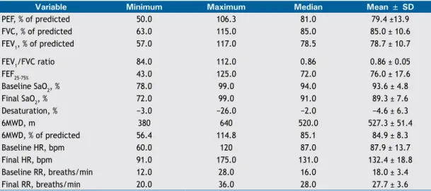
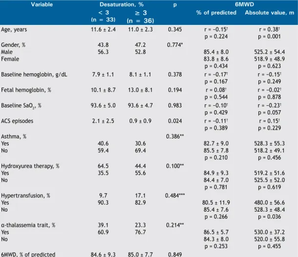
Objective: To evaluate pulmonary function and functional capacity in children and adolescents with sickle cell disease. Methods: This was a cross-sectional study involving 70 children and adolescents (8-15 years of age) with sickle cell disease who underwent pulmonary function tests (spirometry) and functional capacity testing (six-minute walk test). The results of the pulmonary function tests were compared with variables related to the severity of sickle cell disease and history of asthma and of acute chest syndrome. Results: Of the 64 patients who underwent spirometry, 15 (23.4%) showed abnormal results: restrictive lung disease, in 8 (12.5%); and obstructive lung disease, in 7 (10.9%). Of the 69 patients who underwent the six-minute walk test, 18 (26.1%) showed abnormal results regarding the six-minute walk distance as a percentage of the predicted value for age, and there was a ≥ 3% decrease in SpO2 in 36 patients (52.2%). Abnormal pulmonary function was not signiicantly associated with any of the other variables studied, except for hypoxemia and restrictive lung disease. Conclusions: In this sample of children and adolescents with sickle cell disease, there was a signiicant prevalence of abnormal pulmonary function. The high prevalence of respiratory disorders suggests the need for a closer look at the lung function of this population, in childhood and thereafter.
Keywords: Anemia, sickle cell; Respiratory function tests; Exercise test.
Pulmonary function in children and
adolescents with sickle cell disease: have
we paid proper attention to this problem?
Ana Karine Vieira1, Cristina Gonçalves Alvim2,
Maria Cristina Marquez Carneiro3, Cássio da Cunha Ibiapina4
Correspondence to:
Cássio da Cunha Ibiapina. Avenida Professor Alfredo Balena, 110, CEP 30160-042, Belo Horizonte, MG, Brasil. Tel.: 55 31 3409-9772 or 55 31 9976-7871. Fax: 55 31 3409-9772. E-mail: [email protected] Financial support: None.
INTRODUCTION
Sickle cell disease (SCD) is the most common monogenic disease in Brazil. The number of individuals with SCD in Brazil is estimated to range from 25,000 to 30,000.(1)
The incidence of SCD in the state of Minas Gerais, Brazil, is approximately 1:1,400 live births, according to the Minas Gerais State Neonatal Screening Program.(2) The
manifestations of this disease result from a predominance of sickle-shaped red blood cells, which leads to chronic hemolytic disease and vaso-occlusive phenomena.(2) SCD
leads to multisystem impairment, with lung involvement being a major cause of morbidity and mortality.(3)
Some studies have been published on assessment of pulmonary function in adults with SCD, showing that the major abnormality is restrictive lung disease (RLD). (4,5) Since the 1970s, studies have been published on pulmonary function in the pediatric age group with SCD,(6) with conlicting results being reported; however, most studies show that obstructive lung disease (OLD) is the most common abnormality.(7.8) RLD is also common in some pediatric studies, highlighting the importance of pulmonary function testing in such children.(9,10)
Another important respiratory disease affecting children with SCD is asthma. Asthma is known to be a comorbidity impacting the course of SCD, leading to increased morbidity and mortality.(11,12) Several studies have reported an association between asthma and an increased number of vaso-occlusive crises or acute
chest syndrome (ACS) episodes.(11-14) An association between asthma and pulmonary hypertension in children was described by Hagar et al., raising the suspicion of a shared mechanism, possibly associated with chronic hemolysis.(15)
The mortality associated with lung disease is a serious problem in the SCD population.(16,17) It has been demon-strated that abnormalities in pulmonary function tests are early objective signs of the development of chronic lung disease in SCD.(16) It has recently been published that decreased FEV1 is associated with increased mortality in
adults with SCD.(18) Nevertheless, few authors in Brazil have attempted to investigate pulmonary function in patients with SCD.(19-21) We hypothesize that the onset of pulmonary function abnormalities in SCD occurs as early as childhood. To address this challenge, the objective of the present study was to assess abnormalities in pulmonary function and functional capacity in children and adolescents with SCD, by means of spirometry and the six-minute walk test (6MWT), and to compare these abnormalities with clinical and laboratory variables in these patients.
METHODS
This was a descriptive analytical cross-sectional study. We included children and adolescents aged 8 to 15 years who were enrolled in the Minas Gerais State Neonatal
1. Fundação Hemominas, Belo Horizonte (MG) Brasil.
2. Departamento de Pediatria, Faculdade de Medicina, Universidade Federal de Minas Gerais, Belo Horizonte (MG) Brasil.
3. Grupo de Pneumologia Pediátrica, Faculdade de Medicina, Universidade Federal de Minas Gerais, Belo Horizonte (MG) Brasil.
4. Departamento de Pediatria, Faculdade de Medicina, Universidade Federal de Minas Gerais, Belo Horizonte (MG) Brasil.
Submitted: 9 March 2016.
Accepted: 31 October 2016.
Study carried out under the auspices of the Programa de Triagem Neonatal de Minas Gerais and at the Faculdade de Medicina, Universidade Federal de Minas Gerais, Belo Horizonte (MG) Brasil.
J Bras Pneumol. 2016;42(6):409-415