1808-8694/$ - see front matter. © 2014 Associação Brasileira de Otorrinolaringologia e Cirurgia Cérvico-Facial. Published by Elsevier Editora Ltda. All rights reserved.
www.bjorl.org.br
Brazilian Journal of
OTORHINOLARYNGOLOGY
Authors
Associação Brasileira de Otorrinolaringologia e Cirurgia Cér-vico Facial (Brazilian Association of Otolaryngology and Neck and Facial Surgery)
Academia Brasileira de Neurologia (Brazilian Academy of Neurology)
Sociedade Brasileira de Cardiologia (Brazilian Society of Cardiology)
Sociedade Brasileira de Pediatria (Brazilian Society of Pediatrics) Sociedade Brasileira de Pneumologia e Tisiologia (Brazilian Society of Pneumology and Tisiology)
Participants
Zancanella E, Haddad FM, Oliveira LAMP, Nakasato A, Duarte BB, Soares CFP, Cahali MB, Eckeli A, Caramelli B, Drager L, Ramos BD, Nóbrega M, Fagondes SC, Andrada NC
Final development June 11, 2012.
Description of the evidence collection method
An active search was conducted in the PubMed/MEDLINE, EM-BASE, SciELO/LILACS, and Cochrane Library databases using the following key words (MeSH terms): Epworth, Berlin ques-tionnaire, physical examination, body mass index, circumfer-ence, Mallampati, noise, pharynx, airway, Jaw, Diagnosis, Mass Screening, Diagnostic Techniques and Procedures, Diagnostic Tests, Laboratory Techniques and Procedures, Routine; Diagnos-tic Equipment/standards*; Comparative Effectiveness Research, Laryngoscopy, Cephalometry, Tomography, X-Ray Computed, Magnetic Resonance Imaging, Endoscopy, Pulmonary Ventilation, Polysomnography, Actigraphy, Sleep; Monitoring, Physiologic; Monitoring Sleep Apnea Syndromes, Sleep Disorders, Sleep Ap-nea, Obstructive; Sleep Initiation and Maintenance Disorders, Circadian Rhythm, Sleep, REM/physiology*, Snoring, Disorders of Excessive Somnolence, Restless legs Syndrome, signs and symptoms, Fatigue, Headache, Delirium, Dementia, Amnestic, Cognitive Disorders, Mood Disorders, Fatigue Syndrome, Chron-ic; Questionnaires, survey Ambulatory, home care services, lab-oratory techniques and procedures, complications, adverse ef-fects, Obesity, Overweight, Cardiovascular Diseases, Diabetes Mellitus, Stroke, Ischemic Attack, Transient; Gastroesophageal Relux, Pulmonary Disease, Chronic Obstructive, Pre-Eclampsia,
Pregnancy, Premature Birth, Post-menopause, Memory Disorders, Mental Disorders, Cognition Disorders, Neuropsychological Tests, Severity of Illness Index, Accidents, Trafic; Mortality.
Degree of recommendation and strength of evidence A: Experimental or observational trials of higher consistency. B: Experimental or observational trials of lesser consistency. C: Case reports (non-controlled trials).
D: Opinions without critical evaluation, based on consensus-es, physiological studiconsensus-es, or animal models.
Objective
To evaluate the diagnoses of obstructive sleep apnea and pri-mary snoring in adults and children, focusing on data from med-ical history, questionnaires, physmed-ical examination, and labora-tory tests, as well as stimulating their investigation by general practitioners and several specialists.
Introduction
Obstructive sleep apnea (OSA) is characterized by recurrent collapse of the pharynx during sleep, resulting in a substantial decrease in airlow (apnea or hypopnea). Respiratory events trigger intermittent disorders of blood gases (hypoxemia and hypercapnia) and can lead to sympathetic system activation. Obstructive sleep apnea syndrome (OSAS) is associat-ed with many symptoms and comorbidities, which include excessive daytime sleepiness, cognitive problems, obesi-ty, type 2 diabetes mellitus, hypertension, exacerbation of chronic obstructive pulmonary disease (COPD), reduced quality of life, and signiicant increase in risk of industrial and trafic accidents. It is also considered an independent risk factor for cardiovascular disease and ischemic stroke.
Upper airway collapse during sleep is the result of an im-balance between the activity of pharyngeal dilator muscles and negative intraluminal pressure during inspiration. Fac-tors that tend to narrow the pharynx lumen include mucosal adhesive forces, vasomotor tone, neck lexion, jaw open -ing and lower dislocation, force of gravity, increased nasal resistance, Bernoulli effect (the physics principle that ex-plains the tendency of pharyngeal collapse), and increased
Obstructive sleep apnea and primary snoring: diagnosis
GUIDELINE
DOI: 10.5935/1808-8694.2014S001
dynamic compliance. Forces that dilate the pharynx include the thoracic caudal traction by increased pulmonary volume and neck extension.
Despite showing considerable variation between individ-uals, there are components of the disease physiopathology that have been already demonstrated, which include chang-es in the upper airway anatomy, variations in the capacity of the upper airway dilator muscles to respond to respiratory adversities during sleep, changes in cortical arousal thresh-old during an increase in inspiratory negative pressure, vari-ations in the ventilatory control system stability, and chang-es in pulmonary volume.
OSAS is thought to be a progressive disease, and it is hy-pothesized that primary snoring and severe OSAS are oppo-site stages of the same disease. This pathological evolution would occur in the following chronological order: primary snoring, upper airway resistance syndrome, OSA, mild OSAS, moderate OSAS, and severe OSAS. Prompt diagnosis and ap-propriate treatment are important at any of these stages.
1. What is the clinical history of the patient with
OSA? How important are questionnaires?
The most frequent complaints of adult patients with OSA, when compared to non-apneic patients, are the presence of snor-ing, nocturnal choksnor-ing, excessive daytime sleepiness (EDS), impotence, and nocturnal apneas reported by companions (p
< 0.05)1 (B). Other common symptoms include morning head-aches, unrefreshing sleep, fatigue, and cognitive alterations.
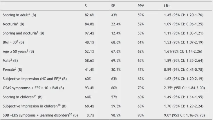
Snoring and nocturia are common complaints in OSA2 (B). Other clinical parameters such as body mass index (BMI), age, and gender are evaluated in Table 1. Men and women aged > 50 years diagnosed with OSA did not differ re-garding the nature or severity of symptoms, as assessed by polysomnography (PSG) or the complaints of snoring, EDS, and perception of impaired diurnal function3 (A).
Associating the subjective impression, which includes the clinical history, with physical examination and the PSG result of the apnea-hypopnea index (AHI) > 10 h allows for increased diagnostic certainty of OSA1 (B).
To differentiate patients with and without apnea among snorers, the presence of OSAS (witnessed apnea, noctur-nal choking, morning headache, or EDS) and alterations in the Epworth sleepiness scale (ESS), which must be ≥ 15 and BMI ≥ 28 kg/m2, are assessed. The sensitivity to identify non-apneic individuals was 93.4% and the speciicity was 60% (p < 0.001)4 (B). This association of criteria is the best way to attain the clinical diagnosis of OSAS. When considering the disease prevalence of 15%, the presence of this association of criteria increases disease probability from 15% to 29% of cas-es, requiring complementary diagnostic conirmation by PSG.
Developed as a screening method for the detection of patients at high risk of OSA in primary care centers, the Ber-lin Questionnaire (BQ) (Box 1) has a sensitivity of 69% to 86% and speciicity of 56% to 95% (positive predictive value of
Table 1 Signs and symptoms suggestive of obstructive sleep apnea (OSA) and their respective likelihood ratios that contribute to the OSA diagnosis.
S SP PPV LR+
Snoring in adult2 (B) 82.6% 43% 59% 1.45 (95% CI: 1.20-1.76)
Nocturia2 (B) 84.8% 22.4% 52% 1.09 (95% CI: 0.96-1.25)
Snoring and nocturia2 (B) 97.4% 12.4% 53% 1.11 (95% CI: 1.03-1.21)
BMI > 302 (B) 48.1% 68.6% 61% 1.53 (95% CI: 1.07-2.19)
Age ≥ 50 years2 (B) 52.1% 67.6% 62% 1.61(95% CI: 1.14-2.26)
Male2 (B) 58.6% 69.5% 65% 1.89 (95% CI: 1.35-2.64)
Female2 (B) 41.4% 30.5% 37% 0.59 (95% CI: 0.45-0.78)
Subjective impression (HC and EF)a (B) 60% 63% 62% 1.62 (95% CI: 1.20-2.19)
OSAS symptomsa + ESS ≥ 10 + BMI (B) 93.4% 60% 70% 2.35a (95% CI: 1.84-3.00)
Snoring in children21 (B) 64% 57% 60% 1.49 (95% CI: 1.14-1.95)
Subjective impression in children20 (B) 68.4% 59.5% 63% 1.70 (95% CI: 1.29-2.24)
SDB +EDS symptoms + learning disorders20 (B) 8.7% 98.9% 90% 9.0a (95% CI: 1.16-69.73)
S, sensitivity; SP, specificity; PPV, positive predictive value; LR+, positive likelihood ratio; BMI, body mass index; SDB, sleep-disordered breathing.
a Obstructive sleep apnea syndrome (OSAS) symptoms: snoring, nocturnal choking, EDS, apneas witnessed by others, morning
77% to 96%)5-9 (B). However, for the assessment of patients in sleep centers, it did not indicate favorable results due to high rates of false-positive and false-negative results, with a sensitivity of 61.5% to 62% and speciicity of 22.6% to 43%, not allowing for an increase in diagnostic certainty10,11 (B). The validation of the Brazilian Portuguese version of the BQ in sleep centers identiied 68.4% of the studied population as high-risk for OSA and 31.6% as low-risk. The sensitivity and speciicity values of the BQ change in relation to AHI, but even in patients at high-risk for OSA, the altered BQ has a positive likelihood ratio (LR +) of 1.44 to 1.4912 (A).
There is an association between alterations at the BQ in the population at high risk for OSA and patients with sys-temic arterial hypertension (SAH) resistant to clinical treat-ment13 (B). Having SAH resistant to clinical treatment is a risk factor for OSA in the Brazilian population, with a sen-sitivity of 44% (31% to 58%), speciicity of 91% (77% to 97%), increasing the diagnostic certainty of OSA from 15% to 46% (LR+ = 4.89; 95% CI: 2.52-9.47)14 (A)15 (B).
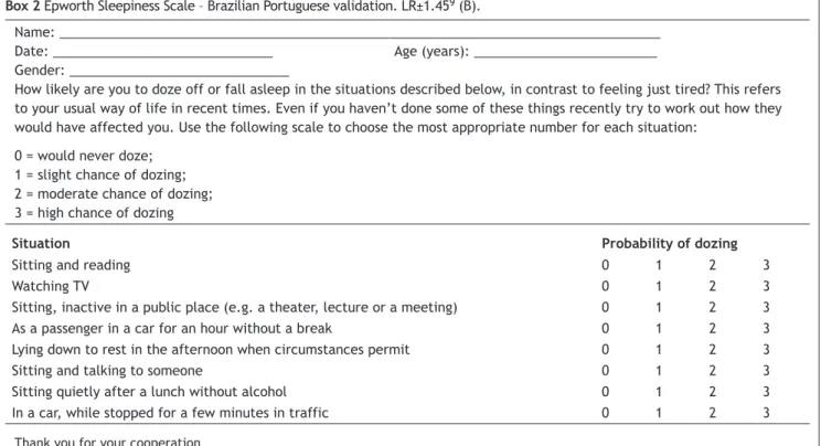
The ESS validated for Brazilian Portuguese16 (B) (Box 2), is very important in the identiication of EDS (ESS > 10), as -sisting in the screening of patients with OSAS, mainly when
associated with other clinical parameters4,10,17,18 (B). Pa-tients with ESS scores > 10 have a 2.5-fold higher risk of having OSA when compared with a normal test17 (B). The prevalence of sleepiness (ESS > 10) increased with OSA se-verity, ranging from 21.4% (AHI < 5/h) to 40.2% (AHI > 30/h) (p < 0.001). However, less than half of patients with moder-ate to severe OSA reported somnolence (45.7%)19 (B). The scale has a sensitivity of 48% and speciicity of 67%, provid -ing a LR+ = 1.45 (1.03-2.06)9 (B).
In children with sleep-disordered breathing (SDB), most associated symptoms are snoring, EDS, learning disorders, as well as somnambulism and somniloquy. Children with report of loud and frequent snoring have a 3.5-fold higher chance of having SDB, and children with EDS have a higher chance of presenting learning disorders and of male gender. The combination of the symptoms snoring or EDS with learning disorders has high speciicity (97% and 98.9%, respectively), but low sensitivity (8.7% and 4.4%, respectively)20 (B).
In preschoolers, the presence of snoring often or almost always has sensitivity of 64% and speciicity of 57%21 (B). Clinical evaluation has sensitivity of 68.4% and speciicity of 59.5% for the diagnosis of OSA in children22 (B).
Box 1 Berlin Questionnaire; LR+ 1.44 – 1.4920 (B).
Category 1
1. Do you snore?
( ) Yes ( ) No ( ) Don’t know
2. Your snoring is:
Slightly louder than breathing? As loud as talking
Louder than talking?
Very loud - can be heard in adjacent rooms?
3. How often do you snore?
Nearly every day 3-4 times a week 1-2 times a week Never or nearly never
4. Has your snoring ever bothered other people?
( ) Yes ( ) No
5. Has anyone noticed that you quit breathing during your sleep?
Nearly every day 3-4 times a week 1-2 times a week Never or nearly never
Category 2
6. How often do you feel tired or fatigued after your sleep?
Nearly every day 3-4 times a week 1-2 times a week Never or nearly never
7. During your waking time, do you feel tired, fatigued or not up to par?
Nearly every day 3-4 times a week 1-2 times a week Never or nearly never
8. Have you ever nodded off or fallen asleep while driving a vehicle?
( ) Yes ( ) No
Category 3
9. Do you have high blood pressure?
( ) Yes ( ) No
( ) I don’t know BMI =
Scoring of questions: Any assigned response is considered positive.
Children with symptoms of SDB may have more EDS (OR = 2.2; 95% CI: 1.7-2.8) and behavioral problems, in-cluding hyperactivity (OR = 2.5; 95% CI: 2.0-3.0), attention deicit disorder (OR = 2.1; 95% CI: 1.7- 2.6), and aggression (OR = 2.1; 95% CI: 1.6 -2.6)23 (B). They may also have alter-ations in growth, central auditory processing, and nocturnal enuresis24,25 (C).
Table 1 compares the diagnostic values of different signs and symptoms suggestive of OSAS. The higher the positive likelihood ratio (LR+), the better. For example: a LR+ = 9 signiies that children with symptoms of SDB, EDS, and learning disorder have a nine-fold higher chance of having a conirmed diagnosis of OSA.
Recommendation
There is an increased likelihood of OSA diagnostic certainty in adults when the presence of symptoms is associated with alterations in ESS and increase in BMI4 (B), whereas in young children the clinical diagnosis of OSA is associated with the presence of SDB symptoms, EDS, and learning disorders20 (B), as highlighted by the a in Table 1, indicating a 2.3-fold and a nine-fold higher chance of OSA diagnosis, respectively.
The main symptoms of adult patients with OSA are snor-ing, nocturnal choksnor-ing, EDS, impotence, and reporting of apneas by companions1 (B). The combination of snoring with nocturia can be used in OSA screening2 (B). There are dif-ferences between men and women older than 50 years re-garding the nature and severity of symptoms3 (A). Patients with SAH resistant to clinical treatment are more likely to have OSA; thus, they should always be assessed to rule out the disease14 (A) 15 (B).
Children with SDB are more likely to have behavioral problems, including hyperactivity, attention deicit, aggres
-sion23 (B), and enuresis24,25 (C), as well as alterations in growth and central auditory processing24,25 (C).
The Berlin Questionnaire (BQ) helps in the screening of patients at high risk of OSA in primary care centers5-8 (B), but it does not allow for a deinite diagnosis of OSA by itself12 (A).
The ESS, along with other clinical parameters, helps to identify patients with OSA4,10,17,18 (B). Although the preva-lence of ESS > 10 increases with OSA severity, less than 50% of patients with moderate to severe OSA have ESS > 1019 (B). The diagnostic contribution of the questionnaires is similar for both ESS and the BQ, with 1.45- 9 (B) and 1.44-1.49-fold12 (A) higher chances of disease when the questionnaire results are altered, respectively.
2. What are the most important indings during
physical examination of patients with OSA and
pri-mary snoring?
The most relevant indings of the physical examination in adult patients with snoring/OSAS are obesity and alterations in the craniofacial skeleton and upper airways (UAs).
In addition to older age (> 50 years11)26 (B) and male gender26-28 (B), obesity markers, particularly increased BMI and neck circumference, are the main predictors of OSA presence1,26-30 (B); however, the association between the degree of obesity and OSAS severity is still controver-sial31,32 (B).
Using the AHI > 10/h, the prevalence of OSA in white men is 3.9%, while in women is 1.2%, maintaining statis-tically signiicant male:female ratio of 3.3:1 (p < 0.0006). This prevalence is modiied when studying premenopausal women (0.6%) or postmenopausal women using hormone replacement therapy (0.5%); postmenopausal women with-Box 2 Epworth Sleepiness Scale – Brazilian Portuguese validation. LR±1.459 (B).
Name: __________________________________________________________________________________ Date: ______________________________ Age (years): _________________________ Gender: ______________________________
How likely are you to doze off or fall asleep in the situations described below, in contrast to feeling just tired? This refers to your usual way of life in recent times. Even if you haven’t done some of these things recently try to work out how they would have affected you. Use the following scale to choose the most appropriate number for each situation:
0 = would never doze; 1 = slight chance of dozing; 2 = moderate chance of dozing; 3 = high chance of dozing
Situation Probability of dozing
Sitting and reading 0 1 2 3
Watching TV 0 1 2 3
Sitting, inactive in a public place (e.g. a theater, lecture or a meeting) 0 1 2 3
As a passenger in a car for an hour without a break 0 1 2 3
Lying down to rest in the afternoon when circumstances permit 0 1 2 3
Sitting and talking to someone 0 1 2 3
Sitting quietly after a lunch without alcohol 0 1 2 3
In a car, while stopped for a few minutes in traffic 0 1 2 3
out hormone replacement present values that are similar to those in men (2.7%)33 (A). In an epidemiological study conducted in the city of São Paulo, using clinical and poly-somnographic criteria, the prevalence of OSA was 32.9% (95% CI: 29.6-36.3%), while maintaining independent associ-ations in men (OR = 4.1; 95% CI 2.9-5.8), obese individuals (OR = 10.5; 95% CI: 7.1-15.7), and postmenopausal women (OR = 21; 95% CI: 1.4-3.9). There is an increase in OSA with increasing age, reaching an OR = 34.5 (95% CI: 18.5-64.2%) when compared with Brazilian groups aged 60-80 years to groups aged 20-29 years30 (B).
BMI < 32.3 kg/m2 was associated with OSAS of 0.4% (95% CI: 0.1-1.2), and BMI ≥ 32.3 kg/m2 was associated with OSAS of 4.8% (95% CI: 2.5-9.0)33 (A). When using BMI ≥ 32.3 kg/m2 in the assessment of the physical examination of patients with snoring/OSA, a sensitivity of 92.5% (95% CI: 89.3%-95.8%) and speciicity of 73.9% (95% CI: 61.2%-86.6%) were observed, increasing the LR+ from 1.532 (B) to 3.5428 (B).
The neck circumference alone has a sensitivity of 60.6% (95% CI: 54.6 to 66.6%) and speciicity of 93.4% (95% CI: 86.3%-100%), providing LR+ of 10.00 (95% CI: 4.53-22.07), increasing diagnostic probability from 15% to 64%28 (B). The association of age > 50 years, neck circumference > 40 cm, and ESS > 10 increases the diagnostic certainty of OSA from 15% to 80% of cases. When applying the Kushida morphometric model to the Brazilian population, it was observed that the mean value of 36.7 cm (31 to 43 cm) can distinguish apneic from non-apneic individuals; in the sample studied, apneic individuals had a mean neck cir-cumference of 40.4 cm (with a standard deviation of 4.1 cm), ranging from 31 to 54 cm34 (B).
Through a morphometric model that associates BMI, neck circumference, and evaluation of craniofacial skele-ton, considering a result found > 70, a sensitivity of 97.6% (95% CI: 95%-98.9%) and speciicity of 100% (95% CI: 92%-100%) were observed. The morphometric model provides a LR+ = 97 (95% CI: 13.79-682). The use of this morpho-metric model should be encouraged, as it increases the likelihood of disease from 15% to 95% of cases28 (B). In the Brazilian population, the morphometric model main-tained the score > 7034 (B).
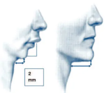
Craniofacial alterations more often related to OSAS are those caused by maxillary and/or mandibular hypoplasia (Fig. 1), which can be identiied by physical examination and conirmed by cephalometry28,35,36 (B). In the Brazilian population, class II dental occlusion (retropositioned lower dental arch) was observed in 26.3% of cases, alterations in the hard palate (narrow or ogival) in 25.1% of cases, and mandibular hypoplasia in 19.7% of cases36 (B) (Fig. 2).
Several anatomical alterations in the upper airways are described in patients with OSA. The most common indings are: nasal alterations; hyperplastic tonsils (Fig. 3); modi-ied Mallampati index classes III and IV (Fig. 4) (inadequate association between the base the tongue and the orophar-ynx); and alterations in the soft palate, uvula, and tonsillar pillars1,32,36,37 (B). In a Brazilian study, the most frequent indings in patients with OSA were the alterations in the soft palate (43.0%), modiied Mallampati index classes III and IV (78.8%), abnormal tonsillar pillars (30.9%), uvular alterations (34.5%), septal deviations grade III (5.8%), and turbinate hypertrophy (49.8%)36 (B). The combination of BMI, modiied Mallampati index, and presence of abnormal
Figure 1 Retrognathia.
Figure 2 Class II dental occlusion (Angle).
Figure 3 Grading system for palatine tonsils.
Figure 4 Modiied Mallampati index.
Grade I
Grade I
Grade II
Grade II
Grade III
Grade III
Grade IV
pharynx anatomy are related to OSA presence and severity in Brazilians38 (B).
Thus, it can be stated that patients with OSA are more obese and have higher values of neck circumference than control patients; however, the more obese patients do not always manifest more severe disease31,32 (B). The preva-lence of OSA in patients with class III obesity was shown to be greater than in the general population32 (B). In a study in the Brazilian population, signiicant predictive factors for OSA in class III obese individuals were: mean age 44.6 ± 10.6 years and increased neck circumference, with a mean of 44.6 ± 5.2 cm32 (B).
Recommendation
On physical examination of patients with snoring/OSA, the following factors must be taken into consideration: neck circumference measurements28 (B), male gender26-28 (B), (as there is a 3.3:1 ratio of men: pre-menopausal wom-an)33 (A), older age (> 50 ± 11 years)26 (B), and BMI val-ues28 (B). The most signiicant individual inding at the physical examination in patients with snoring/OSA is the neck circumference measure. The most relevant asso-ciation in the physical examination includes BMI, neck cir-cumference, and craniofacial skeleton assessment, called the morphometric model2,28,34 (B). At the evaluation of the craniofacial skeleton, the anatomical alterations in the upper airways (UAs)1,32,36,37 (B) and craniofacial ab-normalities28,35,36 (B) must be investigated. It is important to recall that patients with OSA are more obese, but the association between the degree of obesity and OSA severi-ty is still controversial31,32 (B).
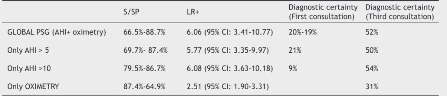
3. When should PSG evaluation be indicated?
PSG is a complementary test considered to be the gold stan-dard, supporting the diagnosis and follow-up of OSAS39 (B). Depending on the parameter alteration found during the ex-amination, there will be different diagnostic probabilities, as described in Table 2. Overall, PSG provides a diagnostic certainty of 20% in a population with low prevalence of the disease (primary care with an estimated prevalence of 4%), while in the tertiary care population, with an estimated prevalence of 15%, the diagnostic certainty reaches 54% if AHI > 1039,40 (B). The variability between nights of sleep can show conlicting results when the same patient is be -ing monitored, which does not rule out the need for a new examination41,42 (B). The AHI correlation between two PSG
assessments in the same patient at 30-day intervals is poor (r = 0.44)41 (B), and this variability of single-night PSG has an impact on diagnosis; in practice, approximately 13% of patients beneit from undergoing a second PSG assessment42 (B). Since the PSG cannot conirm the diagnosis of OSA alone with AHI ranging from 5 to 15, it is necessary to associate the PSG results with the medical history and physical ex-amination indings. Associating the subjective impression, which includes the medical history with physical examina-tion and the PSG result of AHI > 10 h allows for an increase in the diagnostic certainty of AOS1 (B). Considering the pop-ulation with a low prevalence of the disease, this increases the likelihood from 4% to 28%, and when the population has a pretest prevalence of 15%, there is a 63% disease prob-ability. In primary care, patients with neck circumference > 40 cm and PSG alterations will have an increase in the probability of disease from 20% to 71%. In tertiary care, pa-tients with the same alteration in neck circumference and PSG alterations will demonstrate an increase in the proba-bility of disease from 54% to 91%.
PSG can conirm the diagnosis of OSA alone when the AHI ≥ 15; however, this AHI value seldom appears alone, as it is often associated with BMI and neck circumference alterations29 (B).
Epidemiological data including age range and gender show a higher prevalence of OSA in men with AHI > 15/h with OR = 2.7 (95% CI: 2.34-3.12)29 (B) and age ≥ 5026 (B).
Predictive factors obtained from the history and physical examination are suggestive of the presence of OSA, but the disease diagnosis with data concerning their intensity will only be attained by monitoring the patient’s sleep, even in snoring patients43 (B).
The presence of EDS investigated by ESS correlates with an increase in apneic episodes at the PSG (AHI < 5 in 21%, AHI > 5 in 28%, and AHI ≥ 30 in 35% of cases)23 (B). A study that evaluated the ESS in 6,440 patients reported EDS (ESS > 10) in 1,149 patients (46%) with an AHI ≥ 1519 (B).
The presence of snoring has been associated to the OSA diagnosis, presenting a sensitivity of 97.4%, speciicity of 40%, positive predictive value of 82.3%, and negative pre-dictive value of 84.2% for moderate to severe OSAS in a group with BMI > 2544 (B). There is an association of EDS (ESS > 10) and frequent snoring (more than six nights a week) in patients with AHI > 1545 (B).
In the presence of arterial hypertension, the correlation with severe OSA (AHI > 30) increased to 67% and, compared with patients with AHI < 15, an OR = 2.27 (95% CI: 1.76-2.92) was observed46 (B). Another study demonstrated a correla-tion between severe OSAS in patients with BMI > 3047 (B).
Table 2 Diagnostic probability by PSG during part of the night39 (B).
S/SP LR+ Diagnostic certainty
(First consultation)
Diagnostic certainty (Third consultation)
GLOBAL PSG (AHI+ oximetry) 66.5%-88.7% 6.06 (95% CI: 3.41-10.77) 20%-19% 52%
Only AHI > 5 69.7%- 87.4% 5.77 (95% CI: 3.35-9.97) 21% 50%
Only AHI >10 79.5%-86.7% 6.08 (95% CI: 3.63-10.18) 9% 54%
Obesity measured by BMI has been frequently associated with OSAS. Studies indicate that AHI worsens with increas-ing BMI26,44 (B) and demonstrates an association of OSAS in patients with BMI > 3548 (B).
Recommendation
PSG should be indicated in patients with clinical suspicion of OSA and the presence of snoring44,45 (B) associated or unas-sociated with EDS assessed by ESS19,23 (B), neck circumfer-ence > 40 cm, obesity26,44,48 (B), and arterial hypertension46 (B), especially in the context of dificult-to-control hyper -tension14 (A)15 (B). The variability between nights of sleep sometimes requires the performance of a second PSG41,42 (B).
The differential diagnosis between primary snoring and OSA can only be established after sleep monitoring39,43 (B).
4. What are the sleep monitoring modalities and
when should they be requested?
There are four modalities of sleep monitoring: • Type I
Performed in a sleep laboratory with > seven channels for monitoring
• Type II
Non-assisted with > seven channels for monitoring • Type III
Monitoring with four to seven channels • Type IV
Monitoring with one or two channels, of which one is oximetry
The gold standard type-I PSG examination consists of the evaluation through at least seven channels to capture the physiological variables including electroencephalogram, electromyogram (chin and tibial), electrooculogram, air-low, respiratory effort, oxygen saturation, electrocardio -gram, body position, and snoring. It is performed in a sleep laboratory, assisted by a PSG technician, with a minimum of six hours of monitoring; the data are interpreted by a physician qualiied to interpret a report39,49,51 (B) 52,53 (D).
Monitoring with portable equipment is classiied by the number of capture channels available in each device. These tests may be assisted by a PSG technician, allowing for the examination to be performed in the patient’s home54,56 (D). A major limitation is the loss of monitoring channels due to failure, or loosening or disconnection of sensors, which has been estimated at between 4% and 33%, and the variability of equipment and technologies involved in the test57 (D).
Portable sleep monitoring type II (PSM II) (comprehen-sive) comprises at least seven channels, including electro-encephalogram, chin electromyogram, electrooculogram, airlow, respiratory effort, heart rate, and oxygen satura -tion. It allows for the identiication of the different sleep stages with demonstration of statistics and calculations of AHI/h. Its limitation is the fact that it requires the techni-cian to go to the patient’s residence to set up the equip-ment and remove it on the following day, but if a channel is disconnected during the examination, there is no replace-ment54-56 (D) 57 (D). PSM type II has shown similar results for AHI during at-home monitoring when compared to
laborato-ry assessment58 (B). It is estimated to have 70% of sensitivity and 91% of speciicity59 (B).
Portable monitoring type III (cardiopulmonary) uses be-tween four and seven channels, including oxygen saturation, airlow, respiratory effort, and heart rate. It does not assess sleep stages and does not differentiate whether the events occur during the periods of wakefulness or sleep. It demon-strates and differentiates only respiratory events, not al-lowing for the diagnosis of other events, such as lower-limb movements. Some devices can be set up by the patient at home, without the need for a technician55-57 (D)58 (B). In a study of Brazilian patients, when indices were compared to type I monitoring, they showed results with strong correla-tion, with r = 0.876 (95% CI: 0.81-0.91; p < 0.0001) for any value of AHI (> 5, > 15, and > 30)60 (A). Another equipment model presented similar results61 (B).
Type IV monitoring uses one to two channels, and one of them must be oximetry. It does not assess sleep stag-es and dostag-es not differentiate between apnea typstag-es, but demonstrates desaturation. It does not allow for the eval-uation of any data related to sleep55-57 (D)58 (B). In a study with Brazilian patients, similar results were observed when comparing the Type IV portable monitoring performed ei-ther in the sleep laboratory or at home62 (A). Considering the high prevalence of OSA (33% in the population of São Paulo, SP, Brazil)30 (B), type IV portable monitoring in-creases the disease likelihood to 57%62 (A). Using the same type of equipment, studies have compared the rates of AHI with type 1 PSG and observed signiicant correlations, with r = 0.9563 (B) and r = 0.89564 (B). Due to the fact that it is easy to repeat, three assessments were performed with a portable monitor on three consecutive nights, with no signiicant differences found between the values in these three examinations63 (B).
The indication for type III and IV monitoring are still re-stricted to patients with high probability of OSA, whose as-sessment is based on anamnesis, physical examination, and questionnaires. If these types of monitoring do not diagnose OSA, type I or II monitoring is indicated to rule out a false negative result56,57 (D).
PSG for titration of positive airway pressure (PAP) im-plies that the patient will return for a new sleep monitoring, assisted by technician in a sleep laboratory. The choice of treatment with PAP requires the identiication of values at which the pressure produced by the machine can eliminate respiratory events. There is a protocol for the gradual in-crease in positive pressure associated with the placement of appropriate interfaces (mask). The correct PAP equipment to be used by the patient, with pressure identiied by titra -tion and the type of mask to be used, is indicated only after the titration65,66 (D).
Split-night PSG in the irst half of the night for the diagnosis and second half for PAP titulation. This modali-ty does not allow for an accurate patient diagnosis, as it interrupts the evaluation halfway through the night and attempts to ind the adequate pressure for the treatment in only half of the night. It should not be an elective pro-cedure53,65,66 (D).
The initial adherence to PAP comparing the titration of the whole night with the split-night shows similar results when comparing the number of days (78.7 vs. 77.5%), night hours of use (3.9 vs. 3.9 hrs), percentage of nights with use > four hours (52.9% vs. 51.8%)69 (B).
When performing a diagnostic investigation of OSA, the intention is to attain 75% or more of diagnostic certainty70 (D). Considering that the association of clinical and sup-plementary examination still estimates the probability of disease between 25% and 75%, the investigation should be continued, adding other diagnostic methods.
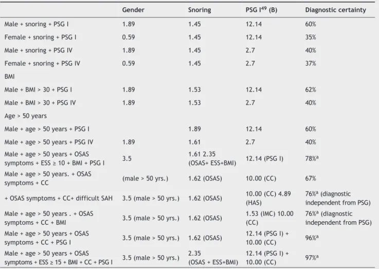
The disease prevalence before the examination inter-feres with the diagnostic certainty in the presence of any altered results; Table 3 compares the diagnostic possibil-ity among the four modalities of sleep monitoring, dif-ferentiating pre-testing low prevalence from high prev-alence. There is a likelihood of OSA diagnostic certainty ≥ 75% when using only PSG I and PSG II in adult popula -tions with high disease prevalence estimated at 32.9%30 (B). Table 4 associates several diagnostic methods, such as signs and symptoms, physical examination, and two types of PSG (I and IV) in a population with low disease prevalence estimated at 4%33 (A) in order to achieve diag-nostic conirmation (diagdiag-nostic certainty > 75%*). A male individual who snores does not demonstrate diagnostic certainty of OSA, even after PSG I is performed; similarly, a male obese individual does not demonstrate diagnostic
certainty OSA, even after PSG I, as described in Table 4. In these two cases, before performing the irst PSG, the neck circumference or the morphometric model should be investigated, as they both have a high positive likeli-hood ratio28 (B), increasing diagnostic certainty. It may be necessary to perform the second PSG, due to AHI vari-ability41,42 (B). However, a thin male individual, younger than 50 years, with symptoms of OSA, neck circumfer-ence > 40 cm, and dificult-to-control hypertension is 76% certain to develop the disease, regardless of the PSG re-sults (Table 4).
Recommendation
Monitored full-night PSG performed in a sleep laboratory is considered the gold standard for OSAS diagnosis39,49,51 (B). The diagnostic probability is similar when performing PSG I and II, as described in Table 3. Portable sleep monitoring assessments still have the limitation of monitoring channel loss due to failure, or loosening or disconnection of sen-sors57 (D), and the need to perform a new PSG I or II to rule out false negatives in cases of high disease probability and normal initial monitoring results56,57 (D).
The choice of treatment with PAP implies a monitored full-night PAP titration performed in a sleep laboratory by a PSG technician65,66 (D). There are no signiicant differ -ences in positive pressure tolerance time during the night, daytime sleepiness improvement, overall improvement, and patient satisfaction when comparing the whole-night PAP ti-tration assisted by a technician with automatic titi-tration69-71 (B), but there are controversies regarding treatment adher-ence67,68 (B).
Table 3 Diagnostic probability by polysomnography (PSG).
S/SP LR+ Diagnostic certainty
(First consultation)
Diagnostic certainty (Third consultation)
GLOBAL automated PSG49 (B)
(AHI + oximetry) 85%-93% 12.14 (95% CI: 5.92 -24.93) 34% 86%
Split-night PSG
(in patients already with AHI > 20)50 (B) 93%-95% 18.60 (95% CI: 7.90-43.78) - 90%
Table 4 Sleep monitoring methods and diagnostic probability according to the prevalence of the population.
S SP LR+ LP HP
PSG I
(part of the night and evaluation of all parameters)39 (B) 66.5% 88.7% 6.06 (95% CI: 3.41-10.77) 20% 75%*
PSG I (full- night with automated analysis)49 (B)
PSG II59 (B) 85% 93% 12.14 (95% CI: 5.92-24.93) 34% 86%*
PSG II59 (B) 70% 91% 7.78 (95% CI: 4.12-14.70) 24% 79%*
PSG III (when AHI > 5)60 (A) 93% 59% 2.27 (95% CI: 1.78-2.89) 09% 53%
PSG III (when AHI > 15)60 (A) 85% 80% 4.25 (95% CI: 2.85-6.34) 15% 68%
5. When should PSG be requested in children?
OSA in children has important conceptual, etiological, and classiication differences when compared to adult apnea.
Snoring is a common complaint reported by the parents; however, the differentiation between primary snoring and OSA in children cannot be made solely based on clinical his-tory data21,72,73 (B). When comparing primary snoring with OSA, statistically different variations can be observed, such as daytime mouth breathing (61% vs. 85%, p = 0.024), wit-nessed apnea (46% vs. 74%, p = 0.013), and respiratory ef-fort (58% vs. 89 %, p = 0.003), but without enough strength to conirm the diagnosis of OSA73 (B).
The presence of syndromic pictures, neuromuscular dis-ease, and obesity are factors to be considered when request-ing PSG assessment in children. Differentiation of pictures of central origin and estimation of apnea severity are important in the prevention of preoperative complications in children after tonsillectomy. Moreover, tonsillar size is not always a good indicative of the need for surgical intervention, and of-ten, parents do not want to the child to undergo surgery and thus underestimate the child’s symptoms74 (D).
Table 6 shows the sensitivity, speciicity, positive pre -dictive value, and negative pre-dictive values of the most common symptoms in children75 (B).
Normal values for PSG in healthy children aged 1 to 15 years are: AI < 1.0 with maximum desaturation of 89%; ex-piratory PCO2 cannot be > 45 mmHg for more than 10% of total sleep time76 (B).
The American Academy of Sleep Medicine believes that the criteria of normality can be used up to the age of 18 years77 (D). Studies have demonstrated that between 13 and 18 years, there is a difference when using the AHI cri-teria recommended for children and those recommended for adults, but this difference does not result in signiicant changes in the classiication of OSA severity when the alter -native criteria for adults are used78 (B ).
The presence of OSA has increased and is recognized as a cause of morbidity, even in young children, with an estimated prevalence of 1% to 4%. Its diagnosis is import-ant, as lack of treatment leads to learning and memory dificulties and decreased weight and height growth rates. In the long-term, it increases the risk of hypertension and depression79 (D).
Recommendation
PSG is recommended for all children with frequent snor-ing and who need to be differentiated from patients with OSA21,72,73 (B). For the diagnosis of OSA, AI > 1, with satura-Table 5 Examples of association of signs and symptoms, physical examination, and polysomnography to diagnose OSAS in a population with low prevalence of disease (estimated at 4%).
Gender Snoring PSG I49 (B) Diagnostic certainty
Male + snoring + PSG I 1.89 1.45 12.14 60%
Female + snoring + PSG I 0.59 1.45 12.14 35%
Male + snoring + PSG IV 1.89 1.45 2.7 40%
Female + snoring + PSG IV 0.59 1.45 2.7 37%
BMI
Male + BMI > 30 + PSG I 1.89 1.53 12.14 62%
Male + BMI > 30 + PSG IV 1.89 1.53 2.7 40%
Age > 50 years
Male + age > 50 years + PSG I 1.89 12.14 60%
Male + age > 50 years + PSG IV 1.89 1.61 2.7 40%
Male + age > 50 years + OSAS
symptoms + ESS ≥ 10 + BMI + PSG I 3.5
1.61 2.35
(OSAS+ ESS+BMI) 12.14 (PSG I) 78%
a
Male + age > 50 years. + OSAS
symptoms + CC (male > 50 yrs.) 1.62 (OSAS) 10.00 (CC) 67%
+ OSAS symptoms + CC+ difficult SAH 3.5 (male > 50 yrs.) 1.62 (OSAS) 10.00 (CC) 4.89 (HAS)
76%a (diagnostic
independent from PSG) Male + age > 50 years . + OSAS
symptoms + CC + BMI 3.5 (male > 50 yrs.) 1.62 (OSAS)
1.53 (IMC) 10.00 (CC)
76%a (diagnostic
independent from PSG) Male + age > 50 years + OSAS
symptoms + CC + PSG I 3.5 (male > 50 yrs.) 1.62 (OSAS)
12.14 (PSG I) +
10.00 (CC) 96%a
Male + age > 50 years + OSAS
symptoms + ESS ≥ 15 + BMI + CC + PSG I 3.5 (male > 50 yrs.) 2.35
(OSAS + ESS+BMI)
12.14 (PSG I) +
tion < 89% and/or expiratory PCO2 > 45 mmHg for over 10% of total sleep time are considered diagnostic criteria76 (B).
The AHI criteria established for children may be used up to the age of 18 years77 (D). Between the ages of 13 and 18 years, the criteria recommended for children or the al-ternative criteria for adults may be used without change in OSAS classiication78 (B).
6. What is the importance of supplementary
exam-inations in the investigation of OSA and snoring?
There are other relevant, currently available tests for the evaluation of OSA and snoring, in addition to the gold stan-dard (PSG). Among them are sleep endoscopy, video-naso-ibrolaryngoscopy with Muller’s maneuver, magnetic reso -nance imaging (MRI) and computed tomography (CT) of the upper airways, and cephalometry80-90 (B).
Much has been discussed regarding the real importance of sleep endoscopy, which is the visualization of the upper airway through a lexible nasal endoscope during pharmaco -logically-induced sleep, to aid in the topographic diagnosis of snoring and OSA. When comparing patients with OSA us-ing lexible video-nasoibrolaryngoscopy in wakefulness and during sleep endoscopy, aiming at the visualization of the pharyngeal obstruction site, similar results can be observed between the two tests in only 25% of cases. There was ob-struction in the hypopharynx during pharmacologically-in-duced sleep in 33% of cases80 (B).
Another study, comparing sleep endoscopy and vid-eo-nasoibroscopy with Muller’s maneuver, showed that the surgical indication during wakefulness was 74%, whereas it was 54% at the sleep endoscopy analysis, corroborating the lack of agreement between examina-tions81 (B). Patients with OSA who were using intraoral mandibular advancement devices underwent sleep endos-copy with and without the use of these devices. It was ob-served that patients who used the device (only those with successful treatments were evaluated) had a signiicantly increased upper airway area82 (B).
When comparing the modiied Mallampati index (MMI) of patients with primary snoring and OSA through sleep endoscopy, it was observed that there was no linear as-sociation between the obstruction level at the sleep en-doscopy and the MMI. Patients with larger tongues (MMI 3
or 4) did not present narrowing at the region of the base of tongue; of this group, 76% had obstruction in the ret-ropalatal region83 (B).
Sleep endoscopy is not signiicantly relevant for the topographic diagnosis of OSA, which does not indicate irrel-evance when assessing the apneic and snoring patient. It is worth mentioning that the criticism of this examination in-cludes the fact that sleep is induced by medications, which can alter the pharyngeal muscle tone, lack of information regarding the patient’s stage of sleep , and speculation on the pharyngeal region with non-segmented anatomical structure80-83 (B).
Muller’s maneuver is quite prevalent among otolar-yngologists in the evaluation of patients with snoring and obstructive sleep apnea, but its real importance has been increasingly questioned. Comparing patients with a history of snoring and patients with OSA demonstrated by the PSG, it became clear that there are no signiicant differences be -tween the two groups when analyzing BMI and retrolingual and retropalatal narrowing analyzed by video-nasoibrola -ryngoscopy with Muller’s maneuver. However, a positive as-sociation between BMI, AHI, and retrolingual obstruction is observed when considering only the group of patients with apnea84 (B). Video-nasoibrolaryngoscopy with Muller’s ma -neuver was performed in the supine and standing positions in OSAS patients, scanning the images on speciic software to minimize the subjectivity of evaluation, subsequently comparing the results with MRI of the pharyngeal region. There is an agreement between the two methods of 93.3% in the retropalatal and of 95.6% in the retrolingual region85 (B). Thus, it can be observed that Muller’s maneuvers do not have the capacity to signiicantly alter management in patients with OSA, especially because the patient is usually evaluated in a non-supine position and during wakefulness. Furthermore, it is noteworthy that there is subjectivity and lack of homogeneity during this assessment, as the inspi-ratory force which the patients present varies signiicantly and evaluation data are yet to be established.
Imaging tests such as MRI, CT, and cephalometry are non-invasive and can be objective in their results; however, their actual relevance continues to prompt studies in sever-al reference centers in the world.
When evaluating patients with OSA and normal patients through MRI of the upper airway, no signiicant differences were observed between the two groups regarding the inter-Table 6 Sensitivity, speciicity, positive predictive value, and negative predictive value of symptoms in children75 (B).
Sensitivity Specificity PPV NPV
Symptoms Present % % % %
Snoring Sometimes 78 36 61 55
Always 95 13 68 55
Difficulty breathing
Sometimes 81 30 54 57
Always 90 33 77 57
Witnessed apnea
Sometimes 59 46 61 44
nal distance between the two mandibular condyles and the mandible bone thickness. However, patients in the apnea group have greater mandibular discrepancy, a smaller inter-nal mandibular length, and a smaller area in the mandibular basal plane than the control group. There were no signif-icant differences in the morphological parameters of the mandible between the obese and nonobese patients with apnea. The volumes of the tongue, soft palate, and later-al pharyngelater-al wlater-alls did not differ signiicantly between the groups86 (B). Ultrafast MRI (0.8 s) was performed in patients with OSAS (AHI > 10) during wakefulness and sleep, and in non-apneic patients (through clinical history and nocturnal oximetry) during wakefulness. It was observed that, during part of the respiratory cycle, the velopharyngeal region is smaller in apneic patients, and that the variation in the velopharyngeal area during the respiratory cycle is greater in apneic patients, particularly during sleep, suggesting a greater plasticity of the upper airway in these patients. Fur-thermore, they veriied that the area of pharyngeal narrow -ing was similar in both the anteroposterior and latero-lat-eral views, both in controls and in apneic individuals during wakefulness; however, during sleep, apneic individuals have maximal circular narrowing of the pharynx. There is also an inverse association between dimensions of the lateral pharyngeal walls and the airway area, probably indicating that the lateral walls are passively compliant as a result of changes in airway caliber. It was observed that the volume of the soft palate and adipose tissue in the parapharyngeal region is higher in apneic patients87 (B).
Focusing on the upper airway CT in the evaluation of patients with sleep-disordered breathing, this imaging study was evaluated in patients with apnea (AHI > 5) and 24 pri-mary snorers (AHI < 5) with oro- and hypopharynx measure-ments, and correlated with the obstructive apnea severity indices and cephalometric studies. Patients with severe OSA had signiicantly greater narrowing in the uvula region during expiration, more inferiorly positioned hyoid bone, larger volume of soft palate, and larger neck circumference when compared with primary snorers and patients with mild to moderate OSA88 (B). When comparing the CT and PSG in patients with obstructive sleep-disordered breathing (1/6 with primary snoring and 5/6 with OSAS), a signiicant asso -ciation between retropalatal and latero-lateral pharyngeal diameters with high rates of AHI was observed. It was also veriied that retropalatal and retroglossal spaces were pre -dictive of the apnea index severity. None of the CT param-eters correlated with intensity of snoring and minimal O2 saturation. BMI correlated positively and signiicantly with retropalatal distance and AHI. From the anatomical stand-point, the latero-lateral retropalatal view is signiicantly associated with impairment of the upper airway caliber in patients with sleep-disordered breathing89 (B). When com-paring the upper airway diameter by CT in patients with OSAS and healthy patients, no correlation existed between PSG parameters in obstructive sleep-disordered breathing (minimum O2 saturation and AHI) and pharyngeal dimen-sions90 (B). There were no signiicant differences between the tomographic measurements of the pharyngeal airway in vigil patients submitted to lateral pharyngoplasty com-pared to uvulopalatopharyngoplasty, although there were differences in clinical and polysomnographic results of this population; therefore, there was no correlation between
polysomnographic and CT parameters in OSAS patients sub-mitted to surgery91 (B). Areas of the nasopharynx, orophar-ynx, and hypopharynx were evaluated during inspiration and expiration, as well as the diameters of the uvula and retropharyngeal tissue to compare the results obtained by CT in patients with apnea (AHI > 10) and without apnea (AHI < 10). It was observed that the retropharyngeal tissue in apneic patients presents more volume than in non-apneic patients, with 10.3 ± 3.6 mm vs. 6.4 ± 2.7 mm, p < 0.01. However, the nasopharynx areas during expiration (228.4 vs. 281.9 mm) and inspiration (195.9 vs. 300.4 mm) of the non-apneic patients were slightly larger, but without statis-tically signiicant differences91 (B).
When studying patients with OSAS and controls by PSG and TC, in which OSAS patients had undergone uvulopal-atopharyngoplasty, it was observed that severely apneic patients had a narrower sectional area of the oropharynx (50 mm2 on average) when compared to the others. The control patients and patients submitted to uvulopalatopha-ryngoplasty without OSAS, i.e., with primary snoring, had minimal pharyngeal area of 110 mm2, on average. Further-more, patients with moderate OSAS submitted to surgery demonstrated values between 60 and 100 mm2 (B).91
Cephalometry is a useful method in the evaluation of apneic and snoring patients93 (C). The choice to request this examination as a routine investigation in a patient with OSA has been the subject of debate, since it has not been proved that it actually changes the therapeutic approach. However, it can be observed that cephalome-try is crucial in the surgical planning of patients submit-ted to orthognathic surgery for the treatment of OSAS. The association between cephalometry and the degree of OSAS severity in adult patients with and without ob-structive sleep apnea was analyzed. It was observed that the length of the upper airway was strongly correlated with the severity of OSA in men (r = 0.72, p < 0.01) and moderately associated in women with OSAS (r = 0.52, p < 0.01)94 (C).
Thus, imaging tests, even during induced sleep, require further studies to assess the applicability of their results as auxiliary examinations when deining conducts. Currently, these tests are more relevant for the development of re-search in this area.
Recommendation
Sleep endoscopy is employed in the clinical investigation of apneic and snoring patients, but controversies still exist regarding its applicability in routine assessment80-83 (B).
Flexible video-nasoibrolaryngoscopy does not change the conduct in patients with snoring and OSA, and there is no ho-mogeneity of results among different observers84,85 (B).
MRI and CT of the pharynx are noninvasive tests and demonstrate that apneic patients have a narrower section-al pharynx area than non-snorers and non-apneic patients, but the location of these strictures varies between individ-uals87,88,90 (B).
7. What are the consequences of OSAS?
Moderate to severe OSAS is an independent predictor of all-cause mortality, with HR = 6.24 (95% CI: 2.01-19.39)95 (A), and this association is not attributable to obesity, age, or other medical chronic conditions, especially in men with severe OSAS aged 40 to 70 years95 (A). Mild OSAS is not an independent risk factor for all-cause mortality, with HR = 0.47 (95% CI: 0.17-1.29)95 (A). An increased risk of coro-nary events or death from cardiovascular causes can be ob-served, regardless of other factors, in patients aged > 50 years95 (A)96,97 (B), with HR = 2.06 (95% CI: 1.10-3.86)98 (A). A self-assessment by standardized questionnaire showed a greater association between OSAS and heart failure and stroke than with coronary heart disease96 (B).
In healthy middle-aged adults and in the elderly, OSAS is associated with increased prevalence of systemic arterial hypertension. Correcting for the main confounding factors (age, gender, BMI, and other measures of adiposity), as well as other potentially relevant variables (tobacco and alcohol consumption), higher AHI and longer desaturation time < 90% were associated with higher risk of hypertension, with OR = 1.37 (95% CI: 1.03-1.83)99 (B). Reduction in systolic dipping related to OSA severity was demonstrated: when AHI < 5, hy-pertension had OR = 3.1 (95% CI: 1.3-7.7), but when AHI > 15, hypertension had OR = 4.4 (95% CI: 1.2-16.31). This lack of systolic dipping can be one of the mechanisms by which OSA contributes to an increase in cardiovascular diseases100 (B).
There is evidence that patients with OSA older than 40 years without underlying arterial hypertension may develop the disease perhaps partly due to the inluence of obesi -ty; however, in patients with AHI > 30, a small inluence of OSA itself cannot be ruled out in the genesis of hyperten-sion46,47,99 (B).
OSAS is an independent risk factor for developing type II diabetes101 (A)102 (B)103 (C), with HR = 1.4 (95% CI: 1.10-1.86), p = 0.008101 (A).
Reduced time of sleep is associated with obesity in adults. Sleep lasting less than ive hours per night is associ -ated with central obesity and increased body fat percentage and body mass index, on average 2.5 kg/m2 (95% CI: 2.0-2.9) for men and 1.8 kg/m2 (95% CI: 1.1-2.4) for women104 (B). Patients with OSAS and metabolic syndrome103 (C) and/ or morbid obesity105 (B) have increased sympathetic tone, with increased cardiovascular risk. Children and adolescents with OSAS and a BMI percentile > 85% assessed by question-naires have lower quality of life106 (B).
OSAS is an independent risk factor for ischemic stroke95,107 (A), with a relative risk of death from stroke (RR) = 5.16 (95% CI: 3.72- 6.60)108 (B). The risk is higher for men with mild to moderate OSAS109 (B), with HR = 6 (95% CI: 2%-10%)110 (A). Menopausal women, aged 50-79 years, of whom 8.3% had sleep duration < ive hours per night and 4.6% had nine hours of sleep per night, were followed-up on average for 7.5 years. There is an association between long sleep duration and risk of ischemic stroke, with RR = 1.70 (95% CI: 1.32-2.21) for those who slept nine hours a night, regardless of the presence of snoring or somnolence. There was no signiicant association between ischemic stroke and short sleep duration111 (A). A reduction in daytime sleepi-ness and BMI was veriied in patients after ischemic stroke; this population may be underdiagnosed for OSAS95 (A).
Untreated OSAS is a contributing factor in motor vehicle accidents112,113 (B). Treatment of OSAS with CPAP reduces the relative risk of collisions, with RR = 0.278 (95% CI: 0.22- 0.35), p < 0.001112 (B).
Sleep apnea is associated with a higher prevalence of psychiatric comorbidities such as depression (21.8%), anxi-ety disorder (16.7%), post-traumatic stress disorder (11.9%), and psychosis and bipolar disorder (3.3%)114 (B).
Several studies have proposed an association between OSAS and neurocognitive dysfunction115,116 (B). When as-sessing elderly individuals considered healthy for their age, with a mean age of 68 years, 58.5% females, the perfor-mance of the PSG showed that 53% had an AHI > 15 events/h and 37% had AHI > 30 events/h, of whom only 9.2% had EDS (ESS > 10). There was no signiicant association between AHI, nocturnal hypoxemia, and cognitive performance, except a tendency toward slower performance in patients with AHI > 30 events/h115 (B). Mild to moderate OSAS has minimal impact on measures related to attention or speed of executive functions and processing speed when compar-ing OSAS individuals with the non-apneic117 (B). OSAS may exacerbate cognitive impairment in dementia caused by Alzheimer’s disease118 (B).
Gastroesophageal relux disease (GERD) is more preva -lent in symptomatic patients with sleep-disordered breath-ing; however, the occurrence of gastroesophageal relux or relux symptoms was not signiicantly inluenced by the se -verity of OSAS. There appears to be an increasing index of microarousal caused by relux in patients with OSAS when compared to the simple snorer119 (B).
Patients with chronic obstructive pulmonary disease (COPD) and OSAS (“overlap syndrome”) have an increased risk of death and hospitalization for COPD exacerbation. At a mean follow-up of 9.4 years (3.3 to 12.7 years) patients with overlap syndrome not treated with CPAP had higher mortality when compared to those using CPAP, with RR = 1.79 (95% CI: 1.16-2.77) and increased tendency to COPD exacerbation requiring hospitalization, with RR = 1.70 (95% CI: 1.21-2.38)120 (B).
In obese OSA patients without COPD, daytime hypercap-nia is associated with OSAS severity, higher BMI, and higher mechanical restriction of the chest wall121 (B).
Pregnant women with OSAS have higher risk of pre-ec-lampsia, premature birth, and medical complications122 (C). For pregnant women with chronic snoring and hypertension, the use of nasal CPAP, while maintaining the usual prenatal care treatment, appears to improve blood pressure and pre-vent complications123 (B).
Recommendation
References
1. Hoffstein V, Szalai JP. Predictive value of clinical features in diagnosing obstructive sleep apnea. Sleep. 1993;16:118-22. 2. Romero E, Krakow B, Haynes P, Ulibarri V. Nocturia and
sno-ring: predictive symptoms for obstructive sleep apnea. Sleep Breath. 2010;14:337-43.
3. Bailes S, Baltzan M, Alapin I, Fichten CS, Libman E. Diagnostic indicators of sleep apnea in older women and men: a prospec-tive study. J Psychosom Res. 2005;59:365-73.
4. Lim PV, Curry AR. The role of history, Epworth Sleepiness Scale Score and body mass index in identifying nonapnoeic snorers. Clin Otolaryngol Allied Sci. 2000;25:244-8.
5. Abrishami A, Khajehdehi A, Chung F. A systematic review of screening questionnaires for obstructive sleep apnea. Can J Anaesth. 2010;57:423-38.
6. Sharma SK, Vasudev C, Sinha S, Banga A, Pandey RM, Handa KK. Validation of the modiied Berlin questionnaire to identify patients at risk for the obstructive sleep apnoea syndrome. Indian J Med Res. 2006;124:281-90.
7. Netzer NC, Stoohs RA, Netzer CM, Clark K, Strohl KP. Using the Berlin Questionnaire to identify patients at risk for the sleep apnea syndrome. Ann Intern Med. 1999;131:485-91.
8. Chung F, Yegneswaran B, Liao P, Chung SA, Vairavanathan S, Islam S, et al. Validation of the Berlin questionnaire and Ameri-can Society of Anesthesiologists checklist as screening tools for obstructive sleep apnea in surgical patients. Anesthesiology. 2008;108:822-30.
9. Olson LG, Cole MF, Ambrogetti A. Correlations among Epworth Sleepiness Scale scores, multiple sleep latency tests and psy-chological symptoms. J Sleep Res. 1998;7:248-53.
10. Friedman M, Wilson MN, Pulver T, Pandya H, Joseph NJ, Lin HC, et al. Screening for obstructive sleep apnea/hypopnea syndro-me: subjective and objective factors. Otolaryngol Head Neck Surg. 2010;142:531-5.
11. Ahmadi N, Chung SA, Gibbs A, Shapiro CM. The Berlin question-naire for sleep apnea in a sleep clinic population: relationship to polysomnographic measurement of respiratory disturbance. Sleep Breath. 2008;12:39-45.
12. Vaz AP, Drummond M, Mota PC, Severo M, Almeida J, Winck JC. Translation of Berlin Questionnaire to Portuguese language and its application in OSA identiication in a sleep disordered breathing clinic. Rev Port Pneumol. 2011;17:59-65.
13. Gus M, Gonçalves SC, Martinez D, de Abreu Silva EO, Moreira LB, Fuchs SC, et al. Risk for Obstructive Sleep Apnea by Berlin Questionnaire, but not day time sleepiness, is associated with resistant hypertension: a case-control study. Am J Hypertens. 2008;21:832-5.
14. Pedrosa RP, Drager LF, Gonzaga CC, Sousa MG, de Paula LK, Amaro AC, et al. Obstructive sleep apnea: the most common secondary cause of hypertension associated with resistant hypertension. Hypertension. 2011;58:811-7.
15. Drager LF, Genta PR, Pedrosa RP, Nerbass FB, Gonzaga CC, Krieger EM, et al. Characteristics and predictors of obstructive sleep apnea in patients with systemic hypertension. Am J Car-diol. 2010;105:1135-9.
16. Bertolazi AN, Fagondes SC, Hoff LS, Pedro VD, Menna Barre-to SS, Johns MW. Portuguese-language version of the Epworth sleepiness scale: validation for usein Brazil. J Bras Pneumol. 2009;35:877-83.
17. Santaolalla Montoya F, Iriondo Bedialauneta JR, Aguirre Larra-coechea U, Martinez Ibargüen A, Sanchez Del Rey A, Sanchez Fernandez JM. The predictivevalue of clinical and epidemiolo-gical parameters in the identiication ofpatients with obstruc -tive sleep apnoea (OSA): a clinical prediction algorithm in the evaluation of OSA. Eur Arch Otorhinolaryngol. 2007;264:637-43. 18. Subramanian S, Hesselbacher SE, Aguilar R, Surani SR. The NA-MES assessment: a novel combined-modality screening tool for obstructive sleep apnea. Sleep Breath. 2011;15:819-26.
19. Kapur VK, Baldwin CM, Resnick HE, Gottlieb DJ, Nieto FJ. Sle-epiness in patients with moderate to severe sleep-disordered breathing. Sleep. 2005;28:472-7.
20. Goodwin JL, Kaemingk KL, Mulvaney SA, Morgan WJ, Quan SF. Clinical screening of school children for polysomnography to detect sleep-disordered breathing-the Tucson Children’s As-sessment of Sleep Apnea study (TuCASA). J Clin Sleep Med. 2005;1:247-54.
21. Montgomery-Downs HE, O’Brien LM, Holbrook CR, Gozal D. Snoring and sleep disordered breathing in young children: sub-jective and obsub-jective correlates. Sleep. 2004;27:87-94. 22. Sproson EL, Hogan AM, Hill CM. Accuracy of clinical assessment
of paediatric obstructive sleep apnoea in two English centres. J Laryngol Otol. 2009;123:1002-9.
23. Gottlieb DJ, Whitney CW, Bonekat WH, Iber C, James GD, Le-bowitz M, et al. Relation of sleepiness to respiratory disturban-ce index: the Sleep Heart Health Study. Am J Respir Crit Care Med. 1999;159:502-7.
24. Goldstein NA, Post JC, Rosenfeld RM, Campbell TF. Impact of tonsillectomy and adenoidectomy on child behavior. Arch Oto-laryngol Head Neck Surg. 2000;126:494-8.
25. Ziliotto KN, dos Santos MF, Monteiro VG, Pradella-Hallinan M, Moreira GA, Pereira LD, et al. Auditory processing assess-ment in children with obstructive sleep apnea syndrome. Braz J Otorhinolaryngol. 2006;72:321-7.
26. Viner S, Szalai JP, Hoffstein V. Are history and physical exami-nation a good screening test for sleep apnea? Ann Intern Med. 1991;115:356-9.
27. Davies RJ, Ali NJ, Stradling JR. Neck circumference and other clinical features in the diagnosis of the obstructive sleep apno-ea syndrome. Thorax. 1992;47:101-5.
28. Kushida CA, Efron B, Guilleminault C. A predictive morphome-tric model for the obstructive sleep apnea syndrome. Ann In-tern Med. 1997;127(8 Pt 1):581-7.
29. Young T, Shahar E, Nieto FJ, Redline S, Newman AB, Gottlieb DJ, et al. Predictors of sleep-disordered breathing in commu-nity-dwelling adults: the Sleep Heart Health Study. Arch Intern Med 2002;162:893-900.
30. Tuik S, Santos-Silva R, Taddei JA, Bittencourt LR. Obstructive sleep apnea syndrome in the Sao Paulo Epidemiologic Sleep Study. Sleep Med. 2010;11:441-6.
31. Fogel RB, Malhotra A, Dalagiorgou G, Robinson MK, Jakab M, Kikinis R, et al. Anatomic and physiologic predictors of apnea severity in morbidly obese subjects. Sleep. 2003;26:150-5.
32. Martinho FL, Tangerina RP, Moura SM, Gregorio LC, Tuik S, Bit -tencourt LR. Systematic head and neck physical examination as a predictor of obstructive sleep apnea in class III obese pa-tients. Braz J Med Biol Res. 2008;41:1093-7.
33. Bixler EO, Vgontzas AN, Ten Have T, Tyson K, Kales A. Effects of age on sleep apnea in men: I. Prevalence and severity. Am J Respir Crit Care Med. 1998;157:144-8.
34. Soares MC, de Azeredo Bittencourt LR, Zonato AI, Gregório LC. Application of the Kushida morphometric model in patients with sleep-disordered breathing. Braz J Otorhinolaryngol. 2006;72:541-8.
35. Tsai WH, Remmers JE, Brant R, Flemons WW, Davies J, Ma-carthur C. A decision rule for diagnostic testing in obstructive sleep apnea. Am J Respir Crit Care Med. 2003;167:1427-32. 36. Zonato AI, Martinho FL, Bittencourt LR, de Oliveira Campones
Brasil O, Gregorio LC, Tuik S. Head and neck physical exami -nation: comparison between nonapneic and obstructive sleep apnea patients. Laryngoscope. 2005;115:1030-4.
37. Friedman M, Tanyeri H, La Rosa M, Landsberg R, Vaidyanathan K, Pieri S, et al. Clinical predictors of obstructive sleep apnea. Laryngoscope. 1999;109:1901-7.