188 189
188 189
ABSTRACT: The purpose of this study was to biochemically compare the decalcifying effects of 1% EDTA (pH 7.4), 1% EGTA (pH 7.4), 1% CDTA (pH 7.4), 1% citric acid solutions (pH 1.0 and 7.4) and saline solution (control) on root dentin. Forty-eight single-rooted teeth were used in this study. The canals were instrumented by the step-back technique and the roots were randomly divided into six equal experimental groups (n = 8) according to the irrigat-ing agent tested. A total of 30 µL of each solution was pipetted into the root canal and allowed to set undisturbed for 5 minutes. After this time, 15 µL of the solutions were removed from each canal using a Hamilton syringe and placed in a container with 5 mL of deionised water. The µg/mL concentration of calcium ion (Ca2+) extracted from
the root canal samples was determined using inductively coupled plasma-atomic emission spectrometry (ICP-AES). Data were analysed by means of the Kruskal-Wallis and Mood’s median tests. Citric acid solution at pH 1.0 removed more calcium than at pH 7.4 and than the other chelating solutions tested (p < 0.05). No differences were observed between EDTA and EGTA. Both EDTA and EGTA removed significantly more calcium than CDTA and cit-ric acid at pH 7.4 (p < 0.05). There were no differences between citcit-ric acid at pH 7.4 and saline solution, which had the least efficacy for Ca2+ extraction (p > 0.05). These results indicate that citric acid at pH 1.0 is a good alternative
as an irrigating solution to remove the smear layer and facilitate the biomechanical procedures. DESCRIPTORS: Chelating agent; Citric acid; Edetic acid; Egtazic acid.
RESUMO: Este trabalho teve como objetivo comparar o efeito desmineralizante do EDTA (pH 7,4), EGTA (pH 7,4), CDTA (pH 7,4), ácido cítrico (pH 1,0 e 7,4) e da solução salina (controle) sobre a dentina radicular. Todas as so-luções teste foram preparadas na concentração de 1%. Quarenta e oito dentes unirradiculares recém-extraídos foram utilizados neste experimento. Após a instrumentação dos canais radiculares pela técnica “step-back”, as raízes foram aleatoriamente divididas em 6 grupos experimentais (n = 8) de acordo com a solução teste utilizada na irrigação final. Em cada grupo, 30 µL da solução teste foram pipetados no interior de cada canal radicular e man-tidos estáveis por 5 minutos. Decorrido esse período, 15 µL da solução foram removidos do canal e depositados em frasco contendo 5 mL de água deionizada. A concentração de Ca2+ (µg/mL) extraída dos espécimes foi determinada
pela espectrometria de absorção de massa (ICP-AES); e os dados foram submetidos à análise estatística pelos testes de Kruskal-Wallis e de mediana de Mood. O ácido cítrico em pH 1,0 foi a solução mais efetiva na remoção de Ca2+
comparativamente às demais soluções-teste (p < 0,05). Nenhuma diferença estatística foi observada entre a ação do EDTA e a do EGTA. Ambos os quelantes removeram significantemente mais Ca2+ que o CDTA e o ácido cítrico
em pH 7,4 (p < 0,05). Não houve diferença significante entre ácido cítrico em pH 7,4 e solução salina (p > 0,05). Os resultados deste estudo indicam que o ácido cítrico em solução de pH 1,0 apresenta-se como uma boa opção para remover a “smear layer” e facilitar o preparo biomecânico do sistema de canal radicular.
DESCRITORES: Quelantes; Ácido cítrico; Ácido edético; Ácido egtázico.
INTRODUCTION
Mechanically prepared dentin surfaces are always covered with the so-called smear layer, a loosely bonded amorphous layer of organic and inorganic debris. Thus, several chelating agents and acids have been used to remove this layer, with varying degrees of success.
These irrigating solutions have been chosen due to their direct action over calcium ions. The calcium present in hydroxyapatite crystals is one of the main inorganic elements of dentin. Any change in the calcium ratio can significantly alter the original proportion of organic and inorganic
* Associate Researcher, PhD, Department of Metallurgy and Material Science, Pontifical Catholic University of Rio de Janeiro. ** Biochemist, MS, Department of Biochemistry, School of Dentistry of Bauru, University of São Paulo.
Demineralization effect of EDTA, EGTA, CDTA and citric acid on
root dentin: a comparative study
Efeito do EDTA, EGTA, CDTA e ácido cítrico na desmineralização
da dentina radicular: estudo comparativo
188 189
188 189
components, which can alter dentin permeability, microhardness and solubility.
Although EDTA has a long-standing history as the agent of choice in Endodontics, research-ers have reported its irritating potential7,9-11. Re-cently, EGTA has also been recommended for root canal biomechanical preparation, since it presents a more precise action on calcium ions than EDTA1,2,9,12,16. Another chelating agent, CDTA, has also been reported to significantly bind the divalent cation. EGTA and CDTA have been used in medicine to obtain calcium-free environments. Recent studies have investigated the application of both chelating agents as an alternative solution to remove the smear layer. Çalt, Serper1 (2000) and Viswanath et al.16 (2003) compared the effect of EDTA and EGTA on removal of the smear layer by using scanning electron microscopy. Cruz-Filho et al.2 (2001) and Sousa-Neto et al.12 (2002) evalu-ated the influence of 15% EDTA, 1% CDTA and 1% EGTA on root dentin microhardness, and dentin adhesiveness and microleakage, respectively. In the study of Cruz-Filho et al.2 (2001), EDTA was associated with a surfactant (Cetavlon). It is im-portant to note that, in these investigations, the EDTA concentration tested was between 15-17%, and 1% was the concentration of EGTA and CDTA solutions. No study has yet compared all chelating agents using the same concentration.
Citric acid has also been studied for its ability as a decalcifying and cleansing solution in root canal irrigation8,13,14,17. However, in some scanning electron microscope studies, it was observed that citric acid left precipitated crystals in the root ca-nal walls3,4. It is a biological and organic acid, with sufficient tissue compatibility6,7,10,11,15.
Studies have demonstrated that the pH and concentration of demineralising solutions are im-portant factors to be considered in their action in removing surface smear layer4,5. A literature review has revealed that different mixtures, concentra-tions, pH, working time and methods have been used to evaluate the irrigating solutions.
Thus, considering these factors, this study was conducted to compare in vitro the decalcifying effect of 1% citric acid, 1% EDTA, 1% EGTA, 1% CDTA and saline solution (control) on root dentin, by using inductively coupled plasma-atomic emis-sion spectrometry (ICP-AES). The pH of the test solutions was 7.4 for all chelating agents and 1.0 and 7.4 for citric acid. The reason to evaluate two different pH values for the citric acid group was
based on the long-standing studies about its best action at lower pH4,5, and inclusion of a second value (pH 7.4) was intended to obtain a more ac-curate comparative result.
MATERIAL AND METHODS
Test materials
The materials used were: 1% EDTA pH 7.4 (eth-ylene-diaminetetra-acetic acid; Merck, Darmstadt, Germany); 1% EGTA pH 7.4 (ethylene-glycol-ether-diaminetetra-acetic acid; Sigma, St. Louis, MO, USA); CDTA (1,2 cyclohexane-diaminetetra-acetic acid, Sigma, St. Louis, MO, USA); 1% citric acid pH 1.0 and 7.4 (Merck, Darmstadt, Germany) and saline solution (control; Merck, Darmstadt, Germany). All test solutions were freshly prepared. The salts were weighed and diluted in deionised water (Permution E.J. Krieger & Cia. Ltda., Curitiba, Brazil), and their pH was adjusted (pH meter B371, Micronal, São Paulo, Brazilian Manufacturing) by addition of hy-drochloric acid (HCl – Merck S.A. Indústria Quími-ca, Rio de Janeiro, Brazil). The molar concentrations of the acid solutions were: EDTA = 0.0263 mol/L, EGTA = 0.0213 mol/L, CDTA = 0.0274 mol/L, and citric acid = 0.0520 mol/L.
Procedure
190 191
190 191
plasma-atomic emission spectrometry (ICP-AES, Spectrometer plasma 400, Pelkin Elmer, Shelton, CT, USA). The data were statistically analysed by using the nonparametric Kruskal-Wallis test and Mood’s median test for multiple comparisons, at the significance level of 5% (p < 0.05).
RESULTS
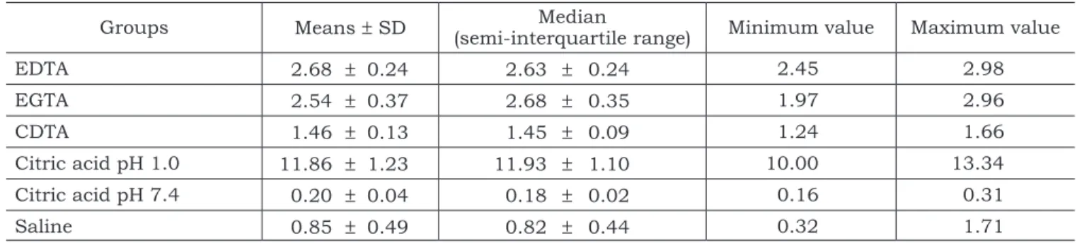
The means, standard deviations (SD), medians (semi-interquartile range) and minimum and maxi-mum values of Ca2+ concentrations (µg/mL) in all experimental groups are shown in Table 1.
The Kruskal-Wallis test shows that there were statistical differences between the acids in terms of Ca2+ concentration. Mood’s median test (Table 2) showed statistically significant difference between citric acid solution at pH 1.0 and the other groups (p < 0.05). This solution removed more calcium than citric acid solution at pH 7.4 and the other chelating solutions tested. Moreover, no differences were noted between EDTA and EGTA. Both che-lating agents removed significantly more calcium than CDTA and citric acid at pH 7.4 (p < 0.05). There were no differences between citric acid at
pH 7.4 and saline solution, which had the least efficacy for Ca2+ extraction (p < 0.05).
DISCUSSION
In this in vitro study, it was noteworthy that citric acid solution is not effective for Ca2+ removal at neutral pH. While EDTA, EGTA and CDTA act as chelating agents at neutral pH, citric acid acts better at lower pH values.
The statistical data showed that 1% citric acid at pH 1.0 was the best solution to remove calcium from the root dentin. Furthermore, considering the concentration factor, even though the 1% cit-ric acid solution (pH 1.0) was employed, i.e., at a lower concentration, better results were obtained when compared to the EDTA, EGTA, and CDTA solution groups. The present results are in agree-ment with those of Hennequin et al.5 (1994) and Haznedaroglu4 (2003).
According to both previous studies, the pH of citric acid solution has been shown to be a more important factor to demineralisation than concen-tration. Sterrett et al.14 (1991) reported that this phenomenon may be due to a balance between the decrease in pH and the increase in viscosity of the
TABLE 1 - Means, standard deviations (SD), medians (semi-interquartile range) and minimum and maximum
values of Ca2+ concentrations (µg/mL) in all experimental groups.
Groups Means ± SD (semi-interquartile range)Median Minimum value Maximum value
EDTA 2.68 ± 0.24 2.63 ± 0.24 2.45 2.98
EGTA 2.54 ± 0.37 2.68 ± 0.35 1.97 2.96
CDTA 1.46 ± 0.13 1.45 ± 0.09 1.24 1.66
Citric acid pH 1.0 11.86 ± 1.23 11.93 ± 1.10 10.00 13.34
Citric acid pH 7.4 0.20 ± 0.04 0.18 ± 0.02 0.16 0.31
Saline 0.85 ± 0.49 0.82 ± 0.44 0.32 1.71
TABLE 2 - Mood’s median test for multiple comparisons.
EDTA EGTA CDTA Citric acid (pH 1.0) Citric acid (pH 7.4) Saline solution
EDTA
-EGTA (n-s)
-CDTA * *
-Citric acid (pH 1.0) * * *
-Citric acid (pH 7.4) * * * *
-Saline solution * * * * (n-s)
190 191
190 191
solution caused by the increase in the constituent concentration. At high concentrations, citrate mo-nopolises such a large portion of the solvent that the amount of solvent available for Ca2+ diffusion is dramatically reduced.
Furthermore, the self-limiting effect associ-ated with pH changes during the demineralisation process of EDTA, EGTA and CDTA can be consid-ered. These chelating agents react with calcium ions in the hydroxyapatite crystals to produce a metallic chelate, and this process is characterised by protonation and complex formation reaction occurs9. As the pH decreases during this process, both the rate of dentin demineralisation and the amount of dentin dissolved decrease.
The irrigating solutions used in this experi-ment were of low concentrations. It should be mentioned that EDTA is currently studied or used in clinical practice with concentrations between 15-17%. In fact, this is the first study comparing EDTA with EGTA, CDTA and citric acid at the same concentration and pH. Comparison between two different pH values of citric acid and the chelat-ing agents was essential to confirm its action at lower pH and concentration values. Few studies have evaluated its action using these parameters so far.
Even though EGTA is reported to bind Ca2+ more specifically than EDTA, in some studies, al-though different analyses and concentrations have been used, EGTA was similar or less potent than EDTA. Çalt, Serper1 (2000) indicated that the ac-tion of 17% EDTA is stronger than that of 17% EGTA for removal of smear layer. However, EGTA did not cause erosion of the intertubular and peri-tubular dentin. Cruz-Filho et al.2 (2001) reported that 1% EGTA, 1% CDTA and 15% EDTAC reduced root dentin microhardness similarly. Viswanath et al.16 (2003), in a SEM study, demonstrated that both EGTA and EDTA completely removed the smear layer.
There was a molar concentration difference between all test solutions, but this difference was smaller among chelating agents. The present study revealed that EDTA and EGTA presented the same effect on dentin Ca2+ extraction. However, CDTA was statistically less effective, though its molar concentration was slightly higher than that of EDTA and EGTA. Note that the citric acid solution had almost double the molar concentration when compared with the other solutions.
Previous reports have shown that 15% EDTAC presented similar action when compared with 1% EGTA and 1% CDTA2, and that EDTA-T was less effective than EDTA8 as a chelating agent. The present data demonstrated that 1% EDTA alone was as efficient as 1% EGTA and better than 1% CDTA. The concentration and associa-tion with other substances can be an important factor on the chelating agents’ action process.
Considering the present results on the similar effect of 1% EDTA and 1% EGTA for removal of smear layer, the previous literature1,2,16, and their cytotoxic effect2,6,7,9-11,16, EGTA should be preferred when a chelating agent is required.
All acid solutions tested in this experiment are considered complexants and chelating agents. Under the conditions of this study, 1% citric acid (pH 1.0) demonstrated to be a good alternative as an irrigating solution to remove the smear layer and facilitate the biomechanical procedures. Scelza et al.8 (2003) also suggested the use of citric acid as an irrigating solution despite the lack of statistical difference between citric acid and 17% EDTA in terms of their efficacy in Ca2+ extraction from root dentin. Citric acid was reported to be inexpensive, effective on anaerobic microorganisms17 and more biocompatible than EDTA and EGTA7,9-11. Sousa11 (2000) and Soares, Sousa10 (2003) reported that, even at 15% and pH 1.0, citric acid was less cy-totoxic than 15% EDTA. The 15% EGTA solution was more biocompatible than EDTA.
Combinations of decalcifying agents and NaOCl solution have been recommended, because no single irrigator is capable of dissolving organic pulpal material and predentin as well as demin-eralising the inorganic calcified portion of the root canal wall. The authors suggested that the acid-ity yielded by utilisation of chemical solutions on endodontic therapy could be minimised by final flushing with water and use of calcium hydroxide sealers, which could neutralize this residual effect. Besides, the application time should also be well controlled in the etching procedure. Attention to these factors will lower the risk of damage to the dental tissue during root canal cleaning and thus increase the success of endodontic therapy.
CONCLUSION
concen-192 PB trations of EDTA and EGTA were found to be more
effective than CDTA; 1% EDTA and 1% EGTA had similar demineralisation effect on dentin. Citric
REFERENCES
1. Çalt S, Serper A. Smear layer removal by EGTA. J Endod 2000;26:459-61.
2. Cruz-Filho AM, Sousa-Neto MD, Saquy PC, Pécora JD. Eval-uation of the effect of EDTAC, CDTA, and EGTA on radicular dentin microhardness. J Endod 2001;27:183-4.
3. Ferrer-Luque CMF, González-López SG, Rodrígues de Mondelo JMN. Utilización del ácido cítrico en la preparación biomecáni-ca del conducto radicular. Endodoncia 1994;12:63-70. 4. Haznedaroglu F. Efficacy of various concentrations of citric
acid at different pH values for smear layer removal. Oral Surg Oral Med Oral Pathol Oral Radiol Endod 2003;96:340-4. 5. Hennequin M, Pajot J, Avignant D. Effects of different pH
val-ues of citric acid solutions on the calcium and phosphorus contents of human root dentin. J Endod 1994;20:551-4. 6. McInnes-Ledoux P, Cleaton-Jones PE, Austin JC. The
pulpal response to dilute citric acid smear removers. J Oral Rehabil 1985;12:215-28.
7. Scelza MFZ, Daniel RLDP, Santos EM, Jaeger MMM. Cyto-toxic effects of 10% citric acid and EDTA-T used as root canal irrigants: an in vitro analysis. J Endod 2001;27:741-3. 8. Scelza MFZ, Teixeira AM, Scelza P. Decalcifying effect of
EDTA-T, 10% citric acid, and 17% EDTA on root canal dentin. Oral Surg Oral Med Oral Pathol Oral Radiol Endod 2003;95:234-6.
9. Segura-Egea JJ, Jimenez-Rubio A, Rios-Santos JV, Velas-co-Ortega E, Calvo-Gutierrez JR. In vitro inhibitory effect of EGTA on macrophage adhesion: endodontic implications. J Endod 2003;29:211-3.
10. Soares AB, Sousa SMG. Avaliação da biocompatibili-dade do EDTA, EGTA e ácido cítrico em tecido subcutâneo de ratos. Rev Odonto Ciência 2003;18:17-22.
11. Sousa SMG. Avaliação da biocompatibilidade do EDTA, EGTA e ácido cítrico pela técnica de exsudação de corantes vitais [Tese de Doutorado]. Bauru: Faculdade de Odontologia de Bauru da USP; 2000.
12. Sousa-Neto MD, Passarinho-Neto JG, Carvalho-Júnior JR, Cruz-Filho AM, Pécora JD, Saquy PC. Evaluation of the effect of EDTA, EGTA and CDTA on dentin adhesiveness and microleakage with different root canal sealers. Braz Dent J 2002;13:123-8.
13. Sterrett JD, Bankey T, Murphy HJ. Dentin demin-eralization. The effects of citric acid concentration and application time. J Clin Periodontol 1993;20:366-70. 14. Sterrett JD, Delaney B, Rizkalla A, Hawkins CH.
Opti-mal citric acid concentration for dentinal demineralization. Quintessence Int 1991;22:371-5.
15. Trujillo Júnior R, Silva FB, Almeida JM, Sousa SMG. Avaliação da biocompatibilidade do ácido cítrico em di-ferentes concentrações – teste edemogênico. Salusvita 2003;22:171-80.
16. Viswanath D, Hegde AM, Munshi AK. The removal of the smear layer using EGTA: a scanning electron micro-scopic study. J Clin Pediatr Dent 2003;28:69-74.
17. Yamaguchi M, Yoshida K, Suzuki R, Nakamura H. Root canal irrigation with citric acid solution. J Endod 1996;22:27-9.