ISSN 0104-6632 Printed in Brazil
www.abeq.org.br/bjche
Vol. 31, No. 04, pp. 867 - 880, October - December, 2014 dx.doi.org/10.1590/0104-6632.20140314s00002473
Brazilian Journal
of Chemical
Engineering
PRODUCTION AND CHARACTERIZATION OF
DI-RHAMNOLIPID PRODUCED BY
Pseudomonas aeruginosa
TMN
T. A. A. Moussa
1, M. S. Mohamed
2*and N. Samak
31Botany Department, Faculty of Science, Cairo University, Egypt. 2
Chemistry Department, Biochemistry Specialty, Faculty of Science, Cairo University, Egypt. Phone: +2 100 1964 580, Fax: +2 02 35727213
*E-mail: mervat_sayed@yahoo.com, mervat@sci.cu.edu.eg 3
Academy of Scientific Research and Technology Scholarship, Cairo University, Egypt.
(Submitted: January 1, 2013 ; Revised: January 30, 2014 ; Accepted: February 13, 2014)
Abstract - Pseudomonas aeruginosa TMN was used to produce rhamnolipid (RL) from a variety of carbon and nitrogen substrates. The most favorable carbon sources for RL production were glucose and glycerol (both at 40 g/L), giving a RL yield of 0.3 and 0.25 g/L, respectively. Meanwhile, sodium nitrate appeared to be the preferable nitrogen source, resulting in a RL production of 0.34g/L. Rhamnolipid production from P. aeruginosa TMN was affected by temperature, pH and agitation rate, with 37 °C, pH 7 and 200 rpm agitation favorable for rhamnolipid production. Fourier transform infrared spectroscopy (FTIR), nuclear magnetic resonance (NMR) and electro spray ionization–mass spectrometry (ESI–MS) analyses indicated that the purified product contained one type of commonly found rhamnolipid, which is L-rhamnosyl-L-rhamnosyl-β -hydroxydecanoyl-β-hydroxydecanoate. The rhamnolipid product can reduce the surface tension of water to 34 mN/m with a critical micelle concentration of nearly 18.75 mg/L and emulsified kerosene by 46%. P. aeruginosa TMN strain is a potential source of rhamnolipid biosurfactant, which could be used for the development of bioremediation processes in the marine environment.
Keywords: Rhamnolipid; Pseudomonas aeruginosa;Optimization; Purification; Glucose.
INTRODUCTION
Environmental pollution caused by petroleum hy-drocarbons represents a great risk to ecosystems. Biodegradation is an effective way to overcome this problem in which microbes utilize the contaminants as a carbon source, leading to the breakdown of the pollution components into low molecular weight compounds (Pirôllo et al., 2008; Zhang et al., 2005).
Synthetic surfactants are unsuitable for bioreme-diation applications since they may cause toxic ef-fects to the environment or result in secondary pol-lution. Thus, biosurfactants, natural products of a variety of microorganisms (Providenti et al., 1995;
Rahman et al., 2002a), appear to be preferable for environmental applications. Biosurfactants have recently received much more attention due to their potential to become an environment-friendly alter-native to conventional synthetic surfactants. Advan-tages of biosurfactants over their synthetic counter-parts include lower toxicity, biodegradability, better environmental compatibility, higher foaming, high selectivity, and specific activity at extreme tempera-tures, pH and salinity (Desai and Banat, 1997; Makkar and Cameotra, 1999; Makkar and Cameotra, 2002; Ilori et al., 2005; Raza et al., 2007; Abouseoud et al., 2008; Fontes et al., 2012).
Brazilian Journal of Chemical Engineering biosurfactants have been the low yield and high
pro-duction cost. Therefore, there is an urgent need to develop an efficient and cost-effective bioprocess for the production of biosurfactants. Biosurfactants are a leading group of valuable microbial natural products with unique biochemical properties. From a biotech-nology prospective, the production of biosurfactants is important owing to their vast applications in food, cosmetics, pharmaceuticals, and the agricultural and petrochemical industries (Robert et al., 1989; Pruthi and Cameotra, 2003; Abouseoud et al., 2008; Nguyen
et al., 2008). They are interesting amphiphilic
bio-macromolecules with various biological functions/ properties such as; enhancer of polycyclic aromatic hydrocarbon bioavailability to microorganisms and, accordingly, their biodegradation, reducer of the interfacial tension by partitioning preferentially at interfaces, effective activity on surfaces, oil recovery, and so on (Desai and Banat, 1997; Pruthi and Cameotra, 2003; Joshi et al., 2008; Nguyen et al., 2008). Microbial surfactants, which are secreted by different groups of bacteria, are composed of lipid, phospholipids, polysaccharide, protein and other biological macromolecules and contain various functional groups including carboxyl, amino and phosphate groups (Kosaric, 1992; Desai and Banat, 1997; Christofi and Ivshina, 2002).
Pseudomonas aeruginosa, a Gram-negative
bac-terium, secretes rhamnolipids when grown under the appropriate conditions (Jarvis and Johnson, 1949). Rhamnolipids are a group of biosurfactants of gly-colipid nature, composed of a hydrophilic head formed by one or two rhamnose molecules, known respectively as monorhamnolipid (monoRL) and di-rhamnolipid (diRL), and a hydrophobic tail which contains one or two fatty acids. The composition of rhamnolipidic homologues is related to many pa-rameters; most importantly strain, media composi-tion, culture conditions, and culture age (Deziel et al., 1999; Mata-Sandoval et al., 1999; Monteiro et al., 2007). Previous work showed that P. aeruginosa
is able to produce six types of rhamnolipids, which possess similar chemical structure and surface activ-ity and have an average molecular weight of 577 (Torrens et al., 1998). Rhamnolipid can reduce the sur-face tension of water from 72 to 30 mN/m (Abalos
et al., 2001) with a critical micelle concentration of 27–54 mg/l (Miller-Maier and Bodour, 1998). Al-though rhamnolipid is not the strongest biosurfactant available, it is well suited for applications in biore-mediation of oil pollutants due to having high emulsification activity and minor antibiotic effects (Mulligan, 2005).
The specific objectives of this study were: to
examine the influence of nutritional requirements; to optimize the environmental conditions for the pro-duction of rhamnolipid by a newly characterized
Pseudomonas aeruginosa TMN strain and the
purifi-cation process; to perform a structural characteriza-tion and determine some associated physicochemical properties, including the critical micelle concentra-tion and the characterizaconcentra-tion of the bioemulsifier pro-duced, based on its solvent specificity and stability.
MATERIALS AND METHODS
Isolation of Biosurfactant-Producing Micro-organisms
Biosurfactant-producing microbial strains were isolated from compost pile waste collected from El-Sharkia region in Delta, Egypt using Cetrimide agar (Difo, U.S.A). The isolated bacterial strains were characterized depending on their morphological, biochemical characteristics by following Bergey’s Manual of Determinative Bacteriology, API 20NE kit (Biomérieux, Mercy, France) and 16S rRNA gene analysis.
Extraction of DNA and PCR
For molecular identification, the selected bacte-rial isolate was referred for 16S rRNA gene sequence analysis. DNA extraction was performed from 2 mL of bacterial cultures collected at the mid-exponential growth phase using Roche Kit (Germany) according to the manufacturer's instructions and run in tripli-cate through the polymerase chain reaction (PCR). Three sets of primers are listed in Table 1.
Table 1: List of primers used in this study.
Name Sequence Size (bp) Reference
27 27f 5′-AGAGTTTGATCCTGGCTCAG-3′ (Lane, 1991)
1492 1492r 5′-TACGGTTACCTTGTTACGACTT-3′
V3F 5′-CCAGACTCCTACGGGAGGCAG-3′ 203 (Chakravorty et al. 2007)
V3
V3R 5′-CGTATTACCGCGGCTGCTG-3′
V6F 5′-TCGATGCAACGCGAAGAA-3′ 124 (Chakravorty et al. 2007)
V6
V6R 5′-ACATTTCACaACACGAGCTGACGA-3′
Sequence homologies were examined using BLAST version 2.2.12 of the National Center for Biotechnology Information (Altschul et al. 1990). Multiple sequence alignments were carried out using ClastalW and a consensus neighbor-joining tree was constructed using Molecular Evolutionary Genetics Analysis (MEGA) software (version 4.0) (Tamura et al. 2007). Finally potent biosurfactant producing iso-lates were maintained on nutrient agar slants for fur-ther studies.
Preparation of Culture Medium
The strain from nutrient agar slants was streaked on a Cetrimide agar plate, which was then incubated at 37 °C for 14–16 h. After that, a single colony was taken from the plate and transferred into 50 mL of liquid broth (LB) to prepare the seed culture. The cultivation condition for the seed culture was 37 °C, 200 rpm, and 14–16 h of incubation time.
Fermentation Medium and Condition
For liquid fermentation, the seed culture (5% in-oculum) was inoculated into a 500-ml flask contain-ing 150 mL of mineral salts (MS) medium consistcontain-ing of (g/L): NH4NO3, 4; KH2PO4, 4.08; Na2HPO4, 5.68;
CaCl2, 7.77 × 10⁻⁴; MgSO4.7H2O, 0.2; sodium EDTA,
1.49 × 10⁻³; FeSO4·7H2O, 5.56 × 10⁻⁴ (Wei and Chu,
1998). In general, the MS medium was amended with 40 g/L glucose as the sole carbon substrate (desig-nated as GMS medium) (Wei and Chu, 1998; Wei et al., 2005). The culture temperature and agitation rate were 37 °C and 200 rpm, respectively. The pH of the medium was initially adjusted to 6.8 with 1.0 M HCl.
Optimization of Biosurfactant Production Carbon Source Optimization
For the experiments exploring the effect of car-bon substrates on rhamnolipid production, the carcar-bon
source in GMS medium (i.e., glucose) was replaced by glycerol (40 g/L), sucrose (40 g/L), hexane (40 g/L), olive oil (80 g/L) and oleic acid (80 g/L) (Wu et al., 2008).
Nitrogen Source Optimization
The nitrogen source in GMS medium (i.e., NH4NO3) was also replaced by NH4Cl (50 mM),
NaNO3 (50 mM), urea (50 mM), and yeast extract
(10 g/L) to investigate the effect of nitrogen source on rhamnolipid production (Wu et al., 2008).
Optimization of pH and Temperature
The optimum pH of MS medium for growth and biosurfactant production of the tested bacterial iso-lates was investigated. The pHs tested were 4, 5, 6, 7, and 8. The pH of the medium was adjusted using a solution of HCl or NaOH (1 M), with the aid of a Sentron 2001 pH meter, and the inoculated medium was amended with optimum carbon substrate and optimum nitrogen source.
The growth and biosurfactant production of the isolated microorganisms were monitored at a range of temperatures to determine the optimum one for each isolate. The tested temperatures were 25, 30, 35, 37, 40 and 45 °C. The inoculated medium was adjusted to the optimum pH and supplied with the optimum carbon substrate and optimum nitrogen source.
Agitation Rate
Brazilian Journal of Chemical Engineering
Dry Weight Cell Determination
After cultivation for 7 days, the collected fermen-tation broth was first centrifuged at 9000g for 15 min to remove bacterial cells. 50 mL of the filtrate was taken to detect surface tension, emulsification index, and oil displacement test, which will be described later. The removed bacterial cells were collected and placed in an oven at 110 °C for 12 h to obtain a dried weight, reported in terms of g/L, which is used to express the microbial concentration.
Extraction and Purification of the Produced Bio-surfactants
The pH of the remaining supernatant was ad-justed to pH 2.0 with 1 N HCl to precipitate rham-nolipid. The precipitate was harvested by centrifuga-tion (9000 g, 20 min) and was then extracted three times with ethyl acetate at room temperature. The organic phase was collected and the solvent was removed in a rotary evaporator, allowing the yield of viscous honey-colored rhamnolipid product (Chen et al., 2005; Wei et al., 2005).
The extracted viscous honey-colored rhamnolipid product was collected for further purification using chromatographic procedures. A sample of viscous honey-colored rhamnolipid was dissolved in chloro-form, mixed with a small amount of silica gel 60 and dried on a rotary evaporator. The column (1.5 cm×35 cm) was packed with silica gel in hexane and not allowed to dry. The sample was applied on the surface of the column and eluted with hexane. After that, it was eluted with solvents of gradually increasing po-larity: hexane > acetone > chloroform > chloroform: methanol (2:1, v/v) >methanol (Darvishi et al., 2011). Each fraction was evaporated on a rotary evapo-rator and its oil displacing area was measured as indicated blow. The fractions that demonstrated the oil displacement test were further separated by thin-layer chromatography (TLC) using aluminum silica gel 60 F254 plates and a chloroform:methanol:20% aqueous acetic acid (65:15:2) solvent system (Koch
et al., 1991). Rhamnolipids can be visualized using
the orcinol test (Koch et al., 1991) or Molisch test, with reagents specific for sugars or fatty acids or with reagents that are used to reveal most organic com-pounds on TLC such as the “ceric dip” (Mechaly et al., 1997).
Structural Characterization
The active fraction was analyzed by Fourier transform infrared spectroscopy (FTIR), nuclear
magnetic resonance (NMR) and electro spray ioni-zation–mass spectrometry (ESI–MS).
Fourier Transform Infrared Spectroscopy (FTIR)
FT-IR spectra of the dried biosurfactants were re-corded on a Bruker 113V FT-IR spectrometer equipped with a mercury–cadmium–telluride (MCT) detector cooled with liquid N2. About 2mg of dried
biomaterial was milled with 200mg of KBr to form a very fine powder. This powder was then compressed into a thin pellet which could be analyzed by FT-IR in the wave number range of 4000–400 cm−1. The analysis of IR spectra was carried out by using OPUS 3.1 (Bruker Optics) software.
Nuclear Magnetic Resonance Analysis (1HNMR)
The 1HNMR spectra of fractions obtained from the fractionation step were recorded on a 500 mHz FT NMR spectrometer (JEOL, JNM-A500), using deuterated methanol as solvent.
Electro Spray Ionization–Mass Spectrometry (ESI– MS)
The analyses were performed with a triple Quadru-pole Quattro LC (Micromass, Manchester, UK) in negative-ion mode. 2 mg of rhamnolipid fraction was dissolved in 1 mL chloroform: methanol (1:1, v/v), and aliquots of 0.1 mL were removed and diluted in 1.9 mL of acetonitrile–water (7:3, v/v), which was introduced by direct infusion with a syringe pump at a flow rate of 10 µL min−1. ESI tandem mass spectra were acquired by mass-selecting the target ion using a quadrupole mass analyzer. The conditions of the analyses were: F1 – capillary 2.03 kV, cone 15V and collision energy 18V for collision induced dissocia-tion (CID); F2 – capillary 2.03 kV, cone 27 V and collision energy 45 V for CID.
Physicochemical Characterization Oil Displacement Test
After that, 15 µL of crude oil was dropped to form a thin oil layer on the surface of the water, and then 10 µL of a test solution was dropped onto the surface of oil. The test was conducted at room temperature (25– 27 °C). The maximum diameter of the clear zone was observed under light and measured. The larger the diameter of the clear zone, the higher the surface activity of the test solution.
Surface Tension Measurement
The surface tension of the aqueous solution at dif-ferent surfactant concentrations was measured by the Du-Nouy ring method (Wei et al., 2005) with a Kruss Tensiometer (Kruss, Hamburg, Germany).The surface tension measurement was carried out at 25 ± 1 °C after dipping the platinum ring in the solution for a while in order to attain equilibrium conditions. The meas-urement was repeated three times and an average value was obtained. The critical micelle concentra-tion (CMC) was then determined from the break point of the surface tension versus log of bulk con-centration curve. For the calibration of the instru-ment, the surface tension of the pure water was measured before each set of experiments.
Measurement of the Critical Micelle Concentra-tion (CMC)
The CMC is an important parameter during the evaluation of activity of biosurfactant. The surface tension of a surfactant reaches the lowest value at its CMC. Above this concentration, no further effect can be observed on the surface activity. Measuring sur-face tension of serially diluted biosurfactant solution, the CMC was determined by plotting the surface tension versus concentration of biosurfactant in the solution. The determination of CMC was performed by several dilutions of crude biosurfactant. These experiments were conducted in three independent replicates and the results presented were the average data.
Determination of the Emulsification Index
A mixture of 2 mL supernatant and 3 mL kero-sene (or diesel) was vertically stirred for 2 min and the height of the emulsion layer was measured after 24 h to determine the emulsification index (Cooper and Goldenberg, 1987). The equation used to deter-mine the emulsification index (E24 (%)) is as follows:
The height of emulsion layer
E24 (%) 100%
The height of total solution
= ×
RESULT AND DISCUSSION
Isolation and Identification of Selected Biosurfac-tant-Producing Bacterial Strain
The bacterial isolate was examined based on its morphological and biochemical characteristics. The isolated strain produced a diffusible green colored fluorescent pigment with excessive amounts of foam when it was grown on glucose-containing MS me-dium. The bacterial strain was a non-spore forming-gram negative, rod-shaped, motile bacterium. The catalase and oxidase tests were positive, growth on triple sugar iron was neutral bottom and neutral slant, and there was no production of hydrogen sulfide (H2S) when grown on MacConkey agar. It
could not use lactose as a carbon source (Lotfabad et al., 2009; Abbasi et al., 2012). Preliminary results indicated that the strain was a member of the genus
Pseudomonas.
Further identification was performed based on the 16S rRNA gene sequence analysis. Partial 16S rRNA sequence alignment revealed that P. aeruginosa
TMN strain was closely related to the species in genus Pseudomonas. P.aeruginosa TMN Strain also exhibited the highest similarity (98%) to P. aeruginosa.
Effects of Carbon Sources on Rhamnolipid Pro-duction
This study started with the investigation of carbon sources on rhamnolipid production and the results are presented in Table 2. The results showed that glucose and glycerol were effective carbon substrates for rhamnolipid production. After cultivation for 7 days, the culture with glucose and glycerol produced 0.3+0.01 and 0.25±0.02 g/L of rhamnolipid, respec-tively. Meanwhile, the dry cell weights for glucose and glycerol were 2.9±0.02 and 1.35±0.01 g/L, re-spectively and this result is according with the lit-erature (Lotfabad et al., 2009; Wei et al., 2005; Wu
et al., 2008). P. aeruginosa TMN was unable to utilize sucrose efficiently, resulting in a poor rhamnolipid yield and dry cell weight of 0.02 and 0.91 g/L, re-spectively. It is likely that the strain may lack the en-zyme (i.e., invertase) for sucrose hydrolysis (Lotfabad
et al., 2009; Wu et al., 2008). Boulton and Ratledge (1987) mentioned that the use of vegetable oil as carbon sources to produce rhamnolipids seemed to be an interesting and low cost alternative, but P.
aeruginosa TMN strain attained a lower rhamnolipid
Brazilian Journal of Chemical Engineering
Table 2: Effect of carbon source on rhamnolipid production (nitrogen source: 50 mM NH4NO3).
Carbon Source Dry cell weight (g/L)
Rhamnolipid Yield (g/L)
Surface Tension (dyne/cm)
P-value
Glucose (40 g/l) 2.9±0.02 0.3±0.01 34±1.2 0.0*
Glycerol (40 g/l) 1.35±0.01 0.25±0.02 46±0.99 0.0*
Sucrose (40 g/l) 0.91±0.03 0.02±0.01 51±0.87 0.0*
Hexane (40 g/l) 0.2±0.01 0.07±0.02 51±0.54 0.0*
Oleic acid (80 g/l) 5.0±0.06 0.13±0.01 35±0.23 0.0*
Olive oil (80 g/l) 5.07±0.04 0.19±0.03 36±0.45 0.0*
Data were statistically described in terms of mean and standard error (±S.E.);*, represent highly significant difference (P<0.001).
The rhamnolipid production and dry cell weight were 0.19±0.03 and 5.07±0.04 g/L for olive oil, re-spectively. The data showed that there seems to be no clear trend between cell yield and rhamnolipid yield, which were, however, strongly dependent on the carbon source used. Santa Anna et al. (2002) showed that glycerol gave much more rhamnolipid yield than different oils. Use of oleic acid as the car-bon source did not improve rhamnolipid production, suggesting that hydrolysis of the oils was not the bottle-neck step and this agrees with the literature
(Wu et al., 2008). Some reports showed that
vegeta-ble oils were more efficient substrates in rhamnolipid production from P. aeruginosa when compared with glucose, glycerol, and hydrocarbons (Maier and Chavez, 2000; Mata-Sandoval et al., 2001).
Moreover, hexane was also inefficient in cell growth and rhamnolipid production; resulting in a low rhamnolipid yield and cell dry weight of only 0.07±0.02 and 0.2± 0.01 g/L, respectively, probably due to its poor biodegradability (Chayabutra et al., 2001).
P. aeruginosa TMN used in this work showed a
different trend, as glucose and glycerol were superior to the olive oils tested in terms of both rhamnolipid yield and productivity (Lotfabad et al., 2009; Anyanwu and Chukwudi, 2010). This suggests that the carbon source preference for rhamnolipid production seems to be strain dependent (Panesar et al., 2009).
On the other hand, the surface tension measure-ment for the biosurfactants produced from different
carbon sources indicated that glucose was the best carbon source. The biosurfactant had a surface ten-sion of 34 dyne/cm and achieved an emulsification index of over 46% for kerosene when NaNO3 was
used as nitrogen source (Wu et al., 2008).
Effects of Nitrogen Sources on Rhamnolipid Pro-duction
Five different organic and inorganic nitrogen sources used in this study are shown in Table 3. It was found that NaNO3 was the most efficient
nitro-gen source for P. aeruginosa TMN, followed by ammonium nitrate, for producing rhamnolipid, giv-ing a high rhamnolipid yield of 0.34+0.01 and cell dry weigth of 0.3±0.01 g/L, respectively. The obser-vation that nitrate was the better inorganic nitrogen source than ammonium ion for rhamnolipid produc-tion is consistent with the findings of comparable studies in the literature (Santos et al., 1984; Arino et al., 1996; Wei and Chu, 1998; Anna et al., 2002; Santos et al., 2002; Jeong et al., 2004). Moreover, using urea and yeast extract as the organic nitrogen sources led to a rhamnolipid yield of 0.3±0.01 and cell dry weight of 0.25±0.01 g/L, respectively. De-spite giving rise to better cell growth, these organic nitrogen sources had poorer performance than NaNO3 in terms of rhamnolipid yield. In fact, it has
been reported that an organic nitrogen source could help cell growth, but was unfavorable for production of glycolipid biosurfactant (Kim et al., 2006).
Table 3: Effect of nitrogen source on rhamnolipid production (carbon source: 40 g/l glucose).
Nitrogen Source Dry cell weight (g/L)
Rhamnolipid Yield (g/L)
Surface Tension (dyne/cm)
P-value
NH4Cl (50 mM) 3.61±0.1 0.04±0.01 37±1.0 0.0*
NaNO3 (50 mM) 4.2±0.03 0.34±0.01 34±0.69 0.0*
Urea (50 mM) 3.89±0.02 0.3±0.01 47±0.47 0.0*
Yeast extract (1%) 2.43±0.11 0.25±0.01 40±0.34 0.0*
NH4NO3 (50 mM) 2.9±0.08 0.3±0.01 36±0.22 0.0*
Surface tension measurements for the biosurfac-tants produced from different nitrogen sources indi-cated that NaNO3 was the best nitrogen source. The
best biosurfactant had a surface tension of 34 dyne/cm and achieved an emulsification index of over 46% for kerosene and this agrees with the literature (Wu
et al., 2008). Hence, in the following experiments,
NaNO3 was chosen as the nitrogen source and
glu-cose as the carbon source to achieve a higher rham-nolipid yield.
Effect of pH
The yield of biosurfactant, investigated with pH varied from 4 to 8, reached the highest at pH 7; how-ever, when the pH reached 8, the biosurfactant yield declined and reached its lowest point. Thus, it can be inferred that P. aeruginosa TMN strain excreted biosurfactant more effectively under neutral condi-tions, as shown in Figure 1. These values agree with those previously reported for rhamnolipid by the au-thors Priya and Usharani (2009). They showed maxi-mum production of rhamnolipid at pH 7 using
Pseu-domonas aeruginosa. Temperature and pH were two
environmental factors that have a major effect on the biological activities of prokaryotes (Yakimov et al., 1995).
Figure 1: Effect of different pHs on Rhamnolipid Yield and Dry cell weight.
Effect of Temperature on Rhamnolipid Production
P. aeruginosa TMN strain was grown in GMS
medium at 25–45 °C to explore the influence of cul-ture temperacul-ture on rhamnolipid production. As indi-cated in Figure 2, rhamnolipid production increased with temperature from 25 to 37 °C, and decreased slightly when the temperature was further increased to 45 °C. P. aeruginosa was unable to grow at 47 °C, leading to negligible rhamnolipid production at that temperature. These results suggest that the optimal
temperature for rhamnolipid production with P.
aeruginosa TMN was in the range of 37–40 °C. This
result is consistent with the findings of comparable studies in the literature (Wei et al., 2005; Priya and Usharani, 2009), which showed maximum biosurfac-tant production at 37 °C.
Figure 2: Effect of different temperatures on Rham-nolipid Yield and Dry cell weight.
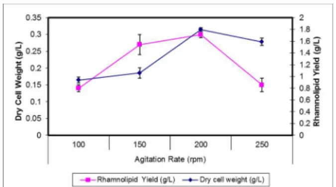
Effect of Agitation Rate on Rhamnolipid Production
Agitation rate affects the mass transfer efficiency of both oxygen and medium components and is con-sidered crucial to the cell growth and biosurfactant formation of the strictly aerobic bacterium P.
aeruginosa, especially when it was grown in a shake
Brazilian Journal of Chemical Engineering
Figure 3: Effect of different agitation rates on rham-nolipid Yield and Dry cell weight.
Structural Characterization
Thin Layer Chromatography (TLC)
The formation of various rhamnolipids was checked by TLC. The crude rhamnolipids contained both monor-hamnolipid and di-rmonor-hamnolipids, but the most active fragment is di-rhamnolipid, which gave a diameter of 10.5 cm in the oil displacement test.
Fourier Transform Infrared Spectroscopy (FTIR)
The infrared spectrum of purified biosurfactant indicated a broad peak at 3429 cm−1, characteristic of O–H stretching vibrations. Absorption around 2922 cm−1 is assigned to the asymmetric C-H stretch of CH2 and CH3 groups of aliphatic chains. The
corre-sponding symmetric stretch is seen at 2853 cm−1. Also, a weak symmetric stretching peak at 1715 cm−1 indicated the presence of ester carbonyl groups (C=O in COOH) in the biosurfactant. The ester carbonyl group was indicated by the band at 1321 cm−1 which
corresponds to C- O deformation vibrations, al-though other groups also absorb in this region.
The presence of the carboxylic acid functional group in the molecule was also confirmed by the medium intensity bands in the region 1455–1386 cm−1 for bending of the hydroxyl (O–H). The absorption peak around 1051cm−1 was also reported as C–O–C stretching in rhamnose (Pornsunthorntawee et al., 2008). The α-pyranyl II sorption band at 834 cm−1 showed the presence of di-rhamnolipid in the mix-ture. These characteristic adsorption bands taken together demonstrate that both have chemical struc-tures identical with those of rhamnolipids, which have rhamnose rings and long hydrocarbon chains. Thus, according to the results of the IR spectra, the rhamnolipids produced by P. aeruginosa TMN be-long to the glycolipid group, which is made up of aliphatic acids and esters. The adsorption bands ob-tained are consistent with the report of Guo et al. (2009) showing the presence of rhamnose rings and hydrocarbon chains. In the FTIR spectrum, we could observe only a minor shoulder; this might be because of the di-rhamnolipid-rich biosurfactant produced by
P. aeruginosa TMN (Rahman et al., 2002b). The
results obtained are consistent with the structure reported by Stanghellini and Miller (1997) consisting of aliphatic acid and the glycolipid moiety.
Nuclear Magnetic Resonance (NMR)
The structures of purified rhamnolipids were con-firmed by 1H-NMR and the results are shown in Table 4. The chemicals shifts were comparable to pre-vious reports (Ramana and Karanth, 1989; Sim et al.,
1997; Wei et al., 2005) and the results were in accordance with the structures as shown by ESI-spectra, Figure 4.
Table 4: 1H-NMR chemical shift data for rhamnolipid components.
1H chemical shift
(ppm)
Multiplicity Assignment
0.882 Singlet –CH3 (on β-hydroxyfatty acids)
1.185, 1.204 Doublet –CH3 (on rhamnose moiety)
1.276 Multiplet –(CH2)5– (on β-hydroxyfatty acids)
1.574 Multiplet –(CH2)–CH(O)–CH2COO (on β-hydroxyfatty acids)
2.545 Multiplet –CH(O)–CH2COO (on β-hydroxyfatty acids)
3.372 Multiplet –(CH2)–CH(O-Rha)–CH2COO (on β-hydroxyfatty acids)
3.335 Multiplet –CH–OH (on rhamnose moiety)
4.130 Multiplet –(CH2)–CH(–O–C=O)–CH2COO (on β-hydroxyfatty acids)
4.938 Singlet –CH–OH (on rhamnose moiety)
Figure 4: ESI mass spectrum and chemical structure of Rha-Rha-C10–C12
rhamnolipid and its fragments.
Electrospray Ionization–Mass Spectrometry (ESI– MS)
In this study, the highest intensity rhamnolipid found was the di-rhamnolipid homologues which correspond to Rha-Rha-C10–C12 with m⁄z of 680; their
fraction at m⁄z 365 is shown in Figure 4. This work is
in agreement with previous reports dealing with production of rhamnolipid surfactant mixtures in which di-rhamnolipids were the predominant species (Arutchelvi and Doble, 2010). On the contrary, few reports (Arino et al., 1996; Sim et al., 1997) describe a rhamnolipid mixture in which monorhamnolipid is the predominant component.
Physicochemical Characterization Oil Displacement Test
The Oil displacement test is an indirect measure-ment of the surface activity of a surfactant sample tested against oil; a larger diameter represents a
higher surface activity of the testing solution (Pornsunthorntawee et al., 2008). The oil spreading method is rapid and easy to carry out, requires no specialized equipment and just a small volume of sample (Płaza et al., 2006). It can be applied when the activity and quantity of biosurfactant is low. Plaza et al. (2004) and Youssef et al. (2006) demon-strated that the oil spreading technique is a reliable method to detect biosurfactant production by diverse microorganisms. The assay was also applied for screening by Huy et al. (1999).
Figure 5 lists the diameters of the clear zones on the oil surface obtained from oil displacement testing with the crude biosurfactant produced from different carbon and nitrogen sources by P. aeruginosa TMN.
Critical Micelle Concentration (CMC)
Brazilian Journal of Chemical Engineering change the surface properties and exhibit functions
such as emulsification, solubilization and foaming even at a relatively low concentration. The surface tension of purified biosurfactant decreased as its concentration increased, until reaching the lowest value, 34 mN m-1. At this point, the concentration of biosurfactant, namely the CMC, was 18.75 mg/L. This CMC was much lower compared with some syn-thetic surfactants. For instance, sodium dodecyl sul-fate (SDS) has a CMC value of 2100 mg/L (Chen et al., 2006). Nitschke and Pastore (2006) reported that the CMC of biosurfactant obtained from Bacillus
subtilis was 33 mg/L. Li et al. (1984) showed that
the CMC of rhamnolipid fermentation liquor was 386 mg/L (Daoshan et al., 2004).
Figure 5: Diameters of the clear zones on the oil surface obtained from oil displacement testing with the crude biosurfactant produced from different car-bon and nitrogen sources by Pseudomonas aerugi-nosa TMN.
Estimation of Emulsification Activity
Emulsification of rhamnolipids depends on the car-bon sources used to produce rhamnolipids (Porn-sunthorntawee et al., 2008). Six carbon sources, i.e., glucose, glycerol, sucrose, hexane, oleic acid, and olive oil were examined for their effectiveness of biosurfactant production. The results of various car-bon sources for emulsification activity under the above mentioned conditions are shown in Figure 6. As evident from the figure, P. aeruginosa TMN was able to grow in a medium containing oleic acid and olive oil with maximum emulsification activity. Other carbon sources (glycerol, glucose, sucrose and hexane) used, showed less emulsification activity, hexane being the lowest, as compared to other car-bon sources. This suggested that there is a carcar-bon source preference of the strain for biosurfactant pro-duction, which seems to be strain dependent (Wu et al., 2008). Most microbial surfactants were substrate specific, solubilizing or emulsifying different hydro-carbons at different rates.
Figure 6: Emulsifying activity (E24%) of biosur-factants obtained from different carbon sources against kerosene.
Poor emulsification of other hydrocarbons might be due to the inability of the biosurfactant to stabilize the microscopic droplets. Also, an inefficient oxygen supply in the flask cultures may be responsible for poor growth with other carbon sources, as biodegra-dation of these oils is known to be an oxygen inten-sive metabolic event (Panesar et al., 2009). On other hand, oleic acid and olive oil are the carbon sources which are taken up very easily compared to others. Among the six carbon sources tested, oleic acid and olive oil were the best carbon sources, with an emul-sification activity of 50%.
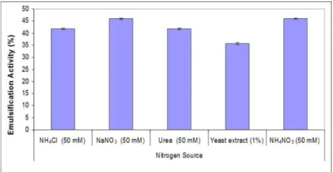
Nitrogen sources such as sodium nitrate, ammo-nium chloride, urea, yeast extract, and ammoammo-nium nitrate were added to the fermentation media. So-dium nitrate and ammonium nitrate were found to be the most effective amongst these nitrogen sources, as shown in Figure 7. Other nitrogen sources (urea, am-monium chloride and yeast extract) gave low emul-sification activity. Among the nitrogen sources, so-dium nitrate and ammonium nitrate gave similar re-sults, but, considering the cost factor, sodium nitrate was the most effective.
CONCLUSIONS
P. aeruginosa TMN capable of effectively
pro-ducing rhamnolipid from various carbon sources was successfully isolated. P. aeruginosa TMN can de-grade vegetable oils to produce biosurfactant and emulsify kerosene. Hence, the strain itself or its biosurfactant product have the potential to be applied in bioremediation of oil pollutants. Among the six carbon substrates and five nitrogen sources exam-ined, glucose and glycerol were the most efficient carbon sources, while NaNO3 was the best nitrogen
source for rhamnolipid production. Rhamnolipid production was optimal in batch cultures when the temperature, pH and agitation rate were controlled at 37 °C, pH 7 and 200 rpm, respectively. FTIR, NMR and ESI-MS analysis showed that the purified prod-uct contained L-rhamnosyl-L-rhamnosyl-β -hydroxy-decanoyl-β-hydroxydecanoate rhamnolipid. In con-clusion, the biosurfactant produced by P. aeruginosa
TMN strain was the preferable surface-active sub-stance, which can be used for potential application in bioremediation of crude oil contamination.
ACKNOWLEDGEMENT
This study was financially supported by the Academy of Scientific Research and Technology, Ministry of Scientific Research, Egypt, under grant Code UA0113.
REFERENCES
Abalos, A., Pinazo, A., Infante, M. R., Casals, M., Garcia, F. and Manresa, A., Physicochemical and antimicrobial properties of new rhamnolipids produced by Pseudomonas aeruginosa AT10 from soybean oil refinery wastes. Langmuir, 17, 1367 (2001).
Abbasi, H., Hamedi, M. M., Lotfabad, T. B., Zahiri, H. S., Sharafi, H., Masoomi, F., Moosavi-Movahedi, A. A., Ortiz, A., Amanlou, M. and Noghabi, K. A., Biosurfactant-producing bacterium, Pseudomonas
aeruginosa MA01 isolated from spoiled apples:
Physicochemical and structural characteristics of isolated biosurfactant. J. Biosci. Bioeng., 113, 211 (2012).
Abouseoud, M., Maachi, R., Amrane, A., Boudergua, S. and Nabi, A., Evaluation of different carbon and nitrogen sources in production of biosurfac-tant by Pseudomonas fluorescens. Desalination, 223, 143 (2008).
Altschul, S. F., Gish, W., Miller, W., Myers, E. W. and Lipman, D. J., Basic local alignment search tool. J. Mol. Biol., 215, 403 (1990).
Anna, L. M. S., Sebastian, G. V., Menezes, E. P., Alves, T. L. M., Santos, A. S., Pereira, N. and Freire, D. M. G., Production of biosurfactants from
Pseu-domonas aeruginosa PA1 isolated in oil
envi-ronments. Braz. J. Chem. Eng., 19, 159 (2002). Anyanwu, U. and Chukwudi, U., Surface activity of
extracellular products of a Pseudomonas aerugi-nosa isolated from petroleum contaminated soil. Inter. J. Environ. Sci., 1, 225 (2010).
Arino, S., Marchal, R. and Vandecasteele, J. P., Identification and production of a rhamnolipidic biosurfactant by a Pseudomonas species. Appl. Microbiol. Biotechnol., 45, 162 (1996).
Arutchelvi, J. and Doble, M., Characterization of glycolipid biosurfactant from Pseudomonas aerugi-nosa CPCL isolated from petroleum-contaminated soil. Lett. Appl. Microbiol., 51, 75 (2010).
Boulton, C. and Ratledge, C., Biosynthesis of lipid precursors to surfactant production. Marcel Dekker Inc., New York, USA (1987).
Chakravorty, S., Helb, D., Burday, M., Connell, N. and Alland, D., A detailed analysis of 16S ribo-somal RNA gene segments for the diagnosis of pathogenic bacteria. J. Microbiol. Methods, 69, 330 (2007).
Chayabutra, C., Wu, J. and Ju, L. K., Rhamnolipid production by Pseudomonas aeruginosa under de nitrification: Effects of limiting nutrients and car-bon substrates. Biotechnol. Bioeng., 72, 25 (2001). Chen, C. C., Riadi, L., Suh, S. J., Ohman, D. E. and
Ju, L. K., Degradation and synthesis kinetics of quorum-sensing autoinducer in Pseudomonas aeruginosa cultivation. J. Biotechnol., 117, 1 (2005). Chen, J., Wang, X. J., Hu, J. D. and Tao, S., Effect of surfactants on biodegradation of PAHs by white-rot fungi. Environ. Sci., 27, 154 (2006).
Christofi, N. and Ivshina, I. B., Microbial surfactants and their use in field studies of soil remediation. J. Appl. Microbiol., 93, 915 (2002).
Cooper, D. G. and Goldenberg, B. G., Surface-active agents from two Bacillus species, Appl. Environ. Microbiol., 53, 224 (1987).
Daoshan, L., Shouliang, L., Yi, L. and Demin, W., The effect of biosurfactant on the interfacial ten-sion and adsorption loss of surfactant in ASP flooding. Coll. Surf., A, 244, 53 (2004).
Brazilian Journal of Chemical Engineering Davis, D. A., Lynch, H. C. and Varley, J., The
appli-cation of foaming for the recovery of surfactin from Bacillus subtilis ATCC 21332 cultures. En-zyme Microb. Technol., 28, 346 (2001).
Desai, J. D. and Banat, I. M., Microbial production of biosurfactants and their commercial potential. Microbiol. Mol. Biol. Rev., 61, 47 (1997).
Deziel, E., Lepine, F., Dennie, D., Boismenu, D., Mamer, O. A. and Villemur, R., Liquid chroma-tography/mass spectrometry analysis of mixtures of rhamnolipids produced by Pseudomonas
aeruginosa strain 57RP grown on mannitol or
naphthalene. Biochim. Biophys. Acta, 1440, 244 (1999).
Fontes, G. C., Ramos, N. M., Amaral, P. F. F., Nele, M. and Coelho, M. A. Z., Renewable resources for biosurfactant production by yarrowia lipolytica. Braz. J. Chem. Eng., 29, 483 (2012). Guo, Y. P., Hu, Y. Y., Gu, R. R. and Lin, H.,
Char-acterization and micellization of rhamnolipidic fractions and crude extracts produced by
Pseu-domonas aeruginosa mutant MIG-N146. J.
Col-loid Interface Sci., 331, 356 (2009).
Huy, N. Q., Jin, S., Amada, K., Haruki, M., Huu, N. B., Hang, D. T., Ha, D. T., Imanaka, T., Morikawa, M. and Kanaya, S., Characterization of petroleum-degrading bacteria from oil-contaminated sites in Vietnam. J. Biosci. Bioeng., 88, 100 (1999). Iqbal, S., Khalid, Z. M. and Malik, K. A., Enhanced
biodegradation and emulsification of crude oil and hyper production of biosurfactants by a gamma ray-induced mutant of Pseudomonas aeruginosa. Lett. Appl. Microbiol., 21, 176 (1995).
Ilori, M. O., Amobi, C. J. and Odocha, A. C., Factors affecting biosurfactant production by oil
degrad-ing Aeromonas spp. isolated from a tropical
envi-ronment. Chemosphere, 61, 985 (2005).
Jarvis, F. G. and Johnson, M. J., A Glyco-lipide pro-duced by Pseudomonas aeruginosa. J. Am. Chem. Soc., 71, 4124 (1949).
Jeong, H. S., Lim, D. J., Hwang, S. H., Ha, S. D. and Kong, J. Y., Rhamnolipid production by
Pseudo-monas aeruginosa immobilized in polyvinyl
al-cohol beads. Biotechnol. Lett., 26, 35 (2004). Joshi, S., Bharucha, C. and Desai, A. J., Production
of biosurfactant and antifungal compound by fermented food isolate Bacillus subtilis 20B. Bio-resource Technol., 99, 4603 (2008).
Kim, H. S., Jeon, J. W., Kim, B. H., Ahn, C. Y., Oh, H. M. and Yoon, B. D., Extracellular production of a glycolipid biosurfactant, mannosylerythritol lipid, by Candida sp. SY16 using fed-batch fer-mentation. Appl. Microbiol. Biotechnol., 70, 391 (2006).
Koch, A. K., Kappeli, O., Fiechter, A. and Reiser, J., Hydrocarbon assimilation and biosurfactant pro-duction in Pseudomonas aeruginosa mutants. J. Bacteriol., 173, 4212 (1991).
Kosaric, N., Biosurfactants in industry. Pure Appl. Chem., 64, 1731 (1992).
Lane, D. J., 16S/23S rRNA Sequencing. In: Nucleic Acid Techniques in Bacterial Systematic. Stacke-brandt E. and Goodfellow, M., Eds., John Wiley and Sons Inc., London, pp. 115 (1991).
Lang, S. and Wullbrandt, D., Rhamnose lipids – biosynthesis, microbial production and applica-tion potential. Appl. Microbiol. Biotechnol., 51, 22 (1999).
Li, Y. Z., Lang, S., Wagner, F., Witte, L. and Wray, V., Formation and identification of interfacial-ac-tive glycolipids from resulting microbial cells of
Arthrobacter sp. and potential use in tertiary oil
recovery. Appl. Environ. Microbiol., 48, 610 (1984).
Lotfabad, T. B., Shourian, M., Roostaazad, R., Na-jafabadi, A. R., Adelzadeh, M. R. and Noghabi, K. A., An efficient biosurfactant-producing bacte-rium Pseudomonas aeruginosa MR01, isolated from oil excavation areas in south of Iran. Coll. Surf. B. Biointer., 69, 183 (2009).
Maier, R. M. and Chavez, G. S., Pseudomonas aerugi-nosa rhamnolipids: Biosynthesis and potential applications. Appl. Microbiol. Biotechnol., 54, 625 (2000).
Makkar, R. S. and Cameotra, S. S., Biosurfactant production by microorganisms on unconventional carbon sources. J. Surfact. Deterg., 2, 237 (1999). Makkar, R. S. and Cameotra, S. S., Effects of various
nutritional supplements on biosurfactant produc-tion by a strain of Bacillus subtilis at 45 °C. J. Surfact. Deterg., 5, 11 (2002).
Mata-Sandoval, J. C., Karns, J. and Torrents, A., High-performance liquid chromatography method for the characterization of rhamnolipid mixtures produced by pseudomonas aeruginosa UG2 on corn oil. J. Chromatogr., A, 864, 211 (1999). Mata-Sandoval, J. C., Karns, J. and Torrents, A.,
Effect of nutritional and environmental conditions on the production and composition of rham-nolipids by Pseudomonas aeruginosa UG2. Mi-crobiol. Res., 155, 249 (2001).
Mechaly, A., Belakhov, V., Shoham, Y. and Baasov, T., An efficient chemical-enzymatic synthesis of 4-nitrophenyl [beta]-xylobioside: A chromogenic substrate for xylanases. Carbohydr. Res., 304, 111 (1997).
surfac-tant quantitation and screening of biosurfacsurfac-tant- biosurfactant-producing microorganisms. J. Microbiol. Meth-ods, 32, 273 (1998).
Monteiro, S. A., Sassaki, G. L., de Souza, L. M., Meira, J. A., de Araujo, J. M., Mitchell, D. A., Ramos, L. P. and Krieger, N., Molecular and structural characterization of the biosurfactant produced by
Pseudomonas aeruginosa DAUPE 614. Chem.
Phys. Lipids, 147, 1 (2007).
Mulligan, C. N., Environmental applications for bio-surfactants. Environ. Pollut., 133, 183 (2005). Nguyen, T. T., Youssef, N. H., McInerney, M. J. and
Sabatinic, D. A., Rhamnolipid biosurfactant mix-tures for environmental remediation. Water Res., 42, 1735 (2008).
Nitschke, M. and Pastore, G. M., Production and properties of a surfactant obtained from Bacillus
subtilis grown on cassava wastewater. Bioresour.
Technol., 97, 336 (2006).
Panesar, R., Panesar, P. S., Hasija, D., Bera, M. B. and Kumar, H., Fermentative potential of
Pseu-domonas aeruginosa strain for biosurfactant
pro-duction. Biol. Forum –Inter. J., 1, 109 (2009). Pirôllo, M. P. S., Mariano, A. P., Lovaglio, R. B.,
Costa, S. G. V. A. O., Walter, V., Hausmann, R. and Contiero, J., Biosurfactant synthesis by
Pseu-domonas aeruginosa LBI isolated from a
hydro-carbon-contaminated site. J. Appl. Microbiol., 105, 1484 (2008).
Płaza, G. A., Zjawiony, I. and Banat, I. M., Use of different methods for detection of thermophilic biosurfactant-producing bacteria from hydrocar-bon-contaminated and bioremediated soils. J. Pe-tro. Sci. Eng., 50, 71 (2006).
Pornsunthorntawee, O., Wongpanit, P., Chavadej, S., Abe, M. and Rujiravanit, R., Structural and phy-sicochemical characterization of crude biosur-factant produced by Pseudomonas aeruginosa
SP4 isolated from petroleum-contaminated soil. Bioresour. Technol., 99, 1589 (2008).
Priya, T. and Usharani, G., Comparative study for biosurfactant production by using Bacillus
sub-tilis and Pseudomonas aeruginosa. Bot. Res.
In-ter., 2, 284 (2009).
Providenti, M. A., Flemming, C. A., Lee, H. and Trevore, J. T., Effect of addition of rhamnolipid biosurfactants or rhamnolipid producing
Pseudo-monas aeruginosa on phenanthrene
mineraliza-tion in soil slurries. FEMS Microbiol. Ecol., 17, 15 (1995).
Pruthi, V. and Cameotra, S. S., Effect of nutrients on optimal production of biosurfactants by Pseudo-monas putida – a Gujarat oil field isolate. J. Sur-fact. Deterg., 6, 65 (2003).
Rahman, K. S., Banat, I. M., Thahira, J., Thayu-manavan, T. and Lakshmanaperumalsamy, P., Bio-remediation of gasoline contaminated soil by a bacterial consortium amended with poultry litter, coir pith and rhamnolipid biosurfactant. Bioresour. Technol., 81, 25 (2002a).
Rahman, K. S. M., Rahman, T. J., McClean, S., Marchant, R. and Banat, I. M., Rhamnolipid biosurfactant production by strains of
Pseudomo-nas aeruginosa using low-cost raw materials.
Biotechnol. Prog., 18, 1277 (2002b).
Ramana, K. V. and Karanth, N. G., Factors affecting biosurfactant production using Pseudomonas
aeruginosa CFTR-6 under submerged conditions.
J. Chem. Tech. Biotechnol., 45, 249 (1989). Raza, Z. A., Khan, M. S. and Khalid, Z. M.,
Evalua-tion of distant carbon sources in biosurfactant production by a gamma ray-induced Pseudomonas
putida mutant. Process Biochem., 42, 686 (2007).
Robert, M., Mercade, E., Bosch, M. P., Parra, J. L., Espuny, M. J., Manresa, M. A. and Guinea, J., Ef-fect of the carbon source on biosurfactant pro-duction by Pseudomonas aeruginosa 44Ti. Bio-technol. Lett., 11, 871 (1989).
Rodrigues, L. R., Teixeira, J. A., van der Mei, H. C. and Oliveira, R., Physicochemical and functional characterization of a biosurfactant produced by
Lactococcus lactis 53. Coll. Surf., B, 49, 79 (2006). Santa Anna, L. M., Sebastian, G. V., Menezes, E. P.,
Alves, T. L. M., Santos, A. S., Pereira Jr., N. and Freire, D. M. G., Production of biosurfactants from
Pseudomonas aeruginosa PA 1 isolated in oil
environments. Braz. J. Chem. Eng., 19, 159-166 (2002).
Santos, L. G., Käppeli, O. and Fiechter,A.,
Pseudo-monas aeruginosa biosurfactant production in
continuous culture with glucose as carbon source. Appl. Environ. Microbiol., 48, 301 (1984). Santos, A. S., Sampaio, A. P., Vasquez, G. S., Santa
Anna, L. M., Pereira, N. and Freire, D. M., Evalua-tion of different carbon and nitrogen sources in production of rhamnolipids by a strain of
Pseu-domonas aeruginosa.Appl. Biochem. Biotechnol.,
98, 1025 (2002).
Sim, L., Ward, O. P and Li, Z. Y., Production and characterization of a biosurfactant isolated from
Pseudomonas aeruginosa UW-1. J. Ind.
Micro-biol. Biotechnol., 19, 232 (1997).
Stanghellini, M. E. and Miller, R. M., Biosurfactants: Their identity and potential efficacy in the biological control of zoosporic plant pathogens. Plant Dis., 81, 4 (1997).
analy-Brazilian Journal of Chemical Engineering sis (MEGA) software version 4.0. Mol. Biol. Evol.,
24, 1596 (2007).
Torrens, J. L., Herman, D. C. and Miller-Maier, R. M., Biosurfactant (rhamnolipid) sorption and the impact on rhamnolipid-facilitated removal of cadmium from various soils under saturated flow conditions. Environ. Sci. Technol., 32, 776 (1998). Urum, K. and Pekdemir, T., Evaluation of biosur-factants for crude oil contaminated soil washing. Chemosphere, 57, 1139 (2004).
Wei, Y. H. and Chu, I. M., Enhancement of surfactin production in iron-enriched media by Bacillus
subtilis ATCC 21332. Enzyme Microb. Technol.,
22, 724 (1998).
Wei, Y. H., Chou, C. L. and Chang, J. S., Rham-nolipid production by indigenous Pseudomonas
aeruginosa J4 originating from petrochemical
wastewater. Biochem. Eng. J., 27, 146 (2005). Wu, J. Y., Yeh, K. L., Lu, W. B., Lin, C. L. and Chang,
J. S., Rhamnolipid production with indigenous
Pseudomonas aeruginosa EM1 isolated from
oil-contaminated site. Bioresour. Technol., 99, 1157 (2008).
Yakimov, M. M., Timmis, K. N., Wray, V. and Fredrickson, H. L., Characterization of a new lipopeptide surfactant produced by thermo toler-ant and halo tolertoler-ant subsurface Bacillus
licheni-formis BAS 50. Appl. Environ. Microbiol., 61,
3276 (1995).
Youssef, N. H., Duncan, K. E., Nagle, D. P., Savage, K. N., Knapp, R. M. and McInerney, M. J., Com-parison of methods to detect biosurfactant pro-duction by diverse microorganisms. J. Microbiol. Methods, 56, 339 (2004).
Zhang, G. L., Wu, Y. T., Qian, X. P. and Meng, Q., Biodegradation of crude oil by Pseudomonas
aeruginosa in the presence of rhamnolipids. J.