eScholarship provides open access, scholarly publishing services to the University of California and delivers a dynamic research platform to scholars worldwide.
Dermatology Online Journal
UC Davis
Peer Reviewed Title:
Dermoscopy in Merkel cell carcinoma: a case report Journal Issue:
Dermatology Online Journal, 20(2)
Author:
Laureano, Andre, Hospital de Curry Cabral – Centro Hospitalar de Lisboa
Cunha, Daniela, Hospital de Curry Cabral – Centro Hospitalar de Lisboa
Pernandes, Candida, Hospital de Curry Cabral – Centro Hospitalar de Lisboa
Cardoso, Jorge, Hospital de Curry Cabral – Centro Hospitalar de Lisboa Publication Date:
2014
Publication Info:
Dermatology Online Journal Permalink:
http://escholarship.org/uc/item/03p4m424
Author Bio: Keywords:
dermoscopy, Merkel cell carcinoma, skin cancer Local Identifier:
doj_21543 Abstract:
Merkel cell carcinoma (MCC) is a rare malignant and primary neuroendocrine carcinoma with several known risk factors. Early diagnosis and aggressive treatment are critical. We report the case of an 82-year old woman with a Merkel cell carcinoma on the face. Clinical and histopathological features are presented. In addition, dermoscopic features and the differential diagnosis of this rare tumor are discussed. Although nodules with atypical dermoscopic vascular pattern and milky-red areas will end up being excised, this report adds more clues to the rarely described dermoscopic morphologic presentation of MCC.
eScholarship provides open access, scholarly publishing services to the University of California and delivers a dynamic research platform to scholars worldwide.
Copyright 2014 by the article author(s). This work is made available under the terms of the Creative Commons Attribution-NonCommercial-NoDerivs3.0 license, http:// creativecommons.org/licenses/by-nc-nd/3.0/
Volume 20 Number 2
February 2014
Case Presentation
Dermoscopy in Merkel cell carcinoma: a case report
André Laureano, Daniela Cunha, Cândida Fernandes, Jorge Cardoso
Dermatology Online Journal 20 (2): 10
Department of Dermatology and Venereology, Hospital de Curry Cabral – Centro Hospitalar de Lisboa
Central, Lisboa, Portugal
Correspondence:
André Laureano, MD
Hospital de Curry Cabral – Centro Hospitalar de Lisboa Central, Lisboa, Portugal
Rua da Beneficência, nº8, 1069-166 Lisboa, Portugal
[email protected]
+351926873106
Abstract
Merkel cell carcinoma (MCC) is a rare malignant and primary neuroendocrine carcinoma with several known risk factors. Early diagnosis and aggressive treatment are critical. We report the case of an 82-year old woman with a Merkel cell carcinoma on the face. Clinical and histopathological features are presented. In addition, dermoscopic features and the differential diagnosis of this rare tumor are discussed. Although nodules with atypical dermoscopic vascular pattern and milky-red areas will end up being excised, this report adds more clues to the rarely described dermoscopic morphologic presentation of MCC.
Keywords: dermoscopy, Merkel cell carcinoma, skin cancer
Case synopsis
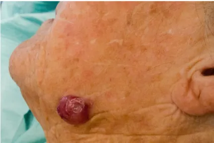
An 82 year-old woman presented with an asymptomatic nodule on her left mandible, which had appeared two months earlier. She had no personal or family history of skin cancer. Physical examination revealed Fitzpatrick skin type III, intense actinic damage, and a solitary, violaceous, shiny nodule located on the left mandible. The nodule was firm, non-ulcerated, with 3 cm maximum diameter.
Figure 1. Fast-growing, asymptomatic, shiny and violaceous nodule on the left mandible
There was no palpable lymphadenopathy. Dermoscopy showed a combination of sharply and poorly focused vessels and a variety of vascular patterns with architectural disruption. Milky-red areas with shiny white areas within and a polymorphous vascular pattern with linear-irregular and large-caliber arborizing vessels were seen.
Figure 2. Dermoscopic features of poorly and sharply focused vessels, milky-red areas, and globules with shiny white areas within are
exhibited..Polymorphous vessels with linear-irregular and large caliber arborizing vessels are evident (Dermlite, DL3, 10x magnification, no contact medium).
Based on the findings of a non-pigmented, fast-growing nodule on a chronically sun-exposed area with an atypical
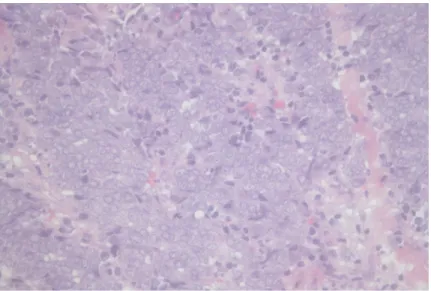
dermoscopic vascular pattern, the differential diagnosis included amelanotic melanoma, basal cell carcinoma (BCC), pyogenic granuloma, and other amelanotic melanocytic tumors. The nodule was excised and histopathological examination revealed small round cells with hyperchromatic nuclei and scanty cytoplasm throughout the dermis.
Figure 3. Small round cells with hyperchromatic nuclei and scanty cytoplasm (hematoxylin and eosin, original magnification x 100)
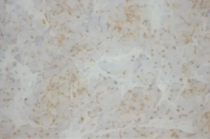
Immunohistochemical staining was positive for chromogranin and cytokeratin-20.
Figure 5. Cytokeratin-20 positive (original magnification x 100)
The diagnosis of Merkel cell carcinoma (MCC) was established based upon the clinical, dermoscopic and histological correlation. Tumor tissue specimens embedded in paraffin tested positive for Merkel cell Polyomavirus (MCPyV) detected using qualitative polymerase chain reaction analysis.
The remaining laboratory and imaging studies were negative and sentinel node biopsy was not performed (T2 cN0 M0 – Stage IIB AJCC). The patient underwent wide local surgical excision and subsequent adjuvant radiotherapy to the primary site (50 Gy). No tumor relapses, lymph node involvement, or distant metastases were seen after three years of follow-up.
Discussion
MCC is a rare malignant and primary neuroendocrine carcinoma with several known risk factors, including MCPyV[1]. Early diagnosis and aggressive treatment are critical, but clinical diagnosis is often delayed owing to the absence of specific
diagnostic criteria at an early stage.Dermoscopy is likely to be a useful diagnostic tool but few reports on the dermoscopic features of MCC have been published[2].
On dermoscopy, this tumor exhibits overlapping features with other skin tumors. The combination of polymorphous vessels and milky-red areas suggest amelanotic melanoma, whereas isolated milky red areas might be seen in pyogenic granuloma; arborizing vessels are typical of basal cell carcinoma. The combination of these structures might also suggest cutaneous metastasis. Nonetheless, other more distinguishing features, such as sharply and poorly focused vessels and shiny white areas within the body of the tumor, were remarkable in this tumor[1,3].
The dermoscopic findings in this case seem to mirror the fast tumor growth. However, further evidence regarding dermoscopy of MCC and its correlation to histological features is required. Neoangiogenesis seems to be vital for tumor growth and deep invasion. Some of the featured dermoscopic structures in MCC, including atypical and polymorphic vessels, might correlate with this phenomenon. Sharply focused large caliber arborizing vessels correspond to new vessels with a more superficial location, whereas out-of-focus vessels are deeply located. Linear-irregular vessels correlate to elongation of capillaries and tortuous, horizontal new vessels. Milky-red areas or globules also correlate with deep tumoral angiogenesis, explaining the
blurred white and pink structures. Shiny white chrysalis-like structures are not yet understood [4].
Conclusion
MCC should be included in the differential diagnosis of a fast-growing pink nodule with atypical dermoscopic vascular pattern. Although these nodules will end up being excised, this report adds more clues to the rarely described dermoscopic morphologic presentation of MCC.
References
1. Dalle S, Parmentier L, Moscarella E, et al. Dermoscopy of a Merkel cell carcinoma. Dermatology. 2012 May;224(2):140-44. [PMID: 22487601]
2. Scalvenzi M, Palmisano F, Ilardi G, et al. Clinical, dermoscopic and histological features of a Merkel cell carcinoma of the hand. J Dermatol Case Rep. 2013 Mar;7(1):15-17. [PMID: 23580909]
3. Harting MS, Ludgate MW, Fullen DR, Johnson TM, Bichakjian CK. Dermatoscopic vascular patterns in cutaneous Merkel cell carcinoma. J Am Acad Dermatol. 2012 Jun;66(6):923-27. [PMID: 21978573]
4. Jalilian C, Chamberlain AJ, Haskett M, Rosendahl C, et al. Clinical and dermoscopic characteristics of Merkel cell carcinoma. Br J Dermatol. 2013;169:294-7. [PMID: 23574613]