Universidade de Aveiro 2015-2016
Departamento de Química
Leonor Sofia
Oliveira Castro
Purificação de anticorpos utilizando sistemas
micelares de duas fases aquosas
Antibodies purification using aqueous micellar
two-phase systems
Universidade de Aveiro 2015-2016
Departamento de Química
Leonor Sofia
Oliveira Castro
Purificação de anticorpos utilizando sistemas
micelares de duas fases aquosas
Antibodies purification using aqueous micellar
two-phase systems
Tese apresentada à Universidade de Aveiro para cumprimento dos requisitos necessários à obtenção do grau de Mestre em Biotecnologia, ramo Biotecnologia Molecular, realizada sob a orientação científica da Doutora Mara Guadalupe Freire Martins, Investigadora Coordenadora do Departamento de Química, CICECO, da Universidade de Aveiro, e coorientação da Doutora Ana Paula Moura Tavares, Estagiária de Pós-Doutoramento do Departamento de Química da Universidade de Aveiro.
o júri
presidente
Professor Doutor João Manuel da Costa Araújo Pereira Coutinho
Professor Catedrático do Departamento de Química da Universidade de Aveiro
Doutora Mara Guadalupe Freire Martins
Investigadora Coordenadora do Departamento de Química, CICECO, da Universidade de Aveiro
Doutor Oscar Rodríguez Figueiras
Investigador, Programa Ramón y Cajal da Universidade de Santiago de Compostela, Espanha
agradecimentos
Antes de mais gostaria de agradecer à Drª Mara Freire por ter aceite orientar-me e pela oportunidade de trabalhar no excelente grupo que é o Path. Gostaria também de agradecer a minha co-orientadora, à Drª Ana Paula pela forma como me acompanhou neste trabalho e por todo o apoio que me deu. A todos os membros do Path, o meu muito obrigado, com vocês aprendi o quão importante é trabalhar em equipa. Um especial obrigado à Filipa, por todo o apoio e paciência ao longo deste ano.
Queria também agradecer à minha irmã Cassandra por ser a minha maior fã, e à Cátia por todo o carinho e apoio, mais do que uma amiga já fazes parte da família. Sem vocês eu não teria chegado até aqui por isso este trabalho também é um pouco vosso, e como tal, espero que este vos inspire da mesma forma que vocês me inspiram.
Aos meus pais, obrigada por acreditarem em mim, por me ensinarem que nada é impossível e por me apoiarem incondicionalmente. Obrigada ainda por aturarem a minha teimosia, mas acima de tudo por me deixarem fazer as minhas próprias escolhas.
palavras-chave Imunoglobulina Y, purificação, sistemas micelares de duas fases aquosas, Triton X-114, líquidos iónicos
resumo Nos últimos anos, terapias à base de anticorpos mono e policlonais têm atraído muita atenção por parte da indústria farmacêutica levando a um aumento da investigação neste campo. A imunoglobulina G (IgG), um dos anticorpos já aprovados pela Food and Drug Administration (FDA) é uma das imunoglobulinas mais abundantes nos mamíferos, cuja obtenção requer o uso de técnicas invasivas. Como alternativa, e devido às semelhanças estruturais com a IgG, surge o uso da imunoglobulina Y (IgY) que se encontra presente na gema de ovo de galinha. Além disso, as quantidades de IgY presente na gema do ovo são bastante altas (100-150 mg/ovo) quando comparadas com a quantidade de IgG no soro (200mg/40mL de sangue). Contudo, o isolamento da IgY a partir da gema de ovo é bastante dispendioso e moroso, uma vez que requer o uso de processos com várias etapas. Tendo em conta estas desvantagens torna-se indispensável o desenvolvimento de um método mais económico e mais biocompatível. Os sistemas micelares de duas fases aquosas, um tipo específico de sistemas de duas fases aquosas que recorrem ao uso de surfactantes, surge como alternativa para a extração, purificação e/ou concentração de (bio)moléculas. Adicionalmente, o uso de líquidos iónicos (LIs) como co-surfactantes pode modificar as propriedades do surfactante e dessa forma conduzir a mudanças na extração das (bio)moléculas. Neste sentido, o objetivo deste trabalho é o desenvolvimento de um novo método para a extração, purificação e concentração da IgY através do uso de sistemas micelares de duas fases aquosas convencionais e mistos com LIs pertencentes à família dos imidazólios e fosfónios a atuar como co-surfactantes. Para tal, foram otimizados parâmetros como a concentração de surfactante e da fração de proteínas solúveis em água, a ausência e presença de (LIs) bem como o efeito da sua concentração e estrutura química. Após toda a otimização, os melhores resultados foram obtidos com o sistema convencional e sistema misto com [C16mim]Cl, onde se obtiveram
purificações de 51.2 % e 64.5% e rendimentos de 100% e 74.7%respetivamente.
Keywords Immunoglobulin Y, purification, aqueous micellar two-phase systems, Triton X-114, ionic liquids
Abstract In the past few years, therapies based on mono- and polyclonal
antibodies have attracted attention from pharmaceutical industries leading to a drastic research increment in this field. Immunoglobulin G (IgG), one of the antibodies already approved by Food and Drug Administration (FDA), is the most abundant mammalian immunoglobulin, whose acquisition requires the use of invasive techniques. As an alternative to the use of mammalian antibodies, Immunoglobulin Y (IgY) from hens’ egg yolk emerges due to its structural similarity to IgG. Furthermore, the IgY amount present in egg’s yolk can be quite high (100-150 mg/egg) when compared with the IgG amount in the blood serum (200 mg/40 mL of blood). However, IgY isolation from the egg’s yolk is quite expensive and time consuming, since it requires multistep processes. Taking these disadvantages into account, the development of a cheaper and more biocompatible technique for IgY extraction and purification becomes mandatory. Aqueous micellar two-phase systems (AMTPS) are a special type of aqueous two-phase systems that comprise micellar solutions of surfactants for (bio)molecules extraction, purification and/or concentration. Additionally, the use of ionic liquids (ILs) as co-surfactants can modify the surfactant properties, leading to changes in the phase separation as well as in the (bio)molecules fractionation. In this sense, this work aims at the development of a new extraction, purification and concentration technique for IgY from egg’s yolk using both conventional and mixed AMTPS with tensioactive ILs belonging to two distinct families, namely imidazolium and phosphonium, acting as co-surfactants. Thus, parameters like the surfactant and the water-soluble proteins fraction (WSPF) concentration, the presence/absence of IL as well as the effect of its concentration and structural features were optimized. After the optimization procedure, the best results were obtained with the conventional system and mixed AMTPS with [C16mim]Cl, were a purification of 51.2 % and 64.5 % and yields of 100%
Contents
List of Abbreviations ... xiii
List of Tables ... xiv
List of Figures ... xv
1. General Introduction ... 3
1.1 Scope and Objectives... 3
1.2 State of the art ... 5
1.2.1. Immunoglobulins structure ... 5
1.2.2. Immunoglobulin Y ... 7
1.2.3. Extraction and purification processes using aqueous (micellar) two-phase systems 10 2. Experimental Section ... 19
2.1. Materials and reagents ... 19
2.2. Methods ... 19
2.2.1. Preparation of the Water Soluble Protein Fraction ... 19
2.2.2. Preparation of the AMTPS for the IgY extraction and purification ... 19
2.2.3. AMTPS preparation for the consecutive IgY purification cycles ... 22
2.2.4. Protein quantification of the micelle-poor phase by SE-HPLC analysis... 22
2.2.5. SDS-PAGE analysis of both micelle-poor and micelle-rich phases... 23
2.2.6. IgY stability studies ... 24
3. Results and Discussion ... 27
3.1. IgY purification using the conventional and mixed AMTPS with ILs acting as co-surfactants ... 27
3.2. Consecutive extraction cycles using mixed AMTPS for the IgY extraction and purification ... 38
xii
3.3. Consecutive extraction cycles for reuse of micelle-rich phase ... 40
3.4. IgY stability studies ... 41
3.5. Conventional vs novel methods for antibodies extraction and purification .... 44
4. Final remarks ... 49
4.1. General conclusions and future work ... 49
5. References... 51
List of Abbreviations
[C10mim]Cl – 1-decyl-3-methylimidazolium chloride
[C12mim]Cl – 1-dodecyl-3-methylimidazolium chloride
[C14mim]Cl – 1-methyl-3-tetradecylimidazolium chloride
[C16mim]Cl – 1-Hexadecyl-3-methylimidazolium Chloride
[C18mim]Cl – 1-Octadecyl-3-methylimidazolium Chloride
[P6,6,6,14]Br – trihexyltetradecylphosphonium bromide
[P6,6,6,14]Cl – trihexyltetradecylphosphonium chloride
[P6,6,6,14]Dec – trihexyltetradecylphosphonium decanoate
[P6,6,6,14]TMPP – trihexyltetradecylphosphonium bis (2,4,4-trimethylpentyl)phosphinate
AMTPS – Aqueous micellar two-phase systems ATPS – Aqueous two-phase systems
C6H8O7 – Citric acid
CMC – Critical micellar concentration DAD – Diode array detector
DTT – Dithiothreitol
FDA – Food and Drug and Administration
FT-IR – Fourrier transform infrared spectroscopy IgG – Immunoglobulin G
IgY – Immunoglobulin Y IL – Ionic liquid
Na2HPO4 – Disodium phosphate
NaH2HPO4 – Sodium dihydrogenphosphate
PEG – Polyethylene glycol
POEAEs – Poly(oxyethyene) alkyl ether
SDS-PAGE – Sodium dodecyl sulphate polyacrylamide gel eletrocphoresis SE-HPLC – Size exclusion - high performance chromatography
TBS – Tris-buffered saline TCA – Trichloroacetic acid
xiv
List of Tables
Table 1: Differences between the mammalian IgG and IgY. Adapted from [1].
List of Figures
Figure 1: General immunoglobulin structure. Adapted from [2].
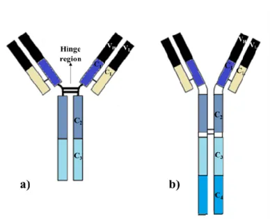
Figure 2: Structure of IgG (a) and IgY (b); representation of H and L chains, hinge region and C and V domains. Adapted from [1].
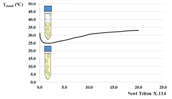
Figure 3: Binodal curve for Triton X-114. At temperatures below the Tcloud, the AMTPS
only has one homogeneous phase and above the Tcloud, the homogeneous mixture is
separated into two phases: one micelle-poor phase and one micelle-rich phase. Adapted from [34].
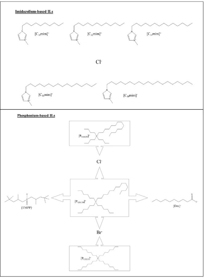
Figure 4: Chemical structure of the anions and cations that composed the imidazolium-based and phosphonium-imidazolium-based ILs applied to the AMTPS.
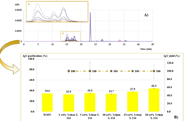
Figure 5: A) Chromatograms for the AMTPS top phase at different surfactant concentrations upon phase separation at 35ºC: ─, WSPF; ─, 1 wt% Triton X-114; ─, 5 wt% Triton X-114; ─, 10 wt% Triton X-114; ─, 15 wt% Triton X-114; and ─, 20 wt% Triton X-114 and B) IgY purification (bars) and yield (line) in percentage (%), obtained for each AMTPS with different surfactant concentrations, (wt%) of Triton X-114, upon phase separation at 35ºC.
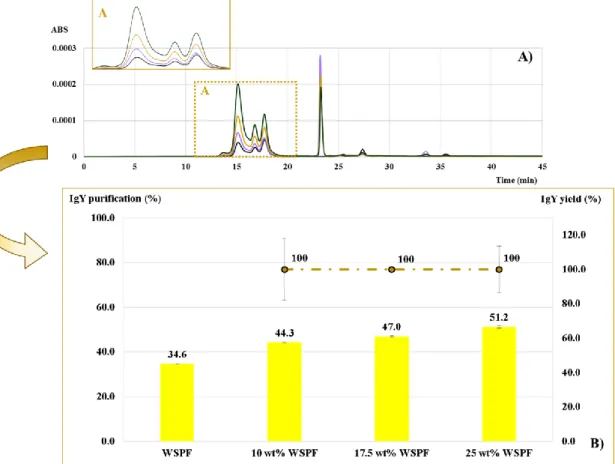
Figure 6: A) Chromatograms for the top phase of the AMTPS at different wt% of WSPF at pH 6 at 35ºC for ─, WSPF; ─,10 wt% WSPF; ─,17.5 wt% WSPF; and ─, 25 wt% WSPF and B) IgY purification (%) (bars) and yield (%) (lines), obtained for each system with different amounts of WSPF (wt%), and 20 wt% of Triton X-114, upon phase separation at 35ºC.
Figure 7: A) Chromatograms for the top-phase of the AMTPS with [Cnmim]Cl-based
ILs at A) 0.3 wt% and B) 0.5 wt% at pH 6 for: ─, WSPF; ─, [C10mim]Cl ; ─, [C12mim]Cl;
─, [C14mim]Cl; ─, [C16mim]Cl; ─, [C18mim]Cl and C) IgY purification (%) (bars) and
yield (%) (line) , obtained for each system with: ─, 0.3 or ─, 0.5 wt% of the imidazolium-based ILs at the optimized system conditions ( 20 wt% of Triton X-114 and 25 wt% of WSPF) and at: , 35ºC; ,37ºC; , 40ºC; and , 50ºC.
xvi
Figure 8: Chromatograms for the AMTPS top-phase at (A) 35ºC and (B) 37ºC with phosphonium-based ILs at 0.3 wt% and pH 6 for: ─, WSPF; ─ , [P6,6,6,14]Cl; ─,
[P6,6,6,14]TMMP; ─, [P6,6,6,14]Br; ─, [P6,6,6,14]Dec; ─, [P8,8,8,8]Br; ─, [P4,4,4,14]Cl.
Figure 9: IgY purification (bars) and yield (line) in percentage (%) obtained for each system with ─, 0.3 and ─, 0.5 wt% of phosphonium-based IL at , 35ºC and , 37ºC for the same system with 20 wt% of Triton X-114 and 25 wt% of WSPF.
Figure 10: SDS-PAGE gel of the supernatant after different protein precipitation methods used in both micelle-poor and micelle-rich phases for the conventional AMTPS with 20 wt% of Triton X-114.
Figure 11: SDS-PAGE gel of the resulting from the protein precipitations both micelle-poor and micelle-rich phase of conventional AMTPS with 20 wt% of Triton X-114 and mixed AMTPS with 0.3 wt% IL acting as co-surfactant.
Figure 12: A) Chromatograms for the top-phase form the extraction cycles with 0.3 wt% of [C14mim]Cl at pH 6 and 37ºC for: ─, WSPF; ─ 1st extraction cycle; *,2nd extraction
cycle;─ ─, 3rd extraction cycle and B) IgY purification (%) (bars) and yield (%) (line) obtained for each extraction cycle with 0.3 wt% of [C14mim]Cl, upon phase separation at
37ºC.
Figure 13: SDS-PAGE gel of the resulting from the protein precipitation of the micelle-poor and micelle-rich phase of the different extraction cycles using 0.3 wt% of [C14mim]Cl.
Figure 14: A) Chromatograms for the top-phase form the extraction cycles with 20 wt% of Triton X-114 and 0.3 wt% of [C14mim]Cl at pH 6 and 37ºC for: ─, WSPF; ─, 1st
extraction cycle and *,2nd extraction cycle and B) IgY purification (%) (bars) and yield
(%) (line) obtained for the new extraction cycle with 0.3 wt% of [C14mim]Cl with
micelle-rich phase reutilization, upon phase separation at 37ºC.
Figure 15: SDS-PAGE gel of some conventional and mixed AMTPS micelle-poor phases with pure IgY at distinct surfactant concentrations; stained with Coomassie G-250. Figure 16: Infrared spectra of pure IgY in conventional and mixed AMTPS. (a) Pure IgY; (b) IgY in AMTPS below the CMC; (c) Pure IgY in AMTPS in the CMC; (d) Pure
IgY in AMTPS with 1 wt% of Triton X-114; (e) Pure IgY in 20 wt% of Triton X-114; (f) Pure IgY in AMTPS with 0.3 wt% of [C14mim]Cl; (g) Pure IgY in AMTPS with 0.3 wt%
1. General Introduction
1.1. Scope and Objectives
In the past few years, therapies based on mono- and polyclonal antibodies have attracted much attention from pharmaceutical industries leading to a drastic research increment in this field. However, any new compound within the medical field must undergo several steps and clinical trials in order to avoid human casualties, and it starts with the Food and Drug Administration (FDA) approval. So far, several antibody-based therapies have been developed in order to treat different disorders and/or diseases, such as cancer [3,4], transplant rejection [5], auto-immune disorders [6], asthma [7] and infectious diseases [8]. Immunoglobulin G (IgG), one of the antibodies already approved by FDA [9], is the most abundant mammalian immunoglobulin and it is responsible for the transition between innate and cell mediated immune responses [2]. However, to acquire IgG from mammal serum, the use of invasive techniques such as animal bleeding is required, leading sometimes to the animal’s death. As an alternative, monoclonal IgG antibody can also be obtained through mammalian culture cells; however, this approach involves high production costs since it needs the acquisition of transfected cells, culture media and laborious procedures [10]. As a substitute for the use of mammalian antibodies, immunoglobulin Y (IgY) from hens’ egg yolk plays a promising role. The IgY molecule has a similar structure to IgG and does not require animals suffering, since it is obtained from the egg yolk. Furthermore, the quantities present in the egg yolk can be quite high (100-150 mg/egg) when compared with the amount of IgG in blood serum (200 mg/40 mL of blood) [1]. IgY also displays phylogenetic distance from mammals, and thus, allowing the production of antibodies against highly conserved mammalian proteins. Moreover, the use of hens as the host for IgY production has lower costs when compared to the use of mammals. The recommendations of European Center for Validation of Alternative Methods specifies that, considering animal welfare reasons, IgY acquired from egg’s yolk should be used instead of mammalian IgG [11].
Currently, the IgY isolation from egg’s yolk is quite expensive and time consuming since it requires multistep processes [12], usually carried out through liquid-liquid extraction using organic solvents, which are hazardous to the environment and non-biocompatible when dealing with proteins [13,14]. Other methods such as precipitation [15], chromatography with the use of affinity ligands [16] or filtration [17] have also been attempted, but are difficult to be used in an industrial scale. In this sense, it is mandatory
4
the development of cheaper and more biocompatible techniques for IgY extraction and purification. For this purpose, the use of aqueous two-phase systems (ATPS), a particular type of liquid-liquid extraction, is a promising alternative. ATPS have already been applied in the extraction of several (bio)molecules, such as proteins [18,19], DNA plasmids [20], antibiotics [21], dyes [22], hormones [23] and cells [24] without the significant denaturation, degradation or loss of biological activity. Aqueous micellar two-phase systems (AMTPS) are a special type of ATPS that comprise micellar solutions of surfactants for (bio)molecules extraction, purification and/or concentration [25–28]. Furthermore, surfactant micellar solutions allow the extraction of (bio)molecules with distinct polarities since they offer, simultaneously, both hydrophobic and hydrophilic environments [26]. Changes on surfactant type, temperature and pH can additionally improve the (bio)molecules separation and purification [29–33]. Additionally, the use of ionic liquids (ILs) as co-surfactants can modify the polymer or surfactant properties, which leads to changes in phase separation and (bio)molecules extraction (yields and selectivity) [34,35]. In this sense, this work aims at the development of a new extraction, purification and concentration technique for IgY using both conventional and novel AMTPS. This study will be accomplished by using the non-ionic surfactant Triton X-114 and tensioactive ILs as co-surfactants, those belonging to two distinct families, the imidazolium- and phosphonium-based ones.
1.2 State of the art
1.2.1. Immunoglobulins structure
Antibodies, also named immunoglobulins (Ig), are glycoproteins secreted by specialized plasma cells, namely the B lymphocytes. Structurally, immunoglobulins are composed of four polypeptides: two heavy chains (H) and two light chains (L), as presented in Figure 1. Those chains have two main regions: the N-terminal region also known as the variable (V) region and a C-terminal or constant (C) region, connected by disulfide bonds. Immunoglobulins can suffer proteolytic digestion and originate two separated fragments, the Fab and the Fc, connected by a hinge region rich in proline, threonine, and serine. The Fc fragment is responsible for the effector properties, such as natural killer cells activation, classical complement pathway activation and phagocytosis. The Fab fragment is the one containing the antigen-binding domains [2].
Figure 1: General immunoglobulin structure. Adapted from [2].
Antibody functions are dependent on the interactions between antibodies and antigens, though they are conditioned by the complexity and number of antigen’s epitopes present [2]. One antigen can be recognized by a large number of lymphocytes T through antigen-presenting cells, such as macrophages, that will consequently activate different plasma cells, thus resulting in a polyclonal response by polyclonal antibodies [2]. If only one B lymphocyte is activated and proliferates, the response will be through monoclonal antibodies with a single specificity. Rabbits, sheep and goats are usually used for the polyclonal antibodies production due to their size. From this group, the most used animal is the rabbit since their maintenance is less expensive. On the other hand, for monoclonal antibodies, mice are the most used species owing to their small size [36]. The mammalian immune system has several clonal lymphocytes populations that exhibit antigen-receptor
6
specificity. Due to this lymphocyte diversity, the immune system will respond to numerous antigens, corresponding to foreign proteins, carbohydrates, peptides, bacterial and viral components [11]. In this sense, depending on the antigen, different types of immunoglobulins will be produced and will circulate in the blood stream. Mammals produce five different types of immunoglobulins: IgG, IgM, IgA, IgD, and IgE; whereas for avian species only three types of immunoglobulins are found: IgY, IgM, and IgA. The last two immunoglobulins are identical to the mammalian correspondent ones and the IgY is similar to the mammalian IgG; nonetheless, with some differences which are presented in Table 1 [36]. When considering the host for antibodies production, it is also important to take into account the main goal of the European Center for Validation and Alternative Methods, which requires the reduction or replacement of laboratory animals. In the case of IgG, non-mammalian species can be used for antibodies production without the use of invasive techniques, namely by the use of cells cultures. As aforementioned, the use of hens as an antibody host not only eliminates the use of invasive techniques but also offers a phylogenetic divergence [11]. Due to this phylogenetic distance between birds and mammals, IgY leads to an enhanced immune response in mammals when compared with IgG.
Table 1: Differences between the mammalian IgG and IgY. Adapted from [1].
Rabbits (IgG) Hens (IgY)
Antibody source Blood serum Egg’s yolk
Type of antibody Polyclonal Polyclonal
Quantities 200 mg/40 mL blood 100-150 mg/egg
Protein A/G binding Yes No
Interaction with mammalian Ig G Yes No
Activation of complement Yes No
1.2.2. Immunoglobulin Y
The hen immune system is different from the mammalian one; in mammals, the transference of the antibodies occurs after birth [2], whilst in the avian immune system, antibodies are transferred to the egg in order to offer the chick an immune protection [1]. The egg is composed of the shell, albumen and yolk, and the yolk constituents are synthesized in the hens liver and then transferred to the blood, where they are carried to be later transferred to the egg [1]. Amongst distinct avian antibodies, IgY is the most prominent one, presenting concentrations ranging from 5 to 15 mg/mL, while IgA and IgM concentrations are 0.3-0.5 mg/mL and 1-3 mg/mL, respectively [37]. Although egg yolk is a highly complex matrix, it can be separated by high-speed centrifugation into granules and plasma. The granules are composed of 70% of α- and β- lipovitellins, 60% phosphovitin and 12% of low-density lipoproteins. On the other hand, the plasma is composed of 33% of low-density lipoprotein fraction and 5% of water soluble protein fraction (WSPF) [1].
IgY and IgG, Figure 2, are similar, only with some differences in their structure: IgG presents 3 C regions in its H chain, whereas IgY holds 4 C regions, which is then translated in a larger molecular weight (180 kDa) when compared with IgG (150 kDa). The addition of an extra C region also leads to the loss of flexibility in IgY since it loses the hinge region [38]. In relation to pH stability, IgY activity suffers a decline at pH 3.5, however, at pH 3, there is a complete loss of the IgY activity [39]. On the other hand, IgG does not suffer any changes in its activity for values above pH 2 [39]. Furthermore, in relation to temperature, IgY suffers a loss of activity at 70ºC and IgG at 75-80ºC [39].
8
Figure 2: Structure of IgG (a) and IgY (b) representation of H and L chains, hinge region and C and V
domains. Adapted from [1].
Despite the clear advantages of IgY, one of the biggest drawbacks of the IgY large-scale application is its problematic isolation from the complex egg yolk matrix. Usually, the methods employed are divided into two distinct steps: the first one involves the recovery of the WSPF, which is rich in immunoglobulins, among other water-soluble proteins [12]. In this sense, precipitation, dilution and freeze and thaw cycles are usually applied to precipitate the yolk’s granules and to acquire the WSPF as the supernatant [12]. Once the WSPF is attained, it is necessary to eliminate the other contaminant proteins in order to collect the pure IgY. Throughout the years, different methods have been investigated to this end [15–17,40,41]. Hatta and co-workers [40] applied natural gums for the precipitation of lipoproteins from egg yolk. For this, a solution with 0.15% of λ-carrageenan was added to the diluted egg yolk and then centrifuged; afterwards, the supernatant was filtered and submitted to three chromatographic cycles with sodium sulphate obtaining IgY at 98% of purity and with a yield of 73%. Kim and Nakai [17] implemented ultrafiltration for IgY purification using diverse membranes with different cut-off values and achieved purification values between 74-99% and yields from 72 to 85%. Moreover, Verodilva and co-workers [16] tested a chromatography column with an affinity ligand for IgY purification and attained a purity around 90% and an yield of 10 mg of IgY/mL of resin. The IgY purification was also pursued using water dilution and salt induced precipitation steps [42]. Zhen and co-workers [42] diluted the egg yolk in water followed by the proteins precipitation using ammonium sulphate and sodium sulphate, followed by an ultrafiltration step using a 100 kDa cut-off membrane. The level
of purity accessed was around 86% after several purification steps [42]. Later on, in 2010, Deignan and co-workers [15] compared different purification methods. For the lipidic fraction removal from the egg yolk, the following approaches were investigated: (i) freeze and thaw cycles at neutral pH, followed by precipitation with 3.5% of polyethylene glycol (PEG) of the diluted egg yolk in a phosphate buffer; (ii) precipitation with dextran sulphate and calcium chloride, and also (iii) precipitation with phosphotungstic acid and magnesium chloride of the diluted egg yolk in a Tris-buffered saline (TBS) solution. All these methods were followed by centrifugation. After the lipids removal, the IgY precipitation was carried out by precipitation with sodium sulphate at pH 6, at two different concentrations, and at 30ºC, followed by centrifugation and dialysis against TBS for the removal of sodium sulphate. The same procedure was also attempted with ammonium sulphate. The precipitation of IgY was also carried out with a solution of 12% of PEG with a molecular weight of 8000 followed by centrifugation and dialysis. The purification results for IgY after the lipids removal were around 60% [15]. More recently, Priyanka and co-workers [41] used a PEG 1500/potassium phosphate ATPS and a three-phase partitioning system with t-butanol and ammonium sulphate for the extraction and purification of IgY. The results obtained showed higher yields for the ATPS (9.0 mg/mL) than for the three-phase partitioning process (6.0 mg/mL). Considering that these methods usually involve multistep approaches and the use of organic solvents (hazardous for the environment and proteins), it becomes urgent the development of an effective, cheaper and more biocompatible method for the IgY purification.
10
1.2.3. Extraction and purification processes using aqueous (micellar) two-phase systems
Advances in biotechnology have created a number of opportunities for (bio)molecules large-scale production and promoted the development of more efficient, cheaper and sustainable techniques. The use of liquid-liquid extraction systems offers the possibility of combining several steps in only one process, namely the compound’s extraction and purification, and involves the transference of components from one phase to the other when immiscible or partially soluble liquid phases are in equilibrium [43]. ATPS comprise liquid-liquid extraction principles and are formed when two different aqueous solutions are mixed above a certain concentration of different components, namely polymer-polymer [44], polymer-salt [19,20,45], IL-salt [46,47], and more recently, polymer-surfactant [48] combinations. These different types of ATPS have already been applied for the recovery and purification of several (bio)molecules, such as proteins [18,19,45,49–51], plasmid DNA [20], antibiotics [21], dyes [22], hormones [23] and cells [24], without the denaturation, degradation or loss of biological activity of the compounds. Human antibodies are another type of biomolecules already extracted using ATPS by combining PEG 3350/dextran [44] and PEG 6000/NaCl [45] pairs. However, to increase the purity levels, additional steps requiring the use of affinity ligands are usually employed [44]. ATPS have other advantages, namely their potential for scale-up and capacity to purify and concentrate various (bio)molecules in a single-step [45]. The (bio)molecules interaction with the system components can be promoted through hydrogen bonding, ionic and hydrophobic interactions, and thus the net effect is different in both phases. This normally allows the preferential migration/partition of the target (bio)molecules to one of the phases [52]. Moreover, the (bio)molecules migration is also influenced by their intrinsic properties, for instance, size, electrochemical properties, surface hydrophobicity/hydrophilicity, conformational characteristics, and extrinsic properties such as the ionic strength, molecular weight, concentration of the phase forming components, pH, and temperature [45,50,53]. Nonetheless, conventional ATPS present some limitations since several of the polymers applied have high viscosities, a limited range of polarities and some can also be quite expensive making it difficult to be employed at an industrial level [54]. In order to overcome these difficulties, Rogers and co-workers [55] reported the possibility of using ILs in the ATPS formation, thus revolutionizing the process for (bio)molecules extraction [23,47,56–61].
ILs are salts composed of a large organic cation and a small organic or inorganic anion, conferring them an asymmetrical ionic structure and higher temperatures of crystallization. As salts, ILs present a negligible vapor pressure and non-flammability, being also characterized by their high chemical and thermal stabilities. However, one of the most interesting properties of ILs is the possibility to combine several anions and cations, which gives them the so called “designer solvents” character [62]. Depending on their cation, ILs belonging to phosphonium, ammonium and cholinium families are usually more compatible with (bio)molecules, allowing them to be more adequate for their extraction [47,63]. Compared to imidazolium-based ILs, the phosphonium-based fluids have a greater ability to form ATPS in the presence of the same anions [47,60]. This behavior can be related to the different affinities of both classes of ILs for water [47,60]. (Bio)molecules extraction using ILs as phase-forming components of ATPS is also dependent on the use of different salts, temperatures and pH values [56]. In some cases, lower pH values improve the purification results since the proteins partitioning can be manipulated by changes in their isoelectric points [61]. When applied in ATPS, ILs allow the broadening of polarities and affinities [62]. In addition to the use of ILs to form IL-salt and IL-polymer ATPS, the use of ILs as co-adjuvants (in low quantities) alongside other polymers can improve the (bio)molecules separation, since it causes alterations in the aqueous phase polarities allowing the manipulation of the (bio)molecules extraction [35,59]. Studies have shown that the addition of 5 wt% of [Cnmim]Cl-based ILs increase
the two-phase region and the (bio)molecules extraction results following the increment of their hydrophobic nature ([C2mim]Cl > [C4mim]Cl >[C6mimCl]) [35]. However, even
with the incorporation of ILs into ATPS, these systems still present some drawbacks, for instance the increased economic impact derived from the use of ILs (e.g., in the previous mentioned study [35] 5 wt% of IL are required in each ATPS). In order to overcome this problem, a step of back extraction is normally added to recover the IL [47]. Furthermore, in some cases the (bio)molecule of interest is extracted in the IL rich-phase, which could lead to biocompatibility issues since the IL can interact negatively with the (bio)molecule. In this sense, AMTPS, a specific type of ATPS, appear as an attractive alternative to these systems since they are only composed of surfactants and water (or a buffer if the control of the system pH is required) as the system components [25–28,64], thus reducing the amount of solvents required for the system preparation. Even when combined with ILs as co-surfactants, significantly lower amounts are employed, ranging from 0.3 to 0.5 wt% [34,65]. Besides, AMTPS are capable of extracting, purifying and even concentrate
12
several biomolecules, specially proteins, without losses on their activity and native conformation, which is commonly justified by the lack of interactions between the system components and the proteins [66]. In particular, the (bio)molecules extraction, separation and purification is mainly dependent on the surfactants micellar aggregation capacity [66]. Surfactants are amphiphilic molecules formed by a hydrophilic (polar) moiety, known as the head, and a hydrophobic (nonpolar) moiety established as the tail. When dissolved in water above its critical micellar concentration (CMC), surfactants aggregate in a noncovalent way and form micelles in order to minimize the water contact with the hydrophobic moiety [67,68]. Moreover, these systems are dependent of specific temperatures and surfactant concentrations. Usually, the experimental procedure applied to determine the binodal curves is based on the method described by Huang [69]. In this method, a solution with a certain concentration of surfactant is set up in a controlled temperature bath and slowly heated until the solution becomes turbid and suffers phase separation. This specific temperature known as the cloud point or Tcloud, when plotted
with the different surfactant concentrations under study, describe the binodal curve obtained for this specific system, similar to that presented in Figure 3. Under an appropriated temperature and surfactant concentration, the system displays one micelle-poor phase and one micelle-rich phase that usually corresponds to the top and bottom phases, respectively. In the particular case of AMTPS of non-ionic surfactants from the poly(oxyethylene) alkyl ether (POEAEs) family, the temperature at which the AMTPS suffers separation depends on the balance between the alkyl chain length and the length of the poly(oxyethylene) chain of the surfactant. For POEAEs with the same alkyl length, the Tcloud increases with the poly(oxyethylene) alkyl chain length increment [30].
Figure 3: Binodal curve for Triton X-114. At temperatures below the Tcloud, the AMTPS only has one
homogeneous phase and above the Tcloud, the homogeneous mixture is separated into two phases: one
micelle-poor phase and one micelle-rich phase. Adapted from [34].
The Tcloud can be modified by the addition of inorganic salts: if the salt has the ability to
promote the salting-out, such as chloride- or sulphate-based salts, the system Tcloud is
lower; on the other hand, if the salt induces the salting-in, e.g. nitrate- or thiocyanate-based salts, the system Tcloud is higher [70,71]. The salt effect on the system Tcloud is
related with the hydration of the surfactant poly(oxyethylene) chains. In this sense, salting-in inducing salts have weaker electric fields and a smaller hydration shell, which leads to an increment on the concentration of free water molecules that form hydrogen bonds with the poly(oxyethylene) chains. On the other hand, salting-out salts possess high electric fields and bind strongly with the water molecules, thus competing with the poly(oxyethylene) chains for the water molecules, which leads to a dehydration of the chains and consequent Tcloud reduction [71]. Moreover, the (bio)molecules extraction is
dependent on their properties, so hydrophobic and hydrophilic proteins should migrate to the micelle-rich and micelle-poor phases, respectively, as firstly demonstrated by Bordier [26] and more recently by Vicente et al. [34]. The (bio)molecules separation/purification is also dependent on repulsive and steric interactions as well as on the molecules size. If the (bio)molecule molecular weight is higher than the micelle size, then the molecule partitions preferably towards the micelle-poor phase due to the micelles exclusion volume effect [25,27,66]. The exact mechanism for this separation phenomenon is not completely understood but it is possibly associated with competitions between entropic and enthalpic
14
factors [29]. It should be noted that the phase separation is reversible and can be associated with temperature variations, since below the Tcloud, the surfactant micelles are
solubilized in water due to hydrogen bonds promoted between the solvent and the poly(oxyethylene) chains. With the temperature raising, the hydrogen bonds are reduced, and the aggregation phenomenon starts [30].
In proteins separation/purification, the most common surfactant reported is Triton X-114 [72] due to its relatively low Tcloud, around 22ºC [28,71]. However, other surfactants could
also be applied in the proteins purification, namely alkylglucosides [31], zwitterionic surfactants [32], triblock copolymer surfactants [29,73], and mixtures of cationic and anionic surfactants [33]. The mixture of different surfactants allows an adequate manipulation of different factors, namely variations on CMC, micelle shape [29,74], (bio)molecules solvation and extraction abilities [33],among others. Nonetheless, when ionic surfactants are employed, it should be taken into account that, in order to avoid protein denaturation, the amount of ionic surfactant in the system should only be high enough to induce changes in the protein partitioning behavior, but without compromise the proteins conformational structure of activity [29]. Changes in pH also change the (bio)molecules extraction, and those alterations could be related with modifications on the electrical charge of the (bio)molecule, which can affect the repulsive and/or attractive interactions between the (bio)molecules and the surfactant [75], consequently promoting a distinct migration behavior.
Some ILs were found to exhibit surface active properties similar to surfactants, having the ability to form micelles as demonstrated by several studies of small angle neutron scattering analysis (SANS), surface tension, conductivity [76], and steady-state fluorescence measurements [77]. The ILs CMC is dependent on the number of methylene (-CH2-) groups present in the aliphatic moiety, and for which ILs with more methylene
groups require a lower concentration in order to form micelles [78]. Different anions also influence the ILs’ CMC even if the surface-active feature arises from the IL cation: the larger the anion, the lower the CMC is, since ions that are less hydrated will be more easily adsorbed and suffer lesser electrostatic repulsions, leading to an easier aggregation [79]. Furthermore, temperature variations lead to changes in CMC values of ILs in aqueous solutions since it also reduces the hydration of the IL anion, favoring the IL aggregation [80]. Considering the IL ability to self-aggregate, the addition of IL to other surfactants can originate mixed micelles [81–83] with different diameters, CMC, and surface charge [84]. The pioneer work of Vicente et al. [34] demonstrated the possibility
of using Triton X-114 and ILs as co-surfactants for the selective extraction of target (bio)molecules, namely cytochrome c and rhodamine 6G. It was demonstrated that the use of different ILs influences the Tcloud of the AMTPS: imidazolium-based ILs require
superior temperatures to create the AMTPS due to their higher hydrophilic nature; on the other hand, phosphonium- and ammonium-based ILs need lower temperatures to promote the same phase separation owing to its more hydrophobic character [34]. Variations on the ILs concentration can also induce changes in the system Tcloud by pronouncing the
previous effects [34]. Additionally, these systems were proven to enhance the extraction performance and to be very selective with a selectivity improvement from 925.25 of conventional AMTPS to 3418.89 for a mixed AMTPS with ILs acting as co-surfactants [34]. The enhanced extraction was also explained by an increment in the surfactant concentration, which increases the system entropy, thus leading to micelles fusion that results in micelles with larger diameters and better separation results [66]. This selectivity can be further explained by hydrophobic, electrostatic and excluded-volume phenomenon’s with hydrophilic proteins presenting an higher affinity to the micelle-poor phase but also with smaller (bio)molecules migrating to the micelle-rich phase [34]. Taking into account the nature and characteristics of AMTPS employing ILs as co-surfactants, it seems that they may be an attractive alternative for IgY purification.
2. Experimental Section
2.1. Materials and reagents
The nonionic surfactant Triton X-114 used in the AMTPS preparation was supplied by Sigma Aldrich/Acros Organics and the McIlvaine buffer components, particularly citric acid (C6H8O7, purity = 99.5%) and disodium phosphate (Na2HPO4, purity = 99%) were
acquired from Panreac and Merck, respectively. For the mixed AMTPS preparation using ILs as co-surfactants, the ILs used, namely 1-decyl-3-methylimidazolium chloride [C10mim]Cl (purity > 98 wt%), 1-dodecyl-3-methylimidazolium chloride [C12mim]Cl
(purity > 98 wt%) and 1-methyl-3-tetradecylimidazolium chloride [C14mim]Cl (purity >
98 wt%) were acquired from Iolitec (Ionic Liquid Technologies, Heilbronn, Germany). All the phosphonium-based ILs, namely trihexyltetradecylphosphonium chloride, [P6,6,6,14]Cl (purity > 93 wt%); trihexyltetradecylphosphonium bromide, [P6,6,6,14]Br
(purity > 96.0 wt%); trihexyltetradecylphosphonium decanoate, [P6,6,6,14]Dec (purity > 97
wt%); and trihexyltetradecylphosphonium bis (2,4,4-trimethylpentyl)phosphinate [P6,6,6,14]TMPP (purity > 93.0 wt%) were kindly offered by Cytec. The components of the
phosphate buffer used as the mobile phase for Size Exclusion High Performance Liquid Chromatography (SE-HPLC) analysis, namely Na2HPO4.7H2O, NaH2HPO4 and NaCl
were attained at Panreac with purities above 98%. The staining solution Coomssie Blue (G-250) was purchase from AMRESCO, whereas the destainning solution components, methanol and acetic acid, were supplied by Fisherchem. The components of the loading and running buffer for the electrophoresis, bromophenol blue, were provided by Merck, Tris base by Pronolab, glycine with 99% of purity by Acros, glycerol by Sigma-Aldrich and dithiothreitol (DTT) by Acros Organics. The gel of electrophoresis used in the SDS-PAGE analysis was purchased from Amersham.
2.2. Methods
2.2.1. Preparation of the Water Soluble Protein Fraction
In order to acquire the IgY-rich WSPF from chicken’s egg yolk, it was followed the standard experimental protocol reported by Liu et al. [85].
2.2.2. Preparation of the AMTPS for the IgY extraction and purification
For the optimization studies of the surfactant and WSPF concentrations, several conventional AMTPS were prepared. Firstly, the surfactant concentration study was
20
carried out by weighting all the systems components into a falcon tube: 1, 5, 10, 15 or 20 wt % of Triton X-114 + 10 wt% of WSPF + McIlvaine buffer pH 6.0 (184.25 mL of C6H8O7.H2O at 0.1M + 315.75 mL of Na2HPO4 at 0.2M) to complete a final volume of
10 mL. Then, the AMTPS were homogenized at 4ºC in a freezer for at least 2 hours, using a rotor apparatus Stuart SB3 at 35 rpm. Afterwards, the systems were left at 35ºC in a Venticell incubator overnight in order to reach the thermodynamic equilibrium and to guarantee the complete phase separation. The result was a micelle-rich phase (bottom phase) and a micelle-poor phase (top phase). These were carefully separated and their volumes and weights collected. After the proper optimization of the surfactant concentration, this procedure was repeated for the WSPF concentration study using 20 wt % of Triton X-114 + 10, 17.5 or 25 wt % of WSPF + McIlvaine buffer (pH 6.0) to complete the final volume of 10 mL. Finally, the complete optimized system was carried out to study the effect of the IL addition as co-surfactant (Figure 4) to the AMTPS: 20 wt % of Triton X-114 + 0.3 or 0.5 wt % of IL + 25 wt % of WSPF + McIlvaine buffer pH 6.0 to complete the final volume of 10 mL. It should be stressed that all studies were performed in triplicate and the respective average and standard deviations were determined. Moreover, when working with mixed AMTPS, an interval of temperatures (35 - 50ºC) was studied due to the Tcloud variations imposed by the ILs addition. For ILs
belonging to the imidazolium family, the phase separation occurs at 37ºC and higher temperatures due to its higher hydrophobicity. Regarding the phosphonium-based ILs, the tests of purification of IgY were carried at 35ºC and 37ºC.
Figure 4: Chemical structure of the anions and cations that composed the imidazolium-based and
22
2.2.3. AMTPS preparation for the consecutive IgY purification cycles
After the optimization of the best ILs for the purification of IgY, new consecutive extraction cycles were applied to the resulting top-phase of the mixed AMTPS. In the case of the AMTPS with 0.3 wt% of [C14mim]Cl, the system top-phase was applied in
consecutive extraction cycles. The new AMTPS composition was constituted by 25 wt% of the AMTPS top-phase, 20 wt% of Triton X-114, 0.3 wt% of IL and the appropriated amount of McIlvaine buffer at pH 6.0 in order to obtain the same concentration of the original AMTPS components, from where the top-phase was attained. A new consecutive cycle was also applied to the micelle-rich phase in an attempt to investigate the possibility of its reuse for a continuous IgY purification using a new WSPF. In this case, an appropriated amount (20.3 wt%) of the micelle-rich phase was added to a new WSPF in order to obtain the second AMTPS components in the same concentration prepared for the first one.
2.2.4. Protein quantification of the micelle-poor phase by SE-HPLC analysis
Proteins from both the WSPF and the micelle-poor phase of each AMTPS were quantified through size exclusion high performance liquid chromatography (SE-HPLC) with a size exclusion column Shodex Protein KW-802.5 (8 mm x 300 mm). Initially, the micelle-poor phase was diluted (1:10) in 100 mM of phosphate buffer +NaCl at 0.3 M and pH 7.0 (mobile phase), injected into the HPLC and run isocratically with a flow rate of 0.5 mL.min-1 at 25ºC. The injection volume was 25µL and the wavelength was set at 280 nm
using a diode array detector (DAD). The IgY quantification was carried out by an external standard calibration method in the range from 0.1 to 1.0 g/L. The chromatograms acquired from the HPLC were used for the determination of the IgY purity and recovery, which were estimated by Eqs 1 and 2, respectively:
IgY purity (%) = IgY area
(Contaminat Proteins+IgY) area× 100 (Eq. 1)
IgY yield (%) =IgY weight𝑇𝑜𝑝 𝑝ℎ𝑎𝑠𝑒
IgY weight𝑖𝑛𝑖𝑡𝑖𝑎𝑙 × 100 (Eq. 2)
where IgY weightTop phase represents: [IgY] in top phase × volume of top phase, and
added IgY weightinitial corresponds to: [IgY] in WSPF × volume of WSPF added. Lastly,
always possible to use the same column for every system prepared. Therefore, and in order to minimize the effect of using a new column, a new calibration curve (depicted in Figure 1 of supporting information) was prepared for each column used. Furthermore, with the column usage the protein separation can be affected so, each set of results were prepared and analyzed at the same time in order to overcome those effects.
2.2.5. SDS-PAGE analysis of both micelle-poor and micelle-rich phases
Considering the impossibility to analyze the AMTPS micelle-rich phase through SE-HPLC due to the surfactant and IL interference with the column and in order to analyze the micelle-rich phase, it was necessary to proceed to the use of sodium dodecyl sulfate polyacrylamide gel electrophoresis (SDS-PAGE) analysis. Thus, the SDS-PAGE analysis allowed the demonstration of the contaminants proteins presence in the micelle-rich phase through the determination of the proteins profile of both phases. In this context, three different methods were evaluated to precipitate the proteins and to allow the removal of all the surfactant and IL used in the AMTPS. The first methodology consisted in adding 500 µL of acetone and 100 µL of a sample from each phase [86]. The second method comprised the addition of 200 µL of 20 % (w/v) of trichloroacetic acid (TCA) in acetone to 100 µL of a sample from each phase [87]. The third method involved 1000 µL of acetone/methanol (8:1) in 100 µL of a sample from each phase [86]. After the precipitation and the discard of the supernatant, the pellet was dissolved in 100 µL of the McIlvaine buffer. All samples were diluted in water so that the amount of protein in each lane was around 0.5 µg/mL. The samples were then diluted in a dissociation buffer consisting of 2.5 mL of 0.5 M Tris-HCl at pH 6.8, 4.0 mL of 10 % (w/v) of a SDS solution, 2.0 mL of glycerol, 2.0 mg of bromophenol blue and 310 mg of dithiothreitol (DTT), followed by a 5 minutes incubation at 95ºC. This step allowed the protein denaturation and the disulfide linkage reduction, thus allowing the breakup of the protein quaternary structure when present in an unfolded state. The electrophoresis was carried out with a polyacrylamide gel (stacking and resolving gels at 4 and 20 %, respectively) from Amersham ECL Gel Box alongside with the running buffer(25 mM Tris, 192 mM glycine and 0.1% of SDS, pH 8.3), which was submitted to a pre-run during 12 minutes at 160 V. Afterwards, the samples and the full-range Amersham rainbow marker (12 to 225 kDa) were loaded into the gel and submitted to a run during 1.30 h at 135 V. Then, the gel was incubated overnight in an IKA KS 4000 ic control orbital shaker with 0.1 % (w/v), 50 % (v/v), 7 % (v/v) and 42.9 % (v/v) of Coomassie Brilliant Blue G-250, methanol, acetic
24
acid and water, respectively, in order to stain the proteins. The final step was the gel distaining with 7 % (v/v), 20 % (v/v) and 73 % (v/v) of acetic acid, methanol and water, respectively, in the same orbital shaker at 80 rpm during 3-6 hours at room temperature.
2.2.6. IgY stability studies
In order to study the effect of the surfactant as well as the IL (as co-surfactant) in the IgY stability, the best (conventional AMTPS with 20 wt % of Triton X-114 and the mixed AMTPS with 0.3 wt % of [C14mim]Cl) and worst (conventional AMTPS with 1 wt% of
Triton X-114 and the mixed AMTPS with 0.3 wt% of [P8,8,8,8]Br) AMTPS towards the
IgY purification, were analyzed by attenuated total reflection Fourier transform infrared spectroscopy (ATR- FT-IR) and SDS-PAGE. It should be stressed that in this step pure IgY was used, which was previously purified from an egg’s yolk by a Pierce Chicken IgY Purification Kit from Thermo Scientific. In both SDS-PAGE and FT-IR analysis only the top-phases were analyzed since the IgY, due to its hydrophilic nature, migrates preferentially towards the micelle-poor (top) layer. Owing to the interferences that protein concentration can display in a SDS-PAGE analysis and considering that AMTPS have the ability to concentrate the IgY, the control system in this case was prepared in the lower concentration that the antibody can be present in the top-phase. The FT-IR analysis were performed in a Perkin Elmer Spectrum Bx spectrophotometer and scanned between [2000-1000] cm-1 with a resolution of 4 cm−1 and 64 scans. These conditions were chosen
since this is the most important interval when studying proteins owing to the localization of the amide I and II regions for the protein secondary structure determination [88]. Background (water vapour), McIlvaine buffer and each AMTPS blank spectra were also subtracted from all samples prior to data analysis in order to eliminate their interference from the samples containing pure IgY.
3. Results and Discussion
3.1. IgY purification using the conventional and mixed AMTPS with ILs acting as co-surfactants
Considering the aspects mentioned before, the main goal of this work is the development of a new IgY purification method using conventional and mixed AMTPS with ILs acting as co-surfactants. In this context, and considering that the biomolecules separation in these systems is dependent on surfactant concentration and temperature [30,70,71], the first step was the optimization of Triton X-114 and WSPF concentration on the IgY purification. Therefore, several conventional and mixed AMTPS in concentrations of 0.3 and 0.5% of IL were prepared, since it has been reported that the addition of an IL into the AMTPS improved significantly the (bio)molecules selectivity [34]. Furthermore, different ILs families were carefully chosen in order to study the effect of tensioactive ILs’ structure, namely the imidazolium-based ILs that possess a more hydrophilic nature and the phosphonium-based ILs with a more hydrophobic character.
As far as the surfactant concentration effect is concerned, the optimization study was evaluated using distinct conventional AMTPS composed of 1, 5, 10, 15 and 20 wt% of Triton X-114 for the IgY extraction and purification, being the results from the micelle-poor phase displayed in Figures 5.A) and B).
28
Figure 5: A) Chromatograms for the AMTPS top phase at different surfactant concentrations upon phase
separation at 35ºC: ─, WSPF; ─, 1 wt% Triton X-114; ─, 5 wt% Triton X-114; ─, 10 wt% Triton X-114;
─, 15 wt% Triton X-114; and ─, 20 wt% Triton X-114 and B) IgY purification (bars) and yield (line) in percentage (%), obtained for each AMTPS with different surfactant concentrations, (wt%) of Triton
X-114, upon phase separation at 35ºC.
In the chromatogram depicted in Figure 5.A), it can be seen the presence of several peaks that correspond to IgY (15 minutes), main contaminant proteins (16, 17 minutes) and other small contaminants (27 and 37 minutes). At 23 minutes it is represented a peak associated with the residual presence of surfactant micelles. The analysis of both Figures 5.A) and B) demonstrates that with the surfactant concentration increase, the IgY purification from the WSPF was improved from 34.6 to 44.3%, a profile accompanied by the complete recovery of the IgY into the micelle-poor (top) phase. This effect could be related with the fact that an increment in the surfactant concentration seems to increase the size of the micelles (i.e. with larger diameters and different shapes [68]), which resulted from the micelles fusion. Consequently, with the increase of the micelles diameter, the system has a greater capacity to concentrate a larger amount of contaminant proteins inside the micelles, which is more pronounced in the bottom phase. On the other hand, and due to the higher molecular weight of IgY, as well as its hydrophilicity, its entrance into the micelles is physically impossible, leading to their separation from the
main contaminant proteins. Both phenomenon, micelles size and hydrophilicity of IgY, are thus contributing to a higher IgY purification in the micelle-poor phase. Thereby, molecules with bigger sizes than the size of the micelles formed, will be located into the micelle-poor phase independently of the biomolecules hydrophobicity [25,27,66]. As a second step of optimization, the WSPF concentration was studied on the IgY purification. In this context, several AMTPS composed of 10, 17.5 and 25 wt % of WSPF were performed. The chromatograms and the IgY purification and recovery are presented in Figures 6.A) and B), respectively. In this study, it was possible to verify that the increment in the WSPF concentration leads to an increase in the IgY purification since the amount of proteins added to the systems is also higher. These results can also suggest the possibly that the micelle-rich phase has not achieved saturation when a lower WSPF concentration was used. Additionally, it was possible to confirm that the systems do not present any IgY losses, as it can be seen in Figure 6.B).
Figure 6: A) Chromatograms for the top phase of the AMTPS at different wt% of WSPF at pH 6 at 35ºC for ─, WSPF; ─,10 wt% WSPF; ─,17.5 wt% WSPF; and ─, 25 wt% WSPF and B) IgY purification (%)
(bars) and yield (%) (lines), obtained for each system with different amounts of WSPF (wt%), and 20 wt% of Triton X-114, upon phase separation at 35ºC.
30
Once the optimization study was finished, and aiming at designing a more efficient AMTPS for the purification of IgY, the mixed AMTPS were tested, namely those using ILs from the phosphonium and imidazolium families as co-surfactants, in order to study their potential in the enhancement of the biomolecules selectivity [34]. In this context, several modifications at the level of the alkyl side chain, the anion moiety and the cation core as well as the ILs’ concentration were studied. The alkyl side chain effect was evaluated through the imidazolium family, [Cnmim]Cl, by varying n between 10 and 18
as presented by the chromatograms in Figure 7.A) and B), where it can be clearly seen a decrease in the protein contaminant peaks (16 and 17 minutes). This decrease is clearly demonstrated by the improvements of the IgY purification and yield as represented in Figure 7.C). Moreover, the anion influence was studied using the phosphonium family, through the cation [P6,6,6,14]+ while using Cl-, Br-, Dec- and TMPP- as the anions
conjugated. This family was also used to investigate the cations’ symmetry and the alkyl side chain by comparing [P6,6,6,14]Br with [P8,8,8,8]Br and [P6,6,6,14]Cl with [P4,4,4,14]Cl,
respectively, since variations on the cation can be related to changes in viscosity, density and atom charge [89]. Thus, changes on the cation in the phosphonium family could be translated in different purification results as presented in Figures 8.A) and B) and Figure 9.A) and B), respectively. It should be stressed that some of the chromatograms obtained in this study are displayed in Figure 2 of Supporting Information. Finally, the ILs concentration effect is visible over Figures 7, 8 and 9.
Figure 7: A) Chromatograms for the top-phase of the AMTPS with [Cnmim]Cl-based ILs at A) 0.3 wt% and B) 0.5 wt% at pH 6 for: ─, WSPF; ─, [C10mim]Cl ; ─, [C12mim]Cl; ─, [C14mim]Cl; ─, [C16mim]Cl;
─, [C18mim]Cl. C) IgY purification (%) (bars) and yield (%) (line) , obtained for each system with: ─,
0.3 or ─, 0.5 wt% of the imidazolium-based ILs at the optimized system conditions ( 20 wt% of Triton X-114 and 25 wt% of WSPF) at: , 35ºC; ,37ºC; , 40ºC; and , 50ºC.
Considering the results evidenced in Figures 7.A) and B), it is possible to verify that the best and worst imidazolium-based AMTPS for the IgY purification are the ones that use [C18mim]Cl and [C12mim]Cl, respectively, being both developed with 0.5 wt% of IL. The
achieved results can be explained by the formation of mixed micelles, since the addition of IL as co-surfactants seems to lead to the incorporation of some IL monomers alongside the Triton X-114 monomers, thus driving the system to exhibit mixed micelles with different diameters and shapes as well as superficial charges [81–84]. However, it is important to notice that the results for the imidazolium-based ILs were not studied at the
32
same temperature due to their Tcloud variation, i.e. when the alkyl side chain of the IL
increases, so does the system hydrophobicity and thus, lowering the temperature required to promote the phase separation [34]. Therefore, when comparing these results, it was mandatory to have this fact into attention since the AMTPS phase separation is temperature dependent [67]. In this sense, it was impossible to exhibit the data for 0.5 wt% of [C10mim]Cl owing to the absence of a phase separation until 50ºC. It is also
important to note that alterations on temperature can also lead to changes in the CMC since as the temperature increases, the hydration shell of the imidazolium heads decreases, leading to an increased hydrophobicity [79,80]. This hydrophobicity increment can affect the phase separation, phase volume (data not shown) as well as the purification results. In this case, it seems that a temperature increment leads to a decrease in the IgY purification (Figure 7). However, the IgY loss should not associated to the temperature increase since this antibody is stable until 70ºC [39]. Furthermore, and according to the results from the IgY yield, it can be seen that the best results are attained for the highest temperatures tested, as 100% of IgY yield was obtained with [C10mim]Cl.
Additionally, and taking into consideration the results for [C16mim]Cl and [C18mim]Cl
obtained in the same temperature (Figure 7), it was possible to conclude that when the IL’s alkyl side chain increased, a better extraction of the contaminants proteins was obtained for the opposite phase of IgY, resulting in the higher antibody purity that is present in the micelle-poor phase. This effect is even more pronounced when the IL concentration increases from 0.3 to 0.5 wt%. Moreover, the increment of the IL’s alkyl side chain can lead to an increase in the IL hydrophobicity, which in turn tends to create problems in its dissolution in the surfactant and buffer and also contributes to a decrease in the CMC values [78]. In summary, it is possible to state that the addition of the imidazolium-based ILs as co-surfactant to the AMTPS can improve the IgY purification from 51.2 % to 68.9 %.
Afterwards and as aforementioned, several studies were carried out incorporating the phosphonium based-ILs in the AMTPS for the extraction and purification of IgY and its results can be seen over Figures 8.A and B) and 9.A) and B).
Figure 8: Chromatograms for the AMTPS top-phase at (A) 35ºC and (B) 37ºC with phosphonium-based ILs at 0.3 wt% and pH 6 for: ─, WSPF; ─ , [P6,6,6,14]Cl; ─, [P6,6,6,14]TMMP; ─, [P6,6,6,14]Br; ─,
34
Figure 9: IgY purification (bars) and yield (line) in percentage (%) obtained for each system with ─, 0.3 and ─, 0.5 wt% of phosphonium-based IL at , 35ºC and , 37ºC
According to Figure 9, the addition of phosphonium-based ILs to the AMTPS reduces the IgY purification (results depicted as bars) in comparison with the results obtained for the conventional system. This effect could be related with the more hydrophobic nature of the IL, which can impose difficulties to the extraction of the hydrophilic contaminant proteins, even though these ILs have shown a better phase separation [90] (data not showed). Furthermore, through the results attained for the longer phosphonium-based ILs, [P6,6,6,14]+, at both concentrations (0.3 and 0.5 wt%), it was possible to verify that the
anion effect does not seem to be significant for the purification of IgY and neither it is for the IL concentration.When the cation effect was concerned, the achieved results at 37ºC showed that the IgY purification followed the increasing tendency: [P4,4,4,14]+ > [P6,6,6,14]+
>[P8,8,8,8]+, whereas the IgY yield presented a decreasing trend: [P6,6,6,14]+ < [P8,8,8,8]+ <
[P4,4,4,14]+. Through the phosphonium family, it was proven once again that the
temperature increase leads to a decrease in the IgY purification as well as a variation of the phase separation and phase volume (data not showed) as it was predicted from literature [67]. Considering these results, it was possible to conclude that the phosphonium-based ILs are less effective for the IgY extraction and purification when compared with imidazolium-based ILs, possibly due to hydrophilic/hydrophobicity differences.
3.1.1. SDS-PAGE analysis of micelle-poor and -rich phases for conventional and mixed AMTPS
In view of the impossibility to analyze the AMTPS micelle-rich phase through SE-HPLC and in order to investigate the AMTPS capacity to extract the contaminant proteins in the micelle-rich phase, it was necessary to proceed to the use of SDS-PAGE analysis. However, since the surfactant (Triton X-114) and co-surfactant (IL) can interfere with the gel running, it was indispensable the proteins precipitation in both AMTPS phases. For this purpose, three different precipitations methods were tested, acetone, acetone/TCA and acetone/methanol being the results presented in Figure 10.
36
Figure 10: SDS-PAGE gel of the supernatant after different protein precipitation methods used in both
micelle-poor and micelle-rich phases for the conventional AMTPS with 20 wt% of Triton X-114.
From the gel analysis, it is easily seen that the method showing better results was the one using acetone/methanol (8:1), exhibiting more defined bands for both IgY heavy (65-68 kDa) and IgY light (25 kDa) chains, as well as the contaminants. Furthermore, in the micelle-poor phase, it is not possible to distinguish the bands between the IgY heavy chain band and the contaminant proteins. Bearing in mind these results, the acetone/methanol precipitation method was also applied to the systems with IL as an approach to verify the IgY and the contaminant proteins migration during the purification tests (Figure 11).
Figure 11: SDS-PAGE gel of the resulting from the protein precipitations both micelle-poor and micelle-rich phase of conventional AMTPS with 20 wt% of Triton X-114
![Figure 1: General immunoglobulin structure. Adapted from [2] .](https://thumb-eu.123doks.com/thumbv2/123dok_br/15779471.1076830/25.892.256.630.481.732/figure-general-immunoglobulin-structure-adapted-from.webp)
![Table 1: Differences between the mammalian IgG and IgY. Adapted from [1].](https://thumb-eu.123doks.com/thumbv2/123dok_br/15779471.1076830/26.892.197.696.689.1133/table-differences-mammalian-igg-igy-adapted.webp)
![Figure 7: A) Chromatograms for the top-phase of the AMTPS with [C n mim]Cl-based ILs at A) 0.3 wt%](https://thumb-eu.123doks.com/thumbv2/123dok_br/15779471.1076830/51.892.160.730.103.717/figure-chromatograms-phase-amtps-mim-cl-based-ils.webp)
![Figure 8 : Chromatograms for the AMTPS top-phase at (A) 35ºC and (B) 37ºC with phosphonium-based ILs at 0.3 wt% and pH 6 for: ─, WSPF; ─ , [P 6,6,6,14 ]Cl; ─, [P 6,6,6,14 ]TMMP; ─, [P 6,6,6,14 ]Br; ─,](https://thumb-eu.123doks.com/thumbv2/123dok_br/15779471.1076830/53.892.153.740.105.550/figure-chromatograms-amtps-phase-phosphonium-based-wspf-tmmp.webp)
