Original Article
Immunohistochemical evaluation of cardiac connexin43
in rats exposed to low-frequency noise
Eduardo Antunes
1, Gonçalo Borrecho
1, Pedro Oliveira
1, José Brito
1, Artur Águas
2, José Martins dos Santos
11Center for Interdisciplinary Research Egas Moniz, Health Sciences Institute, Monte de Caparica, Portugal;
2Department of Anatomy and UMIB of ICBAS, Abel Salazar Institute for Biomedical Sciences, University of Porto, Porto, Portugal
Received July 11, 2013; Accepted July 26, 2013; Epub August 15, 2013; Published September 1, 2013
Abstract: Introduction: Low-frequency noise (LFN) leads to an abnormal proliferation of collagen and development of tissue fibrosis. It has been shown that myocardial fibrosis in association with gap junction remodeling occurs in several cardiac diseases and can be implicated in the development of ventricular tachyarrhythmias. We previously reported a strong development of myocardial fibrosis induced by LFN in rats but it is not known whether LFN induces any modification on cardiac connexin43 (Cx43). Objectives: The aim of this study was to evaluate modifications on cardiac Cx43 induced by LFN in Wistar rats. Methods: Two groups of rats were considered: A LFN-exposed group with 10 rats submitted continuously to LFN during 3 months and a control group with 8 rats. The hearts were sectioned from the ventricular apex to the atria and the mid-ventricular fragment was selected. The immunohistochemical evaluation of Cx43 was performed using the polyclonal antibody connexin-43m diluted 1:1000 overnight at 4°C. Quantifications of Cx43 and muscle were performed with the image J software and the ratio Cx43/muscle was analyzed in the left ventricle, interventricular septum and right ventricle. Results: The ratio Cx43/muscle was signifi-cantly reduced in LFN-exposed rats (p=0.001). The mean value decreased 46.2%, 22.2% and 55.6% respectively in the left ventricle (p=0.008), interventricular septum (p=0.301) and right ventricle (p=0.004). Conclusions: LFN induces modifications on cardiac Cx43 in rats. The Cx43 reduction observed in our study suggests that LFN may in-duce an arrhythmogenic substrate and opens a new investigational area concerning the effects of LFN on the heart. Keywords: Low-frequency noise, connexin43, gap junction, intercalated disks, ventricular myocardium
Introduction
Low-frequency noise (LFN) leads to
pathologi-cal changes in the extracellular matrix,
charac-terized by an abnormal proliferation of collagen
and the development of tissue fibrosis, in the
absence of an inflammatory process [1-7]. We
previously reported a significant fibrotic
devel-opment in ventricular myocardium of rats
exposed to LFN [8] and an increase of
perivas-cular fibrosis in the arterial coronary vessels
after exposure to industrial noise which is rich
in LFN components [9], but it is not known
whether LFN induces modifications on the
elec-trophysiological milieu.
Gap junctions are composed by proteins known
as connexins, form the intercellular pathway for
electrical impulse transmission and are
deter-minants in the genesis of cardiac arrhythmias.
Changes on gap junctional connexin43 (Cx43)
have been implicated in ventricular remodeling
and development of arrhythmias in several
car-diac diseases [10-16]. Additionally,
experimen-tal studies provided evidence that a reduction
of Cx43 expression is critical to increase the
propensity for ventricular tachyarrhythmias
[17-22].
As LFN induces the development of myocardial
fibrosis [8] and perivascular fibrosis [9] and
tak-ing into account that the presence of interstitial
fibrosis in association with a decrease of Cx43
seems to have arrhythmogenic consequences
[23-25], the importance of quantifying this
pro-tein is crucial to establish the occurrence of a
morphological arrhythmogenic substrate indu-
ced by LFN. Thus, the aim of this study was to
evaluate modifications on cardiac Cx43
induced by LFN in Wistar rats.
Materials and methods
Eighteen adult Wistar rats from a Spanish
breeder (Charles River Laboratories España,
SA, Spain) were studied. The animals were
treated in accordance with the EU Commission
on Animal Protection for Experimental and
Scientific Purposes and with the Portuguese
legislation for the same purpose. Ten rats were
continuously exposed to LFN for a period of
three months. The control group of 8 rats was
kept in a silent environment. All the animals
were kept in cages, fed standard rat food and
had free access to water.
The sound signal was emitted by an analog
noise generator and the noise level was the
same as previously reported [26].
The hearts were fixed in 10% buffered formalin,
transversely sectioned from the ventricular
apex to the atria and the mid-ventricular
frag-ment was selected for the study. The samples
were incubated with polyclonal antibody
con-nexin-43m (GJA1) diluted 1:1000 overnight at
4°C for immunohistochemical analysis.
The histological images were acquired with an
optical microscope using 400 x magnifications.
In each section the optical fields were selected
from the left ventricle, the interventricular
sep-tum and the right ventricle. Criteria used to
select each field were defined by the
myocardi-um portions containing the highest
visualiza-tion of Cx43 immunostained intercalated disks.
A total of 146 optical fields were selected from
all the anatomical components, by three
observers, under blinded assessment, and
analyzed using the Image J software that gives
a quantification based on the image color
anal-ysis. The signal intensity threshold value of 140
on the 0 – 255 scale was identified to
distin-guish Cx43 from other structures. All areas with
signal intensity between 0 and 140 were
con-sidered gap junctions at the intercalated disks
and the following parameters were measured:
1- Cx43, 2- muscle. Then a ratio of
Cx43/mus-cle was calculated.
Data are presented as mean ± SD. Comparisons
among groups simultaneously for the three
anatomical regions were performed by
One-Way MANOVA, while comparisons in totum were
performed using a t-test for independent
sam-ples. Statistical tests were applied at the 5%
level of significance.
Results
The histological observation showed
immunos-tained Cx43 at the intercalated disks and
examples of sections from LFN-exposed and
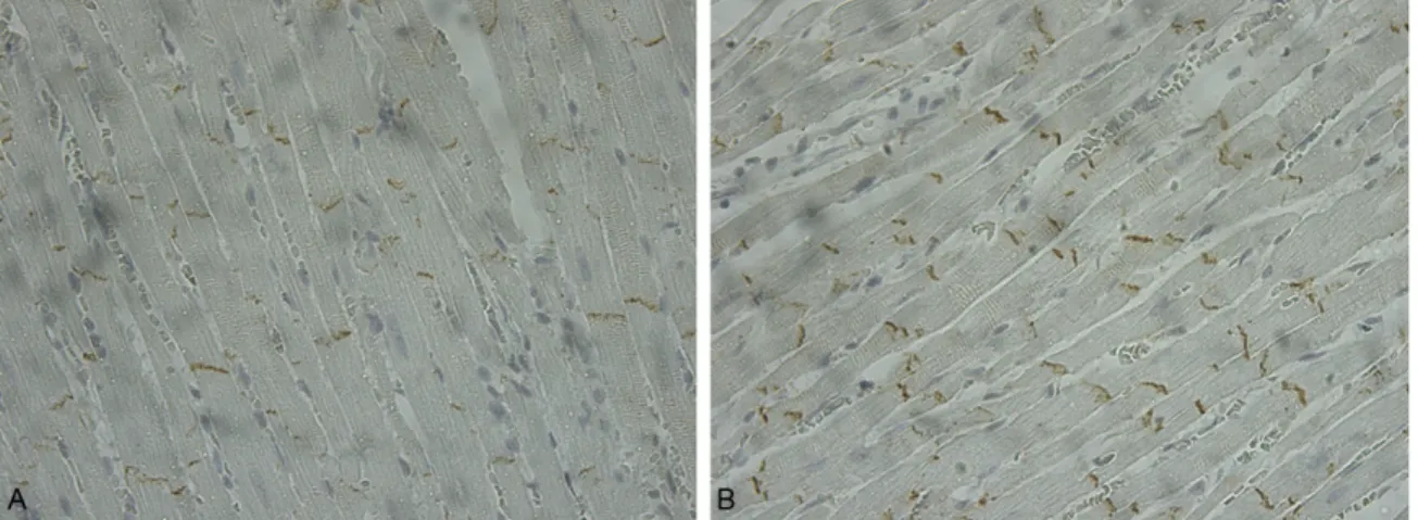
control rats are shown in Figure 1. In general,
less immunoreactive particles were observed
among the samples of LFN exposed rats.
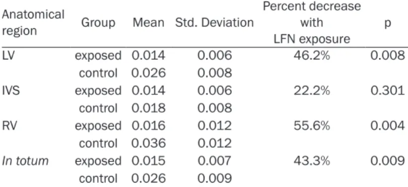
The ratio Cx43/muscle in each anatomical
region is shown in Table 1 and is graphically
depicted in Figure 2. The ratio Cx43/muscle in
totum is also shown in Table 1.
Figure 1. Immunostained connexin43 observed at the intercalated disks in a section taken from the left ventricle of a LFN-exposed rat (A) and control rat (B) (x 400).
The mean values of cardiac muscle were not
significantly different between the exposed and
control animals in any of the anatomical regions
considered in this study (p≥0.664).
The total amount of Cx43 was significantly
reduced in the left ventricle (p=0.011) and in
the right ventricle (p=0.009) of LFN-exposed
animals when compared to
con-trols. No differences were
detect-ed between the two groups
con-cerning the total amount of Cx43
in the interventricular septum (p=
0.237).
The ratio Cx43/muscle in totum
decreased 43.3% among the
LFN-exposed rats (p=0.009). Multiva-
riate comparisons over the three
anatomical regions showed
signifi-cant differences between groups
(p=0.001) with a decrease of
46.2%, 22.2% and 55.6%
respec-tively in the left ventricle
(p=0.008), in the interventricular
septum (p=0.301) and in the right
ventricle (p=0.004).
Discussion
As far as we know this is the first
study concerning the evaluation of
cardiac Cx43 in rats submitted to
LFN.
In humans, gap junction
remodel-ing has been studied in several
pathologies and arrhythmias
[10-16, 27, 28]. The loss of Cx43
expression has been shown to be
a key for the development of an
arrhythmic anatomic substrate in
chronically hypertrophied
myocar-dium [15]. Modifications in gap
junction organization and on Cx43
expression contribute to
conduc-tion disturbances and
develop-ment of arrhythmias in myocardial
infarction [27], non-ischemic
dilat-ed cardiomyopathy [12, 13],
car-diac heart failure [11] and valvular
heart disease [14].
In several studies, a reduction of
Cx43 from 30 to 50% occurs in
ventricular remodeling [14, 23,
Table 1. Ratio Cx43/muscle in each anatomical region and in
totum in LFN-exposed (n=10) and control (n=8) animals
Anatomical
region Group Mean Std. Deviation
Percent decrease with LFN exposure p LV exposed 0.014 0.006 46.2% 0.008 control 0.026 0.008 IVS exposed 0.014 0.006 22.2% 0.301 control 0.018 0.008 RV exposed 0.016 0.012 55.6% 0.004 control 0.036 0.012 In totum exposed 0.015 0.007 43.3% 0.009 control 0.026 0.009
LV = Left Ventricle; IVS = Interventricular Septum; RV = Right Ventricle; LFN = Low-Frequency Noise.
Figure 2. Ratio Cx43/muscle in the left ventricle (LV), interventricular sep-tum (IVS) and right ventricle (RV) in LFN exposed and control animals. A significant reduction was observed in exposed animals in the left ven-tricle (p=0.008) and in the right venven-tricle (p=0.004) but not in the inter-ventricular septum (p=0.301).
29], but changes in gap junction expression
alone are presumably not sufficient for
conduc-tion slowing and enhanced arrhythmogenicity,
apparently because there is a large conduction
reserve [30].
Meanwhile, it is known that an increase of the
intercellular collagen deposits may lead to
anisotropic reentry [23, 24], and a strong
enhancement of arrhythmogenic vulnerability
can be attained by the association of increased
fibrosis with a 50% reduction of Cx43
expres-sion [31].
As LFN induces the development of interstitial
myocardial fibrosis [8] and perivascular fibrosis
[9] we hypothesized that the finding of
signifi-cant modifications on gap junctions after LFN
exposure could lead to a morphological
arrhyth-mogenic substrate.
In our study the measurement of Cx43 was
per-formed in equivalent tissue mass among
exposed and control animals and showed a
sig-nificant reduction in rats exposed to LFN. The
modification was evident at the free ventricular
wall but not in the interventricular septum
sug-gesting that this anatomical region could be
more protected against the effects of LFN.
However, this does not discard the possibility of
a strong alteration of the electrophysiological
milieu. In fact, the observed statistical powers
of 99% for the multiple comparisons between
groups and in excess of 80% for the
compari-sons between groups regarding the left and
right ventricles are noteworthy, in view of the
sample dimension, suggesting the marked
effects of LFN exposure.
Taking into consideration the universal
exis-tence of LFN in modern societies and having in
mind the difficulties to explain some ventricular
tachyarrhythmias without structural heart
dis-ease, the hypothesis of idiopathic ventricular
fibrillation as being also a consequence of gap
junction remodeling mediated through the
effects of LFN, should not be despicable. On
the other hand, in patients with already known
specific cardiac disorders, a reduction of gap
junctions by the exposure to LFN makes the
development of arrhythmic events possible,
carrying out an adverse prognosis.
We still do not know the mechanisms
underly-ing the fibrotic development we previously
reported in rats exposed to LFN [8] or to
indus-trial noise [9] as well as the mechanism of the
Cx43 alteration observed in this study.
Nevertheless, the Cx43 remodeling has been
linked to intrinsic factors occurring on
biosyn-thesis at transcriptional or posttranscriptional
phase [32, 33]. As connexin43 is degraded
through lysosomal or proteasomal pathway
[34, 35] one can also speculate that the Cx43
reduction observed in this study can be related
to an activation of these pathways. Theoretically,
LFN acting as an external mechanical force
could also lead to activation of protein kinases
which might modify the level of Cx43
phosphor-ylation [36, 37].
Regardless of the mechanism how LFN induces
loss of Cx43, our results suggest that the
sig-nificant reduction of this protein can lead to
electrophysiological modifications. The
occur-rence of a significant myocardial and
periarte-rial fibrosis induced respectively by LFN and
industrial noise reported earlier by our group
[8, 9], together with a possible gap junction
remodeling observed in this study, makes the
development of a morphological
arrhythmogen-ic substrate by LFN possible. Further
experi-mental and clinical studies are needed to
eval-uate the functional and arrhythmic
conse-quences. With this study we put forward the
hypothesis of a link between LFN and
ventricu-lar tachyarrhythmias, opening a new
investiga-tional area concerning the effects of LFN on the
heart.
In conclusion, we can state that low frequency
noise induces modifications on cardiac
con-nexin43 in rats. The concon-nexin43 reduction
observed in our study can contribute to a
mor-phological arrhythmogenic substrate.
Disclosure of conflict of interest
None.
Address correspondence to: Eduardo Antunes, CIIEM, Centro de Investigação Interdisciplinar Egas Moniz, Instituto Superior das Ciências da Saúde Egas Moniz, ISCSEM, Quinta da Granja, Monte de Caparica, 2829-511 Caparica, Portugal. E-mail: ejpantunes@sapo.pt
References
[1] Alves-Pereira M, Castelo Branco NAA. Vibroacoustic disease: biological effects of in-frasound and low-frequency noise explained by mechanotransduction cellular signalling. Prog Bioph Mol Biol 2007; 93: 256-279. [2] da Fonseca J, dos Santos JM, Branco NC,
Alves-Pereira M, Grande NR, Oliveira P, Martins AP. Noise-induced gastric lesions: a light and scanning electron microscopy study of the al-terations of the rat gastric mucosa induced by
low frequency noise. Cent Eur J Public Health 2006; 14: 35-38.
[3] Grande NR, Águas AP, Sousa Pereira A, Mon-teiro E, Castelo Branco NAA. Morphological changes in the rat lung parenchyma exposed to low frequency noise. Aviat Space Environ Med 1999; 70: A70-A77.
[4] de Sousa Pereira A, Águas AP, Grande NR, Mirones J, Monteiro E, Castelo Branco NAA. The effect of chronic exposure to low frequen-cy noise on rat tracheal epithelia. Aviat Space Environ Med 1999; 70: A186-A190.
[5] Martins dos Santos J, Grande NR, Castelo Branco NAA, Zagalo C, Oliveira P, Alves-Pereira M. Lymphatic lesions and vibroacoustic dis-ease. Eur J Lymphol 2004; 12: 17-20.
[6] Oliveira PM, Pereira da Mata AD, Martins dos Santos JA, da Silva Marques DN, Branco NC, Silveira JM, Correia da Fonseca JC. Low-fre-quency noise effects on the parotid gland of the Wistar rat. Oral Dis 2007; 13: 468-73. [7] Martins dos Santos J, Grande NR, Castelo
Branco NA, Zagalo C, Oliveira P. Vascular le-sions and vibroacoustic disease. Eur J Anat 2002; 6: 17-21.
[8] Antunes E, Borrecho G, Oliveira P, Oliveira MJR, Brito J, Águas A, Martins dos Santos J. Myocar-dial Fibrosis in Rats Exposed to Low Frequency Noise. Acta Cardiol 2013; 68: 241-245. [9] Antunes E, Oliveira P, Oliveira MJR, Brito J,
Águas A, Martins dos Santos J. Histomorpho-metric evaluation of the coronary artery ves-sels in rats submitted to industrial noise. Acta Cardiol 2013; 68: 285-289.
[10] Smith JH, Green CR, Peters NS, Rothery S, Sev-ers NJ. Altered Patterns of gap junction distri-bution in ischemic heart disease. An immuno-histochemical study of human myocardium using laser scanning confocal microscopy. Am J Pathol 1991; 139: 871-878.
[11] Kostin S, Rieger M, Dammer S, Hein S, Richter M, Klovekorn WP, Bauer EP, Schaper J. Gap junction remodeling and altered connexin43 expression in the failing human heart. Mol Cell Biochem 2003; 242: 135-144.
[12] Kitamura H, Ohnishi Y, Yoshida A, Okajima K, Azumi H, Ishida A, Galeano EJ, Kubo S, Hayashi Y, Itoh H, Yokoyama M. Heterogeneous loss of connexin43 protein in nonischemic dilated car-diomyopathy with ventricular tachycardia. J Cardiovasc Electrophysiol 2002; 13: 865-887. [13] Kitamura H, Ohnishi Y, Yoshida A, Okajima K,
Azumi H, Ishida A, Galeano EJ, Kubo S, Fuku-zawa K, Takano T, Yokoyama M. Correlation of connexin43 expression and late ventricular po-tentials in nonischemic dilated cardiomyopa-thy. Circ J 2003; 67: 1017-1021.
[14] Kostin S, Dammer S, Hein S, Klovekorn WP, Bauer EP, Schaper J. Connexin 43 expression
and distribution in compensated and decom-pensated cardiac hypertrophy in patients with aortic stenosis. Cardiovasc Res 2004; 62: 426-436.
[15] Peters NS, Green CR, Poole-Wilson PA, Severs NJ. Reduced content of connexin43 gap junc-tions in ventricular myocardium from hypertro-phied and ischemic human hearts. Circulation 1993; 88: 864-875.
[16] Takeuchi S, Akita T, Takagishi Y, Watanabe E, Sasano C, Honjo H, Kodama I. Disorganization of gap junction distribution in dilated atria of patients with chronic atrial fibrillation. Circ J 2006; 70: 575-582.
[17] Gutstein DE, Morley GE, Tammaddon H, Vaidya D, Schneider MD, Chen J, Chien KR, Stuhl-mann H, Fishman GI. Conduction slowing and sudden arrhythmic death in mice with cardiac-restricted inactivation of connexin43. Circ Res 2001; 88: 333-339.
[18] Danik SB, Liu F, Zhang J, Suk HJ, Morley GE, Fishman GI, Gutstein DE. Modulation of cardi-ac gap junction expression and arrhythmic susceptibility. Circ Res 2004; 95: 1035-1041. [19] van Rijen HVM, Eckardt D, Degen J, Theis M,
Ott T, Willecke K, Jonsma HJ, Opthof T, Bakker JMT. Slow conduction and enhanced anisotro-py increase the propensity for ventricular tachyarrhythmias in adult mice with induced deletion of connexin43. Circulation 2004; 109: 1048-1055.
[20] Uzzaman M, Honjo H, Tagagishi Y, Emdad L, Magee AI, Severs NJ, Kodama I. Remodeling of gap junctional coupling in hypertrophied right ventricles of rats with monocrotaline-induced pulmonary hypertension. Circ Res 2000; 86: 871-879.
[21] Mayama T, Matsumura K, Lin H, Ogawa K, Imanaga I. Remodeling of cardiac gap junction connexin 43 and arrhythmogenesis. Exp Clin Cardiol 2007; 12: 67-76.
[22] Lin H, Ogawa K, Imanaga I, Tribulova N. Re-modeling of connexin 43 in the diabetic rat heart. Mol Cell Biochem 2006; 290: 69-78. [23] Peters NS, Coromilas J, Severs NJ, Wit AL.
Dis-turbed connexin43 gap junction distribution correlates with the location of reentrant cir-cuits in the epicardial border zone of healing canine infarcts that cause ventricular tachycar-dia. Circulation 1997; 97: 988-996.
[24] Kawara T, Derksen R, de Groot JR, Coronel R, Tasseron S, Linnenbank AC, Hauer RN, Kirkels H, Janse MJ, de Bakker JM. Activation delay after premature stimulation in chronically dis-eased human myocardium relates to the archi-tecture of interstitial fibrosis. Circulation 2001; 104: 3069-3075.
[25] La Vecchia L, Ometto R, Bedogni F, Finocchi G, Mosele GM, Bozzola L, Bevilacqua P, Vincenzi
M. Ventricular late potentials, interstitial fibro-sis, and right ventricular function in patients with ventricular tachycardia and normal left ventricular function. Am J Cardiol 1998; 81: 790-792.
[26] da Fonseca J, Martins dos Santos J, Castelo-Branco N, Alves-Pereira M, Grande N, Oliveira P. Noise-induced duodenal lesions. Eur J Anat 2005; 9: 29-33.
[27] Kieken F, Mutsaers N, Dolmotova E, Virgil K, Wit AL, Kellezi A, Hirst-Jensen BJ, Duffy HS, Sorgen PL. Structural and molecular mecha-nisms of gap junction remodeling in epicardial border zone myocytes following myocardial in-fartion. Circ Res 2009; 104: 1103-1112. [28] Basso C, Czarnowska E, Barbera ML, Bauce B,
Beffagna G, Wlodarska EK, Pilichou K, Ramon-do A, Lorenzon A, Wozniek O, CorraRamon-do D, Daliento L, Danieli GA, Valente M, Nava A, Thiene G, Rampazzo A. Ultrastructural evi-dence of intercalated disc remodeling in ar-rhythmogenic right ventricular cardiomyopa-thy: an electron microscopy investigation on endomyocardial biopsies. Eur Heart J 2006; 27: 1847-1854.
[29] Kaprielian RR, Gunning M, Dupont E, Shep-pard MN, Rothery SM, Underwood R, Pennell DJ, Fox K, Pepper J, Poole-Wilson PA, Severs NJ. Downregulation of immunodetectable con-nexin43 and decreased gap junction size in the pathogenesis of chronic hibernation in the human left ventricle. Circulation 1998; 97: 651-660.
[30] van Rijen HV, de Bakker JM, van Veen TA. Hy-poxia, electrical uncoupling, and conduction
slowing: Role of conduction reserve. Cardio-vasc Res 2005; 66: 9-11.
[31] Jansen JA, van Veen AA, Bosch AA, van der Na-gel R, Vos MA, Bakker JM, van Rijen HV. Ar-rhythmia vulnerability of aged haploinsufficient Cx43 mice is determinant by heterogeneous downregulation of Cx43 combined with in-creased fibrosis. Circulation 2008; 118: S494. [32] Teunissen BE, Bierhuisen MF. Transcriptional
control of myocardial connexins. Cardiovasc Res 2004; 62: 246-255.
[33] Yang B, Lin H, Xiao J, Lu Y, Luo X, Li BT, Zhang Y, Xu C, Bai Y, Wang H, Chen G, Wang Z. The muscle-specific micro-RNA miR-1 regulates cardiac arrhythmogenic potential by targeting GJA1 and KCNJ2. Nat Med 2007; 13: 486-491.
[34] Laing JG, Tadros PN, Green K, Saffitz JE, Beyer EC. Proteolysis of connexin43-containing gap junctions in normal and heat-stressed cardiac myocytes. Cardiovasc Res 1998; 38: 711-718. [35] Berthoud VM, Minogue PJ, Laing JG, Beyer EC.
Pathways for degradation of connexins and gap junctions. Cardiovasc Res 2004; 62: 256-267.
[36] Saffitz JE, Kleber AG. Effects of mechanical forces and mediators of hypertrophy on remod-eling of gap junction in the heart. Circ Res 2004: 94: 585-591.
[37] Yamada K, Green KG, Samarel AM, Saffitz JE. Distinct pathways regulate expression of car-diac electrical and mechanical junction pro-teins in response to stretch. Circ Res 2005; 97: 346-353.