2018
UNIVERSIDADE DE LISBOA
FACULDADE DE CIÊNCIAS
DEPARTAMENTO DE QUÍMICA E BIOQUÍMICA
Regulation of the alternative splicing of RAC1b in tumour cells
Cláudia Alexandra da Silva Rodrigues
Mestrado em Bioquímica
Especialização em Bioquímica Médica
Dissertação orientada por:
iii
Acknowledgments
I would like to use this chapter to express how grateful I am for having had this amazing internship opportunity at the Oncobiology and Signalling Pathways Lab from the Department of Human Genetics at the National Health Institute Dr. Ricardo Jorge, and for having had the chance to meet so many wonderful people and professionals who led me through this internship period.
I will start by expressing my deepest gratitude and special thanks to National Health Institute Dr. Ricardo Jorge and to my supervisors, doctors Peter Jordan and Vânia Gonçalves, for having given me this opportunity to learn and grow professionally, in such a great work environment. Vânia, thank you for having had the patience and care to teach and guide me through all the experimental work. I am so grateful for having had such a nice and awesome person as my supervisor, I truly learned a lot from you, and for that, I will always be thankful. I would also like to thank Dr. Peter Jordan and Dr. Paulo Matos whom, in spite of being extraordinarily busy with their duties, always took time out to hear me out, to guide and keep me on the correct path. I feel very lucky for having had such kind, nice and understanding people to guide me through this work. I would also like to express my gratitude towards Patrícia, you are an amazing person who gives out great professional and personal advice. Thank you for always being so willing to help, for caring and for being such a great person! A huge thank you to the rest of girls at the lab, Andreia, Joana, Márcia, Ana and Cláudia, for always being so kind, welcoming and willing to help. All of you showed me that working can also be fun, in a professional way of course!
I also want to express my deepest gratitude towards the most important people in my life, my parents, for making this opportunity possible and for always believing in me even when I didn’t. I wouldn’t be here without all of your hard work, support and encouragement! Thank you for being such amazing parents! I also need to acknowledge the crucial role of my closest family and friends during this stage of my live. I appreciate how my family continuously supported my choices and how they were always there for me when I needed it. From my friends, I am especially grateful for Joana, Raquel, Madalena and Catarina, whom have been putting up with me for a while now! Thank you for being such great friends, without you my academic experience wouldn’t have been the same!
I perceived this opportunity as a big milestone in my career development. I will try to use my gained skills and knowledge in the best possible way, and I will continue to work on their improvement.
iv
Resumo
A expressão génica é o mecanismo pelo qual a informação codificada num gene é convertida num produto funcional. A regulação deste processo permite que as células expressem genes diferencialmente consoante o seu tipo, fase de desenvolvimento ou mesmo em resposta a estímulos externos. Portanto, através da regulação da expressão génica, as células conseguem ativar genes seletivamente dependendo das suas necessidades e funções. Um dos processos cruciais envolvidos neste processo regulatório é o splicing do pré-mRNA, que consiste na remoção dos intrões e junção dos exões numa sequência codificante contígua. Esta reação de splicing é catalisada pelo spliceossoma, um complexo ribonucleoproteico que reconhece e interage com as sequências de consenso nos limites dos exões e dos intrões, de forma a direcionar a excisão dos intrões e a ligação dos exões do RNA. Na maioria dos genes humanos a inclusão ou exclusão dos exões no transcrito final ocorre de forma diferencial, tornando-se assim possível a produção de múltiplos mRNAs diferentes a partir de um único gene. Este processo é denominado de splicing alternativo e é um dos mecanismos que modula a regulação da expressão proteica e a produção de um proteoma complexo e diversificado em eucariotas mais complexos. Durante o processo de splicing alternativo a decisão de qual exão é removido e qual é incluído no mRNA final, para além das sequencias de consenso é também fortemente influenciada pela interação entre elementos regulatórios cis e os fatores trans. Os elementos cis ocorrem tanto nas regiões exónicas como nas regiões intrónicas e podem promover a inclusão do exão através do recrutamento do spliceossoma (sequências promotoras) ou promover a sua exclusão por interferência com a ligação do spliceossoma às sequências de consenso (sequências silenciadoras). As sequências promotoras são, geralmente, ligadas por fatores que atuam em trans positivos, como é o caso das proteínas SR, enquanto que as sequências silenciadoras são normalmente ligadas por fatores que atuam em trans negativos, como é o caso das proteínas hnRNPs. Para além da presença de elementos regulatórios cis e fatores trans, uma variedade de outros fatores podem influenciar o splicing alternativo, como é o caso da presença de estruturas secundárias no mRNA, da presença de microRNAs, da arquitetura dos exões e intrões, da força relativa das sequências de consenso no local de splicing e ainda da velocidade de elongação durante a síntese do pré-mRNA pela RNA polimerase II. A soma das contribuições de cada um destes elementos define o potencial de reconhecimento de um exão e a sua respetiva afinidade pelo spliceossoma. Devido ao seu papel central na expressão e função de proteínas, é expectável que problemas ao nível da regulação do splicing alternativo possam resultar no desenvolvimento de doenças. Um exemplo disso é a sobreexpressão da variante de splicing hiperativa do gene RAC1, RAC1b, em diversos tumores malignos. A variante RAC1b é caracterizada pela inserção de um exão (exão 3b) extra entre os exões 3 e 4 de RAC1. Estes 19 aminoácidos extra codificados pelo exão 3b, para além de uma regulação diferente também conferem uma seletividade na sinalização a jusante de RAC1b. Esta variante promove a produção de ROS e a via de sinalização do NF-kB em detrimento de outras vias clássicas ativadas por RAC1, promovendo a progressão do ciclo celular e a sobrevivência das células. Em cancro colorretal, a variante RAC1b encontra-se sobre expressa num subgrupo específico de tumores que também apresentam uma mutação oncogénica em BRAF (BRAFV600E), tendo sido demonstrado a existência de uma cooperação entre estes eventos no sentido de promover a sobrevivência das células tumorais. Até agora, sabe-se que o splicing alternativo de RAC1 é regulado por duas proteínas SR, SRSF1 e SRSF3. SRSF1 promove a inclusão do exão alternativo 3b, ao contrário de SRSF3 que promove a sua exclusão. Ambos os fatores são regulados por vias de sinalização a montante, nomeadamente, os níveis proteicos de SRSF1 aumentam quando a via de sinalização PI3K é inibida, enquanto que a via β-catenin/TCF4 estimula a expressão de SRSF3. É provável que existam outros elementos que regulem o splicing alternativo de RAC1, e recentemente, as proteínas PTBP1 e ESRP1 foram descritas como possíveis modeladoras deste evento em diferentes tipos de células, enquanto que a nucleoporina RANBP2 foi relacionada com a distribuição de proteínas SR fosforiladas, as quais são responsáveis
v pela regulação da expressão de RAC1b. Neste trabalho experimental foram estudados estes três possíveis modeladores do splicing alternativo de RAC1 em células colorretais.
Para estudar estes possíveis novos mecanismos de regulação da expressão de RAC1b em células colorretais, começámos por construir vetores de expressão para PTBP1 e ESRP1, sendo que o vetor de expressão para RANBP2 já se encontrava disponível no laboratório de acolhimento. Posteriormente, os possíveis efeitos da sobreexpressão de PTBP1, de ESRP1 e de RANBP2 no
splicing alternativo de RAC1 foram determinados através do uso de um minigene RAC1.
Basicamente, cada plasmídeo que codificava as proteínas em estudo foi co-transfetado em células com o minigene RAC1. Os resultados foram observados através de um RT-PCR semi-quantitativo, com
primers específicos para os transcritos RAC1 e RAC1b derivados do minigene RAC1. A expressão
endógena de RAC1b foi também avaliada por Western blot (WB) através do uso de um anticorpo específico contra RAC1b. De acordo com os resultados, os efeitos significativos que foram observados para ESRP1 e RANBP2 foram confirmados ao nível endógeno, através do uso de siRNAs comercialmente disponíveis de forma a silenciar a sua expressão. Os resultados foram mais uma vez obtidos através de um RT-PCR semi-quantitativo, mas desta vez os primers utilizados foram específicos para os transcritos endógenos. Os níveis proteicos de RAC1b endógeno foram também avaliados através de WB. As experiências foram realizadas principalmente em células epiteliais normais do cólon, NCM460, mas confirmadas com células HeLa e HT29 para determinar se os resultados observados eram dependentes da linha celular. A localização celular das proteínas transfetadas foi visualizada através de ensaios de imunofluorescência em células NCM460 e HeLa.
A sobreexpressão de PTBP1, nas células NCM460, não afetou significativamente a inclusão do exão 3b no transcrito final. No entanto, a sobreexpressão de ESRP1 e de RANBP2 promoveu a exclusão do exão 3b. As experiências de imunofluorescência mostraram que a expressão de PTBP1 e ESRP1 ocorre tanto no núcleo como no citoplasma, enquanto que a localização de RANBP2 se restringe essencialmente à membrana nuclear. Esta observação vai de encontro com o esperado, dado que PTBP1 e ESRP1 são ambos fatores de splicing, enquanto que RANBP2 faz parte do complexo do poro nuclear. Nas células NCM460, o silenciamento de ESRP1 diminuiu a expressão endógena de RAC1b, enquanto que a depleção de RANBP2 levou ao aumento de RAC1b. O efeito da depleção de ESRP1 no splicing de RAC1 não corroborou os resultados obtidos nas experiências de sobreexpressão, dando assim a indicação de que este fator de splicing, devido ao seu papel na manutenção do fenótipo epitelial, pode ser altamente regulado por um mecanismo de feedback negativo, no qual regula a sua própria expressão. Os resultados obtidos para as células HeLa e HT29 seguiram a mesma tendência observada nas células NCM460. Assim, no geral, o silenciamento de ESRP1 diminuiu a expressão endógena de RAC1b, enquanto que a depleção de RANBP2 levou ao seu aumento, ambos independentemente da linha celular utilizada, sugerindo que tanto ESRP1 como RANBP2 são reguladores gerais da expressão de RAC1b.
Em conclusão, esta tese forneceu fortes evidências de que ESRP1 e RANBP2 estão envolvidos na regulação da expressão de RAC1b e identificou pela primeira vez outros fatores, para além de SRSF1 e de SRSF3, envolvidos na regulação da expressão de RAC1b em células colorretais. Mais experiências são necessárias para esclarecer como é que estas proteínas estão a regular o splicing alternativo de RAC1. Este conhecimento será útil na caracterização dos mecanismos de regulação de
splicing alternativo de RAC1 e, eventualmente, para desenvolver moduladores farmacológicos
eficazes que possam restaurar a sinalização normal de RAC1 em células tumorais.
vi
Abstract
Regulation of gene expression allows cells to differentially express genes in different cell types, developmental stages or even in response to external conditions. Alternative splicing is a crucial regulatory process in the pathway of gene expression and the mechanism by which multiple protein isoforms can be generated from a single gene. This way, complex organisms can regulate protein expression and generate a more diverse proteome from a given gene number within the genome. Due to its central role in modulating gene expression, it is not surprising that aberrant regulation of alternative splicing is associated with human disease. On example is the selective overexpression of a hyperactive splice variant of the RAC1 gene, RAC1b, in several malignant tumours. RAC1b promotes reactive oxygen species production and the NF-kB pathway activation, but not other classical RAC1 signalling pathways, and stimulates cell cycle progression and cell survival. In colorectal cancer, RAC1b was found to be overexpressed in a specific subgroup, namely in 80% of tumours with mutation in the oncogene BRAF, suggesting that both events cooperate to promote the survival of colorectal cells. Previous studies in colorectal cells showed that the splicing factor SRSF1 acts as an enhancer of RAC1 alternative splicing by promoting the inclusion of alternative exon 3b, while SRSF3 acts as a silencer by promoting the skipping of the exon 3b. Besides SRSF1 and SRSF3 it is likely that RAC1 alternative splicing can be regulated by additional factors, and recently, PTBP1 and ESRP1 were described as possible modulators of the alternative splicing of RAC1b in different cell types while RANBP2 was shown to be a modulator of the distribution of phosphorylated SR proteins, which are known to regulate RAC1b expression.
To study whether these factors regulate RAC1b expression in colorectal cells, we started by analysing the effects of PTBP1, ESRP1 and RANBP2 overexpression on RAC1 alternative splicing. For that, each expression vector encoding the proteins in study was co-transfected with the RAC1 minigene into cells. The results were then assessed through a semi-quantitative RT-PCR, with specific primers for RAC1 and RAC1b transcripts derived from the RAC1 minigene. Effects on endogenous RAC1b expression was also assayed by Western blot and cellular localization of the transfected proteins assessed by immunofluorescence. Positive effects were then confirmed at the endogenous protein level through the use of commercially available siRNAs to deplete the regulators. The experiments were mainly performed in NCM460 colon cells but confirmed using HeLa and HT29 cells to determine if the observed results were cell line dependent. As expected, immunofluorescence experiments showed that both PTBP1 and ESRP1 can be found in the nucleus and cytoplasm, while RANBP2 is found at the nuclear membrane and also at the cytoplasm. RANBP2 overexpression was found to promote the skipping of the alternative exon 3b, while its depletion promoted exon 3b inclusion. Thus, RANBP2 emerged as a candidate negative regulator of RAC1 alternative splicing that promotes the skipping of exon 3b. In the case of ESRP1, both overexpression and depletion promoted the skipping of the alternative exon 3b. ESRP1 overexpression might interfere with a negative feedback mechanism, in which ESRP1 regulates its own expression, due to its role in maintaining the epithelial phenotype. Based on the depletion experiment, ESRP1 can also be considered a candidate positive regulator of RAC1 alternative splicing, which promotes the inclusion of exon 3b, unlike RANBP2. No significant effect of PTBP1 on RAC1 alternative splicing was observed.
In conclusion, this thesis provided strong evidence that ESRP1 and RANBP2 are involved in RAC1b expression regulation and identified for the first time other factors besides SRSF1 and SRSF3 that are involved in the regulation of RAC1b expression in colorectal cells. Further experiments are needed to clarify how these proteins are regulating RAC1 alternative splicing. This knowledge will be useful to characterize RAC1 alternative splicing regulation mechanisms and eventually to develop effective pharmacological modulators that can restore normal RAC1 signalling in tumour cells.
vii
Table of contents
Acknowledgments ... iii
Resumo ... iv
Abstract ... vi
List of tables and figures ... viii
List of abbreviations ... ix
1. Introduction ... 1
1.1. Molecular mechanism of pre-mRNA Splicing ... 1
1.2. Regulation of pre-mRNA splicing ... 4
1.3. Effects of alternative splicing ... 6
1.4. The small GTPase RAC1 and its splice variant RAC1b ... 7
1.5. Colorectal cancer and RAC1b alternative splicing... 9
1.6. PTBP1, ESRP1 and RANBP2 as possible modulators of RAC1b alternative splicing... 10
2. Objectives ... 11
3. Experimental Procedures ... 12
3.1. Polymerase Chain Reaction (PCR) ... 12
3.1.1. PTBP1 and ESRP1 amplification ... 13
3.1.2. Agarose gel electrophoresis ... 13
3.2. Cloning ... 14
3.2.1. PTBP1 and ESRP1 cloning ... 14
3.2.4. Sequencing ... 17
3.3. Cell Culture ... 18
3.4. Cell Transfection ... 20
3.5. Cell lysis ... 24
3.6. SDS-PAGE and Western blot ... 25
3.7. Semi-quantitative RT-PCR ... 28
3.8. Immunofluorescence assay ... 29
3.9. Data Treatment ... 30
4. Results ... 32
4.1. Production of expression vectors for PTBP1 and ESRP1 ... 32
4.2. The effect of PTBP1, ESRP1 and RANBP2 overexpression in the alternative splicing of RAC1 ... 33
4.2.1. Subcellular localization of PTBP1, ESRP1 and RANBP2 ... 36
4.3. The effect of ESRP1 and RANBP2 depletion in the alternative splicing of endogenous RAC1 transcripts ... 37
5. Discussion, Conclusions and Future Perspectives ... 42
viii
List of tables and figures
Table 3.1 - Primers sequence used for the amplification of ESRP1 and PTBP1 cDNA sequences. .... 13
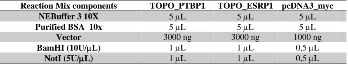
Table 3.2 - Composition of the reaction mixes for the digestion of the TOPO vectors, TOPO_PTBP1 and TOPO_ESRP1, and the pcDNA3_myc vector. ... 15
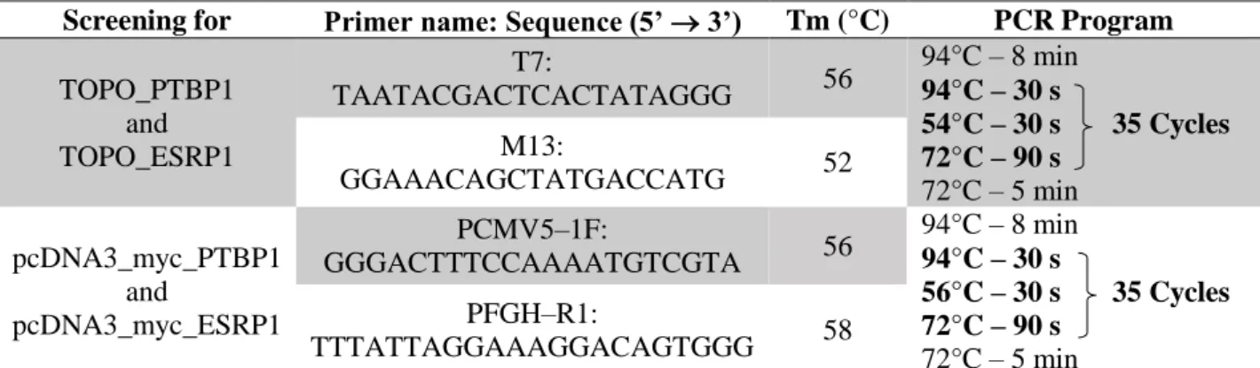
Table 3.3 - Primers and PCR programs used for the screening of the integration of PTBP1 and ESRP1 into the pCR™2.1-TOPO® vector and the pcDNA3_myc expression vector. ... 17
Table 3.4 - Primers used to sequence the vectors cloned with ESRP1, PTBP1 and RANBP2. ... 18
Table 3.5 - Information on every cell line used on this experimental work, including their origin, type and culture conditions. ... 20
Table 3.6 - Co-transfection conditions of pcDNA3_EGFP, pcDNA3_myc_PTBP1, pcDNA3_myc_ESRP1 or pkTol2Chy_RANBP2_1-3224 with RAC1 minigene (MG) in NCM460, HeLa and HT29 cell lines. Transfection conditions of pcDNA3_EGFP or pkTol2Chy_RANBP2_1-3224 in NCM460, Caco2, HT29 and HEK 293 cell lines. All transfections and co-transfections were performed in 6-well plates. ... 23
Table 3.7 - Transfection conditions of siGFP (Eurofins Genomics), siESRP1 (Santa Cruz Biotechnology) and siRANBP2 (Santa Cruz Biotechnology) for NCM460, HeLa and HT29 cell lines, all performed in 24-well plates. ... 24
Table 3.8 - Cell lysis conditions for every transfection preformed in this experimental work... 25
Table 3.9 - Recipes of the polyacrylamide gels ... 26
Table 3.10 - Antibodies used in Western blot and respective dilutions... 27
Table 3.11 - Primers and PCR programs used for the semi-quantitative RT-PCRs of RAC1b from the minigene (MG), endogenous RAC1b, ESRP1 and RANBP2. ... 29
Figure 1.1 – Consensus sequences that define exon/intron boundaries ... 1
Figure 1.2 – Schematic representation of the transesterification steps of RNA splicing ... 2
Figure 1.3 – Spliceosome assembly and catalysis ... 2
Figure 1.4 – Schematic representation of the five main types of alternative splicing events ... 3
Figure 1.5 – Schematic representation of regulatory cis-elements ... 5
Figure 1.6 – Schematic representation of RAC1 activation and regulation ... 8
Figure 1.7 – Schematic representation of the human RAC1 gene ... 8
Figure 3.1 – Western blot transference ... 27
Figure 3.2 – Antigen detection methods ... 30
Figure 4.1 – PTBP1 and ESRP1 cDNA amplification ... 32
Figure 4.2 – PCR screenings. ... 33
Figure 4.3 – RAC1 minigene... 34
Figure 4.4 – Western blot analysis of the transfected tagged-proteins expression ... 34
Figure 4.5 – Analysis of RAC1 exon 3b inclusion after PTBP1, ESRP1 and RANBP2 overexpression in NCM460 cells ... 35
Figure 4.6 – Analysis of RAC1 exon 3b inclusion after PTBP1, ESRP1 and RANBP2 overexpression in HeLa cells ... 36
Figure 4.7 – Subcellular localization of transfected PTBP1, ESRP1 and RANBP2 ... 37
Figure 4.8 – Time course experiment of siRNA-mediated depletion of ESRP1 and RANBP2 ... 38
Figure 4.9 – Analysis of endogenous RAC1b expression after depletion of ESRP1 and RANBP2 in NCM460 cells ... 39
Figure 4.10 – ESRP1 and RANBP2 depletion in NCM460, HeLa and HT29 after 72 h incubation ... 40
ix
List of abbreviations
A Adenosine
APS Ammonium persulfate
ATP Adenosine triphosphate BAP Bacterial alkaline phosphatase
bp Base pairs
BPS Branch-point site
BSA Bovine serum albumin
C Cytosine
cDNA mRNA-complementary deoxyribonucleic acid
CO2 Carbon dioxide
CRC Colorectal cancer
CTD Carboxy terminal domain DAPI 4',6-diamidino-2-phenylindole ddNTP Chain-terminating dideoxy nucleotide DMEM Dulbecco's Modified Eagle's Medium DMSO Dimethyl sulfoxide
DNA Deoxyribonucleic acid dNTP Deoxyribonucleotide dsDNA Double-stranded DNA
DTT Dithiothreitol
ECL Enhanced Chemiluminescence EDTA Ethylenediamine tetraacetic acid EMT Epithelial-to-mesenchymal transition ERK Extracellular signal-regulated kinase ESE Exonic splicing enhancer
ESRP1 Epithelial Splicing Regulatory Protein 1 ESS Exonic splicing silencer
EtBr Ethidium bromide
FBS Foetal bovine serum
G Guanosine
GAPs GTPase-activating protein
GDP Guanosine diphosphate
GEF Guanine nucleotide exchange factor GFP Green fluorescent protein
GSK3β Glycogen synthase kinase-3 beta GTP Guanosine triphosphate
GTPases Guanosine triphosphate phosphohydrolases
H2O2 Hydrogen peroxide
HCl Hydrochloric acid
hnRNP Heterogeneous nuclear ribonucleoprotein HRP Horseradish peroxidase
IF Immunofluorescence assay
IRES Internal ribosome entry site ISE Intronic splicing enhancer ISS Intronic splicing silencer JNK c-Jun N-terminal kinase
x
kb Kilobase
KCl Potassium chloride
kDa Kilodalton
KH2PO4 Monopotassium phosphate
LB Luria Bertani
mA Milliampere
MgCl2 Magnesium chloride
mL Millilitre
mM Millimolar
mRNA Messenger ribonucleic acid
Myc Epitope tag derived from the c-myc gene product Na2HPO4.2H2O Sodium phosphate dibasic dihydrate
NaCl Sodium Chloride
ncRNA Non-coding RNAs
NF-kB Nuclear factor kappa-light-chain-enhancer of activated B cells
ng Nanogram
nm Nanometre
NMD Nonsense-mediated mRNA-decay
NP-40 Nonidet P-40
NPC Nuclear pore complexes
ºC Degree Celsius
OIS Oncogene-induce senescence PAGE Polyacrylamide gel electrophoresis PAK p21 activating kinase
PBS Phosphate-buffered saline PCR Polymerase Chain Reaction
PFA Formaldehyde
PI3K Phosphatidylinositol 3 kinase Pol II RNA polymerase II
pre-mRNA Precursor mRNA
PTBP1 Polypyrimidine tract binding protein 1 PTC Premature termination codons
PVDF Polyvinylidene fluoride membrane
RAC1 Ras-related C3 botulinum toxin substrate 1
RAC1MG RAC1 minigene
RANBP2 RAN binding protein 2
Rho-GDI Rho-GDP dissociation inhibitor RISC RNA-induced silencing complex
RNA Ribonucleic acid
ROS Reactive oxygen species RRM RNA recognition motif
RT-PCR Reverse transcription polymerase chain reaction SDS Sodium dodecyl sulphate
SDS-PAGE Sodium dodecyl sulphate-polyacrylamide gel electrophoresis
SF1 Splicing factor 1
siRNA Short/small interfering ribonucleic acid snRNPs Small nuclear ribonucleoproteins SR proteins Serine/arginine-rich protein
xi SRPK1 Serine/arginine-rich protein-specific kinase 1
SRSF1 Serine/arginine-rich splicing factor 1 SRSF3 Serine/arginine-rich splicing factor 3 ssDNA Single-stranded deoxyribonucleic acid
T Thymidine TBE Tris-Borate-EDTA TBS Tris-buffered saline TCF4 Transcription factor 4 TEMED Tetramethylethylenediamine Tm Melting temperature Tris Tris(hydroxymethyl)aminomethane U Uridine
U2AF U2 auxiliary factor UTRs Untranslated regions
UV Ultraviolet
V Volt
v/v Volume per volume
WB Western blot
Wnt Wingless
X-Gal 5-bromo-4-chloro-3-indolyl-β-D-galactopyranoside
μg Microgram
1
1. Introduction
Gene expression is a complex mechanism by which the information encoded in a gene is converted into a functional product. In the case of protein coding genes, this process consists in the transcription of the gene into a precursor mRNA (pre-mRNA) that is processed (originating a functional mRNA) and transported to the cytoplasm, where it is translated into protein (Lodish 2016). Regulation of this multistep process allows cells to differentially express genes in different cell types, developmental stages or even in response to external conditions (Lodish 2016). Thus, by regulating gene expression cells can activate genes selectively depending on their needs and purpose. Pre-mRNA splicing, one of the RNA processing steps, is a crucial regulatory point in the pathway of gene expression and will be described in more detail in this work.
1.1. Molecular mechanism of pre-mRNA Splicing
In most eukaryotic genes, coding regions (exons) are interrupted by noncoding regions (introns), and during transcription both are copied into the pre-mRNA (Matlin et al. 2005; Lodish 2016). One of the steps of RNA processing is pre-mRNA splicing, where introns are removed, and exons are joined to form a contiguous coding sequence (Matlin et al. 2005; Lodish 2016). The pre-mRNA splicing reaction is directed by four conserved sequences that define the exon/intron boundaries (Figure 1.1). These four consensus sequences include: the exon–intron junction at the intron’s 5′ end (GU, 5′ splice site or splice donor site); the exon–intron junction at the intron’s 3′ end (AG, 3′ splice site or splice acceptor site); the branch-point site (BPS) sequence; and the polypyrimidine tract (Figure 1.1) (Matlin et al. 2005).
Figure 1.1 – Consensus sequences that define exon/intron boundaries. Y=U or C; R=G or A. (adapted from (Matlin et al.
2005))
Succinctly, the splicing reaction comprises two consecutive steps of transesterification (Papasaikas and Valcárcel 2016). In the first step, a nucleophilic attack on the phosphate group between the 5' exon–intron junction is carried out by the 2' hydroxyl group of an adenosine residue from the BPS, generating a 2'-5' phosphodiester bond and, consequently, a lariat intermediate. In the second step, the free 3' hydroxyl of the 5' exon attacks the phosphate group between the intron and the 3' exon, splicing the two exons together and releasing the intron lariat (Figure 1.2) (Papasaikas and Valcárcel 2016). This reaction is catalysed by the spliceosome, a ribonucleoprotein complex composed of five small nuclear ribonucleoprotein complexes (snRNPs) - U1, U2, U4, U5 and U6 - and several other non-snRNPs associated factors (House and Lynch 2008; Chen and Manley 2009; Wang et al. 2015). The five snRNPs are assembled around one small nuclear RNA, U1 - U6 snRNAs, that use RNA-RNA base pairing to direct the snRNPs to the consensus sequences at the exon/intron boundaries (Figure 1.1) and direct RNA excision and ligation. Several snRNPs also interact with each other to ensure that the distant regions of the substrate required for splicing catalysis are correctly juxtaposed (House and Lynch 2008; Chen and Manley 2009; Wang et al. 2015). The process of pre-mRNA splicing begins with the assembly of complex E, which is defined by the base pairing of U1 snRNA to the 5′ splice site, the binding of splicing factor 1 (SF1) to the branch site, in an
ATP-2 independent manner, and the recruitment of U2 auxiliary factor (U2AF) heterodimer (comprising U2AF65 and U2AF35) to the polypyrimidine tract and 3′ splice site. This complex is converted into the ATP-dependent pre-spliceosome A complex by the replacement of SF1 by U2 snRNP at the branch point. Further recruitment of the U4/U6–U5 tri-snRNP leads to the formation of the B complex, which contains all spliceosomal subunits that carry out pre-mRNA splicing. Finally, the C complex assembles by extensive conformational changes and remodelling of both the snRNPs and the protein components that are present in the B complex, including the loss of U1 and U4 snRNPs, to produce an active site that is capable of catalysing the transesterification chemistry required for exon ligation and lariat release (Figure 1.3) (Chen and Manley 2009; Wang et al. 2015; Wang et al. 2015).
Figure 1.2 – Schematic representation of the transesterification steps of RNA splicing. Each step involves a nucleophilic
attack on the terminal phosphodiester bonds of the intron. In the first step this is carried out by the 2′ hydroxyl of the branch point site (BPS) and in the second step by the 3′ hydroxyl of the 5′ exon. (adapted from (Papasaikas and Valcárcel 2016))
Figure 1.3 – Spliceosome assembly and catalysis. Canonical stepwise assembly of the spliceosome on pre-mRNA
substrates indicating the transitions from the E through P complexes. 5’SS: 5’ Splice site; BPS: Branch point site; 3’SS: 3’ Splice site; snRNPs: U1, U2, U4, U5 and U6; U2AF: U2 auxiliary factor; SF1: Splicing factor 1. (adapted from (Papasaikas and Valcárcel 2016))
The process described above is referred to as constitutive splicing (Figure 1.4A). With the recent technologic advances allowing genome-wide transcript analysis, it has become clear that more than 90% of human genes can generate mRNAs with a differential inclusion or exclusion of exons in the final mRNA product (Poulos et al. 2011). This process is called alternative splicing (House and Lynch 2008) and is a mechanism by which multiple protein isoforms can be generated from a single gene. This process is predicted to occur in most mammalian genes, being an important mechanism by which complex organisms can regulate protein expression and generate a more diverse proteome from a given gene number within the genome (House and Lynch 2008; Wang et al. 2015).
3 There are 7 main types of alternative splicing that can lead to the production of different isoform transcripts: (1) cassette exon (skipped exon); (2) intron retention; (3) mutually exclusive exons; (4) alternative 3' splice site; (5) alternative 5' splice site; (6) alternative first exon and (7) alternative last exon (Figure 1.4 B-H) (Wang et al. 2015; Iñiguez and Hernández 2017). The most common alternative splicing pattern type is the cassette exon (skipped exon), with a prevalence of approximately 30% (Wang et al. 2015). This splice event occurs when one exon is spliced out of the primary transcript together with its flanking introns (Koscielny et al. 2009). In human transcripts intron retention occurs mainly in the untranslated regions (UTRs) and has been associated with weaker splice sites and short intron length (Wang et al. 2015). This pattern happens when a reported intron, or a part of it, is not removed and retained as part of the mature mRNA (Iñiguez and Hernández 2017). The splicing event known as mutually exclusive exons refers to the case in which two consecutive exons are never simultaneously included in the mature mRNA transcript (Koscielny et al. 2009). The alternative 3' splice site pattern, also known as alternative acceptor sites, corresponds to the case in which two or more splice sites are available near the 5' end of an exon, resulting in the use of an alternative 3' splice junction (acceptor site), therefore changing the 5' boundary of the downstream exon (Koscielny et al. 2009). The alternative 5' splice site pattern also called alternative donor sites, occurs when two or more splice sites are recognized near the 3' end of an exon, resulting in the use of an alternative 5' splice junction (donor site), therefore changing the 3' boundary of the upstream exon (Koscielny et al. 2009). In alternative first exon, two or more first exons exist that are mutually exclusive and spliced to the same second exons in each variant. This might be explained by the usage of an alternative promoter; however, additional biological evidence is needed to support this theory (Koscielny et al. 2009). In alternative last exon, the penultimate exon of each splice variant is identical, but one of mutually exclusive last exons is chosen. This pattern may result in an alternative polyadenylation usage or the differential presence of miRNA binding sites in the resulting mature mRNA (Koscielny et al. 2009). It is important to mention that each of the types of alternative splicing summarized above can occur within both translated and untranslated regions of transcripts. This means that alternative exons within 5’ and 3’ UTRs can either add or remove RNA regulatory motifs and, thereby, modulate the stability and translation of transcripts (Iñiguez and Hernández 2017).
Figure 1.4 – Schematic representation of the five main types of alternative splicing events. (A) Constitutive splicing; (B)
mutually exclusive exons; (C) cassette alternative exon; (D) alternative 3' splice site; (E) alternative 5' splice site; and (F) intron retention.
4
1.2. Regulation of pre-mRNA splicing
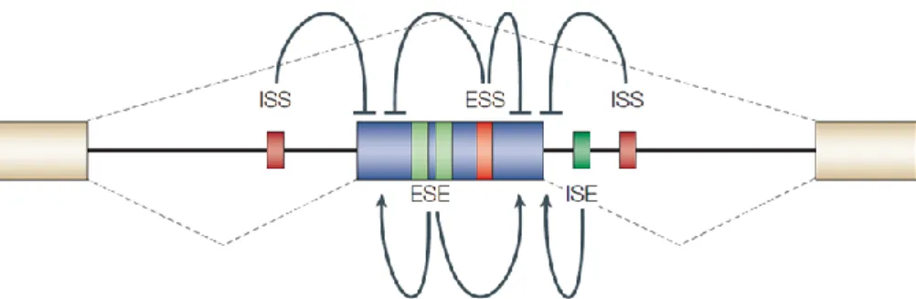
As described above, consensus sequences direct the spliceosome assembly on the pre-mRNA. However, in higher eukaryotes these sequence elements are highly degenerate, which indicates that additional marks are required for the spliceosome to identify the “real” splice sites among the numerous pseudo sites found in any pre-mRNA transcript (De Conti et al. 2013). At the most basic level, the relative strength of the splice sites (i.e., how near to consensus is their sequence) plays a major role in determining the inclusion or exclusion of exons (De Conti et al. 2013). Usually, the stronger the splice site, the more often it is used. However, a pair of ‘strong’ splice sites is not sufficient to define an exon; many ‘pseudo-exons’ that are flanked by predicted splice sites are not spliced or remain cryptic until specifically activated, e.g. in the above mentioned modes of ‘alternative 3' splice site’ or ‘alternative 5' splice site’(De Conti et al. 2013; Kelemen et al. 2013). So, in order to efficiently recognize the “real” splicing sites, the spliceosome is further guided by supplementary cis-acting elements that recruit both positive and negative splicing regulatory factors (trans-cis-acting factors), which either strengthen or weaken spliceosome interaction, in a process called exon definition (De Conti et al. 2013). These auxiliary splicing elements are highly variable in sequence, but they are important in defining both constitutive and alternative exons (Kelemen et al. 2013). Thus, the decision of which exon is removed, and which exon is included in the mature mRNA during the process of alternative splicing, is strongly influenced by the interaction between cis-acting elements and trans-acting factors (Hertel 2008; House and Lynch 2008; Kelemen et al. 2013; Wang et al. 2015). Cis-acting auxiliary sequences occur within both exonic and intronic regions and can either promote recruitment of the spliceosome and exon inclusion (splicing enhancers) or disrupt assembly of the splicing machinery and cause exon skipping (splicing silencers) (House and Lynch 2008; Wang et al. 2015). Depending on the position and function of the cis-regulatory elements, they are divided into four categories (Figure 1.5): exonic splicing enhancers (ESEs), exonic splicing silencers (ESSs), intronic splicing enhancers (ISEs) and intronic splicing silencers (ISSs) (Wang et al. 2015). Splicing enhancers (ESE and ISE) are, usually, bound by positive trans-acting factors, such as members of the SR (serine/arginine-rich) family of nuclear phosphoproteins, whereas splicing silencers (ESS and ISS) are, generally, bound by negative acting factors, such as members of the family of heterogeneous nuclear ribonucleoproteins (hnRNPs) (House and Lynch 2008; Kelemen et al. 2013; Wang et al. 2015). SR proteins promote exon inclusion by recruiting the splicing machinery to the adjacent intron, while hnRNPs repress spliceosomal assembly, for example, by multimerization along exons, looping out exons or by blocking the recruitment of snRNPs (House and Lynch 2008; Kelemen et al. 2013; Wang et al. 2015). SR proteins binding sites are present not only within alternatively spliced exons, but also within constitutively spliced exons, defining a crucial role in productive spliceosome assembly (House and Lynch 2008). Even though it is a general rule that SR proteins and hnRNPs promote or antagonize exon inclusion, respectively, there are numerous exceptions to this rule. For example, the splicing of the transcript encoding GTPase RAC1, is regulated by the antagonic effect of two SR-proteins, SRSF3 and SRSF1 previously known as SRp20 and ASF/SF2, respectively (Manley and Krainer 2010), whereas the myosin phosphatase targeting subunit-1 (MYPT1), is regulated by an antagonism between the two hnRNPs, PTB and TIA-1 (Shukla 2005; Gonçalves et al. 2009). In general, the cis-acting elements function additively and enhancing elements tend to play dominant roles in constitutive splicing, while the silencers are relatively more important in the control of alternative splicing (House and Lynch 2008; Kelemen et al. 2013; Wang et al. 2015). Typically, silencers and enhancers are present within the vicinity of potential exon/intron junctions, suggesting that the interplay between activating and repressing cis-acting elements modulates the probability of exon inclusion (House and Lynch 2008; Kelemen et al. 2013).
5
Figure 1.5 – Schematic representation of regulatory cis-elements. ESE: exonic splicing enhancer; ESS: exonic splicing
silencer; ISE: intronic splicing enhancer; ISS: intronic splicing silencers. Enhancers can activate adjacent splice sites or antagonize silencers, whereas silencers can repress splice sites or enhancers. Exon inclusion or skipping is determined by the balance of these competing influences, which in turn might be determined by relative concentrations of the activator and repressor proteins. Dashed lines: Two alternative splicing pathways, with the middle exon either included or excluded. (from (Matlin et al. 2005))
Beside the presence of regulatory cis-elements, a variety of other factors can influence alternative splicing. First, the assembly of local RNA secondary structures has been shown to interfere with the recognition of splice sites and cis-acting sequences by protein factors (Hertel 2008; Kelemen et al. 2013). This is because the recognition of these elements by RNA-binding proteins depends on the single-stranded structure of the pre-mRNA. If the structure is not single-stranded, the sequence elements might be concealed within the stable helices of the secondary RNA structure, becoming unavailable to the protein factors, influencing, therefore, pre-mRNA splicing (Hertel 2008; Kelemen et al. 2013). Second, the expression level of key splicing factors in a given cell will affect the interplay between activating and repressing effects on the spliceosome. Besides transcriptional regulation of splicing factor genes, non-coding RNAs (ncRNAs), including microRNA and small interfering RNA, have also been shown to regulate alternative splicing, generally through the modulation of the expression of key splicing factors during development and differentiation (Luco and Misteli 2011).
Third, exon/ intron architecture of a gene also influences splice site recognition, which has been shown to be more efficient when introns or exons are small (Hertel 2008; De Conti et al. 2013). Exon skipping is promoted by big exons or large flanking introns. These observations suggested that splice sites are recognized across an optimal nucleotide length and predicted that intron length significantly influences the efficiency of pre-mRNA splicing and alternative splice site choice (Hertel 2008; De Conti et al. 2013).
Fourth, the type of promoter used to drive transcription by RNA polymerase II can also impact the level of alternative splicing of a downstream exon. Two non-exclusive models were proposed to explain this effect: the recruitment model and the kinetic model (Hertel 2008; Kelemen et al. 2013; Braunschweig et al. 2013). The recruitment model assumes splicing factors assemble at the carboxy terminal domain (CTD) of RNA polymerase II and are released onto the nascent pre-mRNA during transcription. As these factors influence splice sites in a concentration dependent manner, the pre-loading of the CTD influences alternative exon usage (Hertel 2008; Kelemen et al. 2013; Braunschweig et al. 2013). The kinetic model postulates that protein complexes need time to assemble on an exon during its recognition. Everything that slows down a polymerase would give more time for the recruitment of the regulatory complexes and would favour alternative exon usage, as these exons usually depend more strongly on auxiliary factors (Hertel 2008; Kelemen et al. 2013; Braunschweig et al. 2013). Fundamentally, these mechanisms influence patterns of alternative splicing via the variations in RNA polymerase II elongation and recruitment of splicing factors. These mechanisms also offer an attractive explanation for how epigenetic marks and chromatin structure can change alternative splicing. (Naftelberg et al. 2015) A very interesting case is the one of Hu proteins that can induce local histone hyperacetylation by association with their target sequences on the pre-mRNA
6 surrounding alternative exons of Nf1 and Fas genes. This hyperacetylation favours higher elongation rates, which in turn decreases exon 23a inclusion in the Nf1 transcript, generating a chromatin-mediated reinforcement of the primary splicing decision (Zhou et al. 2011).
Overall, every exon has a specific set of identity elements, including the strength of the splice sites, the presence or absence of splicing enhancers or silencers, the presence or absence of local RNA secondary structures, the exon/intron architecture and the process of pre-mRNA synthesis by RNA polymerase II, that permit its recognition by the spliceosome. The sum of contributions from each of these identity elements defines the overall recognition potential of an exon or the overall binding affinity for the spliceosome.
Different cell types or distinct biological processes such as the cell cycle, tissue differentiation or developmental stages, will exhibit fluctuations in the expression level, of spliceosomal components and splicing activator/repressors (Hertel 2008). A further layer is added by signal transduction pathways in response to extracellular signals, in which cells can regulate gene expression through the use of alternative splicing (Shin and Manley 2004; Blaustein et al. 2007; Gonçalves et al. 2017). This signal-mediated splicing regulation is operated through the activation of intricate networks of signal transduction pathways (Kalyna et al. 2012) that influence trans-acting splicing regulatory factors through post-translational modification, including protein phosphorylation. This can result in the alteration of their activity or cellular localization (Shin and Manley 2004; Blaustein et al. 2007; Gonçalves et al. 2017). Such alterations may influence splicing efficiency or induce alternative splicing (as described in 1.5). As a result, the same exon in these different scenarios has the same structural properties but the cellular recognition potential can vary, meaning that exons that are alternatively included in one cell type or biological process can be alternatively excluded in another (Hertel 2008). Essentially, regulation of alternative splicing can be achieved through modulating any one of the exon recognition components. The interplay between the several regulatory sequences, complexes, processes, pathways, and factors mention above establishes the presence of a combinatorial control in alternative splicing (Smith and Valcárcel 2000).
1.3. Effects of alternative splicing
An important consequence of alternative splicing is phenotypic complexity by increasing transcriptomic and proteomic diversity (Stamm et al. 2005; Hertel 2008; Cieply and Carstens 2015). The magnitude of these effects ranges from a complete loss of function or acquisition of a new function to very subtle modulations, due to variation in various aspects of protein function, including, their binding properties, intracellular localization, enzymatic activity, stability and regulation by posttranslational modifications (Stamm et al. 2005; Hertel 2008; Cieply and Carstens 2015). In addition to proteome modulation, alternative splicing is also a crucial regulatory stage in the pathway of gene expression. For example, as mentioned above, alternative exons within UTRs can modulate the stability and translation of transcripts. Up to one third of human alternative splicing events introduce premature termination codons (PTC), which are recognized and lead to the degradation of transcripts by the nonsense-mediated mRNA-decay (NMD) pathway (Lewis et al. 2003; Lareau et al. 2007a). Thus, the sensitivity of mRNA transcripts to NMD is modulated by alternative splicing events (Kalyna et al. 2012). One interesting example are the ‘poison exons’ found in transcripts from splicing factors. The inclusion of these exons introduces a PTC in the mRNA sequence, resulting in the transcript degradation by NMD. The inclusion or exclusion of this exon will indirectly influence the alternative splicing of the splicing factors targets (Lareau et al. 2007b; Rossbach et al. 2009).
Due to its central role in protein expression and function, alternative splicing seems to be an important mechanism in defining biological function. As so, it is not surprising that aberrant regulation of alternative splicing leads to human disease (Zhang and Manley 2013). Mutations in the consensus
7 splice site sequences, in the splicing regulatory sequences, in the splicing machinery and in the regulatory splicing factors genes have been suggested to cause aberrant splicing (Daguenet et al. 2015). In genetic diseases these types of mutation will interfere with the splice site recognition efficiency which can lead to exon skipping, intron retention or the introduction of a new splice site within an exon or intron (Daguenet et al. 2015). This deregulation in splicing has the potential to originate protein isoforms that ultimately contribute to human disease (Wang et al. 2015). Alterations in regulatory splicing factors’ cellular concentration, composition, localization and activity have been suggested to be the cause of aberrant splicing, in multifactorial diseases, such as cancer (Wang et al. 2015). In genetic disease, an immediate cause for aberrant splicing is the alteration of the splice site recognition efficiency, while irregularities in protein isoforms in different systems ultimately contribute to multifactorial diseases (Wang et al. 2015). The potential roles for splicing in cancer are well documented and include changes in genes associated with cell migration, regulation of cell growth, hormone responsiveness, apoptosis and response to chemotherapy (Shkreta et al. 2013). Alternative splicing has been implicated in nearly all aspects of cancer development, thus, understanding the basic mechanisms and patterns of splicing in tumour progress will shed light on the biology of cancer and lay the foundation for diagnostic, prognostic and therapeutic(Kim and Kim 2012).
1.4. The small GTPase RAC1 and its splice variant RAC1b
Ras-related C3 botulinum toxin substrate 1 (RAC1) is a member of the Rac family of guanosine triphosphate phosphohydrolases (GTPases), a subfamily of the Rho family of small GTPases, which are best known for their role in regulating the actin cytoskeleton and gene expression (Marei and Malliri 2017). RAC1 exist in two different conformational states, an inactive GDP-bound form and an active GTP-bound form (Jordan et al. 1999; Matos et al. 2000). The interconversion between the two states occurs through a cycle of guanine exchange and GTP hydrolysis. The conformational changes induced upon GTP binding involve two important regulatory protein regions of RAC1, known as Switch I and Switch II. Consequently, the switch regions provide binding domains for both regulatory and effector proteins. In the active state, these regulatory regions enable their interaction with downstream effectors, allowing these GTPases to function as molecular switches (Wennerberg 2005). The transition between the active and inactive states of RAC1 occurs at the plasma membrane following appropriate cellular signals and is tightly controlled and spatially regulated by guanine nucleotide exchange factors (GEFs) which convert RAC1 to its active form, GTPase-activating proteins (GAPs), which inactivate RAC1, and Rho-GDP dissociation inhibitors (Rho-GDIs) that bind to and remove RAC1 from the plasma membrane, keeping it inactive in the cytoplasm and blocking its activation by GEFs (Figure 1.6) (Symons and Settleman 2000). Following its activation, RAC1 interacts with downstream effector proteins and activates signalling cascades that trigger various cellular responses such as secretory processes, phagocytosis of apoptotic cells, epithelial cell polarization, neuron adhesion, migration and differentiation, and growth-factor induced formation of membrane ruffles.
The mammalian RAC1 gene is composed of seven coding exons that after undergoing alternative splicing can originate two different transcripts, RAC1 and RAC1b (Figure 1.7) (Gonçalves et al. 2009). RAC1b is characterized by the insertion of an additional exon (exon 3b) between exons 3 and 4 of RAC1. Consequently, RAC1b contains 57 additional nucleotides that result in an in-frame insertion of 19 amino acid residues between codons 75 and 76 of RAC1, near an important regulatory region of the GTPase, the switch II domain, changing the regulation and signalling properties of the protein (Jordan et al. 1999).
8
Figure 1.6 – Schematic representation of RAC1 activation and regulation. RAC1 GTPase cycles between inactive
GDP-bound and active GTP-GDP-bound states. RAC1 activation is facilitated by the action of GEFs, which promotes GDP dissociation from RAC1 and allows GTP to bind instead. Through the association with GAPs, the intrinsic GTPase activity of RAC1 is accelerated, thereby inactivating RAC1. Through association with RhoGDIs RAC1 can be sequestered in its inactive state.
RAC1b is a highly activated variant of RAC1 because, despite the lower levels of expression compared to RAC1, RAC1b exists predominantly in the active GTP-bound state. This is essentially due to RAC1b disability to interact with Rho-GDI, which keeps this GTPase constitutively membrane-bound, a location that favours the interaction with activators, and consequently promotes the active GTP-bound state (Matos et al. 2003). Moreover, RAC1b shows impaired intrinsic GTPase activity and increased GDP to GTP exchange rates, although this variant can still be down regulated by activated GAPs and it is influenced by GEFs action (Matos et al. 2003; Fiegen et al. 2004; Singh et al. 2004).
Figure 1.7 – Schematic representation of the human RAC1 gene. RAC1 gene has seven coding exons, including the
alternative exon 3b, that after undergoing the alternative splicing event can originate two alternative transcripts, RAC1 and RAC1b. (from (Gonçalves et al. 2009))
In addition, the 19 extra amino acids of RAC1b seem to confer a selective downstream signalling to this variant, since several pathways activated by RAC1 are not activated by RAC1b (Matos et al. 2003). Unlike RAC1, GTP-bound RAC1b is unable to induce lamellipodia formation, which means that this isoform doesn’t have the ability to induce actin cytoskeleton reorganization. Additionally, RAC1b is incapable to activate PAK1 effector and stimulate the JNK cascade, two well-established RAC1 signalling pathways (Matos et al. 2003; Fiegen et al. 2004; Singh et al. 2004). On the other hand, RAC1b was shown to bind more effectively than RAC1 to proteins that can promote loss of epithelial cell structure and increased cell proliferation, such as p120 catenin and RACK1 (Orlichenko et al. 2010). RAC1b also favours specific pathways conducting to the production of reactive oxygen species (ROS) and NF-kB canonical pathway activation (Matos et al. 2003; Matos and Jordan 2005; Radisky et al. 2005). This RAC1 isoform was found to be overexpressed in several malignant tumours including colorectal, breast, lung, thyroid and pancreas (Schnelzer et al. 2000; Matos and Jordan 2008; Stallings-Mann et al. 2012; Silva et al. 2013; Mehner et al. 2014).
9
1.5. Colorectal cancer and RAC1b alternative splicing
Colorectal cancer (CRC) is one of the most common types of cancer worldwide, representing in 2012 the third highest incidence, with 1.4 million cases, after lung and breast cancer (Ferlay et al. 2015). The main risk factors identified for the development of CRC include family histories of either colorectal cancer or inflammatory bowel disease, but the disease burden at the population-level is mainly accounted for by modifiable life-style factors such as smoking, excessive alcohol consumption, high consumption of red and processed meat, obesity, and diabetes (Brenner et al. 2014).
Normal gastrointestinal epithelium is organized along a crypt-villus axis. A pool of colon stem and progenitor cells, the most undifferentiated cell types that are able of self-renewal and pluripotency, are located at the bottom of the crypt. These cells differentiate along the crypt-villus axis, into all epithelial colon lineages. In about 14 days they arrive at the top of the villus and undergo programmed cell death (apoptosis) (Peifer 2002; Kosinski et al. 2007). During colorectal adenocarcinoma development, some progenitor cells acquire sequential genetic and epigenetic mutations in specific oncogenes and/or tumour suppressor genes, conferring them a selective advantage on proliferation and self-renewal (Vogelstein et al. 1988; Ewing et al. 2014). So, the normal epithelium becomes a hyperproliferative mucosa and subsequently gives rise to a benign adenoma that evolves into carcinoma and metastasis in about 10 years (Vogelstein et al. 1988).
In colorectal cancer, RAC1b is overexpressed in a specific subgroup, namely in 80% of tumours with mutation in the oncogene BRAF (BRAFV600E), suggesting that both events cooperate to promote the survival of colorectal cells (Matos et al. 2008; Matos et al. 2016). Activating mutations in the BRAF gene have been found to induce oncogene-induce senescence (OIS), an important tumour suppressing mechanism at early stages of cancer. In colorectal cancer, RAC1b overexpression was found to antagonize OIS, suggesting that this hyperactive splice variant is selected in early stages of tumour development. Knowing that RAC1b expression was found to be increased in patients with inflammatory bowel diseases or in mouse models of acute colitis and that chronic inflammation is a known risk factor for colorectal cancer, the authors suggested that RAC1b overexpression in BRAFV600E-initiated tumour cells could be triggering escape from OIS, leading to cancer progression (Henriques et al. 2015). In another study, RAC1b was reported to be a putative accelerator of tumour progression by positively regulating the expression of proliferation-promoting genes through Wnt pathway activation and decreasing the adhesive properties of colorectal cancer cells by negatively regulation of E-cadherin expression (Esufali et al. 2007). RAC1b overexpression was also associated with a poor outcome of patients with wild-type KRAS/BRAF metastatic colorectal cancer treated with FOLFOX/XELOX chemotherapy (Alonso-Espinaco et al. 2014). As a result of its hyperactive properties and selective overexpression in cancerous tissue, RAC1b has been highlighted as a promising therapeutic target, being, therefore, important to understand the basic mechanisms underlining the regulation of its expression.
Previous studies in this sense showed that in colorectal cells, RAC1 alternative splicing event is regulated by two SR proteins with antagonistic roles, SRSF1 and SRSF3 (Gonçalves et al. 2009). SRSF1 acts as an enhancer by promoting the inclusion of alternative exon 3b, while SRSF3 acts as a silencer by promoting the skipping of the exon 3b (Gonçalves et al. 2009). Both splicing factors were found to be regulated by upstream signalling pathways: the inhibition of the phosphatidylinositol 3-kinase pathway increased protein levels of SRSF1 and promoted RAC1b, whereas activation of β-catenin/TCF4 increased expression of SRSF3 and inhibited that of RAC1b (Gonçalves et al. 2009). Thus, extracellular stimuli might induce or sustain RAC1b overexpression in tumour cells through signal transduction pathways. For example, the protein kinases SRPK1 and GSK3β were also found required to sustain RAC1b levels and both were shown to act upon the phosphorylation of splicing factor SRSF1 (Goncalves et al. 2014). However, besides SRSF1 and SRSF3 it is likely that RAC1
10 alternative splicing can be regulated by additional factors, and recently, PTBP1 and ESRP1 were described as possible modulators of the alternative splicing of RAC1b in different cell types while RANBP2 was shown to be a modulator of the distribution of phosphorylated SR proteins, which are known to regulate RAC1b expression (Saitoh et al. 2012; Ishii et al. 2014; Hollander et al. 2016; Vecchione et al. 2016).
1.6. PTBP1, ESRP1 and RANBP2 as possible modulators of RAC1b alternative splicing
Polypyrimidine tract-binding protein 1 (PTBP1), also known as hnRNP I, is a member of a subfamily of ubiquitously expressed hnRNPs and contains four RNA recognition motif (RRM) domains that bind to the polypyrimidine track of mRNAs introns (Oberstrass 2005). PTBP1 shuttles between the nucleus and the cytoplasm, intervening in almost all steps of mRNA metabolism, such as alternative splicing, mRNA transport, cytoplasmic localization, translation initiation in internal ribosome entry site (IRES) and regulation of RNA stability (Fu et al. 2018). This RNA-binding protein is also involved in several biological processes, including cell structure and motility, protein targeting and localization, protein metabolism and modification, muscle contraction, cell cycle and immunity (Fu et al. 2018). Numerous studies have reported that PTBP1 is overexpressed in several different types of cancer, including brain, colorectal, ovarian, gastric and breast cancer (McCutcheon et al. 2004; He et al. 2007; Cheung et al. 2009; He et al. 2014; Takahashi et al. 2015; Sugiyama et al. 2016). Furthermore, high expression of PTBP1 has been demonstrated to be associated with aggressive behaviour of several types of cancer, especially in glioma and ovarian tumours (He et al. 2007; Cheung et al. 2009). In colorectal cancer PTBP1 was shown to facilitate cancer migration and invasion activities by promoting the inclusion of cortactin exon 11 (Wang et al. 2017). In another study PTBP1 was associated with metastasis of colorectal cancer cells by downregulating ATG10, an autophagy-related gene (Jo et al. 2017). PTBP1 was also positively associated with cancer progression properties, such as invasion or proliferation, in colorectal cancer through upregulation of PKM2 (plays a central role in metabolism and growth, promoting cell migration) and CD44 (induces a metastatic phenotype in tumour cells) variants (Takahashi et al. 2015). Furthermore, it was shown, in HCT116 (human epithelial colorectal carcinoma cells with KRAS mutation) that the depletion of PTBP1 and PTBP2 promoted the skipping of exon 3b in RAC1 pre-mRNA (Hollander et al. 2016). In this work the effect of PTBP1 on RAC1b splicing event was further analysed in other colorectal cell lines.
Epithelial splicing regulatory protein 1 (ESRP1) is an epithelial cell-specific RNA-binding protein from the hnRNP family that regulates alternative splicing events associated with epithelial phenotypes (Hayakawa et al. 2016; Jeong et al. 2017). ESRP1 binds preferentially to UGG-rich repeats and plays crucial roles during organogenesis (Hayakawa et al. 2016). This protein regulates the alternative splicing of multiple genes, including CD44, CTNND1, ENAH and FGFR2, all transcripts that undergo changes in splicing during the epithelial-to-mesenchymal transition (EMT), a process by which epithelial cells lose their polarity and acquire motile and invasive phenotypes (Kalluri and Weinberg 2009; Jeong et al. 2017). This protein has dual roles in cancer progression, depending on the context of microenvironments surrounding cancer cells. In some situations, ESRP1 expression is favoured as it supports cell survival; in other situations, downregulation of ESRP1 is favoured as this facilitates cell invasion (Hayakawa et al. 2016). Both of these scenarios were observed in colorectal cancer: on one hand downregulation of ESRP1 promoted EMT and consequently tumour progression (Deloria et al. 2016). On the other hand, ESRP1 overexpression enhanced fibroblast growth factor receptor (FGFR1/2) signalling, Akt activation, and Snail upregulation, thus stimulate growth of cancer epithelial cells and promote colorectal cancer progression. Moreover, ESRP1 promoted the ability of colorectal cells to generate macrometastases in mice livers (Fagoonee et al. 2017). In SAS and HSC4 cells, both tongue squamous cell carcinoma, ESRP1 was found to suppress RAC1b expression (Ishii et
11 al. 2014). In this work, we studied whether the effect of ESRP1 on RAC1b alternative splicing can also be observed in colorectal cells.
Ran-binding protein 2 (RANBP2), also known as Nup358, is a cytosolic component of the filaments that attach to the cytoplasmic ring of the nuclear pore complexes (NPC). NPC are large protein channels that act as mediators of molecular exchange between the nucleus and the cytoplasm of eukaryotic cells (Raices and D’Angelo 2012; Ibarra and Hetzer 2015). RANBP2 plays major roles in nuclear export and import by providing a docking site for Ran and its cofactors. This protein also mediates SUMOylation of Ran cofactor, RanGAP1, as well as of various cargo proteins (Matunis et al. 1998; Pichler et al. 2002; Forler et al. 2004; Bernad et al. 2004; Reverter and Lima 2005; Hutten et al. 2009). In addition, during mitosis, this nucleoporin is found at kinetochores where it is involved in spindle formation and chromosome segregation (Salina et al. 2003; Joseph and Dasso 2008). Furthermore, RANBP2 binds to the kinesin motors KIF5B and KIF5C, linking the NPC to the cytoskeleton (Cai et al. 2001). RANBP2 has been associated with cancer in different, and contradicting, manners. This protein was shown to act as a tumour suppressor due to its role in preventing chromosome segregation errors (Dawlaty et al. 2008; Navarro and Bachant 2008). However, in another study, RANBP2 was identified as a candidate oncogene overexpressed in the subgroup of human colorectal cancers with microsatellite instability (Gylfe et al. 2013). Corroborating with this information, in another study this nucleoprotein was also reported to be overexpressed in human colorectal cancers with microsatellite instability (Dunican et al.). Consistent with an oncogenic function, RANBP2 was found to protect BRAFV600E mutant colon cancers cells from undergoing mitotic cell death (Vecchione et al. 2016). All this information suggested that somehow RANBP2 is involved in the survival of human colorectal cancers with microsatellite instability. Furthermore, the speckled distribution of phosphorylated pre-mRNA processing factors, like SRSF1 and SRSF3 (known regulators of RAC1b splicing event), was found to be regulated by the nucleocytoplasmic transport system in mammalian cells (Saitoh et al. 2012). Although RANBP2 is not a splicing factor itself, the gathered information prompted the investigation of its role in RAC1b overexpression in colorectal cancer cells.
2. Objectives
RAC1b was found to be overexpressed in a subgroup of colon tumours also characterized by the presence of an oncogenic mutation in BRAF (Matos et al. 2008). Together, these two alterations stimulate signalling pathways that promote the proliferation and survival of malignant cells (Matos et al. 2008). Due to its hyperactive properties and selective overexpression in cancerous tissue, RAC1b is a promising therapeutic target, for this subgroup of tumour patients. Therefore, understanding the basic mechanisms underlining its expression regulation is important to identify potential therapeutic agents. PTBP1 and ESRP1 were described in the literature as possible modulators of the alternative splicing of RAC1b while RANBP2 was shown to be a modulator of the distribution of phosphorylated SR proteins, which are known to regulate RAC1b expression (Saitoh et al. 2012; Ishii et al. 2014; Hollander et al. 2016; Vecchione et al. 2016). With the objective of studying whether these factors can regulate RAC1b expression in colorectal cells, this work was divided into 3 different parts:
(1) Provide or generate expression vectors as tools to study the effect of PTBP1,
ESRP1, and RANBP2.
(2) Overexpress PTBP1, ESRP1 and RANBP2 in colorectal cells and determine their
effect on alternative splicing of a RAC1 minigene.
(3) Deplete the endogenous expression of PTBP1, ESRP1 and RANBP2 in colorectal
12
3. Experimental Procedures
3.1. Polymerase Chain Reaction (PCR)
Polymerase chain reaction (PCR) is an invaluable tool for molecular biology research since it provides a rapid mean for DNA identification and analysis through the amplification of a specific DNA region/sequence in vitro (Wilson and Walker 2009).
Several components are required to perform a PCR. First, a double-stranded DNA (dsDNA) template that includes the target sequence to be amplified is essential. For each strand, is necessary, a small oligonucleotide, also known as primer, that provides a starting point for DNA synthesis. The primers are chemically synthesized according to the known template DNA sequence and flank the region to be amplified. One primer will have the same sequence as the DNA template - forward primer - and the other will be reverse and complementary - reverse primer. When designing primers there are some constraints that should be considered, such as, the primer length (16-28 base pairs (bp)), the primer length difference (± 3 nt), the GC content (40-60%), the melting temperature (Tm, 50-62°C), the melting temperature difference (± 5°C) and the sequence complementarity between the pair of primers or even in the same primer (possibility of secondary structures, like dimers and hairpins) (Wilson and Walker 2009; Pestana et al. 2010; Chuang et al. 2013). Finally, a DNA polymerase is also needed to synthesize a new DNA strand in the presence of deoxyribonucleotides (dNTPs), a buffer that provides a suitable chemical environment for the polymerase performance and magnesium ions as co-factors to increase the yield of the reaction (Pestana et al. 2010).
PCR can be separated in 3 basic steps: DNA denaturation, primer annealing and polymerase extension. In the first step, high temperatures (94-96°C) are used to break the hydrogen bonds that connect the two DNA strands. This results in the separation of the dsDNA originating two single-stranded DNA (ssDNA) templates. The time necessary for denaturation depends on the size of the DNA fragment to be amplified: the longer the fragment, the longer it takes to be denaturated. In the second step the temperature is lowered until the melting temperature of the primers is reached, allowing them to bind to their complementary sequences on the ssDNA template. In the final step, the polymerase synthesizes the missing strands starting from the annealed primers in a process called elongation. The temperature of elongation depends on the used polymerase (for Taq and Pfu the ideal temperature is 72°C) and the time depends both on DNA polymerase efficiency and on DNA fragment length to be amplified (Taq needs approximately 1 minute to elongate 1000 bp, while Pfu only elongates 500 bp in the same time). Hence, in a short time, exact replicates of the target sequence are produced, and at the end of several cycles, the amount of target sequence is significantly increased enabling further analysis (Pestana et al. 2010).
In general, every PCR reaction mix preformed in this experimental work had a final volume of 25 L. Each primer was used at 0,2 M and Go Taq G2 Flexi at 0,02 U/L. The volume of DNA added depended on the type and purpose of the PCR and buffer B (10 mM Tris-HCl pH 9 1,5 M; 50 mM MgCl2; 1,5 mM KCl; 0,1% Bacta Gelatin (DifcoLab)); 0,2 mM of each dNTP) was added to make up the final volume. For all PCRs, a mock reaction was made with water instead of DNA to ensure the amplification resulted from the DNA template and not from genomic DNA or possible contaminants in the mix. The thermocycler C1000 TouchTM Thermal Cycler (Bio-Rad) was used to run the amplification programs.