Title:
3D echoendoscopy and miniprobes for rectal cancer staging
Authors:
Fernando Castro-Poças, Mário Dinis-Ribeiro, Anabela Rocha, Tarcísio Araújo, Isabel Pedroto
DOI: 10.17235/reed.2018.4453/2016 Link:PubMed (Epub ahead of print)
Please cite this article as:
Castro-Poças Fernando, Dinis-Ribeiro Mário, Rocha Anabela, Araújo Tarcísio, Pedroto Isabel. 3D echoendoscopy and miniprobes for rectal cancer staging. Rev Esp Enferm Dig 2018. doi:
10.17235/reed.2018.4453/2016.
This is a PDF file of an unedited manuscript that has been accepted for publication. As a service to our customers we are providing this early version of the manuscript. The manuscript will undergo
copyediting, typesetting, and review of the resulting proof before it is published in its final form. Please note that during the production process errors may be discovered which could affect the content, and all legal disclaimers that apply to the journal pertain.
OR 4453
3D echoendoscopy and miniprobes for rectal cancer staging
Fernando Castro-Poças1,2,5, Mário Dinis-Ribeiro3, Anabela Rocha2,4, Tarcísio Araújo5and Isabel Pedroto2,5
Departments of1Ultrasound and5Gastroenterology. Santo António Hospital. Porto Hospital Center. Porto, Portugal.2Institute of Ciências Biomédicas Abel Salazar. University of Porto. Porto, Portugal.3Center for Health Technology and Services Research. Faculty of Medicine. University of Porto. Porto, Portugal.4Unit of Digestive Surgery. Service of General Surgery. Santo António Hospital. Porto Hospital Center. Porto, Portugal
Received: 27/05/2017 Accepted: 02/12/2017
Correspondence: Fernando Castro-Poças. Departments of Ultrasound and Gastroenterology. Santo António Hospital. Porto Hospital Center. Largo do Prof. Abel Salazar. 4099-001 Porto, Portugal
e-mail:[email protected] ABSTRACT
Background: Rectal cancer staging using rigid probes or echoendoscopes has some limitations. The aim of the study was to compare rectal cancer preoperative staging using conventional endoluminal ultrasonography with three-dimensional endoscopic ultrasonography and miniprobes.
Materials and methods: Sixty patients were included and evaluated with: a) a conventional echoendoscope (7.5 and 12 MHz); b) miniprobes (12 MHz); and c) the Easy 3D Freescan software for three-dimensional endoscopic ultrasonography. The reference or gold standard was conventional endoluminal ultrasonography in all cases and pathological assessment for those without preoperative therapy. The differences in T and N staging accuracy in both longitudinal and circumferential extension were evaluated.
Results: With regard to T staging, conventional endoluminal ultrasonography had an accuracy of 85% (compared to pathological analysis), and the agreement between miniprobes vs conventional endoluminal ultrasonography (kappa = 0.81) and three-dimensional endoscopic ultrasonography vs conventional endoluminal ultrasonography (k = 0.87) was significant. In addition, miniprobes had an accuracy of 82% and three-dimensional endoscopic ultrasonography had a higher accuracy (96%). With regard to N staging, conventional endoluminal ultrasonography had an accuracy of 91% with a sensitivity of 78%. However, the agreement between miniprobes and conventional endoluminal ultrasonography and three-dimensional endoscopic ultrasonography and conventional endoluminal ultrasonography (k = 0.70) was lower. Interestingly, miniprobes had a lower accuracy of 81% whereas three-dimensional endoscopic ultrasonography had an accuracy of 100% without any false negative. No false positives were observed in any of the techniques. Accuracy for T and N staging was not influenced by longitudinal or circumferential extensions of the tumor in all types of endoscopic ultrasonography analyzed.
Conclusions: Miniprobes and especially three-dimensional endoscopic ultrasonography may be relevant during rectal cancer staging.
Key words: Human colon. Miniprobes. Endoscopic Ultrasonography. Intestinal wall. INTRODUCTION
Magnetic resonance imaging, endoluminal ultrasound and computed tomography are imaging tools commonly used to evaluate rectal tumor staging (1). Rectal endoluminal ultrasonography (US) with rigid probes or endoscopic ultrasonography (EUS) is the preferential method for local staging of rectal carcinoma (RC) and decisively influences the therapeutic approach of patients (2). Nevertheless, its accuracy varies significantly, ranging from 63% to 95% for T staging and from 64% to 80% for N staging (1,3-5). It is well known that operator expertise is an important factor in the accuracy of RC by endoluminal US (6). However, tumor anatomical characteristics (e.g., stenosis), lymph node location (e.g., pelvic lateral nodes) or criteria (e.g., size) may also influence the final result (4,7).
Conventional EUS (C-EUS) does not provide whole tumor assessment for all stenosing tumors. Miniprobe EUS (mp-EUS) may easily overcome these obstacles due to its reduced
diameter and flexibility (8,9). mp-EUS may be the most adequate ultrasonographic tool for the differential diagnosis of tumors limited to the mucosa (T1m) or with involvement of the submucosa (T1sm) as it uses high frequencies and thus resolves the limitations of C-EUS (10,11).
C-EUS provides only two-dimensional images of the lesions and structures. Three-dimensional EUS (3D-EUS) may be able to provide simultaneous spatial information of different planes and even a multi-plane vision. It also allows the isolation of structures or lesions of interest and presents them in different perspectives, changing their texture and/or transparency (12-15). However, the application of 3D-EUS in this setting has rarely been reported (12,14,16,17). Therefore, we aimed to compare RC preoperative staging using C-EUS with 3D-EUS and mp-EUS.
METHODS
Selection of participants
The prospective study was approved by the Ethics Committee for Health of our hospital. Patients with RC (defined as located up to 15 cm from the anal verge) that underwent endoscopic ultrasonography staging of the disease gave their written informed consent prior to inclusion in the study. Patients less than 18 years of age, pregnant women or individuals unable to give informed consent were excluded.
Procedures
Two enemas were given one hour before the procedures. Three types of equipment were used in staging: a) a conventional echoendoscope with frequencies of 7.5 and 12 MHz (Olympus GF-UM20®); b) mp-EUS with 12 MHz (Olympus UM-2R®); and c) 3D-EUS with the Easy 3D Freescan software from Echotech®. The assessment for T and N stages (defined in accordance with TNM staging [18]) was initiated with mp-EUS followed by C-EUS. 3D-EUS was the final technique performed and images were acquired via a conventional echoendoscope. The identification of lymph nodes in the perirectal space was conducted from the distal rectum to the iliac vessels. The differences in the ability of the three techniques to assess the entire lesion were also assessed.
Reference tests
Patients were divided in two groups according to the use of neoadjuvant therapy with radio/chemotherapy. Prior assessment with a conventional echoendoscopy was performed before neoadjuvant therapy and was considered as a reference in all cases. Pathological assessment after surgery was considered as the gold standard for those who did not receive preoperative therapy.
Statistical analysis
The PASW version 21 software was used for the analysis. The kappa coefficient was used to estimate the agreement between techniques and accuracy; this was calculated as the proportion of true results versus the total number of patients. In addition, sensitivity and specificity were estimated as the proportion of true positive or negative cases, respectively. The relationship between longitudinal or circumferential extent of the tumor and staging accuracy was evaluated for all three techniques using a Spearman’s correlation.
RESULTS
Sixty patients aged between 34 and 89 years (mean value of 63.8 11.8) were assessed, and 36 (60%) patients were male. All patients underwent surgery, 27 (45%) without preoperative adjuvant therapy.
Mp-EUS assessed the lesion in its entirety in 97% of cases, whereas this was achieved less frequently via C-EUS and 3D-EUS, in 85% of cases (p = 0.01). Staging was not possible using C-EUS in 15% (n = 9) of cases vs 3% (n = 2) of cases with mp-EUS, due to tumor stenosis. In these cases where it was not possible to evaluate the whole of the tumor, T and N stages results were based on the tumor extent that was observed.
T Staging
Comparison of T staging by mp-EUS, 3D-EUS and C-EUS in all patients (n = 60) (Table 1)
Both the mp-EUS and 3D-EUS techniques were highly concordant with C-EUS for T staging, with Kappa coefficients of 0.81 and 0.87. Global accuracy of T staging by mp-EUS compared to C-EUS was 86.7% and 81.7% compared to 3D-EUS.
Comparison of T staging obtained using different EUS techniques and anatomopathological staging (APS) (n = 27) (Table 2)
T staging by C-EUS, mp-EUS and 3D-EUS was concordant with APS with Kappa coefficient values of 0.81, 0.76 and 0.95, respectively.
Simultaneous comparison of T staging using the three EUS techniques and T APS (n = 27) (Table 2)
There were no significant differences with regard to the simultaneous comparison of the three techniques (p = 0.75).
N Staging
Comparison of N staging by mp-EUS, 3D-EUS and C-EUS staging in all patients (n = 60) (Table 3)
The mp-EUS and 3D-EUS analyses were concordant with C-EUS for N staging, with kappa coefficients of 0.65 and 0.79, respectively.
The sensitivity of C-EUS compared to mp-EUS and 3D-EUS for the identification of metastasized nodes was 73.1% and 92.3%, respectively. The corresponding values for specificity were 91.2% and 79.4%, positive predictive values were 86.4% and 77.4%, negative predictive values were 81.6% and 93.1% and global accuracy was 83.3% and 85%, respectively.
Comparison of N staging obtained via different EUS techniques and APS (n = 21) (Table 4)
Anatomopathological information with regard to the presence of metastasized nodes was available in 21 (77.8%) cases. Of the 27 patients that underwent surgery, it was not possible to obtain this information in six (22.2%) cases as a transanal resection was performed. N staging by C-EUS, mp-EUS and 3D-EUS and pathological analysis were concordant with respective kappa coefficients of 0.80, 0.59 and 1. The sensitivity of C-EUS, mp-EUS and 3D-EUS for the identification of metastasized nodes was 77.8%, 55.6% and 100%, respectively. The corresponding values for specificity and positive predictive values were 100% for the three techniques, whereas the negative predictive values were 85.7%, 75% and 100% and global accuracy was 90.5%, 81% and 100%, respectively.
Simultaneous comparison of N staging accuracy using the three EUS techniques and N APS (n = 21) (Table 4)
There were no significant differences between the three techniques (2= 4.42; p = 0.11). DISCUSSION
The continuous technological development has led to the application of new techniques associated with endoscopic ultrasonography (19). Good results were obtained in this study with mini-probes and three-dimensional endoscopic ultrasonography.
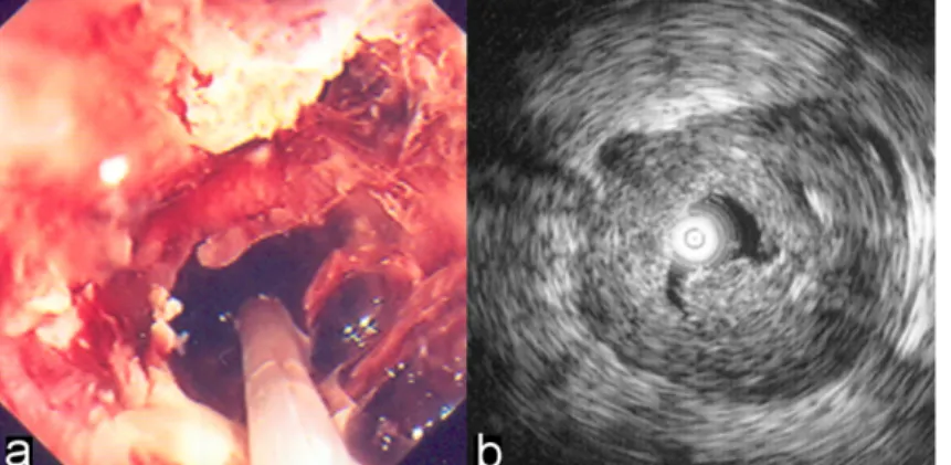
There was a significant agreement with regard to T staging between miniprobes, 3D and conventional EUS. These values range between 77 and 93% in previous reports (12,14,16,17). Our findings confirm the high accuracy of 3D-EUS, which is higher than that of conventional EUS. However, stenosing rectal cancer cannot be assessed either by C-EUS or 3D-EUS in up to 21.6% of cases (20), the rate was 15% in our study. Interestingly, miniprobe-EUS allowed the identification of most lesions in their entirety. Miniprobes can in fact transpose stenosing RC (Fig. 1), overcoming the rigid end and “large” diameter characteristics of C-EUS, which may inhibit RC staging (4,7).
Both 3D-EUS and mp-EUS had a high accuracy (Fig. 2) for T1 staging, which is in agreement with previous reports (21,22). With regard to T2 staging, 3D was superior to mp-EUS, which tended to overstage tumors (12,16,17,22-24). A very high accuracy for T3 staging was reported for 3D or mp-EUS (12,16,17,23-26), whereas mp-EUS had the lowest accuracy in relation to the higher ultrasound frequency for T4 staging, which is in line with current evidence (17,24-27). The use of 3D for T staging of rectal cancer had the best accuracy with mp and allowed the staging of stenosing tumors, although with some limitations for large masses (e.g., T4 staging).
With regard to lymph node diagnosis with EUS, the results obtained with mp-EUS and 3D-EUS were also encouraging since they were in significant concordance with C-3D-EUS, with a global accuracy of 90.5%. The highest sensitivity was obtained with 3D-EUS (100%), as well as a very high negative predictive value (100%). This not only represents the high capacity of 3D-EUS to identify lymph nodes but also reinforces our option for not imposing a cut-off value for node diameter in order to classify it as metastasized. However, there is no consensus with regard to this matter (28,29). In fact, this trend to improved accuracy was
also observed in other studies (12,17,30,31).
In conclusion, Mp-EUS and 3D-EUS are valid techniques compared to C-EUS for RC staging. We suggest that Mp-EUS may be an alternative to C-EUS due to the possibility of staging a carcinoma during colonoscopy with interesting T-staging results, and it can usually assess the entire tumor mass. This is an advantage for stenosing masses. More importantly, even though the results were not statistically significant in the comparison of 3D-EUS with C-EUS, this technique may become the gold standard method in RC staging. This will ultimately improve the clinical decisions taken with regard to these patients, particularly for T2 vs T3 staging, which is highly relevant in patient management.
REFERENCES
1. Muthusamy VR, Chang KJ. Optimal methods for staging rectal cancer. Clin Cancer Res 2007;13:6877-84s. DOI: 10.1158/1078-0432.CCR-07-1137
2. Berton F, Gola G, Wilson SR. Perspective on the role of transrectal and transvaginal sonography of tumors of the rectum and anal canal. Am J Roentgenol 2008;190:1495-504. DOI: 10.2214/AJR.07.3188
3. Puli SR, Bechtold ML, Reddy JB, et al. How good is endoscopic ultrasound in differentiating various T stages of rectal cancer? Meta-analysis and systematic review. Ann Surg Oncol 2009;16:254-65. DOI: 10.1245/s10434-008-0231-5
4. Heo S. Multimodal imaging evaluation in staging of rectal cancer. World J Gastrointest Endosc 2014;20:42-4. DOI: 10.3748/wjg.v20.i15.4244
5. Schaffzin D, Wong W. Endorectal ultrasound in the preoperative evaluation of rectal cancer. Clin Colorectal Cancer 2004;4:124-32. DOI: 10.3816/CCC.2004.n.015
6. Bipat S, Glas AS, Slors FJ, et al. Rectal cancer: Local staging and assessment of lymph node involvement with endoluminal US, CT, and MR imaging - A meta-analysis. Radiology 2004;232:773-83. DOI: 10.1148/radiol.2323031368
7. Saranovic D, Barisic G, Krivocapic G, et al. Endoanal ultrasound evaluation of anorectal diseases and disorders: Technique, indications, results and limitations. Eur J Radiol 2007;61:480-9. DOI: 10.1016/j.ejrad.2006.07.033
8. Tsung PC, Park JH, Kim YS, et al. Miniprobe endoscopic ultrasonography has limitations in determining the T stage in early colorectal cancer. Gut Liver 2013;7:163-8. DOI:
10.5009/gnl.2013.7.2.163
9. Hurlstone DP, Brown S, Cross SS, et al. Endoscopic ultrasound miniprobe staging of colorectal cancer: Can management be modified? Endoscopy 2014;37:710-4.
10. Haji A, Adams K, Bjarnason I, et al. High-frequency miniprobe ultrasound before endoscopic resection of colorectal polyps - Is it useful? Dis Colon Rectum 2014;57:378-82. 11. Hurlstone DP, Brown S, Cross SS, et al. High magnification chromoscopic colonoscopy or high frequency 20 MHz miniprobe endoscopic ultrasound staging for early colorectal neoplasia: A comparative prospective analysis. Gut 2005;54:1585-9. DOI: 10.1136/gut.2005.069849
12. Kolev NY, Tonev AY, Ignatov VL, et al. The role of 3-D endorectal ultrasound in rectal cancer: Our experience. Int Surg 2014;99:106-11. DOI: 10.9738/INTSURG-D-13-00227.1 13. Santoro GA. Preoperative staging of rectal cancer: Role of 3D endorectal ultrasonography. Acta Chir Iugosl 2012;59:57-61. DOI: 10.2298/ACI1202057S
14. Kim JC, Kim HC, Yu Cs, et al. Efficacy of 3-dimensional endorectal ultrasonography compared with conventional ultrasonography in preoperative rectal cancer staging. Am J Surg 2006;192:89-97. DOI: 10.1016/j.amjsurg.2006.01.054
15. Giovannini M, Bories E, Pesenti C, et al. Three-dimensional endorectal ultrasound using a new freehand software program: Results in 35 patients with rectal cancer. Endoscopy 2006;38:339-43. DOI: 10.1055/s-2005-870412
16. Kim JC, Cho YK, Kim SY, et al. Comparative study of three-dimensional and conventional endorectal ultrasonography used in rectal cancer staging. Surg Endosc 2002;16:1280-5. DOI: 10.1007/s00464-001-8277-5
17. Manger T, Stroh C. Accuracy of endorectal ultrasonography in the preoperative of rectal cancer. Tech Coloproctol 2004;8:14-5s. DOI: 10.1007/s10151-004-0099-8
18. The new TNM classification in gastroenterology (1997). Endoscopy 1998;30:643-9. 19. Săftoiu A. State-of-the-art imaging techniques in endoscopic ultrasound. World J Gastrointest Endosc 2011;17:691. DOI: 10.3748/wjg.v17.i6.691
20. Beer-Gabel M, Assulin Y, Zmora O, et al. A new rectal ultrasonographic method for the staging of rectal cancer. Dis Colon Rectum 2009;52:1475-80. DOI: 10.1007/DCR.0b013e3181a7b69d
21. Tsung P, Park J, Kim Y, et al. Miniprobe endoscopic ultrasonography has limitations in determining the T stage in early colorectal cancer. Gut Liver 2013;7:163-8. DOI: 10.5009/gnl.2013.7.2.163
22. Stergiou N, Haji-Kermani N, Schneider C, et al. Staging of colonic neoplasms by colonoscopic miniprobe ultrasonography. Int J Colorectal Dis 2003;18:445-9. DOI: 10.1007/s00384-003-0506-z
23. Harewood G, Wiersema M, Nelson H, et al. A prospective, blinded assessment of the impact of preoperative staging on the management of rectal cancer. Gastroenterology 2002;123:24-32. DOI: 10.1053/gast.2002.34163
24. Akahoshi K, Yoshinaga S, Soejima A, et al. Transit endoscopic ultrasound of colorectal cancer using a 12 MHz catheter probe. Br J Radiol 2001;74:1017-22. DOI: 10.1259/bjr.74.887.741017
25. Tseng L, Jao Y, Mo L. Preoperative staging of colorectal cancer with a ballon-sheathed miniprobe. Endoscopy 2002;34:564-8. DOI: 10.1055/s-2002-33218
26. Tseng LJ, Mo LR, Thian LT, et al. Pre-operative staging of recto-sigmoid colon carcinoma by upper gastrointestinal endoscopic ultrasonography. Hepatogastroenterology 1999;46:891-3.
27. Akasu T, Sugihara K, Moriya Y, et al. Limitations and pitfalls of transrectal ultrasonography for staging of rectal cancer. Dis Colon Rectum 1997;40:10-5. DOI: 10.1007/BF02062014
28. Marone P, De Bellis M, D’Angelo V, et al. Role of endoscopic ultrasonography in the loco-regional staging of patients with rectal cancer. World J Gastrointest Endosc 2015;7:688-701. DOI: 10.4253/wjge.v7.i7.688
29. Kim J, Yu C, Jung H, et al. Source of errors in the evaluation of early rectal cancer by endoluminal ultrasonography. Dis Colon Rectum 2001;44:1302-9. DOI: 10.1007/BF02234788 30. Bianchi PP, Ceriani C, Rottoli M, et al. Endoscopic ultrasonography and magnetic resonance in preoperative staging of rectal cancer: Comparison with histologic findings. J Gastrointest Surg 2005;9:1222-7. DOI: 10.1016/j.gassur.2005.07.008
31. Santoro GA, D’Elia A, Battistella G, et al. The use of a dedicated rectosigmoidoscope for ultrasound staging of tumors of the upper and middle third of the rectum. Colorectal Dis 2007;9:61-6. DOI: 10.1111/j.1463-1318.2006.01012.x
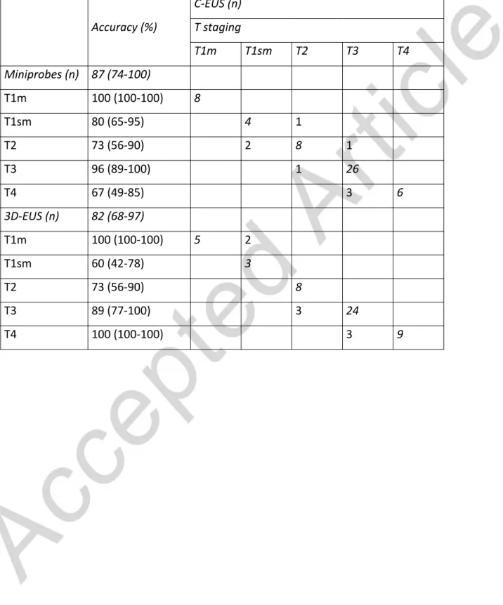
Table 1. Accuracy and agreement for T staging using conventional (C-EUS) as a reference for miniprobes (accuracy = 87%, kappa = 0.81) and 3D-EUS (accuracy = 82%, kappa = 0.87). Total n = 60 Accuracy (%) C-EUS (n) T staging T1m T1sm T2 T3 T4 Miniprobes (n) 87 (74-100) T1m 100 (100-100) 8 T1sm 80 (65-95) 4 1 T2 73 (56-90) 2 8 1 T3 96 (89-100) 1 26 T4 67 (49-85) 3 6 3D-EUS (n) 82 (68-97) T1m 100 (100-100) 5 2 T1sm 60 (42-78) 3 T2 73 (56-90) 8 T3 89 (77-100) 3 24 T4 100 (100-100) 3 9
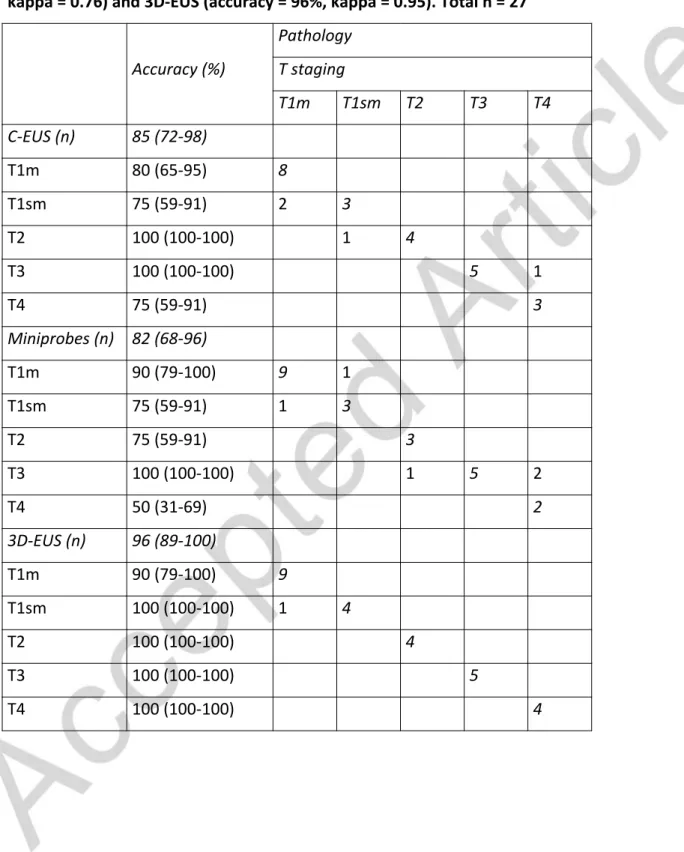
Table 2. Accuracy and agreement for T staging using pathology as a reference for conventional EUS (C-EUS) (accuracy = 85%, kappa = 0.81), miniprobes (accuracy = 82%, kappa = 0.76) and 3D-EUS (accuracy = 96%, kappa = 0.95). Total n = 27
Accuracy (%) Pathology T staging T1m T1sm T2 T3 T4 C-EUS (n) 85 (72-98) T1m 80 (65-95) 8 T1sm 75 (59-91) 2 3 T2 100 (100-100) 1 4 T3 100 (100-100) 5 1 T4 75 (59-91) 3 Miniprobes (n) 82 (68-96) T1m 90 (79-100) 9 1 T1sm 75 (59-91) 1 3 T2 75 (59-91) 3 T3 100 (100-100) 1 5 2 T4 50 (31-69) 2 3D-EUS (n) 96 (89-100) T1m 90 (79-100) 9 T1sm 100 (100-100) 1 4 T2 100 (100-100) 4 T3 100 (100-100) 5 T4 100 (100-100) 4
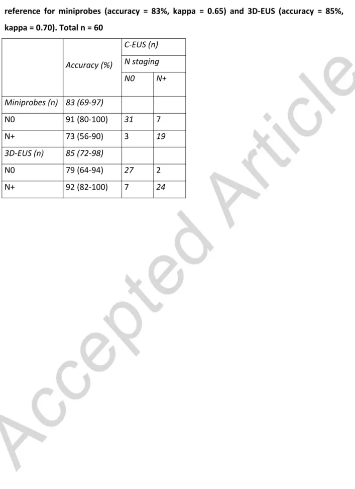
Table 3. Accuracy and agreement for N staging using conventional EUS (C-EUS) as a reference for miniprobes (accuracy = 83%, kappa = 0.65) and 3D-EUS (accuracy = 85%, kappa = 0.70). Total n = 60 Accuracy (%) C-EUS (n) N staging N0 N+ Miniprobes (n) 83 (69-97) N0 91 (80-100) 31 7 N+ 73 (56-90) 3 19 3D-EUS (n) 85 (72-98) N0 79 (64-94) 27 2 N+ 92 (82-100) 7 24
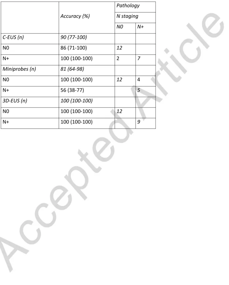
Table 4. Accuracy and agreement for N staging using pathology as a reference for conventional EUS (C-EUS) (accuracy = 90%, kappa = 0.80), miniprobes (accuracy = 81%, kappa = 0.59) and 3D-EUS (accuracy = 100%, kappa=1). Total n = 21
Accuracy (%) Pathology N staging N0 N+ C-EUS (n) 90 (77-100) N0 86 (71-100) 12 N+ 100 (100-100) 2 7 Miniprobes (n) 81 (64-98) N0 100 (100-100) 12 4 N+ 56 (38-77) 5 3D-EUS (n) 100 (100-100) N0 100 (100-100) 12 N+ 100 (100-100) 9
Fig. 1. Stenosing RC. A. Observation by colonoscopy with a miniprobe inserted in the lumen. B. Ultrasonographic image showing the invasion by perirectal fat.
Fig. 2. RC staging by 3D-EUS with invasion of the submucosa but without surpassing it, as confirmed by the surgical sample.