Characterization of the
Exoproteome of Two
Morphologically Distinct
Cyanobacteria
Nuno Manuel de Resende Rego Macedo Martins
Mestrado de Biologia Celular e Molecular
Departamento de Biologia 2013
Orientador
Professora Doutora Paula Tamagnini, Professora Associada da Faculdade
de Ciências do Porto; Líder de grupo em IBMC- Instituto de Biologia Celular
e Molecular, Universidade do Porto
Co orientador
Doutor Paulo Oliveira, Bolseiro de Pós-doutoramento em IBMC- Instituto de
Biologia Celular e Molecular, Universidade do Porto
Dissertação de candidatura ao grau de Mestre em Biologia Celular e Molecular submetida à Faculdade
de Ciências da Universidade do Porto.
O presente trabalho foi desenvolvido sob a orientação científica da Professora Doutora Paula Tamagnini e co-orientação do Doutor Paulo Oliveira e foi realizado no Instituto de Biologia Molecular e celular da Universidade do Porto.
Dissertation for applying to a Master’s Degree in Molecular and Cell Biology, submitted to the Faculty of Sciences of the University of Porto.
The present work was developed under the scientific supervision of Professor Paula Tamagnini and co-supervision of Doctor Paulo Oliveira and was done at the Institute Molecular and Cellular Biology of University of Porto.
Acknowledgements
I would like to express my deepest gratitude to my mentor Dr. Paulo Oliveira. He was a true inspiration and example in lab and taught me so much. He was truly like a Mr. Miyagi to me.
It is with immense gratitude that I acknowledge the support of my supervisor Professor Paula Tamagnini in the writing of the thesis and for opening my eyes for the wonders of the cyanobacterial world, and of course for giving me the opportunity to work in this amazing lab.
I feel compelled to acknowledge Professor Phillip Wright for the opportunity to attend to his department in the University of Sheffield and to use all the mass spectrometry equipment. In the same note I want to thank Dr. Narciso Couto and Dr. Caroline Evans for the immense help with mass spec related issues always with great dedication. And of course I want to thank Dr. Sara Pereira for the help with my protein samples. And I will not forget all the help the Sheffield PhD students gave me, especially Joseph Longworth and David Russo with lab advices; Andrew Landels with all the bioinformatic and of course a special mention to Ben Strutton without whom I would have lived in the streets for a week.
I will never forget the daily brainstorming sessions with Pedro Ferreira which always gave me great ideas which usually allowed me to overcome several setbacks. I also would like to thank everybody in our group for always making me feel welcome and giving me advice everytime it was needed.
Last but not least, I want to give a special thank for my family, they were crucial in my life and in my education, especially my mom and dad who gave me the amazing privilege of education and taught me almost everything I know (maybe not in cyanobacteria per se).
Resumo
Neste trabalho foram investigadas as proteínas extracelulares (exoproteoma) numa cianobactéria unicelular, Synechocystis sp. PCC 6803 e numa filamentosa, Anabaena sp. PCC 7120. A análise foi feita em meios com diferentes fontes de azoto; nitrato e amónia em ambas as espécies e sem nenhuma fonte de azoto combinado apenas em Anabaena sp. PCC 7120, uma vez que Synechocystis sp. PCC 6803 não é capaz de fixar azoto atmosférico.
A identificação de proteínas extracelulares foi feita utilizando uma análise de espectrometria de massa ESI-ion trap. Foram identificadas 117 proteínas extracelulares em Anabaena sp. PCC 7120 e 30 em Synechocystis sp. PCC 6803. Embora, para uma grande parte das proteínas identificadas não seja conhecida a função, foi notório a presença de proteínas relacionadas com o processamento e aquisição de nutrientes do meio. Foram também identificadas proteínas envolvidas na defesa de stresse oxidativo em culturas de Anabaena sp. PCC 7120. Foi, ainda, analisada a atividade destas no meio de cultura.
Para avaliar a contaminação do meio extracelular com proteínas intracelulares, foram gerados mutantes com expressão de GFP direccionada para o periplasma para, posteriormente, verificar a presença da GFP no exoproteoma. Foram também gerados, para ambas as espécies, mutantes sem o poro da membrana extracelular (TolC) da via de secreção do tipo I, posteriormente o padrão de proteínas destes mutantes será comparado com o da respetiva estirpe selvagem.
Uma análise mais detalhada das proteínas identificadas neste trabalho, conjuntamente com uma caracterização das mesmas vai proporcionar um melhor conhecimento das proteínas secretadas para o meio extracelular, por cianobactérias, e quais as suas funções na sobrevivência destas bactérias.
Abstract
In this work it was investigated the extracellular proteins (exoproteome) in a unicellular cyanobacterium, Synechocystis sp. PCC 6803 and a filamentous one, Anabaena sp. PCC 7120. The analysis was done in the presence of different nitrogen sources; nitrate and ammonium in both species and without combined nitrogen only in Anabaena sp. PCC 7120 since
Synechocystis sp. PCC 6803 is incapable of nitrogen fixation.
The identification of the extracellular proteins was done with an ESI-ion trap mass spectrometry analysis. A total of 117 extracellular proteins were identified for Anabaena sp. PCC 7120 and 30 for Synechocystis sp. PCC 6803. Although most of the proteins identified had unknown functions it was notorious the presence of proteins related to the processing and acquisition of nutrients present in the medium. It was also identified proteins involved in the defense of oxidative stress in the Anabaena sp. PCC 7120 cultures. These proteins were further analyzed to confirm if indeed these proteins have activity in the extracellular milieu.
To evaluate the contamination of the extracellular milieu with intracellular proteins, mutants expressing GFP exported to the periplasm were generated to ultimately verify the presence of the GFP in the exoproteome. Deletion mutants for the outer membrane pore (TolC) of the secretion pathway Type I were also generated for both species. In the future these mutants will be used to compare their extracellular proteins pattern against their respective wild type strain.
The follow-up of the proteins identified in this work together with the characterization of the proteins identified will provide a better understanding of the mechanisms of survival of these bacteria in different environments.
Table of contents
Introduction ... 1
Protein Secretion ... 2
State of the art in cyanobacterial exoproteomes ... 8
Objectives ... 9
Material and Methods ... 10
Organisms and Growth Conditions ... 10
Growth Measurements ... 10
Preparing Escherichia coli XL1 blue competent cells ... 10
Escherichia coli transformation ... 11
Synechocystis sp. PCC 6803 transformation ... 11
Triparental mating ... 11
Exoproteome extraction and concentration ... 12
Proteome extraction ... 12
SDS-Polyacrylamide gel electrophoresis (SDS-PAGE) ... 13
Superoxide Dismutase (SOD) and Catalase activity in-gel assay ... 13
2D-Gel Electrophoresis ... 14
In-Gel Trypsin Digestion ... 15
Mass Spectrometry Bioinformatic Data Analysis ... 16
DNA isolation ... 16
DNA electrophoresis... 17
PCR – Polymerase Chain Reaction ... 17
Oligonucleotides Used... 18
DNA Digestion ... 18
DNA Ligation ... 19
DNA Purification and quantification ... 19
Plasmid Preparation ... 19
DNA Sequencing ... 19
Strategy for the generation of periplasmic GFP mutants ... 20
Strategy for the generation of ΔtolC mutants ... 21
Exoproteome Identification ... 23
Superoxide Dismutase and Catalase activities: ... 34
Cyanobacterial periplasmic containing GFP mutants ... 39
ΔTolC mutant generation ... 40
Conclusions ... 42
Future Perspectives ... 43
References ... 45
List of tables and illustrations
Fig 1- Models for type II secretion and type 4 pilus biogenesis………..…………6Fig 2- Models for the Archimedes’ screw…………..………..………….7
Table 1- Oligonucleotides used in this work………..………..18
Fig 3- Scheme of the four constructs cloned in the pRL25C plasmid………..………..20
Fig 4- Scheme of the cloning strategy used for the pSK+ with the flanking regions of slr1270...21
Fig 5- SDS-PAGE separation of the exoproteome samples………..………23
Fig 6- 2D-gel separation of the Anabaena sp. PCC 7120 exoproteome. Anabaena sp. PCC 7120 was grown for 10 days in BG110……….……….…24
Fig 7- 2D-gel separation of the Anabaena sp. PCC 7120 exoproteome. Anabaena sp. PCC 7120 was grown for 6 days in BG11……….………..25
Fig 8- 2D-gel separation of the Anabaena sp. PCC 7120 exoproteome. Anabaena sp. PCC 7120 was grown for 6 days in BG110 supplemented with NH4Cl……….…………..…………25
Fig 9- SDS-PAGE separation of the Anabaena sp. PCC 7120 exoproteome……..…..………..26
Fig 10- SDS-PAGE separation of the Synechocystis sp. PCC 6803 exoproteome….…………27
Table 2- List of proteins identified in Anabaena sp. PCC 7120 exoproteome……….…………28
Table 3- List of proteins identified in Synechocystis sp. PCC 6803 exoproteome….…………32
Fig 11- SOD activity gel……….……….35
Fig 12- SOD activity gel treated with 5mM H2O2 ……….………..36
Fig 13- Catalase activity gel………..……….38
Fig 14- Confocal fluorescent microscopy of the Anabaena sp. PCC 7120 mutants….….……..40
Fig 15- DNA electrophoresis with the results from the colony PCR of the Synechocystis sp. PCC 6803 Δslr1270 mutants………..…..41
General abbreviations
ABC ATP Binding Cassette
APS Ammonium persulphate
ATP Adenosine triphosphate
DAB 3,3'-Diaminobenzidine
DNA Desoxyribonucleic acid
DTT Dithiothreitol
EDTA Ethylenediaminetetraacetic acid
GFP Green Fluorescent Protein
GSP General Secretory Pathway
HSP Horseradish peroxidase
IM Inner membrane
LB Lysogeny broth
LC Liquid Chromatography
MFP Membrane fusion protein
MS Mass Spectrometry
NBT Nitroblue tetrazolium
OM Outer membrane
OMP Outer membrane protein
PAGE Polyacrylamide gel electrophoresis PCC Pasteur Culture Collection
PCR Polymerase chain reaction
pSK+ Plasmid Bluescript SK+
PTM Post Translation Modification
RNA Ribonucleic acid
RND Resistance Nodulation-cell Division
RPM Revolutions per minute
SDS Sodium dodecyl sulphate
SOB Super Optimal Broth
SOD Superoxide Dismutase
SP Signal Peptide
TaT Twin-arginine Transportation TEMED Tetramethylethylenediamine
Introduction
Cyanobacteria are an ancient (over 3 billion years old) and diverse group of prokaryotes. Their ability to perform oxygenic photosynthesis together with their ability to adapt to environmental conditions, contribute to their worldwide distribution. They are extensively studied because of their variety and for their ability to convert solar power into energy and atmospheric carbon dioxide into organic molecules [1].
Within the many species of cyanobacteria, the unicellular Synechocystis sp. PCC 6803 has become a model of study. This strain was isolated from a fresh water lake and deposited in Pasteur culture collection in 1968 [2]. Besides being one of the most well studied cyanobacteria, it has the capacity of natural transformation with exogenous DNA, including its capacity of integration that DNA into the genome by double recombination [3]. Another important mark for its wide use was its genome sequencing in 1996, establishing itself as the first photosynthetic organism with its genome completely sequenced [4, 5]. Although it has several advantages for scientific research it lacks certain characteristics present in other cyanobacteria like multicellularity, nitrogen fixation capacity or cellular differentiation.
Another well studied cyanobacterium, the filamentous Anabaena sp. PCC 7120 (also known as
Nostoc sp. PCC 7120) has also been used because of its ability to fix atmospheric nitrogen in
differentiated cells, the heterocysts. Its genome has also been fully sequenced, and it became available in 2001 [6, 7].
Although both organisms are cyanobacteria, they are different. Besides all the characteristics already mentioned, one of the most important differences between them, relevant for this work, is the morphology of the cell wall. While Synechocystis sp. PCC 6803 is unicellular with a cell wall composed of a plasma membrane and an outer membrane, both limiting the periplasmic space, and a thin peptidoglycan layer between both membranes, Anabaena sp. PCC 7120 is a multicellular strain that has an individual plasma membrane for each cell and a common outer membrane for the whole filament with a dynamic continuous periplasm [8, 9]. Another very important distinction is the presence of a Surface-layer (S-layer) in Synechocystis sp. PCC 6803 and its absence in Anabaena sp. PCC 7120 [10]. The S-layer consists in repetition of structural proteins throughout the cell surface anchored to the outside of the outer membrane and it has several functions such as cell stabilization and protection against environmental factors [11]. Although Anabaena sp. PCC 7120 lacks the S-layer, it has an extra cellular matrix, which
consists of a dynamic mixture of polysaccharides, proteins, cell remnants and secondary metabolites [12, 13].
Protein secretion is an essential mechanism for bacterial survival; it is involved in several aspects of bacterial life such as motility, nutrient acquisition and pathogen secretion. Although there are a lot of proteomic studies for both cyanobacteria mentioned above, including total proteomics [14], periplasm proteomics [15], cell and outer membrane proteomics [16-19] , and even thylakoid membrane proteomics [20], the content of proteins accumulated in the extracellular milieu (Exoproteome) is yet to be fully identified. Only a few studies have been carried out in Synechocystis sp. PCC 6803 trying to explore its exoproteome [21-23], but there are still many proteins left to be identified. In Anabaena sp. PCC 7120 the exoproteome is still to be identified, although it has been partially characterized in a similar filamentous cyanobacterium, Nostoc commune [12, 24, 25]. The significant differences between both species are likely to influence which proteins belong to their respective exoproteomes. Because
Anabaena sp. PCC 7120 is a filamentous strain it is possible to have cell-cell interaction with
proteins travelling through the periplasm without being released to the growth medium [9], which can affect the composition of the exoproteome. In addition, it has been documented that the S-layer proteins present in Synechocystis sp. PCC 6803 are often released to the growth media, thus also influencing the composition of the exoproteome [26].
It is important to establish the clear difference between the exoproteome, the sum of all proteins present in the extracellular space, and the secretome, all the proteins transported from the interior of the cell out into the medium. Thus, it is possible for a protein to be part of the exoproteome but not to be part of the secretome, in the case when it is accumulated in the growth medium by some sort of leakage of the periplasm or cell lysis instead of being actively transported [27]. It is also important to make the distinction between the terms export, which is when a protein is transported through a membrane whether it is the plasma membrane (thus arriving the periplasm) or the outer membrane (reaching the extracellular space), and the term secretion, which is the transport of a protein all the way through the cell wall and into the growth medium [27].
Protein Secretion
Gram-negative bacteria face an extra obstacle when it comes to protein secretion, the outer membrane. While the proteins are usually exported through the inner membrane quite easily [28], they face two new obstacles when they are secreted through the outer membrane. The periplasm does not have ATP or other sources of energy so the secretion mechanisms need to
be self-energized or harness energy from the inner membrane [28]. In addition, proteins acquire tertiary structure in the periplasm, thus making the secretion even more difficult [28].
In Gram-negative bacteria proteins can be secreted in one-step, crossing the inner and outer membranes inside the same protein complex, or it can be in two-steps. In the latter secretion pathway, proteins are first exported to the periplasm through the general secretory pathway (GSP) or Sec pathway and then secreted to the extracellular space [28-32].
There are six branches of protein secretion in Gram-negative bacteria [28]: two of them consist in the secretion of the proteins after the first export into the periplasm, these are considered terminal branches of the GSP in a two-steps secretion process (Sec-dependent) [28-30]. The other four are named Type I to IV. Types I and III are considered full Sec-independent while types II and IV can be Sec-dependent or Sec-independent [28-30].
Protein export to the periplasm
The first step of the Sec-dependent pathways is the export of the proteins to the periplasm, usually done by the Sec system. The Sec system involves a first step of recognition of an N-terminal signal peptide of the protein by a ribonucleotide complex comprising of the Ffh protein (not yet identified in Anabaena sp. PCC 7120, slr1531 in Synechocystis sp. PCC 6803) and the RNA ffs (identified in both species) and by the chaperone SecB protein (not yet identified in either of the cyanobacterial strains used). The protein is then sent to the translocation unit consisting of SecA (alr4851 in Anabaena sp. PCC 7120, sll0616 in Synechocystis sp. PCC 6803), SecY (all4197, sll1814), SecE (all4197, ssl3335), SecG (not yet identified in Anabaena sp. PCC 7120, ssr3307) [4, 6, 33]. After this process proteins may stay in the periplasm or be further secreted to the medium by the secretion terminal branches of the Sec pathway [28, 29]. Although the Sec pathway is the most important pathway involved in secretion, there is also another cytoplasm to periplasm export pathway relevant in cyanobacteria, the twin-arginine pathway (TaT) [9, 34, 35]. This pathway is involved in the export of proteins in a similar way of the Sec pathway, but it recognizes a different N-terminal signal peptide which has two arginine’s together [9, 34].
Autotransporters and single accessory pathway
The autotransporter pathway is a terminal branch of the Sec pathway. These proteins do not require accessory factors to be secreted since they have all the domains necessary to cross the outer membrane in their sequence. To trespass the outer membrane these proteins have C-terminal β-barrel domain that insert themselves in the outer membrane forming a pore structure similar to porins [28, 36]. The rest of the sequence is then secreted inside the pore and can
become anchored in the extracellular side of the outer membrane or be fully released [36]. The sequence of the protein which is secreted inside the β-barrel pore does not seem to be important, since in other works it has been substituted for different proteins which were secreted [37, 38].
Some proteins do not have the β-barrel domain necessary for secretion; they instead use single accessory factors to be secreted. These single accessory factors work in a way similar to the autotransporter, forming a β-barrel pore in the outer membrane and the soon to be secreted protein is translocated inside the pore into the medium [28]. Although both systems are very similar in function the β-barrel of the autotransporters and of the single accessory factors have very low homology [28]. Most of these single accessory factors seem to have homology with protein-translocating porin found in chloroplasts [39]. In Synechocystis sp. PCC 6803 it was identified and confirmed homologies of these protein-translocating porins, the synToc75 (slr1227) were homologous on other Gram-negative species are involved in the secretion of Hemolysin proteins [39]. In Anabaena sp. PCC 7120 there is a homologue of this transporter, the Omp85 (alr2268) [40].
Type I Secretion
The type I secretion system is a one-step, sec-independent pathway. In this secretion system the substrate protein are recognized, usually by their C-terminal signal peptide (~60 aminoacids) by an inner membrane ABC-Transporter; then they travel inside a membrane fusion protein (MFP) without going to the periplasmic space, finally they are secreted by an outer membrane protein (OMP), a protein of the TolC family [35, 41, 42]. Although each of the three parts of the complex are necessary for the secretion to occur it is the ABC-transporter that makes the selection of which proteins are going to be secreted [35]. In the same bacteria there are usually different ABC-transporters and MFP’s; on the other side there is usually only one OMP in each species. Usually these different components can assembly in different combinations depending on the substrate to be secreted [35, 42]. While the ABC-transporter and the MFP are already together upon the substrate recognition, the OMP only assembles after the recognition occurs [42]. The OMP can be assembled in a different complex comprising of a MFP with an RND exporter instead of an ABC-transporter. This RND is a drug export inner membrane protein involved in processes of detoxification. This means that the OMP does not mediate the secretion of only proteins but other molecules too [41].
The ABC-transporters in the type I secretion work both in the recognition of the protein substrate and to energize the secretion process [43]. The structure of the ABC-transporters comprise two transmembrane domains and two nucleotide-binding domains which can be arranged in different
combinations [42]. The nucleotide-binding domains are more highly conserved than the transmembrane domains. This shows that the specificity of the ABC-transporter-substrate is given by the transmembrane domains while the nucleotide-binding domains have ATPase activity necessary to energize the secretion process [41]. Because the ABC-transporters are usually responsible for the recognition of one specific substrate, they are usually coded in gene clusters with their MFP and their substrate [43]. There are exceptions where the ABC-transporters and their MFP are in the same gene cluster but there is no secreted protein nearby. In these cases usually the ABC-transporters are not specific and can secrete more than one type of protein [43]. The ABC-transporters usually recognize Glycine-rich repeats (GGXGXD) present in the last 60 aminoacids, usually repeated 4-36 times. They are often present in toxins like the RTX, Hemolysin and lipases [43].
The MFP complex role in type I secretion is to make a bridge between the ABC-transporter and the OMP. It has an N-terminal fragment in the cytoplasm, a transmembrane segment and a large periplasmic domain which binds to the OMP after the substrate is recognized by the ABC-transporter [41]. In some cases the MFP N-terminal fragment is known to be essential for substrate recognition for Type I secretion [42]. Although at the moment there is no information about other functions of the MFP complex, it is postulated that it might have a more complex role; this evidence is supported by the discovery of MFP complex in Gram-positive bacteria, which do not have outer membrane [35].
The OMP function is act as a pore to secrete the substrate protein transported from the MFP. The best studied OMP is the TolC from Escherichia coli which is the third complement of several Type I secretion trios together with ABC-transporters and MFP’s [41]. Because the TolC is ubiquitous in all Gram-negative bacteria and is important both in protein secretion and drug efflux, it has been well characterized and its structure is well known [44]. The TolC works by alternating from a closed position to an open position when it is triggered by its substrate. To do so the coiled-coils twist like an iris opening, changing its hole aperture from 3.5 Å to 16 to 20 Å [45]. This aperture is then wide enough for proteins in their secondary structure [41]. There is also the unconfirmed hypothesis that the TolC can act as a single accessory factor to secrete proteins of the periplasm independent of the ABC-Transporter-MFP complex [41].
In Anabaena sp. PCC 7120 and Synechocystis sp. PCC 6803 there is only one TolC homologue that acts as OMP for protein secretion, alr2887 and slr1270 [5, 6]. However at least in Synechocystis sp. PCC 6803 there are four confirmed substrates for Type I secretion, this is indication that like in most Gram-negative bacteria there is only one OMP that assembles in different ABC-transporters-MFP complexes [44].
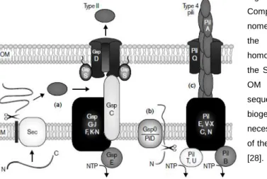
Type II Secretion
Type II secretion also known as main terminal branch of the Sec pathway, is mainly a Sec-dependent, although not completely Sec-dependent, secretion pathway. The type II secretion system is extremely similar to the Type IV pilus biogenesis (also known as type 4 pilus biogenesis); they even share most of the proteins in some bacteria [46, 47]. Type IV pilus biogenesis pathway is also important in the motility and adhesion of bacteria, and in the uptake of DNA and the natural transformation [47, 48]. However while many proteins are identical in the type II secretion and in the type IV pilus biogenesis there are some differences described as shown on Figure 1 [28].
Fig 1- Models for type II secretion and type 4 pilus biogenesis. Components of the type II secreton are indicated using the Gsp nomenclature, and type 4 pilus proteins are labeled according to the Pil system. Similar shading and location indicates homologous components. (a) Type II substrates cross the IM via the Sec system. GspC may transmit energy from the IM to the OM complex. (b) GspO cleaves their amino-terminal leader sequence on the cytoplasmic face of the IM. (c) Type 4 pilus biogenesis requires the OM secretin PilQ, but no energy is necessary for secretion, which is presumably driven by the force of the PilA assembly towards the outer membrane. Adapted from [28].
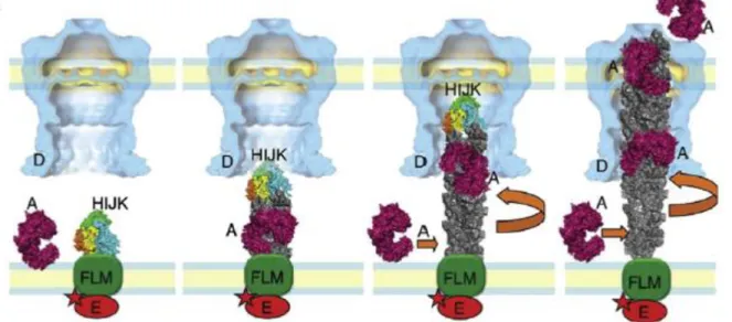
Recently it has been described that both systems can work together. In that model the pilin polypeptide (PilA) is constantly being assembled in the Pil complex of the inner membrane (PilE, PilC, PilN, PilV-X) and grows from the base to the outer membrane and trespasses the outer membrane inside the secretin channel (PilQ) in a rotation motion, which will be responsible for the twitching motility of the cell. At the same time proteins present in the periplasm, exported in the Sec pathway, attach to the PilA subunits and are driven to the medium together with the PilA complex rotation motion. All of these go through the secretin pore in a way similar to the Archimedes’ screw model (Figure 2). That way the protein present in the periplasm is secreted in the Type II secretion system in a dependent way, while the PilA is secreted in a Sec-independent way using the type IV pilus biogenesis [46].
Fig 2- Models for the Archimedes’ screw. The minor pseudopilin complex HIJK binds to the inner membrane complex to initiate the PilA assembly. The PilA (grey) starts to form the complex in a rotation upwards motion into the outer membrane pore. While the PilA is being assembled a periplasmic protein exported by the Sec pathway binds to the PilA and is secreted together with the PilA. Adapted from [46].
In Synechocystis sp. PCC 6803 it is not known the details of the Type II secretion system, whether it occurs like the Archimedes’ screw model or in parallel of the type IV pilus biogenesis using only some of the same proteins. However most of the Pil proteins of the type IV pilus biogenesis have been identified in Synechocystis sp. PCC 6803 with the most important being the sll1694 (PilA, major pillin protein); slr1227 (PilQ; Secretin pore) and the slr1120 (PilD, leader peptidase/N-metilase of the PilA) [49]. However, a mutant secreting lichenase in the Type II pathway was already achieved in Synechocystis sp. PCC 6803 [22]. To do so it was made a fusion of the active lichenase protein with the N-terminal leader peptide of the PilA
Synechocystis sp. PCC 6803 gene (sll1694) [23]. More important, that work discovered that the
particular sequence of the leader peptide was not essential for the secretion but instead the relevant characteristic was the positive global charge of the N-terminal sequence [23]. This indicates that there is type II secretion in Synechocystis sp. PCC 6803 because not only the PilA (part of the Type IV pilus biogenesis and motility) is recognized for secretion. However there is no information that can elucidate whatever this Type II secretion occurs together with the Type IV pilus biogenesis and motility or in a parallel way.
In filamentous Anabaena sp. PCC 7120 there is no pillus therefor there is no Type IV pilus biogenesis. However there are homologues of the Pil main structures such as the inner membrane complex ant the outer membrane porin in the genome of Anabaena sp. PCC 7120 with the exception of the PilA which seems to be absent [6]. This highly suggests that although
there is no pilus formation the type II secretion still exists in Anabaena sp. PCC 7120 (as described in Figure 1 (a)).
Type III and Type IV secretion
Type III secretion consist in the translocation of virulence factors from the Gram-negative bacteria to the eukaryotic host. This virulence factors can be DNA or secreted proteins. The secretion is mediated by a needle-like apparatus connected to the bacteria outer membrane and inner membrane by a transperiplasmic ring and projects itself to the host membrane. The proteins and DNA then travel inside the hollow needle into the host cytoplasm [28, 50]. The Type IV secretion is a mechanism analogue to the infection with plasmid DNA and virulence factors found in Agrobacterium tumefaciens and it is also often used in conjugation techniques mediated by E. coli [51]. The type-III is only Sec-dependent while the Type IV secretion can be either Sec-independent or Sec-dependent [50, 52].
Both secretion systems occur mainly in eukaryotic pathogens, thus they are absent in cyanobacteria [50, 52].
State of the art in cyanobacterial exoproteomes
Although most studies involving extracellular proteins have the intention of studying secretion, it is very hard to identify whether a protein present in the extracellular milieu came from active secretion or periplasmic leakage or even cell lysis. Therefore many studies identify the whole exoproteome. That setback is clear in a study where the objective was to identify the exoproteome of Synechocystis salina [13], in which 9 of the 14 proteins identified (including the ones with highest confidence) were components of the phycobilisome, which is well established to be anchored to the thylakoid membrane [53]. Although the authors did several controls to try to prevent leakage of internal proteins to the media the results show how difficult it is to fully prevent contamination with intracellular content [13]. It is expected the same could happen in
Synechocystis sp. PCC 6803 or in Anabaena sp. PCC 7120.
At the moment there are no studies about Anabaena sp. PCC 7120 exoproteome and only one study was done in Synechocystis sp. PCC 6803 [21]. This study only analyzed a fraction of the exoproteome with N-terminal sequencing techniques which leaves most of the extracellular proteins yet to be revealed. In that particular study only 7 proteins were identified from which only two had known function; the pilin polypeptide PilA1 (Sll1694) and the Slr0924 which corresponds to a plant chloroplast protein Tic22 that is involved in protein import into thylakoid membranes [54]. Nevertheless, there are at least four proteins like the Hemolysin sll1951
confirmed to be secreted by the Type I secretion method that were not identified in that study [41, 55, 56]. This indicates that the exoproteome of both species are yet to be fully unveiled.
Objectives
The first goal of this work was to identify the exoproteomes of a filamentous and a unicellular cyanobacteria, Anabaena sp. PCC 7120 and Synechocystis sp. PCC 6803 grown with different nitrogen sources. Anabaena sp. PCC 7120 was grown with atmospheric nitrogen, nitrate and ammonia whereas Synechocystis sp. PCC 6803 was grown only with nitrate and ammonia, since this strain cannot fix atmospheric nitrogen.
To assess the possibility of periplasmic leakage, mutants with periplasmic GFP were generated, and the presence of the GFP in the media will be evaluated in the future.
Once the identification of the exoproteome, which contained the secretome, was performed it is important to understand which set of proteins constitute the secretome. To identify which proteins are secreted by type I secretion mechanism, deletion mutants for the outer membrane pore (TolC) were generated for both cyanobacteria.
Material and Methods
Organisms and Growth Conditions
Synechocystis sp. PCC 6803 and Anabaena sp. PCC 7120, as well as their respective mutants
were grown in liquid medium at 25⁰C, with constant aeration and a regime of 16h light (40 µmol photons m-2 s-1) /8h dark. Cyanobacteria were also grown in agar plates, at the same temperature and light conditions. They were grown in variations of BG110 [57] with different nitrogen sources. For both species it was used BG11 (BG110+1.5g/L NaNO3) and BG110+NH4Cl and BG110 only for the Anabaena sp. PCC 7120; due to the incapacity of Synechocystis sp. PCC6803 to fix atmospheric nitrogen. All the cyanobacterial mediums were supplemented with 10mM HEPES buffer.
Escherichia coli was grown in LB at 37⁰C, either in an orbital shaker at 180RPM for liquid
cultures or on plates.
To make solid medium 1.5% Bacteriological Agar or 1.5% Difco® Agar Noble was added to LB medium or to BG11, respectively.
Growth Measurements
The growth of the cyanobacteria culture was monitored by measuring the Chlorophyll a content. For that purpose, a sample of the culture was centrifuged, and the pellet resuspended in 100% methanol. The suspension was then left at 4 ⁰C overnight, before being centrifuged and the supernatant’s absorbance measured at 665nm in a Shimadzu® UVmini-1240 spectrophotometer. An extinction coefficient of 13.43 was used to calculate Chlorophyll a concentration, as described in [58].
The growth of Synechocystis sp. PCC 6803 was also monitored by optical density (OD) at 750nm, in a Shimadzu® UVmini-1240 spectrophotometer.
Preparing Escherichia coli XL1 blue competent cells
To prepare competent cells it was used a variation of the Hanahan protocol [59]. A seed stock of
Escherichia coli XL1 blue previously prepared competent cells were used. That stock was grown
in 1000mL Erlenmeyer flasks containing 250mL of SOB media at 37⁰C to an OD600nm of 0.5. The cells were then centrifuged at 3875g and 4ºC for 10 minutes. The supernatant was then discarded and the pellet was resuspended in 80mL of ice cold CCMB80 buffer [60] and incubated on ice for 20 minutes. Cells were centrifuged again in the same conditions as described above. Once again the supernatant was discarded and the pellet resuspended in
10mL of ice cold CCMB80 buffer. The OD600nm of the cells suspension was measured and adjusted to a final OD of 2. The cells were then transferred to Eppendorf tubes as 100µL aliquots and stored at -80⁰C.
Escherichia coli transformation
E. coli was transformed using a method based on a procedure described by Hanahan [59]. To
make the transformation, 10µL of a given ligation was added to a 100µL aliquot of E. coli cells previously thawed on ice and the mixture was incubated there for 30 minutes. Then the cells were transferred to a 42⁰C water-bath for 30 seconds, after which they were quickly transferred onto ice for 2 minutes. Once the incubation on ice was over, warm LB medium was added to the cells, which were left to recover in an orbital shaker at 37⁰C for 2h. Finally, cells were plated in LB plates supplemented with the appropriate antibiotic.
Synechocystis sp. PCC 6803 transformation
Synechocystis was transformed using a method based on a procedure described by Williams
[61]. Synechocystis sp. PCC 6803 cells were grown in BG11 up to an OD750nm of 0.55. Then 50mL of culture were harvested and centrifuged at 3875g for 10 minutes. The pellet was resuspended in fresh BG11 to a final OD750nm of 2.60. 500µL of Synechocystis sp. PCC 6803 suspension was transferred to 1.5mL Eppendorf tubes and incubated with 10µg of plasmid in standard growth conditions for 6h. Every 30 minutes the tubes were gently inverted to obtain a homogeneous distribution of cells and DNA in the transformation process. In the end of the incubation step, cells were plated on BG11 plates without antibiotic, which were left overnight in normal growth conditions. The following day cells were washed out of the plate with 1mL of liquid BG11. The final cell suspension was then plated in fresh BG11 plates with the appropriate antibiotic. They were plated in three dilutions with 10x dilution factor.
Triparental mating
Some cyanobacteria as it is the case of Anabaena sp. PCC 7120 are not naturally transformable. To circumvent this difficulty, plasmids harboring constructs of interest can be transferred into Anabaena sp. PCC 7120 by conjugation, making use of a well-established triparental mating technique [62]. To do so the E. coli strains DH5α harboring the plasmid of interest in addition to the helper plasmid pRL623, and the E. coli HB101, harboring the conjugative plasmid pRL443, were grown overnight in LB medium supplemented with the appropriate antibiotics.
The following day, cultures of both strains were inoculated in 25mL of fresh LB medium supplemented with the appropriate antibiotics to an initial OD600nm of 0.1. The cultures were then grown in an orbital shaker at 37ºC for 4h. At the end of this period, cultures were centrifuged at 2100g for 10 minutes and washed twice with LB medium. After the last wash, E.
coli cells were centrifuged and the pellet of the HB101 cells was resuspended in 1mL of LB
medium. The HB101 cell suspension was then used to resuspend the DH5α cells, resulting in a cell suspension containing both E. coli strains. One additional washing step was carried out and the pellet containing HB101 and DH5α was finally resuspended in 300µL of LB medium. The cell suspension was left resting quietly in a tube rack for approximately 1hour, promoting appropriate conditions for the first conjugation event between the two E. coli strains, while the cyanobacterial strain was being prepared for the subsequent steps of the mating. 50mL of the cyanobacterial strain were harvested in mid exponential phase by centrifugation for 10 minutes at 3875g. Then the cyanobacterial pellet was washed and resuspended in the least volume possible of BG11 medium. The cyanobacterial cell suspension was mixed with both E. coli strains and
mating was let to occur for another hour. After that the mixture, now containing all three strains, was plated on BG11 plates supplemented with 5% LB medium and plates were placed under ordinary cyanobacterial growth conditions.
Approximately 30h later plates were washed with 1mL of BG11. The cells washed out from the plate were then transferred and plated onto fresh BG11 plates supplemented with 25µg/mL Kanamycin (for the Synechocystis sp. PCC 6803 conjugation) or 40µg/mL Neomycin (for the
Anabaena sp. PCC 7120 conjugation), in a dilution series of 1%, 10% and undiluted.
Exoproteome extraction and concentration
To analyze the exoproteome, 200mL of each culture were harvested by day 6 of growth, except for Anabaena sp. PCC 7120 grown without combined nitrogen which was taken at day 14 due to its slower growth rate in BG110. The samples were then centrifuged to separate cells from the respective medium. The supernatant containing the exoproteome was then filtered through 0.2 µm pore size filters (Whatman®) to further remove any cell contaminant. The proteins present in the filtered supernatant were then concentrated with Amicon Ultra 15 mL Centrifugal Filter (Millipore®) with 3kDa Molecular Weight Cut-Off by centrifugation at 4000g. Concentrated exoproteome samples were either used immediately for further analysis or saved at -20ºC.
Proteome extraction
To obtain cell-free extracts cyanobacterial cells were grown in previous described conditions. Then 50mL of culture was centrifuged for 10 minutes at 3875g and 4ºC. The pellet was
resuspended with 2mL of Protein extraction buffer [10 mM HEPES, 0.5% triton X-100, 10mM EDTA, 2 mM DTT, pH 8.0, supplemented with protease inhibitor cocktail (Roche Diagnostics GmbH)], followed by a centrifugation step in a refrigerated microcentrifuge for 5min at 11000g and 4ºC. After this washing step, the pellet was finally resuspended in 1.5mL of the same buffer. Cells were disrupted by sonication (Branson Sonifier 250) with cycles of 30 seconds sonication followed by incubation on ice for 2min; a total of 5 cycles were performed for each sample. Each cycle had a duty cycle of 50%. Cell debris and unbroken cells were separated from the extracts by centrifugation 10min at 11000g at 4ºC. Cell free extracts were either used immediately for analysis or stored at -20ºC.
The determination of the protein content was performed using in-plate BCA™ Protein Assay (Pierce), using bovine serum albumin (BSA) as standard.
SDS-Polyacrylamide gel electrophoresis (SDS-PAGE)
Polyacrylamide gels were made accordingly to [63] with either 10 or 12% (w/v) acrylamide/Bis-acrylamide (29:1) concentration in the separating gel (Bio-Rad; 1.5mm thick).
Samples for electrophoresis were prepared by mixing the respective sample with loading buffer in a 4:1 proportion. Then samples were heated at 95ºC for 5min, cooled to room temperature and loaded in the wells, as well as the protein marker Precision Plus Protein™ All Blue Standards, (Bio-Rad®). The electrophoresis was then performed with constant electric current of 20 mA per gel and stopped when the bromophenol blue reached the bottom of the gel.
The gel was then incubated with fixation buffer (40% (v/v) methanol and 7% (v/v) acetic acid) for one hour and later transferred on to a staining solution containing Coomassie blue (Brilliant Blue G - Colloidal Concentrate, Sigma-Aldrich®). Staining was carried out in an orbital shaker (60 RPM) overnight, after which the gel was destained with deionized water with several washes.
After the gel was thoroughly destained it was scanned in a Molecular Imager GS800 calibrated densitometer (Bio-Rad).
Superoxide Dismutase (SOD) and Catalase activity in-gel assay
Native-gel electrophoresis
The gels for SOD and Catalase activity assays were made the same way as described for “SDS-PAGE” gels but without the adition of SDS to any component, including the separating and stacking gel, sample buffer and running buffer. If the gel was meant to assay the SOD activity a
gel containing a final concentration of 10% polyacrylamide was chosen, while catalase activity was assayed in 7.5% polyacrylamide gels.
Samples were prepared by mixing the respective extract with native sample buffer (125mM Tris-HCl pH 6,8; 80% (v/v) glycerol; 0.02% (v/v) bromophenolblue) in a 3:1 proportion. For SOD and catalase activity assays 100µg and 60µg of cell free extracts were loaded, respectively. For the exoproteome samples one tenth of the concentrated extracellular medium, corresponding to a total culture volume of 20mL, was used for both activity gels.
Electrophoresis was carried out with a constant voltage of 100V until the bromophenol blue reached the bottom of the gel.
In-gel Zymography
To assay the catalase activity the gel was incubated for 45 min in a 20mL solution of 50µg/mL of HSP (Horseradish Peroxidase) in 50mM KPi. Then 10.25µL of hydrogen peroxide was added to the incubation mixture and incubated for another 10 min in constant agitation. After that period the gel was washed twice with water and developed in a solution containing 0.5mg/mL of DAB (3,3’-diaminobenzidine tetrahydrochloride) in 50mM KH2PO4 until the gel got a brown coloration. The SOD activity was done accordingly to [64], the gel was incubated in 20mL of a 2.5mM NBT (nitroblue tetrazolium) solution for 20 min in darkness with agitation. Then 30mL of SOD development solution (86µM Riboflavin; 28mM TEMED dissolved in 36mM KH2PO4) [65] was added to the incubation mixture and it was left with agitation and obscurity for another 15min. The gel was finally exposed to the light of a 60W lamp until the bands appeared.
2D-Gel Electrophoresis
The isoelectric focusing step was performed using 7cm pH 3-10 nonlinear strips (ReadyStrip™ IPG strips, Bio-Rad). The strips were first hydrated overnight (at least 16h) with 100µL of hydration buffer (Bio-Rad) with 25µL of each exoproteome sample. To perform the hydration step, strips were placed upside down on top of the previously described mixture, which was lying on a Rehydration tray (GE Healthcare) and then covered with mineral oil.
The isoelectric focusing step was carried out by putting the hydrated strips into an IEF running unit (Bio-Rad). An electric field with a gradient of 0-2000V for a total of 8000Vh and maximum intensity of 50mA per strip was applied.
After the electrofocusing is done, the strips were incubated in hydration buffer supplemented with 10mg/mL of DTT for 15 min, and then they are incubated for another 15 min in hydration buffer supplemented with 25mg/mL iodoacetamide. The strips are then loaded on top of a 10%
SDS-polyacrylamide gel prepared as described above with the exception that the well comb was not used. A mixture of 1% Agarose with 0.02% bromophenol blue was added on top of the strip, immobilizing it. After the Agarose was gelified the gel was transferred to the electrophoresis tank. Electrophoresis settings and gel fixation and staining were performed as previously described in the “SDS-PAGE” section.
In-Gel Trypsin Digestion
The SDS-polyacrylamide gel pieces were cut out of the gel using a clean scalpel with the least amount of gel possible extracting the whole Coomassie stained band. The gel piece was then put on a siliconized tube (LoBind Tube, Eppendorf®) and covered with 200µL of 200mM ammonium bicarbonate in 40% (v/v) acetonitrile and incubated at 37ºC for 30min. The solution was then discarded and the step was repeated once more. Then the gel piece was dried in a vacuum concentrator. To perform the reduction and alkylation steps the dried gel piece was incubated in 200µL of 10mM DTT for 1 hour at 56ºC and then centrifuged at 13000g for 10 seconds. The liquid was discarded. Then 200µL of 55mM Iodoacetamide was added to the gel piece of interest and incubated for 30 min in darkness at room temperature. At the end of this step all liquid was discarded. The gel piece under treatment was washed twice with 200µL of 50mM ammonium bicarbonate and once with 200µL of 50mM ammonium bicarbonate in 40% (v/v) acetonitrile. The gel pieces were centrifuged again for 10 seconds at 13000g and all liquid was discarded, before being dried in a vacuum concentrator.
To digest the peptides present in each gel piece, 0.4µg of Trypsin (Promega) dissolved in 40mM ammonium bicarbonate in 9% (v/v) acetonitrile, was added to the gel pieces and incubated overnight at 37ºC.
The following day the liquid was collected to a new siliconized tube. 20µL of 100% acetonitrile were added to the gel piece and incubated at 37ºC for 15min. Then an additional 50µL of 5% formic acid was added to the gel piece, which was still immersed in 100% acetonitrile, and incubated at 37ºC for another 15min. At the end of the incubation step the supernatant was recovered to the new siliconized tube, finally containing the extracted peptides. These were completely dried in the vacuum dryer and stored at -20ºC until further analysis by the Liquid Chromatographer copulated with tandem Mass Spectrometer (LC-MS-MS).
Mass Spectrometry Readings
The mass spectrometry readings were done in an amaZon ETD (Bruker®) mass spectrometer with electrospray ionization and ion trap mass analyzer (ESI-ion trap) copulated with a liquid chromatographer (LC) UltiMate 3000 rapid separation (Dionex®).
The dried digested peptides from the gel piece were dissolved in 10µL Buffer C (3% Acetonitrile, 0.1% Trifluoroacetic acid). Then they were centrifuged at 17000g for 10 minutes and transferred to the LC vial, from which the LC was programmed to inject 7µL of sample.
The LC was configured to perform a 90 minute binary gradient with increasing concentration of the organic Buffer B (97% Acetonitrile, 0.1% Formic Acid) relative to the aqueous Buffer A (3% Acetonitrile, 0.1% Formic Acid) with a constant pump flow of 0.300µL/min. The gradient started with 0 % buffer B for 5 minutes, followed by a linear ramp from 5 to 40% buffer B for 70min, then a isocratic wash with 90% buffer B for 7 min, and column re-equilibration at 5% B for 8 min. Between every two sample injections, the LC-MS-MS run a 40 minute isocratic wash with 100% Buffer A to act as a blank and purge the system.
The MS ionization source was calibrated for a capillarity voltage of 3600V and an end plate offset of 500V. The trap scan range was between 300 to 1500m/z and 4 precursor ions were automatically chosen for the MS/MS analysis.
Mass Spectrometry Bioinformatic Data Analysis
After the completion of the mass spectrometry analysis data, .mgf files were created using an in-house algorithm. The peptides, present in each sample were then identified using the software EasyProt (developed between the Biomedical Proteomics Research Group of the Human Protein Science department at the University of Geneva, the Swiss Center for Applied Human Toxicology and the Swiss Institute of Bioinformatics), and the UniProt databases, from July 2013, of each organism used. The parameters used allowed for peptide and MS/MS tolerance up to 0.6 Da and two defined missed cleavages. A set of protein level modifications were defined, methyl-thiol of cysteins (+46Da; Fixed) and oxidation of methionine (+16Da; Variable). Data quality filtering was performed allowing only peptide identification with a Z-score > 5 and a minimum length of 6 aminoacid. To determine which proteins were present in the sample it was chosen a false discovery rate of no more than 1%, calculated using a reverse target-decoy [66] a further stringency of a minimum of two peptides identified per protein was applied. For the results shown in this work a further restriction of 50 minimum protein Z-score was applied in order to increase the results accuracy.
DNA isolation
Genomic DNA was extracted according to a previously described method [67]. In summary, 50mL of cell culture was harvested and centrifuged for 10 minutes at 3875g. The cell pellet was washed twice with storage buffer (150mM NaCl; 1mM EDTA; 10mM Tris HCl, pH 8,0). After the
last centrifugation the pellet was resuspended in 250µL of Resuspension Buffer (10mM EDTA; 50mM Tris HCl, pH8,0). To the cell suspension it was added: 350mg of 0.6mm diameter glass beads (Sigma®), 25 µL of 10% SDS and 500 µL of phenol/chloroform 1:1 (pH 7.5). The mixture was thoroughly vortexed for 30 seconds, followed by incubation on ice for another 30 seconds. These 1min cycles were repeated in a total of seven times. In the end the mixture was centrifuged at 11000g for 10 minutes at 4ºC to separate the aqueous and organic phases. The aqueous phase was then transferred to a new Eppendorf tube and mixed with 500µL of chloroform, to perform another extraction as previously described. The DNA present in the aqueous phase was precipitated with one tenth of its volume of 3M sodium acetate (pH 5.2) and 2.5 volumes of 100% ethanol. The mixture was incubated at -20 ⁰C for 30 minutes for the DNA to precipitate, and a centrifugation at 11000g and 4ºC for 30min was carried out to pellet the genomic DNA. The supernatant was discarded and the pellet was washed with 70% ethanol. The dried pellet was dissolved in 50µL of deionized water. DNA concentration was determined using a Nanodrop ND-1000 (Thermo Scientific®).
DNA electrophoresis
DNA electrophoresis was carried out in 0.8% agarose gels containing sodium borate buffer (5mM Na2B4O7·10H2O) and stained with ethidium bromide as described in [68].
PCR – Polymerase Chain Reaction
The PCR’s were carried out with GoTaq® DNA polymerase (Promega) kit, following the manufacturer’s instructions. The final magnesium chloride and dNTP concentrations in each reaction were 2mM and 0.2mM, respectively, while 1u of the enzyme was used. Oligonucleotides were included at a final concentration of 1µM. The PCR reactions were carried out in a thermocycler (Bio-Rad) using the following profile: 2 minutes denaturation step at 95ºC; then 35 cycles of 30 seconds at 95ºC, 30 seconds of annealing temperature (table 1) and then 72ºC for 1 minute for every kb of the target DNA; in the end of the cycles another 7 minutes of extension step at 72ºC.
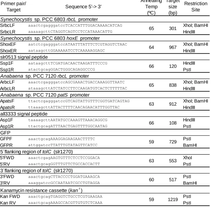
Oligonucleotides Used
Table 1- Oligonucleotides used in this work.DNA Digestion
All DNA digestions performed in this work were made accordingly with the protocol supplied by the manufacturer (Fermentas®). In case of double restriction reactions the protocol followed was
according to the DoubleDigest™ Fermentas® tool
(http://www.thermoscientificbio.com/webtools/doubledigest/). Primer pair/ Target Sequence 5'-> 3' Annealing Temp (ºC) Target size (bp) Restriction Site Synechocystis sp. PCC 6803 rbcL promoter
SrbcLF aaactcgagggatccTCACCATTTGGACAAAACATCAG XhoI; BamHI
SrbcLR aaaaagcttCTAGGTCAGTCCTCCATAAACATTG HindIII
Synechocystis sp. PCC 6803 hoxE promoter
ShoxEF aatctcgagggatccATAATTTATTTCTCGTAGGTCTAAC XhoI; BamHI
ShoxER aataagcttGGAAAAATCCTCAAAAAGGAGC HindIII
slr0513 signal peptide
Ssp1F aataagcttTCGATGACAACTAAGATTTCCCG HindIII
Ssp1R atactgcagGGACTGGGCAGAGGCCCG PstI
Anabaena sp. PCC 7120 rbcL promoter
ArbcLF aaactcgagggatccAGCGAAACTGACCAAAGGTTAATC XhoI; BamHI
ArbcLR ataaagcttATCTATCCTTCCAAGATGTCACTCTTTTTAC HindIII
Anabaena sp. PCC 7120 patS promoter
ApatsF ttactcgagggatccGTCAGTATTGTTTCGGTGATCAGTAG XhoI; BamHI
ApatsR ttaaagcttATTACTTTTCAACAGAACATTTGGTTAC HindIII
all3333 signal peptide
Asp1F taaaagcttAATATGCCAAAGTTAAACAGGCG HindIII
Asp1R ttactgcagATTTAACTGAGTTTTGGCAATAG PstI
GFP
GFPF aaactgcagAAAGGAGAAGAACTTTTC PstI GFPR attggatccTTATTTGTATAGTTCATCC BamHI 5´flanking region of tolC (slr1270)
5'FWD aaactcgagAAGTGTTTCTCCTCCGGACA XhoI
5'RV aaactgcagGGTTTGTTCTGCCACCACTT PstI 3´flanking region of tolC (slr1270)
3'FWD aaactgcagCTTACCCCTGGATGAAAGCA PstI
3'RV aaaggatccGCCAATAATCGCCTGTAGGA BamHI Kanamycin resistance cassette (kanr)
Kan FWD aaactgcagTGAGGTCTGCCTCGTGAAGAA PstI
Kan RV aaactgcagAAAGCCACGTTGTGTCTCAAA PstI
65 301 64 967 66 120 65 838 63 912 66 108 59 729 63 553 60 517 59 1219
DNA Ligation
All DNA ligations were made with 1u T4 DNA ligase (Thermo Scientific®) according to the instructions of the manufacturer (Thermo Scientific®). In every ligation 100ng of vector were used in 20µL reactions while the amount of insert used was 3 times the molar concentration of the vector.
The ligations were performed at 22⁰C for at least 2h, followed by an inactivation step at 65⁰C for 10 minutes before transformation in E. coli.
DNA Purification and quantification
DNA purification from gel or from enzymatic reactions was carried out with a NZYGelpure (Nzytech, Lda.) kit following the manufacturer instructions. DNA concentration was determined using a Nanodrop ND-1000 (Thermo Scientific®)
Plasmid Preparation
Plasmid preparation was done with a GenElute™ plasmid miniprep kit (SIGMA®) following the manufacturer instructions and using 5mL of an overnight grown culture. DNA concentration was determined using a Nanodrop ND-1000 (Thermo Scientific®).
DNA Sequencing
The DNA sequencing was performed by Stab Vida Company.
Confocal Microscopy
Anabaena sp. PCC 7120 harboring pRL25C, a self-replicating plasmid expressing a fusion
protein with the N-terminal signal peptide from the periplasmic nrtA (all3333) together with GFP, under the control of either the patS or the rbcL promoters, were studied using a Leica SP2 AOBS SE laser scanning confocal microscope. Cells grown either in non-nitrogen fixing or nitrogen-fixing conditions were loaded on a 1% low-melting-point agarose bed, dissolved in
BG11 or BG110, respectively, and covered with a clover slip. The GFP emission, collected
between 500 and 540 nm, was observed when cells were exposed to an Ar laser beam of 488 nm, while cyanobacterial autofluorescence, acquired between 640 and 700 nm, was visualized after excitation at 633 nm, using a HeNe laser. Wild-type cells were used to define the basal autofluorescence signal in the GFP channel, and the same acquisition settings were used throughout the various experiments.
Strategy for the generation of periplasmic GFP mutants
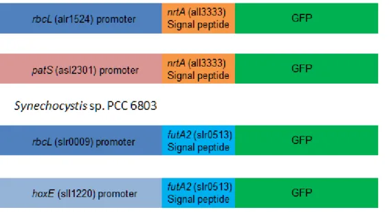
Various pRL25C based plasmids were constructed, containing different well characterized native promoters to drive expression of the signal peptide fused with the GFP encoding gene, and were transferred by conjugation into the corresponding cyanobacterium. For the Anabaena sp. PCC 7120 mutants the promoters chosen were the rbcL (alr1524 – encodes the large subunit of RuBisCO) promoter, which is a strong constitutive promoter [69], and the patS (asl2301) promoter, which is activated in cells that initiate differentiation upon deprivation of fixed nitrogen [70], in addition, the signal peptide chosen was the one from NrtA (all3333), a well described periplasmic protein involved in nitrate import [9]. For the Synechocystis mutants the promoters chosen were the rbcL (slr0009 – encodes the large subunit of RuBisCO) promoter, which is a strong constitutive promoter [69] and the hoxE (sll1220 – encodes one of the subunits of the bidirectional hydrogenase) promoter, which is a weak constitutive promoter; additionally, the signal peptide chosen was from FutA2 (slr0513), which is a periplasmic iron binding protein [71]. In the end two constructs for each strain were obtained as shown in figure 3.
Fig 3 – Scheme of the four constructs cloned in the pRL25C plasmid.
To isolate the Anabaena sp. PCC 7120 fragments rbcL; patS and nrtA signal peptide it was made a PCR using the Anabaena sp. PCC 7120 genome and the oligonucleotides from table 1. To isolate the Synechocystis fragments rbcL; hoxE and futA2 signal peptide it was made a PCR using the Anabaena sp. PCC 7120 genome and the oligonucleotides from table 1. To isolate the
GFP it was made a PCR using as template an in-house plasmid containing GFP gene and the oligonucleotides from table 1.
The constructs were cloned inside a Bluescript SK+ plasmid and transformed in Escherichia coli XL1 blue. The plasmid was then sent to sequencing to verify there were no errors in the PCR amplification. After the confirmation the constructs were excised of the Bluescript SK+ and cloned in the final plasmid (pRL25C) and transformed into E. coli DH5α already with the helper plasmid pRL623. The mutants were then ready to conjugate the pRL25C with the constructs into the appropriate cyanobacteria using the Triparental mating technique.
Strategy for the generation of ΔtolC mutants
The Anabaena sp. PCC 7120 ΔtolC mutant was already being made in our lab, so it was only necessary to make the Synechocystis sp. PCC 6803 one. For that purpose, it was constructed a pSK+ plasmid with a 5´ region of the ORF of the slr1270 gene (region between 122-675bp of the ORF) followed by a Kanamycin resistance cassette and a 3´ region of the same ORF (region between 1020-1503 bp of the ORF). This construct was made with the objective of making double homologous recombination with the gene present in the chromosomes that way disrupting the ORF and preventing the production of TolC.
To make the mutant the ORF regions were amplified with the primer 5´FWD and 5´REV for the 5´fragment and 3´FWD and 3´REV for the 3´Fragment (table 1). The inserts 5´and 3´were cloned inside the plasmid pSK+ as schemed on figure 4.
After the cloning the plasmid was sent to sequencing to verify the correct sequence of the flanking regions of the slr1270.
To make the selection marker, the Kanamycin cassette was amplified using the Kan FWD and the Kan REV primers (table 1). After the amplification it was digested with PstI and cloned between the 5´fragment and the 3´fragment previously cloned in the pSK+. This new plasmid was then transformed in E. coli XL1 blue which was plated on LB with kanamycin. Because only mutants with functional Kanamycin cassette would grow in the plates, there is no need to sequence the cassette. The mutant growth on LB with Kanamycin is proof enough the cassette is working.
The final plasmid inside the E. coli mutants was then extracted and used to transform
Synechocystis sp. PCC 6803. The Synechocystis sp. PCC 6803 mutants were then plated in
BG11 with increasing concentration of Kanamycin to promote selective pressure of the cassette and thus promoting the copy segregation on all the chromosomes.
Results and Discussion
Exoproteome Identification
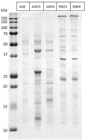
In order to identify the peptides present in the different exoproteome samples the first step is to determine the complexity of the exoproteomes to decide the strategy for the mass spectrometry identification. To have an idea of the protein pattern for each exoproteome sample SDS-polyacrylamide gels were used to separate the various samples
Fig 5- SDS-PAGE separation of the exoproteome samples. First lane is the molecular marker, the lanes two to six are Anabaena sp. PCC 7120 grown in BG110 (AN2); Anabaena sp. PCC 7120 grown in BG11 (ANO3);Anabaena sp. PCC 7120 grown in BG110
supplemented with NH4Cl (ANH4); Synechocystis sp. PCC 6803 grown in BG11 (SNO3) and Synechocystis sp. PCC 6803 grown in
BG110 supplemented with NH4Cl (SNH4) respectively.
The SDS-PAGE results showed that each sample have few bands and thus a small array of different proteins. For that reason it was decided that the strategy for the exoproteome identification was to separate the exoproteome samples on a regular SDS-polyacrylamide gel, followed by cutting out gel bands and finally to proceed with the identification of the peptides present in each band by MS. This approach has the advantage of giving second validation to the MS technique because excised bands contain proteins of a given molecular mass that can be
compared to the theoretical mass of the proteins identified. If the molecular mass is similar, the confidence of that correct protein identification is increased. However, it has the disadvantage of analyzing only fractions of the gel which possess proteins that can be detected by the staining method in use (in this case, Coomassie Blue staining). That can make some of the low abundant or less stained proteins to be left out of the analysis.
It was also noticed that although the band pattern changes noticeably in the different Anabaena sp. PCC 7120 samples grown in different nitrogen source they are similar in both Synechocystis sp. PCC 6803 samples grown with nitrate or ammonia (figure 5). For that reason it was decided that Synechocystis sp. PCC 6803 grown in ammonia would not be chosen for further analysis, to prioritize the resources for other conditions.
To validate this strategy for MS analysis it was important to know how complex the exoproteomes were. If each sample was not enriched in multiple proteins and peptides, simple enough, that most proteins could be separated and resolved only according to their mass in an SDS-polyacrylamide gel, then each band may be cut out, processed and analyzed directly in the LC-MS-MS. Alternatively, if the sample was too complex and each band on an SDS-polyacrylamide gel represented several different proteins, then the sample should be submitted to further separation prior to the injection. To confirm that the samples are in fact of low complexity it is important to know if each band represents only a few different proteins instead of being the sum of many proteins with the same mass. 2D-gel electrophoresis was performed for all exoproteome samples.
Fig 6- 2D-gel separation of the Anabaena sp. PCC 7120 exoproteome. Anabaena sp. PCC 7120 was grown for 10 days in BG110. The isoelectric focusing strips have a
pH range of 3 (left) to 10 (right) non-linear, and the gel has 10% polyacrylamide.
Fig 7- 2D-gel separation of the Anabaena sp. PCC 7120 exoproteome. Anabaena sp. PCC 7120 was grown for 6 days in BG11. The isoelectric focusing strips have a pH range of 3 (left) to 10 (right) non-linear, and the gel has 10% polyacrylamide.
Fig 8- 2D-gel separation of the Anabaena sp. PCC 7120 exoproteome. Anabaena sp. PCC 7120 was grown for 6 days in BG110 supplemented with NH4Cl. The isoelectric
focusing strips have a pH range of 3 (left) to 10 (right) non-linear, and the gel has 10% polyacrylamide.
After the 2D-Gel analysis (figures 6-8) was completed, it was possible to conclude that all exoproteome samples of Anabaena sp. PCC 7120 are of relative low complexity. The same was concluded for the Synechocystis sp. PCC 6803 samples (data not shown). In fact, each band detected on the SDS-polyacrylamide gels (see below) is composed either by a single peptide or, in the worst case, by a relatively low number of peptides (see figures 6-8). Therefore, it was decided that all the exoproteome samples should be analyzed in the LC-MS-MS after a simple SDS-polyacrylamide gel electrophoresis separation, after which bands were excised and processed for identification.
To separate the proteins for MS analysis the samples were separated on a new SDS-PAGE this time loading the maximum amount of protein possible in each well. The most visible bands of each condition, with the exception of the SNH4 for reasons previously referred, were then chosen for analysis.
Fig 9- SDS-PAGE separation of the Anabaena sp. PCC 7120 exoproteome. The gel on left is the exoproteome of Anabaena sp. PCC 7120 grown 14 days in BG110 (AN2). The gel on the middle is the exoproteome of Anabaena sp. PCC 7120 grown 6 days in
BG11 (ANO3). The gel on the right is the exoproteome of Anabaena sp. PCC 7120 grown 6 days in BG110 supplemented with NH4Cl
(ANH4). First lane of each gel is the molecular marker with the corresponding weights marked in kDa, on the left. Second lane is the exoproteome with the red rectangles pointing to the gel locations where the gel was cut for further process and MS identification. Inside the rectangles it was written the code of each fraction.