TOXICITY ASSESSMENT AND CHEMICAL PROPERTIES OF DOLOMITE FOR USE IN DENTISTRY
Gisele Fernandes DIAS1
Fabiana Bucholdz Teixeira ALVES1
André Vitor Chaves de ANDRADE2
Aline Mara FERREIRA3
Fábio André dos SANTOS1
ABSTRACT
The dolomite (DMT) can affect the metabolism of calcium and hydroxyl ions mineralization. To evalu-ate the toxicity, chemical properties and release of calcium and magnesium ions about 4 samples of DMT: Bioficina® - DMT I, Flora Pinhais® - DMT II, Dolomitex® - DMT III and Gran-White - DMT IV and hy-droxide calcium PA (Biodinâmica® - HCA). Through bioassay Artemia Salina, hydrogen potential (pH), atomic absorption, X-ray diffraction (XRD) and X-ray fluorescence (XRF) of 4 samples of DMT was verified that the samples are suitable for potential use in dental materials. XRD and XRF techniques allowed to characterize the spatial conformation of the unit cell of dolomite, crystalline phases, mass percentage of chemical elements present in the samples. The presence of crystalline phases in addi-tion to DMT has been identified as quartz and calcite. Impurities were detected in small amounts (Fe, K, Sr, Tm, S, Cu) and HCa to expectations about 100% portlandite. The pH was measured at concen-trations of 1000 µg/mL;750 µg/mL;500 µg/mL;250 µg/ml and 100 ug/ml of the diluted crude extract of the sample at initial (0) and the periods of 24 and 168 hours wich were characterized in alkalinity pattern. In the interpretation of XRD and XRF tests have been detected the presence of silica, calcite and impurities besides the pure DMT in trace amounts in 2 samples while HCa to expectations of ap-proximately 100% portlandite. To determine the toxicity was used the alternative method of lethal
concentration 50 (CL50) with the bioassay model Artemia Salina. It resulted in low level of toxicity of
DMTs with insignificant difference between times 24 and 48 hours. There was a release of 100ppm calcium and 32 ppm magnesium ions. None of the samples showed a significant percentage of other constituents considered harmful to health. It could be concluded that DMT is non-toxic, alkaline pH,
1 Professor, Department of Restorative Dentistry, School of Dentistry, Ponta Grossa State University, School of Dentistry, ,Ponta Grossa, Paraná, Brazil
considerable release of calcium ions, with crystalline phase that is characterized as a potential dental use.
KEY WORDS: CHEMICAL PROPERTIES. TOXICITY. PH. IONS. CALCIFICATION. MINERALS. INORGANIC
PAR-TICLES.
AVALIAÇÃO DA TOXICIDADE E PROPRIEDADES QUÍMICAS DA DOLOMITA PARA USO
NA ODONTOLOGIA
RESUMO
A dolomita (DMT) afeta o metabolismo da mineralização por dissociação em íons cálcio e hidroxila. Avaliar a toxicidade, propriedades químicas e liberação de íons cálcio e magnésio de 4 amostras comerciais: Bio-ficina® - DMT I, Flora Pinhais® - DMT II, Dolomitex® - DMT III e Gran-White–DMT IV e do hidróxido de cálcio PA(Biodinâmica® - HCa). Com bioensaio Artemia Salina, potencial Hidrogeniônico(pH), absorção atômica, difração de raios X(DRX) e fluorescência de raios X(FRX) foi verificada a adequação do uso potencial para materiais odontológicos. DRX e FRX caracterizaram as fases cristalinas e percentual em massa de elemen-tos químicos. Foi identificada a presença de fases cristalinas além da DMT, como quartzo e calcita. Impu-rezas foram detectadas e o HCa correspondeu às expectativas de aproximadamente 100% portlandita. O pH foi aferido em concentrações de 1000µg/mL;750µg/mL;500µg/mL;250 µg/mL e 100µg/mL do extrato bruto diluído das amostras nos tempos inicial (0) e 24 e 168horas. Sendo caracterizados no padrão de al-calinidade, DRX e FRX detectaram a presença de sílica, calcita e impurezas em quantidades mínimas em 2 das amostras e o HCa correspondeu às expectativas de aproximadamente 100% de portlandita. A toxici-dade resultou em baixo índice com diferença insignificante entre os tempos 24 e 48h. Em média, ocorreu a liberação de íons: 100ppm de cálcio e 32ppm de magnésio. Nenhuma das amostras apresentou porcenta-gem significativa de constituintes considerados prejudiciais à saúde. Pôde-se concluir que a DMT é atóxica, pH alcalino, considerável liberação de íons cálcio, de fase cristalina que a caracteriza como potencial uso odontológico.
PALAVRAS-CHAVE: PROPRIEDADES QUÍMICAS. TOXICIDADE. PH. ÍONS. CALCIFICAÇÃO. MINERAIS.
PARTÍCULAS INORGÂNICAS
INTRODUCTION
Minerals are named according to the geographic location where they occur, however, the dolomite (DMT) was named by the French geologist Deodat Dolomieu (1750-1801) one of the first students of this mineral in the Alpine area in northern Italy, now known as Alps Dolomitas1. DMT known as gran-white is
a hexagonal crystal composed of double calcium and magnesium carbonate (CaMg (CO3)2) wich contains: CaCO3 (calcium carbonate) 54.35%, MgCO3 (magnesium carbonate) 45.65 %, CaO (calcium oxide) 30.4%, MgO (magnesium oxide) 21.9% and CO2 (carbon dioxide) 47.7%2.
The origin of the DMT is a major geological puzzle, being generally of secondary origin formed from limestone by replacing part of Ca by Mg3 which can be found in lakes and caves in various locations
worldwide. The extensive review by Müller et al.4 (1972) for the formation of Ca-Mg carbonates it became
clear that the aquatic chemistry for the formation of carbonates mainly Mg/Ca-Mg/Ca, which plays an important role in the mineralogy of the precipitated carbonates from marine waters and non-marine.
DMT is important raw material easily crushed to a fine powder used as agricultural limestone by farmers to reduce the acidity of the soil and also to adjust the deficiency Mg5. Moreover, it can be used in
cosmetics, facial creams and cream dental baby6. It can be used as a Ca supplement and Mg7. Although, its
clinical effectiveness is not fully proven. It is empirically known for its alkalizing action, anti-inflammatory, analgesic, decongestant, toning, relaxing and revitalizing muscular7. Some studies with DMT and some
toxicity inducing greater cytotoxicity in human cells lung8; antiviral activity compared to the avian influenza
H5N39; mineral food supplement used as an aid in the prevention and treatment of osteoporosis in effect
of increasing the bone mass10. Cordeiro e Moreira11 (2004) investigated the effect of DMT in bone cavities
surgically created healing process and is described as a repairing material for its rich chemical composition of Ca carbonate and Mg constituents from the inorganic bone matrix which may suggest stimulating action osteogenesis. Concomitantly, the supposed anti-inflammatory and analgesic activity attributed to the mineral could play a supporting role in the bone repair process. Yamamoto et al.12 (2010) tested the
antibacterial activity of DMT in dentifrices to Gram-positive bacteria and Gram-negative and concluded that the obtained compound had functions dental abrasive and antibacterial agent can contribute to oral hygiene.
DMT is formed from calcium oxide, which when in contact with water is converted to calcium hydroxide and this process is responsible for raising the pH of dissociation in calcium ions and hidroxila13. The release
capacity of the ions is of clinical relevance since it is related to the ability to induce repair proven in other dental materials such as MTA and calcium hydroxide. Another major factor is that studies have revealed significant amounts of Mg in the composition of the dentin, enamel, and cementum14 being also present in
the DMT. What may suggest affect mineralization metabolism for possible effects on crystallisation process the mineral inorganic phase15.
Because of the composition of DMT described in the literature evidenced by high Ca and Mg carbonate content, the study aimed to evaluate the toxicity and chemical properties for the adequacy of DMT for use in the composition of dental materials.
MATERIAL AND METHOD
This research was approved by the Ethics in Research Committee, protocol #1.565.693.
Sample
The sample consisted of following samples dolomite (DMT) and calcium hydroxide (HCA): Bioficina® (DMT I), Plant Pinhais® (DMT II) Dolomitex® (DMT III) and Gran-White (DMT IV) and calcium hydroxide PA Biodinâmica® (HCA).
Study Design
The toxicity hydrogen potential (pH), release of calcium and magnesium ions, X-ray diffraction (XRD) and X-ray fluorescence (XRF) were studied in four commercial samples of DMT and HCa.
Toxicity
Bioassay of Artemia Salina for determining the toxicity is an alternative method of estimating the lethal concentration 50 (CL50) in Artemia salina model based on the technique previously described16. In
The temperature was maintained between 28 to 30°C; pH between 8 and 9 and artificial lighting. Oxygen saturation state was achieved with the aid of an oxygen pump. After 48h, the larvae reached metanauplii the microcrustacean which is more sensitive to treatment. The larvae were not fed. The bioassay Artemia
salina is to distribute 10 larvae (n = 10) in each test tube with 5ml of saline in graded concentrations of 25 ug
/ ml, 50ug / ml, 100ug / ml, 150 ug / ml, 250 ug / ml, 500ug / ml, 750 ug / ml and 1000 ug / ml of each sample. Larvae of Artemia salina were incubated in different concentrations of crude extract dissolved during 24 hours under artificial light and controlled temperature. After this, 10 larvae were transferred to test tubes containing saline (negative control), potassium dichromate 0.1% (positive control) and dolomite in different concentrations. The result was obtained by counting after 24 and 48h the number of metanauplius dead and alive in each test tube by macroscopic visualization with magnifying glass and descriptive statistical analysis was done by counting the number of survivors and the percentage of dead. Microcrustaceans were considered dead when they showed no movement after 10 seconds of observation16. To calculate toxicity
was determined CL50, toxic agent concentration that causes 50% mortality of organisms exposed within 24 or 48 hours. The estimate was performed by regression analysis Probit, which is suitable for data sets in which the dependent variable is measured in units of type Yes or No. In this case the answer is represented by the percentage of survivors, births, etc. The test was performed in duplicate.
Analysis hydrogenionic potential (pH)
The analysis was made by dispersing a commercial sample of DMT and HCa in 1 ml of distilled water. Five different concentrations for each sample were analyzed: 100µg/mL, 250µg/mL, 500 µg/mL, 750 µg/mL and 1000 µg/mL. Initially the pH was measured as distilled water. Then the pH of the prepared dispersions were measured after contact with the scanning electrode (pH initial, 0 h) and at times 24h and 168h. During these periods samples were stored in at 37 ° C. For analysis of pH analysis was used ANOVA 1 factor with post Tukey test. It adopted a significance level of p <5% and used the GraphPad Prism 5.0 software.
Release of calcium and magnesium ions
It was used the official method of atomic absorption, which is based on solubilization of sources of Ca that are present in the sample with concentrated acid to after determining the concentration of Ca element by this method. First, 0,500g to each DMT was transferred to beakers of 100mL. After added 20mL of hydrochloric acid 1 + 1 and boiled for 10 minutes, then transferred to 100 mL volumetric flasks and completed to volume with distilled water. An alíquota of the extract of DMTs was used and transferred to balloons of 50mL. And then 5L of lanthanum oxide was added to 5% and completed to volume with water. Lanthanum was added for its ability to eliminate interference from other ions. After preparation of the sample, atomic absorption spectrometer with flame was placed under the conditions required for the determination of Ca as the operating manual. The device was calibrated and immediate reading was held. The bulk is the standard that is used to make the curve of the equipment, so obtained a Ca and Mg reference value, respectively 10,25ppm and 2, 04 ppm.
Structural characterization
The crystal structure of DMT samples and commercial HCa were analyzed by X-ray diffraction (XRD). Data were collected on a Shimadzu XRD 6000 diffractometer operating with copper tube at 40 kV and 30 mA. Divergence and scattering slits equal to 1 and slit reception equal to 0.15 mm were used. The kβ radiation was eliminated by use of graphite monochromator. The scan was performed in the range of 3 to 120th in continuous scan mode, the ratio of 1.00 / min. The crystalline phases were identified by
comparing the diffraction pattern observed with padrõss present in the crystallographic information files (crystallographicinformation file - cif) available on Structures Database Crystalline Inorganic (Inorganic Crystal StructureDatabase - ICSD)17, 18. In this work we used the structural models of ICSD # 10404 chips to
DMT (calcium carbonate and magnesium, CaMg (CO3) 2), ICSD # 1011159 for quartz (silicon dioxide, SiO2), ICSD # 18166 for calcite (calcium carbonate, CaCO3) and ICSD # 1,001,768 to portlandite (Ca(OH)2). We used the pseudo-Voigt profile function of Thompson and Hastings19 (1987), for adjustment of the diffraction peaks.
The phenomenological model of Janez1 (2001), incorporated in the profile function, allowed the adjustment
of anisotropic broadening of diffraction peaks. For quantitative phase analysis model was adopted Howard and Hill20 (1987). The quality of refinement was monitored by visual examination of the Rietveld plot, it is
possible to observe the profile, calculated and the difference between the two curves; and the statistical indicators RWP (Rietveld profile index), Chi2 (chi-square) reduced, which are related to the adjustment by the diffraction profile and RF2 and the quality of the adjustment of the crystal structure (atomic positions, the unit cell parameters, parameters of atomic displacement and occupation fator).
X-ray fluorescence (XRF)
It was used to test the fluorescence spectrophotometer X-ray energy dispersive Shimadzu EDX-720, with collimator 10mm, energy band Na-U. The device voltage ranges from 5 to 50kV and the tube current between 1 and 1000 mA. The detector system is a Si (Li) semiconductor cooled with liquid N -196 °C. The time for each sample was 100s, 15 kV and 100s in the Ti-U power strip with 50 kV. The measurements were conducted under 30 Pa pressure. Under the initial conditions that must be vacuum atmosphere and the sample in powder form, the samples were placed in the sample holder of the instrument which already indicates the amount of material required for testing , since only the material surface is analyzed. This sample holder is in a polyethylene film for sample protection. The results were evaluated with the XCOM software which is a code developed to calculate X-ray attenuation coefficients and gamma crosssections interaction for pure elements (Z = 1-100), the compound at a range of wide energy (1 keV -100 GeV)21.
RESULTS
Test toxicity
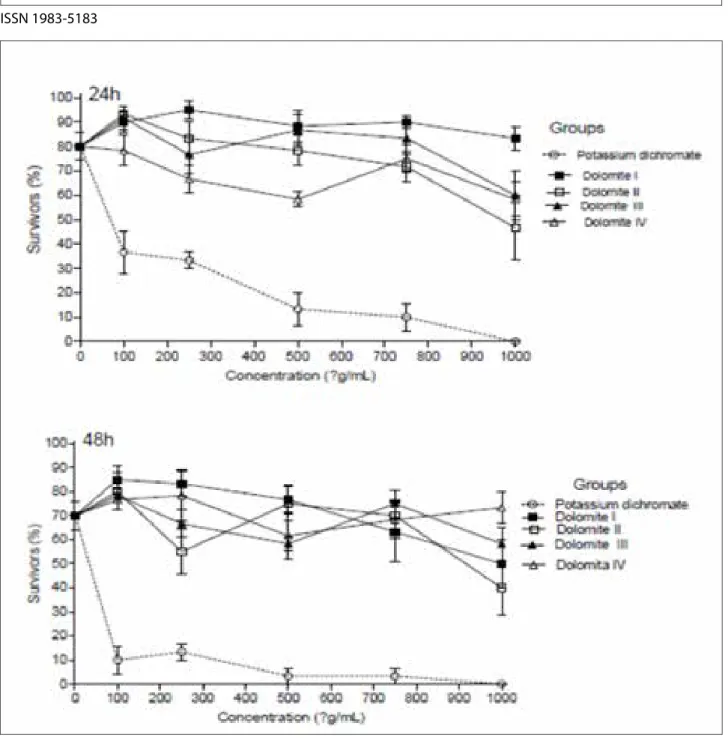
The 4 commercial samples of DMT were not considered toxic. After 24h, as can be seen in Figure 1, the positive control which in this case is potassium dichromate presented distant values studied DMTs with high toxicity 38,12 μg/mL, while the smallest value among them was DMT II with 1243,80 μg / mL. The analysis of 48h showed no significant differences. We can observe the standard error for each concentration, both in reviews of 24 hours and in 48 hours. According to Meyer et al.15 (1982) which established a relationship
between the degree of toxicity and the median lethal dose (CL50) by plant extracts on larvae of A. salina since it is considered that when the above values are checked 1000μg / mL these are considered non-toxic. Thus, they can not be classified as toxic thus presenting no damage to the body.
Figure 1 - * Lethal Concentration 50 (LC50) determined in each evaluation period with the regression analysis Probit - Finney method (log-normal distribution). 24h: Potassium dichromate (positive control): LC50 = 38,12μg / ml; DMT I: LC50 = 3438.48 x106 / mL; DMT II: LC
50 = 1243,80μg / mL;
DMT III: LC50 = 2727,42μg / mL; DMT IV: LC50 = 24.34 x10⁶ / mL. 48h: Potassium dichromate (positive control): LC
50 = 1,52μg / mL; DMT I: LC50 =
1544.55 ug / mL; DMT II: LC50 = 1710,90μg / mL; DMT III: LC50 = 9762.32 ug / ml; DMT IV: LC50 = 1,57 x10⁶ /
Analysis hydrogenionic potential (pH)
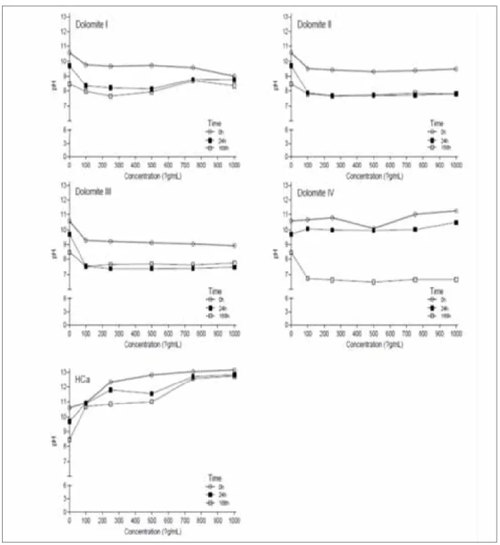
Figure 2 shows the pH values obtained for samples in different concentrations studied. In all cases the pH values of the dispersions are associated with basic environments. In this sense, for the DMT I, DMT II and DMT III samples the pH was between about 8:10, significant differences were observed between the concentrations studied. For IV DMT sample was increased to pH0h time and a fall in the pH to values close to 7 for pH168h but not statistically significant. The highest pH values were observed for HCa sample (especially in concentrations I, II and III) with values close to 12. The higher concentrations of calcium hydroxide (levels
I, II and III) were significantly different from the pH of the distilled water and all four different concentrations of DMT in the three different times. No difference between the DMTs and between their concentrations. In DMT IV T there was a fall in pH0h initial time to the end time of pH168 h but not statistically significant. Keeping up with basic pH.
Figure 2 - pH analysis at different times and different concentrations of calcium hydroxide and dolomite *.
AD: distilled water. Concentration I: 1000 µg/mL; II Concentration: 750 µg/mL; III Concentration: 500 µg/mL; IV Concentration: 250 µg/mL; V Con-centration: 100 µg/mL. One-way ANOVA with post test Tukey. *H. Calcium concentrations in I, II and III differ from all other groups*
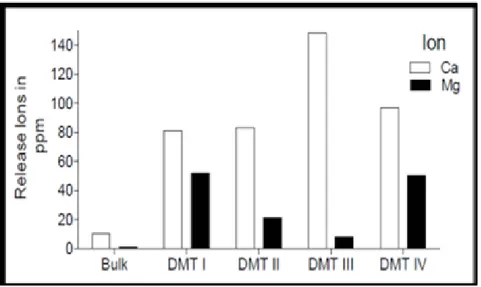
The DMTs analyzed showed large release of calcium ions, especially DMT III with a value of 147,70 ppm in the DMT IV with 99,43 ppm, DMT II with 83,17ppm and DMT I with 81,18 ppm respectively. The magnesium release was more moderate, with the highest value in the DMT I with 50,98 ppm (Figure 3).
Figure 3 - Analysis of the release of calcium and magnesium ions (ppm) by the atomic absorption method
XRD analysis and Rietveld structure refinement
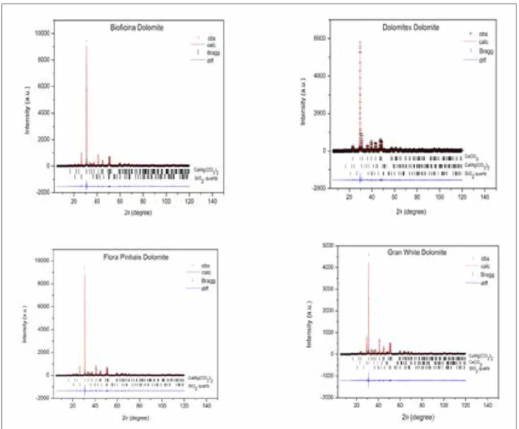
Figure 4 shows graphs obtained by Rietveld refinement method for samples of DMTs and HCa. It can be observed a significant convergence between experimental data and calculated which demonstrates the good quality adjustments. Quality indicators of refinement present also show the good quality of the Rietveld analysis. The identification phase shows that DMT I and II samples have as a major phase the mineral dolomite (ICSD # 10404), but also noted the presence of certain amount of quartz (ICSD # 1011159) in his compositions. On the other hand, the sample showed DMT III as major phase calcite (ICSD # 18166) and in this case, dolomite phase is present as a secondary phase together with quartz. DMT IV sample also showed these three crystal phases as in the sample DMT III. However, the dolomite stage is the majority in DMT IV. Sample I and II presented in a dolomite composition (calcium carbonate and magnesium - CaMg (CO2)3) and silica (silicon dioxide - SiO2) whereas the samples III and IV showed addition calcite phase (calcium
carbonate - CaCO3). HCa corresponded to the presence of only portlandite (calcium hydroxide - Ca (OH)2). Table 1 gives the percentage by mass of the products under study regarding the presence of pure DMT, quartz and calcium carbonate obtained from XRD.
Figure 4 - Rietveld graphics for commercial samples of DMT and HCa TABLE 1 – Percentage by mass of the samples used in the study DMT obtained by XRD
Percentage by mass (%)
Dolomite pure Quartz Calcium carbonate
DMT I DMT II 88% 89% 12% 11% -DMT III DMT IV 26% 85% 1,7% 2% 72% 13% Chemical composition by XRF
Table 2 shows the results obtained by XRF analysis. In all cases, it was observed that Ca is the most abundant element of the samples studied, with some variation in their amounts (between 62.79% and 98.97%). In addition, significant differences were identified for the presence of other elements and the proportion. The DMT I, DMT II and IV samples present in their compositions significant amounts of Mg (28.91%, 29.35%, 26.05% respectively), which could be associated with the presence of major phase of dolomite. Failure to observe this element in DMT III sample and the high concentration of Ca (97.70%) were associated with major phase of calcite. Si element is present in all commercial samples of DMT, which could be associated with the presence of quartz in the samples. However, it is present in a quantity in the sample DMT III (1.84%) and DMT IV (1.66%) compared to samples DMT I (7.16%) and DMT II (9.98%). These results corroborate those observed in the structural analysis by XRD. Adding to the impurities commonly found in derivatives of minerals have been identified in all samples, especially in DMT IV (Gran-White) but none of these impurities (Fe, K, Sr, Tm, S, Cu) was present in significant amounts .
TABLE 2 - Elemental analysis * by XRF.
Dolomite Bioficina® Dolomite Flora Pinhais®
Dolomitex ® Gran White Calcium hydroxide .A Biodinâmica® DMT I DMT II DMT III DMT IV HCa (%) (%) (%) (%) (%) Ca 63,08 62,79 97,70 71,32 98,9 Mg 28,91 29,35 - 26,05 -Si 7,16 9,98 1,84 1,66 -S 0,56 0,58 0,40 0,49 0,35 Fe 0,25 0,25 - 0,17 0,09 Sr 0,011 0,02 0,47 0,03 0,36 Tm - 0,01 - 0,01 -K - - - 0,24 0,18 Cu - - - - 0,01 Co - - - - 0,008
* (Ca = Calcium, Magnesium Mg = S = sulfur; Si = Silicon, Fe = iron, Sr = Strontium; Tm = Tullius; K = potassium; Cu = Copper, Co = Cobalt).
DISCUSSION
XRD analysis identifies and confirms provides mass percentage of the components of the material. In this methodology, it was possible to identify the presence of pure DMT and associated materials (calcium carbonate and quartz) present in DMTs. The predominant presence of the DMT has been identified in most samples (DMT I, II and IV). DMT III showed a high percentage of calcium carbonate (72%), and the value of 26% pure DMT. The XRD method and Rietveld refinement allowed the precise identification of the crystalline phases and percentages by weight. The result was confirmed by XRF analysis and atomic absorption.
The crystalline phases are forms such as atoms, molecules and ions are organized within a fixedly material, regular and repetitive22. As those responsible for the stability and density of material9, 23. Commercial
samples analyzed showed the crystalline phases: DMT, calcite and silica. The predominant presence of DMT phase was identified in all samples. Because of the composition of DMT rich in calcium carbonate and magnesium, which are important minerals in inorganic bone formation and is the subject of other research areas due to the stimulating action osteogenesis of anti-inflammatory and analgesec activity23. The samples
II and III showed DMT calcite phase consisting of calcium carbonate. Since this belongs to the group of carbonates, inorganic salts, chemical composition of carbonate ion (CO3)-2, anhydrous which crystallize in the rhombohedral system24. In many cases, the crystallization of carbonate from solutions rich in Ca and
Mg tends not to form calcite but stratified crystals consisting of carbonate ions layers alternating with DMT layer of Mg and ions.
Samples
DMT is composed of calcium oxide, which in contact with the liquid medium becomes calcium hydroxide giving a higher alkalinity due to the reaction with the oxide9. Calcium is very important in dentistry
for their ability the mineralization present in mineralized tissue barriers, pulp protection, pulpotomies and biological apical sealing9, 23, 24. The Ca ion has the ability to activate the ATPase calcium-dependent in this
area to be mineralized25.
The measurement of pH and calcium release of DMT aimed to confirm the antimicrobial capacity and the mineralization. Through this, when using the atomic absorption spectrometer could observe the large release of calcium ions from the samples. All samples analyzed showed high value, highlighted the DMT III, followed by IV, II and I, respectively. According to Stuart et al.25 (2006) thealkaline pH promotes the
elimination of bactéria such as Enterococcus faecalis, who can survive after chemical-surgical preparation and able to induce or maintain inflammation. When the pH is close to 11, the microorganisms have more difficulty in survive25, 26, 27. Another factor of the alkaline pH, is the correlation between the stimulation of
tissue repair process by deposition resulting mineralized tissue release calcium ions9 .
The study showed the lack of toxicity in the samples. In the first evaluation DMT I appears with the largest value of 3438.48 x106 μg / mL, followed by IV (24,34 x10⁶ μg / mL), III (2727,42μg / ml) and II
(1243,8042μg / ml) respectively. When assessing the toxicological effects to the potential use of application of DMT in dentistry from the methodology of toxicity can characterize the degree of virulence of harmful substances to the body as a whole or part28 and highlight the harmful health effect. Toxicity can be assessed
by several methods, such as cell counts, determining proliferation rates, synthesis of cellular products or determining enzymatic activities31. In this method, values above 1000 ug / ml are considered nontoxic. The
use of the bioassay with saline artemias considered indicative of the presence of toxic substances and it is easy to handle in the laboratory with rapid response and reliable results17, 29. Apart from that, the toxicity test
using Artemia salina is often used to evaluate compounds used in pharmacology30 and values above 1000
ug / ml indicate low toxicity.
In XRF analysis impurities commonly found in minerals derivatives were identified in samples, particularly in DMT IV corresponding to Gran-white, but any of the elements found beyond those identified by XRD analysis (Fe, K, Sr, Tm, S, Cu and Co) showed significant amounts toxic. In studies on the MTA (mineral trioxide aggregate), ceramic biomaterial widely used in dentistry it was observed that the Ca ions when in contact with tissue necrosis determine area to form carbon dioxide35. This, with the calcium hydroxide
formed calcite crystals (calcium carbonate) which serve as core of calcification31, 32, 33. The alkalinity of the
medium stimulates the connective tissue to secrete the glycoprotein fibronectin, which combined with the calcite crystals stimulate the formation of collagen type I associated with calcium induces mineralization33.
Natural medicine uses nature’s resources for the treatment of several diseases, however, scientifically grounded studies should confirm to achieve potential results of pharmacological use of materials. The study of toxicity and physicochemical properties of the Brazilian dolomitic limestone is the first step to propose a pharmaceutical formulation that can be directed to the endodontic treatment of primary teeth or to contribute to the remineralization of dentin.
CONCLUSION
It is concluded that the DMT is non-toxic, able to release significant amount of Ca and alkaline pH. It presents favorable chemical composition of employment in dentistry, why identified in its composition
elements related to common remineralization process in dental materials and non-significant percentage of impurities. Therefore, further studies will be done to examine the possibility of using DMT as a constituent of dental material.
ACKNOWLEDGEMENTS
This study was funded by CAPES.CONFLICT OF INTEREST
The authors declare no conflict of interest.
REFERENCES
1. SUMRADA J. [Ziga Zois and Deodat de Dolomieu]. Kronika (Ljubljana, Slovenia) 2001 49(1-2):65-72.
2. VAN Berlo D, Haberzettl P, Gerloff K, Li H, Scherbart AM, Albrecht C, et al. Investigation of the cytotoxic and proinflammatory effects of cement dusts in rat alveolar macrophages.
Chemical research in toxicology 2009 Sep;22(9):1548-58.
3. ROBERTS JA, Kenward PA, Fowle DA, Goldstein RH, González LA, Moore DS. Surface chemistry allows for abiotic precipitation of dolomite at low temperature. Proc Natl Acad
Sci USA 2013 110(36):14540-5.
4. MÜLLER G, Irion G, Förstner U. Formation and diagenesis of inorganic Ca−Mg carbona-tes in the lacustrine environment. Naturwissenschaften 1972 April 01;59(4):158-64. 5. CHEN GC, He ZL, Stoffella PJ, Yang XE, Yu S, Yang JY, et al. Leaching potential of heavy
metals (Cd, Ni, Pb, Cu and Zn) from acidic sandy soil amended with dolomite phosphate rock (DPR) fertilizers. Journal of trace elements in medicine and biology : organ of the So -ciety for Minerals and Trace Elements (GMS) 2006 20(2):127-33.
6. SLOMSK G, Odle TD. Gale encyclopedia of alternative medicine. 2005 [Acesso em: 19
março 2018]; Disponível em: http://www.encyclopedia.com/doc/1g2-3435100269.html.
7. MIZOGUCHI T, Nagasawa S, Takahashi N, Yagasaki H, Ito M. Dolomite supplementation improves bone metabolism through modulation of calcium-regulating hormone secre-tion in ovariectomized rats. Journal of bone and mineral metabolism 2005 23(2):140-6. 8. CASADO AI, Alonso-Zarza AM, La Iglesia Á. Morphology and origin of dolomite in
pa-leosols and lacustrine sequences. Examples from the Miocene of the Madrid Basin.
Sedi-mentary Geology 2014 2014/10/01/;312(1):50-62.
9. PATIL G, Khan MI, Patel DK, Sultana S, Prasad R, Ahmad I. Evaluation of cytotoxic, oxi-dative stress, proinflammatory and genotoxic responses of micro- and nano-particles of
dolomite on human lung epithelial cells A(549). Environmental toxicology and pharmaco
10. MOTOIKE K, Hirano S, Yamana H, Onda T, Maeda T, Ito T, et al. Antiviral activities of heated dolomite powder. Biocontrol science 2008 Dec;13(4):131-8.
11. CORDEIRO APB, Moreira LMA. Proliferação celular e quebras cromossômicas em cé-lulas submetidas à ação da dolomita brasileira (gran-white) in vitro. R Ci Méd Biol 2004 jul/dez ;3(2):181-7.
12. YAMAMOTO O, Ohira T, Alvarez K, Fukuda M. Antibacterial characteristics of CaCO3– MgO composites. Mater Sci Eng, B 2010 173(1):208-12.
13. DUARTE MA, Demarchi AC, Yamashita JC, Kuga MC, Fraga Sde C. pH and calcium ion release of 2 root-end filling materials. Oral surgery, oral medicine, oral pathology, oral
radiology, and endodontics 2003 Mar;95(3):345-7.
14. LEGEROS RZ, Kijkowska R, Bautista C, Legeros JP. Synergistic effects of magnesium and carbonate on properties of biological and synthetic apatites. Connective tissue
re-search 1995 33(1-3):203-9.
15. MEYER BN, Ferrigni NR, Putnam JE, Jacobsen LB, Nichols DE, Mclaughlin JL. Brine shrimp: a convenient general bioassay for active plant constituents. Planta medica 1982 May;45(5):31-4.
16. PELLOSI DS, Batistela VR, Souza VR, Scarminio IS, Caetano W, Hioka N. Evaluation of the photodynamic activity of xanthene dyes on artemia salina described by chemome-tric approaches. An Acad Bras Ciênc 2013 85(4):1267-74.
17. TOBY BH. EXPGUI, a graphical user interface for GSAS. J Appl Cryst 2001 34(2):210-13. 18. LARSON AC, Von Dreele R, Gsas B. General structure analysis system. California: Lance;
1994.
19. THOMPSON P, Cox DE, Hastings JB. Rietveld refinement of Debye-Scherrer synchro-tron X-ray data from Al2O3. J Appl Cryst 1987 20(2):79-83.
20. HIL R, Howard C. Quantitative phase analysis from neutron powder diffraction data using the rietveld method. J Appl Cryst 1987 20(6):467-74.
21. PIRES LF, Prandel LV, Saab SC. The effect of wetting and drying cycles on soil chemical composition and their impact on bulk density evaluation: An analysis by using XCOM data and gamma-ray computed tomography. Geoderma 2014 213(1):512-20.
22. HOLLAND R. Histochemical response of amputed pulps to calcium hydroxide. Rev Bras
Pesqui Med Biol 1971 Jan.–Apr.;4(1):83.
23. LEONARDO MR, Almeida WA, Bezerra Silva LA, Utrilla LS. Histopathological obser-vations of periapical repair in teeth with radiolucent areas submitted to two different methods of root canal treatment. J Endod 1995 21(3):137-41.
24. TRONSTAD L, Andreasen JO, Hasselgren G, Kristerson L, Riis I. pH changes in dental tissues after root canal filling with calcium hydroxide. J Endod 1981 Jan;7(1):17-21. 25. STUART CH, Schwartz SA, Beeson TJ, Owatz CB. Enterococcus faecalis: its role in root
Feb;32(2):93-26. MCHUGH CP, Zhang P, Michalek S, Eleazer PD. pH required to kill Enterococcus faecalis in vitro. J Endod 2004 Apr;30(4):218-9.
27. OKABE T, Sakamoto M, Takeuchi H, Matsushima K. Effects of pH on mineralization abi-lity of human dental pulp cells. J Endod 2006 Mar;32(3):198-201.
28. GUERRA R. Ecotoxicological and chemical evaluation of phenolic compounds in in-dustrial effluents. Chemosphere 2001 Sep;44(8):1737-47.
29. LIWARSKA-BIZUKOJC E, Miksch K, Malachowska-Jutsz A, Kalka J. Acute toxicity and genotoxicity of five selected anionic and nonionic surfactants. Chemosphere 2005 Mar;58(9):1249-53.
30. SHEN Z, Szlufarska I, Brown PE, Xu H. Investigation of the Role of Polysaccharide in the Dolomite Growth at Low Temperature by Using Atomistic Simulations. Langmuir : the
ACS journal of surfaces and colloids 2015 Sep 29;31(38):10435-42.
31. LAMEU EL. Análise e caracterização de calcitas por difração de raios X. In: In Anais Do XVIII EAIC. Ponta Grossa 2009.
32. SINHORETI MAC, Vitti RP, Correr-Sobrinho L. Biomateriais na odontologia: panorama atual e perspectivas futuras. Rev assoc paul cir dent 2013 67(3):178-86.
33. PEIXOTO EMA. Silício. Química nova escola [Periódico on-line].2001; (14). Acesso em:
19 março 2018. Disponível em: http://qnesc.sbq.org.br/online/qnesc14/v14a12.pdf.
RECEBIDO EM 16/08/2017 ACEITO EM 23/10/2017