Magda Alexandra
Carvalho Henriques
Curcumin and a new Rutheno(II)curcumin Complex:
characterisation and evaluation of the biological
potential
Curcumina e um novo complexo
Ruteno(II)curcumina: caracterização e avaliação das
suas potencialidades biológicas
Magda Alexandra
Carvalho Henriques
Curcumin and a new Rutheno(II)curcumin Complex:
characterisation and evaluation of the biological
potential
Curcumina e um novo complexo
Ruteno(II)curcumina: caracterização e avaliação das
suas potencialidades biológicas
Tese apresentada à Universidade de Aveiro para cumprimento dos requisitos necessários à obtenção do grau de Mestre em Bioquímica, ramo Métodos Biomoleculares, realizada sob a orientação científica da Doutora Susana Isabel Fonseca de Almeida Santos Braga, Equiparada a Investigadora Principal do Departamento de Química da Universidade de Aveiro e da Doutora Maria do Amparo Ferreira Faustino, Professora Auxiliar do Departamento de Química da Universidade de Aveiro.
o júri
presidente Doutora Maria do Rosário Gonçalves dos Reis Marques Domingues
Professora Associada com Agregação do Departamento de Química da Universidade de Aveiro
Doutora Teresa Margarida dos Santos
Professora Auxiliar do Departamento de Química da Universidade de Aveiro
Doutora Susana Isabel Fonseca de Almeida Santos Braga
agradecimentos Às minhas orientadoras, Doutora Susana Braga e Doutora Maria do Amparo Faustino, pela ajuda que me deram ao longo deste ano e principalmente por toda a paciência que tiveram. Muito obrigada por terem estado sempre disponíveis para mim e por me terem sempre acompanhado não só a nível “profissional” como pessoal, por me terem feito sentir em casa mesmo estando no laboratório. O vosso apoio foi a chave para o êxito deste trabalho.
À Doutora Margarida Fardilha e à Juliana Felgueiras pela ajuda no desenvolvimento do desenho experimental dos ensaios de citotoxicidade e por todos os ensinamentos na cultura de células. Obrigada pela forma carinhosa como me acolheram e me integraram no vosso grupo de trabalho. Foi uma ótima ajuda para que tudo corresse pelo melhor.
Ao Professor Doutor Artur Silva pela disponibilidade para ajudar na análise dos espectros de RMN e em tudo o que foi preciso.
Ao Doutor Flávio Figueira pela ajuda nas purificações e cristalizações. Foi uma mais-valia numa questão tão importante do meu trabalho.
A todas as pessoas do Departamento de Química e do iBiMED da Universidade de Aveiro que tornaram possível a realização deste trabalho.
Aos meus pais, aos meus irmãos e ao Rui o maior agradecimento. Para vos agradecer tudo o que já fizeram por mim eu precisava de uma tese inteira e não apenas de uma secção nos agradecimentos, vocês foram o meu pilar ao longo destes anos. Obrigada por terem sempre acreditado em mim e por me ajudarem para conseguir fazer sempre mais e melhor. Obrigada por me mostrarem que não interessam as notas ou os graus que eu consiga, que aquilo que interessa é eu seguir o meu sonho e ser feliz. Obrigada por todos os sacrifícios que já fizeram por mim. Obrigada por não me deixarem sentir mal mesmo quando as coisas correm menos bem. Obrigada por estarem sempre presentes. Muito, muito obrigada, sem vocês isto nunca teria sido possível.
A todos os meus amigos e à minha segunda família, por toda a disponibilidade para me apoiarem nas minhas derrotas e por festejarem comigo as minhas vitórias.
palavras-chave Curcumina, ruténio(II), cancro da próstata, avaliação da citotoxicidade, terapia fotodinâmica, intercalação com o ADN, caracterização espectroscópica
resumo A curcumina é um pigmento de cor amarela, da família dos polifenóis, obtido a partir dos rizomas da planta Curcuma longa. É dotada de diversas propriedades biológicas que se relacionam com a vasta gama de alvos moleculares que possui, sendo de destacar a sua atividade anticancerígena já provada em diversas linhas celulares cancerígenas. No entanto, a atividade biológica da curcumina é limitada pela sua baixa biodisponibilidade que se deve principalmente à sua fraca solubilidade e rápida degradação em condições fisiológicas. Uma estratégia para contornar as limitações terapêuticas da curcumina é a sua complexação com iões metálicos, nomeadamente com complexos de ruténio(II).
Neste trabalho foi preparado um novo complexo de curcumina com ruténio(II) e tritiaciclononano [9anoS3] com o objetivo de aumentar a solubilidade da curcumina em meio fisiológico. A curcumina foi extraída e purificada a partir de rizomas secos e pulverizados de Curcuma longa sendo depois desprotonada e coordenada com o precursor [Ru(II)(9anoS3)(DMSO)Cl2] (DMSO=dimetilsulfóxido). A estrutura e pureza do complexo formado, [Ru(II)(9aneS3)(curcumina)(S-DMSO)]Cl foi avaliada por Ressonância Magnética Nuclear de 1H e 13C, espectrometria de massa e análise elementar. A afinidade do novo complexo para ligação ao ácido desoxirribonucleico (ADN) foi estudada por ensaios de titulação e por determinação da variação na temperatura de desnaturação do ADN de esperma de salmão. Este estudo permitiu determinar que o complexo tem a capacidade de intercalar com o ADN com uma constante de ligação comparável aos intercaladores de ADN já conhecidos (4.00x10⁵ M-1). A atividade citotóxica deste complexo contra o cancro de próstata foi estudada in vitro, usando como controlo positivo a curcumina. Mais ainda, aproveitando o potencial fotossensibilizador da curcumina, os ensaios foram feitos na presença e na ausência de luz. Para isso, usaram-se culturas celulares humanas isoladas de carcinoma de próstata (PC-3) e a toxicidade foi avaliada em linhas celulares prostáticas não tumorais (PNT-2). Com estes ensaios verificou-se que o complexo preparado não tem efeito citotóxico nem fototóxico nas concentrações usadas para as linhas celulares estudas. Mais, observou-se que a curcumina apresenta, no escuro, efeito citotóxico nas concentrações usadas e ainda, que este efeito é fortemente potenciado na presença de luz, sendo um potencial agente para terapia fotodinâmica em cancro de próstata.
keywords Curcumin, ruthenium(II), prostate cancer, cytotoxicity evaluation, photodynamic therapy, DNA intercalation, spectroscopic characterisation
abstract Curcumin is a yellow pigment, of the family of polyphenols, obtained from the rhizomes of the Curcuma longa. It is provided with several biological properties which relate with the wide range of molecular targets that possesses, especially the anticancer activity already proven in various cell lines. However, the biological activity of curcumin is limited by their low bioavailability which is mainly because their poor solubility and rapid degradation in physiological conditions. One promising strategy to circumvent the therapeutic limitations of curcumin is the binding with metal ions, namely with ruthenium(II) complexes.
In this work, was prepared a new complex of curcumin with ruthenium(II) and trithiacyclononane (9aneS3) with the aim of increasing the solubility of curcumin in physiological medium. The curcumina was extracted and purified from the powder rhizomes of Curcuma longa being after deprotonated and coordinated with the precursor [Ru(II)(9aneS3)(DMSO)Cl2] (DMSO=dimethylsulfoxide). The structure and purity of the prepared complex, [Ru(II)(9aneS3)(curcumina)(S -DMSO)]Cl was evaluated by 1H and 13C Nuclear Magnetic Resonance spectroscopies, mass spectrometry and elemental analysis. The affinity of the new complex to bind to deoxyribonucleic acid (DNA) was studied by titration assays and determination of the variation on sperm salmon DNA melting temperature. This study allowed to determine that the complex has ability to intercalate with DNA with a binding constant comparable with classical intercalators. The cytotoxic activity of this complex against prostate cancer was studied in vitro, using curcumin as a positive control. Moreover, taking advantage of the photosensitizer potential of curcumin, the assays were made under dark and light conditions. For this, human cell cultures were used isolated from prostate carcinoma (PC-3) and the toxicity was evaluated in non-tumour prostate cells (PNT-2). With these tests, it was found that the new complex do not have any cytotoxic or phototoxic effect in the same concentrations range tested for cell lines studied. Further, was observed that the curcumin presents, in dark, cytotoxic effect and, that this effect is strongly potentiated by light presence, with a potential for photodynamic therapy in prostate cancer.
Contents
Contents ... viii
List of figures ... x
List of tables ... xiv
List of Abbreviations, symbols and acronyms ... xvi
1. Introduction ... 1
1.1. Curcuma longa Linn ... 1
1.1.1. Extraction, purification and identification of curcuminoids ... 2
1.2. Curcumin ... 3
1.2.1. Molecular targets ... 6
1.2.2. Bioavailability ... 7
1.2.3. Binding to metals and effects on stability, bioavailability and cytotoxicity ... 8
1.3. Metal complexes in cancer treatment...11
1.3.1. Platinum complexes ...12
1.3.2. Ruthenium complexes ...13
1.4. Cancer ...15
1.5. Prostate cancer ...16
1.6. Photodynamic therapy in cancer treatment ...18
1.6.1. Curcumin in photodynamic therapy ...19
1.7. Aims of this work ...20
2. Experimental section ...23
2.1. Equipment ...23
2.2. Materials ...24
2.3. Methods ...25
2.3.1. Extraction and isolation of curcumin ...25
2.3.2. Preparation of [Ru([9]aneS3)(curcumin)(S-DMSO)]Cl complex (Ru-curc complex) ...26
2.3.3. Fluorescence quantum yield ...27
2.3.4. Singlet oxygen measurements ...27
2.3.5. DNA binding studies ...28
2.3.6. Biological assays ...30
2.3.7. Statistical analysis ...32
3. Results and discussion ...33
3.1. Extraction and isolation of curcumin ...33
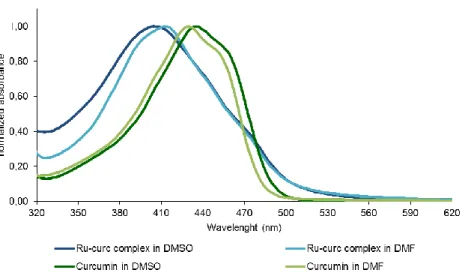
3.3. Photophysical properties...39
3.3.1. Absorption spectra ...39
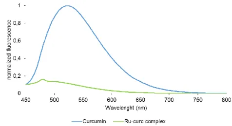
3.3.2. Fluorescence spectra ...40
3.4. Singlet oxygen studies ...41
3.5. DNA binding studies ...42
3.5.1. Absorption spectral studies ...43
3.5.2. Thermal denaturation of DNA ...45
3.6. Biological evaluation ...47
3.6.1. Cellular Viability ...47
4. Conclusions and future prospects ...56
4.1. Conclusions ...56
4.2. Future prospects ...57
References ...59
Appendix ...77
List of figures
Figure 1 - Chemical structures of the three curcuminoids present in the plant Curcuma longa L. ... 2 Figure 2 - Keto-enol tautomeric equilibrium of curcumin. ... 4 Figure 3 - Curcumin degradation products after exposure to light. ... 5 Figure 4 - Human diseases against which curcumin has exhibited activity. Adapted from He and collaborators.43 ... 5 Figure 5 – Synthesis of [Ruthenium(II)(R)(curcumin)X] complexes already tested in several human tumour and non-tumour cell lines. Where, R=p-cymene, benzene or hexamethylbenzene and X=Cl or PTA. i) NaOCH3, MeOH... 9 Figure 6 - Platinum complexes approved worldwide for clinical use. ...12 Figure 7 – Ruthenium(III) based anticancer complexes NAMI-A and KP1019 in clinical trials ...14 Figure 8 - Structure of macrocyclic ligand 1,4,7-trithiacyclononane ([9]aneS3). ...14
Figure 9 - Molecular structure of [Ru(II)(9aneS3)(S-DMSO)Cl2]. ...15
Figure 10 - Photobleaching reaction of 9,10-dimethylanthracene by 1O
2 ...28
Figure 11 – Carbon labeling scheme for the curcumin ...34 Figure 12 - 1H NMR spectrum recorded in CDCl
3 for the curcumin ...34
Figure 13 - 13C NMR spectrum recorded in CDCl
3 of curcumin. ...35
Figure 14 – Reactional scheme for the two-step synthesis of Ru-curc complex. ...36 Figure 15 - 1H NMR spectrum recorded in CDCl
3 of Ru-curc complex. ...37
Figure 16 - 13C NMR spectrum recorded in CDCl
3 of the Ru-curc complex. ...38
Figure 17 – Normalized UV-Vis absorption spectra of curcumin and Ru-curc complex in DMF and DMSO at 25 ºC. ...40 Figure 18 - Normalized fluorescence emission spectra (λexc 430 nm) of curcumin and
Ru-curc complex in DMF. ...41 Figure 19 - First-order plots for the photooxidation of DMA (30 μM) photosensitized by TPP (green squares), curcumin (yellow triangles) and Ru-curc complex (grey crosses) in DMF, under irradiation at 430 nm at the irradiance of 30 mW/cm2. Values
represent mean ± standard deviation of three independent experiments. ...42 Figure 20 – UV-Vis absorption spectra of curcumin (30 μM) with increasing concentrations of sp-DNA in PBS. ...44 Figure 21 – UV-Vis absorption spectra of Ru-curc complex (30 μM) with increasing concentrations of sp-DNA in PBS. ...44
Figure 22 - Comparison of the thermal denaturation curves for pure sp-DNA (grey), sp-DNA/curcumin (orange) and sp-DNA/Ru-curc complex (blue) mixtures in 10:1 molar ratio. Curves were recorded under equilibrium conditions in 10 mM PBS at pH 7.2. ...46 Figure 23 - Alamar Blue (AB) assay for PNT-2 cell viability. PNT-2 cells were seeded at varying densities in a 96-well plate. The AB solution (10% of cell culture volume) was added to the plate and relative absorbance units at 540 and 630 nm were measured at 1, 2, 4, 6, 8 and 24 h. ...47 Figure 24 - Alamar Blue (AB) assay for PC-3 cell viability. PC-3 cells were seeded at varying densities in a 96-well plate. The AB solution (10% of cell culture volume) was added to the plate and relative absorbance units at 540 and 630 nm were measured at 1, 2, 4, 6, 8 and 24 h. ...48 Figure 25 - Effect of curcumin and Ru-curc complex on cell viability of PNT-2 cell line. PNT-2 cells were treated with different concentrations of curcumin, Ru-curc complex or vehicle control DMSO at 1% for 72 h at 37 ºC in dark. The AB assay was used to determine cell viability. Data are representative of three independent trials and are expressed as the mean ± SD. *P-value<0.05 and **P-value<0.001, compared to the group control. ...49 Figure 26 - Effect of curcumin and Ru-curc complex on cell viability of PC-3 cell line. PC-3 cells were treated with different concentrations of curcumin and Ru-curc complex or vehicle control DMSO at 1% for 72 h at 37 ºC in Dark. The AB assay was used to determine cell viability. Data are representative of three independent trials and are expressed as the mean ± SD. **P-value<0.001 and ****P-value<0.0001, compared to the group control. ...49 Figure 27 - Effect of curcumin on cell viability of PNT-2 and PC-3 cell lines in dark conditions. PNT-2 and PC-3 cells were treated with different concentrations of curcumin or vehicle control DMSO at 1% for 72 h at 37 ºC in dark conditions. The AB assay was used to determine cell viability. Data are representative of three independent trials and are expressed as the mean ± SD. *P-value<0.05, **P-value<0.001 and ****P-value<0.0001, compared to the group control. ...50 Figure 28 - Effect of curcumin on cell viability of PNT-2 cell line in dark and with irradiation. PNT-2 cells were treated with different concentrations of curcumin or vehicle control DMSO at 1% for 72 h at 37 ºC in dark or with irradiation. The AB assay was used to determine cell viability. Data are representative of three independent trials and are expressed as the mean ± SD. *P-value<0.05, **P-value<0.001 and ****P-value<0.0001, compared to the group control. ...51
Figure 29 – Effect of curcumin on cell viability of PC-3 cell line in dark and with irradition. PC-3 cells were treated with different concentrations of curcumin or vehicle control DMSO at 1% for 72 h at 37 ºC in Dark or with irradiation whit white light at an irradiance of 10 mW/cm2 and a total light dose of 6 J/cm2. The AB assay was used to determine cell proliferation. Data are representative of three independent trials and are expressed as the mean ± SD. *P-value<0.05, **P-value<0.001 and ****P-value<0.0001, compared to the group control. ...52 Figure 30 - Effect of curcumin on cell viability of PNT-2 and PC-3 cell lines in light conditions. PNT-2 and PC-3 cells were treated with different concentrations of curcumin or vehicle control DMSO at 1% for 72 h at 37 ºC in light conditions. The AB assay was used to determine cell viability. Data are representative of three independent trials and are expressed as the mean ± SD. **P-value<0.001 and ****P-value<0.0001, compared to the group control. ...53 Figure 31 - Effect of Ru-curc complex on cell viability of PNT-2 cell line. PNT-2 cells were treated with different concentrations of Ru-curc complex or vehicle control 1% DMSO for 72 h in dark or exposure to white light at an irradiance of 10 mW/cm2 and a total light dose of 6 J/cm2 light dose. The AB assay was used to determine cell proliferation. Data are representative of three independent trials and are expressed as the mean ± SD. ...54 Figure 32 - Effect of Ru-curc complex on cell viability of PC-3 cell line. PC-3 cells were treated with different concentrations of Ru-curc complex or vehicle control 1% DMSO for 72 h in dark or exposure to white light at an irradiance of 10 mW/cm2 and a total light dose of 6 J/cm2 light dose. The AB assay was used to determine cell viability. Data are representative of three independent trials and are expressed as the mean ± SD. ...54 Figure 33 – Examples of O-coordinated ligands with ruthenium(II) metal centers. FL=flavonate. ...56 Figure A 1 – Calibration curve for determination of the molar absorptivity of the curcumin in DMSO. ...77 Figure A 2 – Calibration curve for determination of the molar absorptivity of the curcumin in DMF. ...77 Figure A 3 – Calibration curve for determination of the molar absorptivity of the Ru-curc complex in DMSO. ...78 Figure A 4 - Calibration curve for determination of the molar absorptivity of the Ru-curc complex in DMF. ...78
List of tables
Table 1 - Molecular targets and cell processes modulated by curcumin. Curcumin interacts directly or indirectly with various molecular targets, including transcription factors, growth factors, receptors, cytokines, anti-apoptotic proteins, pro-apoptotic proteins, enzymes, protein kinases, and adhesion molecules, altering their expression. ... 6 Table 2 – Cytotoxic activities of curcumin and [Ru(II)(R)(curcumin)X] complexes described in literature, where R= p-cymene, benzene or hexamethylbenzene and X= Cl or PTA. IC50 values of complexes in human cell lines. Cisplatin was tested as a positive control. ...11 Table 3 - Potential targets for prostate cancer chemoprevention. ...18 Table 4- 1H NMR chemical shifts for the ligand curcumin and the Ru-curc complex in
CDCl3. ...37
Table 5 - 13C NMR chemical shifts for the ligand curcumin and the Ru-curc complex.
...39 Table 6 - Kinetic parameters, Kobs (s-1) of the photooxidation reaction of DMA by TPP,
curcumin and Ru-curc complex after exposure to light of 430 nm at the irradiance of 30 mW/cm2 and 1O
2 quantum yield in DMF...42
Table 7 Ligand-based absorption spectral properties and binding constant of compounds to sp-DNA. ...45 Table 8 – Comparison of thermal denaturation for pure sp-DNA, sp-DNA/curcumin or sp-DNA/Ru-curc complex mixtures. The values for Tm were obtained from the midpoint of melting curves for pure sp-DNA, sp-DNA/curcumin and sp-DNA/Ru-curc complex mixtures (10:1). ...46 Table 9 - The IC50 values (μM) of curcumin and Ru-curc complex towards PNT-2 and
List of Abbreviations, symbols and acronyms
δ chemical shift (ppm)
ΦF fluorescence quantum yields
ΦΔ singlet oxygen quantum yields
13C NMR carbon Nuclear Magnetic Resonance 1H NMR proton Nuclear Magnetic Resonance J coupling constant
1J coupling constant over one bonds 2J coupling constant over two bonds 3J coupling constant over three bonds 1O
2 singlet state of oxygen 3O2 triplet state of oxygen
A2780 human ovarian cancer cell line
A2780cisR human ovarian platinum-resistant cancer cell line A431 human epidermoid carcinoma cell line
A549 human lung adenocarcinoma cell line
AB alamarblue
A absorbance
ADT androgen-deprivation therapy
AMC-HN3 human head and neck cancer cell line AP-1 activator protein 1
AR androgen receptor
AUC integrated area under the fluorescence curves
Bax b-cell lymphoma 2-associated X protein
Bcl-2 b-cell lymphoma 2
Bcl-XL b-cell lymphoma-extra large
CC column chromatography
CDCl3 deuterated chloroform
Cyclin-CDK cyclin dependent kinase complex
CNE1 human nasopharyngeal carcinoma cell line
COSY homonuclear correlation spectroscopy (1H/1H) COX-2 cyclooxygenase-2 d doublet dd doublet of doublets DMA 9,10-dimethylanthracene DMF N,N-dimethylformamide DMSO dimethylsulfoxide
DNA deoxyribonucleic acid
DRE digital rectal examination
EGF epidermal growth factor
EGFR epidermal growth factor receptor
ESI-MS electrospray ionization mass spectrometry
FBS fetal bovine serum
FDA food and drug administration
FGF fibroblast growth factor
FGFR fibroblast growth factor receptor
GFR growth factor receptor
HaCaT aneuploid immortal keratinocyte cell line HCT116 human colon cancer cell line
HEK293 human embryonic kidney cell line
HMBC heteronuclear multiple bond correlation (13C/1H)
HPLC high performance liquid chromatography
HSQC heteronuclear single quantum correlation (13C/1H)
Hz hertz
IC50 drug concentration needed to reduce cell viability by 50%
IL Interleukin
iNOS inducible nitric oxide synthase
JNK c-jun N-terminal kinase Kb binding constant
Kobs observed rate constant
m multiplet
MAPK mitogen activated protein kinase
MCF7 breast cancer cell line MeOH methanol
MMP matrix metalloproteinase
MS mass spectrometry
NAMI-A new antitumour metastasis inhibitor A
NaOCH3 sodium methoxide
NF-kB nuclear factor kappa B
Nfr-2 nuclear factor erythroid 2-related factor 2
NMR nuclear magnetic resonance
OD optical density
OMe methoxyl group
PBS phosphate buffered saline
PC-3 prostate cancer cell line
PCa prostate cancer
PDGF platelet-derived growth factor
PDGFR platelet-derived growth factor receptor
PDT photodynamic therapy
PKA protein kinase A
PNT-2 prostatic epithelial cell line
PPAR-γ peroxisome proliferator-activated receptor gamma
ppm parts per million PS photosensitizer
PSA prostate-specific antigen
PTA 1,3,5-triaza-7-phosphoadamantane Rf retardation factor
ROS reactive oxygen species
RPMI roswell park memorial institute
Ru(II) ruthenium(II)
Ru-curc [ru([9]anes3)(curcumin)(S-dmso)]Cl complex s singlet
S-DMSO dimethylsulfoxide coordinated by the sulphur atom sp-DNA deoxyribonucleic acid of salmon sperm
STAT-3 signal transducer and activator of transcription 3
TB trypan blue
TLC thin-layer chromatography
Tm melting temperature TMS tetramethylsilane
TNF tumour necrosis factor
TPP 5,10,15,20-tetraphenylporphyrin tPSA total prostate-specific antigen
TRUS transrectal ultrasonography
U87 human primary glioblastoma cell line
USA United States of America
UV-Vis ultraviolet-visible spectroscopy VEGF vascular endothelial growth factor
1. Introduction
1.1. Curcuma longa Linn
Curcuma longa L., commonly known as turmeric or saffron-of-India, is a
perennial plant of the Zingiberaceae family.1,2 This plant is widely grown in South
and Southeast Asia, especially in China and India. The part of the plant that is usually used is the rhizome, which can be eaten fresh or dried.3 The use of Curcuma longa L. rhizomes is referred in Ayurvedic medicine (the characteristic medicinal
system of Ancient India) as a home remedy for various diseases.4 Currently,
turmeric is considered as a functional food because of the therapeutic properties of their active components, namely anti-inflammatory, antioxidant and anticancer activity. This plant is also effective in the treatment of circulatory, liver and dermatological diseases (e.g., psoriasis).5,6
The powder extracted from the dried rhizome of turmeric has a yellow colour and its chemical composition is influenced by various factors, such as the geographical origin of the plant, the climate of farming, the environment and soil composition. It contains mainly starch and, in a lesser extent, proteins, lipids, fibres, curcuminoids, and essential oils, such as the α-phellandrene (1%), the sabinene (0.6%), the cineole (1%), the borneol (0.5%), the zingiberene (25%), and the sesquiterpenes (53%).7,8 The essential oils have been isolated from Curcuma longa
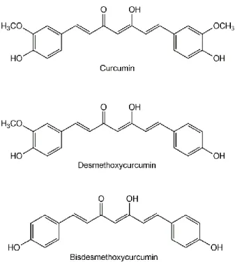
by distillation by steam entrainment. The curcuminoids, in amounts varying between 2% and 9% of the turmeric powder, are the active substances of the plant and comprise three structurally related phenolic compounds: [(1E,6E)-1,7-bis(4-hydroxy-3-methoxyphenyl)-hepta-1,6-diene-3,5-dione)], known under the designation of curcumin, desmethoxycurcumin [(1E,6E)-1-(4-hydroxy-3-methoxyphenyl)-7-(4-hydroxyphenyl)-hepta-1,6-diene-3,5-dione)] and bisdesmethoxycurcumin [(1E,6E)-1,7-bis(4-hydroxyphenyl)-hepta-1,6-diene-3,5-dione] (Figure 1).9 The commercially available curcumin is a mixture of these three
curcuminoids, in which the most abundant constituent is the curcumin (~77%), followed by desmethoxycurcumin (~18%) and the less abundant bisdesmethoxycurcumin (~5%).10,11
Figure 1 - Chemical structures of the three curcuminoids present in the plant Curcuma
longa L.
Curcuminoids have an intense yellow colour and are poorly soluble in water at physiological and acidic pH, but easily soluble in alkaline solutions.12 They are
quite soluble in dimethylsulfoxide (DMSO), acetone, dichloromethane, methanol and ethanol. These pigments are sensitive to light, high temperature and oxidative conditions, decomposing easily.13
1.1.1. Extraction, purification and identification of curcuminoids
The biological relevance of curcuminoids pigments led to the development of several methods for their extraction, separation and identification. The conventional extraction methods are based on the combined maceration with ultrasound, heat, pressure or enzymatic treatment.14–18 Another way to obtain the compounds is to use a Soxhlet apparatus, allowing extraction and filtration to be done in the same step.19 However, all methods require long extraction times, high energy
consumption and high volume of organic solvent. In addition, they exhibit low extraction efficiency and losses with heat because curcuminoids are heat-sensitive substances. New extraction methods more adapted to this reality, such as microwave-assisted extraction, are under development.20
Once extracted, the pigments can be separated by thin-layer chromatography (TLC),7,10,21 column chromatography (CC)10,22 and high
performance liquid chromatography (HPLC).10,23 CC has been the most commonly
employed method for separating curcumin from turmeric. In CC, the mixture of curcuminoids adsorbed in silica gel is eluted using mixtures of organic solvents, such as dichloromethane/ethyl acetate or methanol/chloroform. The evaluation of the purity of the separated compounds can be made by ultraviolet-visible (UV-Vis) spectrophotometry but the HPLC is the most used technique.24,25 Typically, reverse
phase columns with 18 carbons are used, as well as acetonitrile/water or chloroform/methanol solvent mixtures as the mobile phase.
1.2. Curcumin
Curcumin has a scientific history of nearly two centuries that began in 1815, date of the first report of its isolation from the rhizome of Curcuma longa, being described simply as "a matter of yellow color".26 Later, it was found that this extract
was in fact a mixture of various curcuminoids with oils and resins derived from curcuma rhizomes. In 1870, Daube27 found a method to isolate curcumin in the pure
and crystalline form and described the crystals he observed under the optical microscope. Its chemical structure only was determined in 1910 by Milobedzka and Lampe.28 In 1913, it was reported a well succeeded preparation of curcumin in
laboratory for the first time.29 The curcumin research then slowed and the
resurgence of curcumin interest emerged in the year 1987 when Kuttan and his colleagues30 reported that this polyphenolic compound had anticancer activity.
Since then, the research for characterization and isolation of this compound has increased rapidly.31
The structure of curcumin consists of two methoxyl groups linked to each other, at the orto position of the phenolic rings, which are linked together by a hepta-unsaturated bridge.32 In fact, it is an α,β-unsaturated β-diketo bridge that exhibits
keto-enol tautomerism and can exist in different types of conformers depending on the nature of the solvent where it is dissolved (Figure 2).32,33 Yellow curcumin
changes to dark red colour at alkaline pH and under physiological conditions the λmáx for curcumin is observed at 420 nm.34
Figure 2 - Keto-enol tautomeric equilibrium of curcumin.
The degradation kinetics of curcumin under various pH conditions and its stability in physiological matrices are already reported.35 When curcumin was
incubated in 0.1 M phosphate buffer (PBS), pH 7.2, approximately 90% decomposed within 30 min. Moreover, on exposure to light curcumin degrades into several products, namely trans-6-(4’-hydroxy-3’-methoxyphenyl)-2,4-dioxo-5-hexanal, ferulic acid, feruloyl methane and vanillin (Figure 3).35,36 Under acidic
conditions, the degradation of curcumin is much slower, with less than 20% of total curcuminoids decomposed after 1 h. Degradation of curcumin has also been demonstrated upon addition to cultured cells.35 Further, it is known that the
degradation products resulting from the photodegradation of curcumin are the same as occur during chemical degradation of the polyphenol. However, the degradation is significantly decreased when curcumin is attached to lipids, liposomes, albumins, cyclodextrin, surfactants, polymers and many other systems.
Figure 3 - Curcumin degradation products after exposure to light.
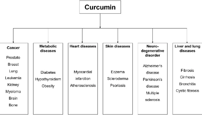
As indicated above, curcumin possesses several biological properties, namely anti-inflammatory,37,38 antioxidant39,40 and anticancer effects.41,42 Therefore,
curcumin is able to act as a chemopreventive agent, as well as a potential therapeutic agent for the many organ and tissue chronic disorders (Figure 4).
Figure 4 - Human diseases against which curcumin has exhibited activity. Adapted from He and collaborators.43
1.2.1. Molecular targets
The studies published in the last three decades show that the curcumin molecule is highly pleiotropic, i.e. capable of modulating the biological activity of a series of signalling molecules, affecting many signalling pathways (Table 1).44,45
Table 1 - Molecular targets and cell processes modulated by curcumin. Curcumin interacts directly or indirectly with various molecular targets, including transcription factors, growth factors, receptors, cytokines, anti-apoptotic proteins, pro-apoptotic proteins, enzymes, protein kinases, and adhesion molecules, altering their expression.
Family Molecular target Effect of curcumin in
molecule activity Reference
Transcriptional factors NF-kB ↓ 46 AP-1 ↓ 47,48 Nrf-2 ↑ 49 PPAR-γ ↑ 50 STAT-3 ↓ 51–53 Inflammatory cytokines TNF-α ↓ 54 IL-1,2,5,6,8,12,18 ↓ 55,56 Growth factors PDGF ↓ 57 EGF ↓ 58 FGF ↓ 59,60 VEGF ↓ 61–63 Receptors AR ↓ 64,65 EGFR ↓ 50,66–68 Kinases PKA ↓ 69 MAPK ↓ 69 JNK ↓ 70 Enzymes iNOS ↓ 71 MMP ↓ 72,73 COX-2 ↓ 74 Telomerase ↓ 75 Anti-apoptotic proteins Bcl-2 ↓ 76–79 Bcl-XL ↓ 76–78,80
Pro-apoptotic proteins Bax ↑
76–79
Caspase 3 ↑ 81,82
List of abbreviations on pages xvi-xix. Upward arrows – increased activity; downward arrows – decreased.
This curcumin feature is directly related to its ability to regulate many targets either by direct interaction with transcription factors, growth factors and their receptors, nuclear factors, hormones and hormone receptors, cytokines, or by controlling the expression of genes that regulate cell proliferation and apoptosis.83
Curcumin may associate with serum albumin through hydrophobic interactions,84
being transported to cells where it exerts its pharmalogical effects. Curcumin enters the cytoplasm and is able to accumulate in the plasma membrane, endoplasmic reticulum and nuclear envelope.85 The complexity of the mechanisms associated
with the action of curcumin justify its efficiency in combating various multifactorial diseases, such as cancer, metabolic diseases and others, as indicated above. Curcumin can act against three important steps in carcinogenesis, tumour promotion, angiogenesis and tumour growth by modulation of several molecular targets.
1.2.2. Bioavailability
Despite the wide range of biological activities of curcumin, some studies report limitations its use as a therapeutic agent, mainly due to its poor bioavailability. Curcumin has a relatively low absorption rate at the intestine, undergoes rapid metabolism in liver and has a short biological half-life.86 Curcumin is metabolized by
both conjugation and reduction pathways in the body resulting in formation of several metabolites. Furthermore, this low bioavailability is further enhanced by the poor solubility of curcumin in water and in physiological conditions, like many other natural polyphenols. The kinetics and availability of curcumin in vivo have quite distinct profiles depending on the administration routes.
Oral intake of curcumin is the most common form of contact with this compound given that it is typically incorporated into food. Nevertheless, the bioavailability of curcumin administered by this route is extremely poor.8 Less than
1% of oral curcumin enters in the plasma and the small amount of curcumin absorbed is subjected to conjugations, like sulfation and glucuronidation in the liver. The major reasons for this are the very low solubility at physiological conditions, the poor absorption in the body, the rapid metabolism with short biological half-life and the rapid systemic elimination.87,88 The poor absorption following oral administration
of curcumin implies that very high doses (>3.6 g per day in humans) are required to produce any medicinal effect. Studies have shown that 99% of the curcumin found in plasma is in the form of glucuronide conjugates,89,90 and that most of these are
biologically inactive. Indeed, many studies highlight the issue of having extremely low concentrations of free curcumin in booth plasma and urine after oral administration.91 There are several published methods suggesting that low
bioavailability of oral curcumin could be circumvent. These include the use of adjuvants like piperine,92 curcumin structural analogues,93 and development of
improved delivery technologies, such as polymeric micelles94 and nanoparticles.95
Additionally, parenteric forms – intraperitoneal, intravenous, intramuscular and subcutaneous - are under development. These require specific formulations to stabilize curcumin in an aqueous matrix.89,96–102 Several formulations available, allowing repeated systemic injections of curcumin reported in preclinical studies for intraperitoneal, intravenous, intramuscular and subcutaneous administration, proved that these formulations offer platforms for this otherwise poorly soluble drug.
1.2.3. Binding to metals and effects on stability, bioavailability and
cytotoxicity
The use of natural products as ligands for coordination with metal ions has been subject of intensive research, since several metal ions allow to mitigate the inherent limitations of natural compounds. The metal ions allow to increase the stability of the natural compounds under physiological conditions. Following these strategy of coordinating a metal centre with natural compounds with therapeutic properties, there are a few literature reports on coordination of metal ions with curcumin.103–109 One of the main advantages of this coordination is the stabilisation of curcumin - an important issue when considering its potential application in medicine. The protocol used to prepare almost all of these metal complexes involves a first step of deprotonation of curcumin, thereby stabilising it in the enol form.110 In
a second step, it is made react, in appropriate molar ratio, with metal halogenates, namely of zinc(II), iron(II), nickel(II), copper(II) and ruthenium(II). Regarding coordination of the curcumin with ruthenium metal, the published reports focus
complexes were studied and their cytotoxic activity evaluated against several cell lines but without being particular light irradiation.
Figure 5 – Synthesis of [Ruthenium(II)(R)(curcumin)X] complexes already tested in several human tumour and non-tumour cell lines. Where, R=p-cymene, benzene or hexamethylbenzene and X=Cl or PTA. i) NaOCH3, MeOH
The preparation of [Ru(II)(p-cymene)(curcumin)Cl] was described in 2012 by two different research teams in Italy.104,105 Caruso and co-workers104 studied the
cytotoxicity of this complex, at 72 h of incubation, against the human tumour cell lines: breast (MCF7), colon-rectal (HCT116), ovarian (A2780), ovarian platinum-resistant (A2780cisR), lung (A549) and glioblastoma (U87) and compared them with that of cisplatin. Their results demonstrated that the complex has greater effect in the HCT116 cell line, with an IC50 of 14 mM, followed by MCF7 (20 mM), A2780
(24 mM) and A2780cisR (27 mM) cell lines; U87 (30 mM) and A549 (65 mM) were less sensitive. The effects of the complex on the four most sensitive cell lines tested (HCT116, MCF7, A2780 and CP9) were compared to those of cisplatin that were still significantly more potent against these cell lines than the [Ru(II)(p-cymene)(curcumin)Cl] complex as shown in Table 2.
On the other hand, the group of Bonfili and co-workers105 studied the same
curcumin complex [Ru(II)(p-cymene)(curcumin)Cl] along with two other ruthenium(II) complexes: [Ru(II)(benzene)(curcumin)Cl] and the [Ru(II)(hexamethylbenzene)(curcumin)Cl], against the human colorectal cancer
HCT116 cell line. The authors pointed out that after 24 h of incubation the complexes only exhibited a mild cell growth inhibition (20-30%), which was comparable or lower than the found for pure curcumin. The DNA binding ability of the complexes and their ability to regulate the activity of the proteasome were also studied. Complexes with p-cymene and benzene had shown greater DNA binding affinity than hexamethylbenzene complex and pure curcumin. The p-cymene complex also showed to be the most potent in inhibiting proteasome in comparison with other compounds and pro-apoptotic events were observed, thus representing a potentially new, nontoxic and effective compound in cancer therapy.
More recently, Pettinari and their collaborators studied the synthesis and biological activity of new ruthenium(II) complexes: the [Ru(II)(p-cymene)(curcumin)PTA] and the [Ru(II)(hexamethylbenzene)(curcumin)PTA].106
The cytotoxicity of this complexes was evaluated at 72 h of incubation against ovarian cancer cell lines with and without cisplatinum resistance (A2780cisR and A2780 cell lines, respectively), as well as on a non-tumour human embryonic kidney cell line, HEK293. Cisplatin was used as reference drug, once it is used in the clinic to treat ovarian cancer. The results showed that the prepared complexes possess high anti-tumoural activity, inhibiting tumour growth at lower concentrations than the reference cisplatin. In general, the new complexes are much more selective towards the tumoural cell lines (with marginal activity towards the healthy cell line), unlike cisplatin. Besides the excellent cytotoxic results, these authors demonstrated that the solubility in water of the novel complex is exceeding cisplatin. All these findings are summarised in Table 2.
Collectively, these results demonstrate that the coordination of curcumin with various precursors of Ru(II) is the starting point for the development of new anticancer drugs. That is, the coordination of curcumin with the Ru(II) metal centre may increase in some cases their selectivity in respect to the action on tumour cells.
In general, these studies also show that metal complexes of Ru(II) further improve the stability of curcumin in solution and under light, and therefore can be suited for phototherapeutic application. Furthermore, such complexes could allow the study of cellular localization due to curcumin stabilization in vitro.
Table 2 – Cytotoxic activities of curcumin and [Ru(II)(R)(curcumin)X] complexes described in literature, where R= p-cymene, benzene or hexamethylbenzene and X= Cl or PTA. IC50 values of complexes in human cell lines. Cisplatin was tested as a positive control.
Complex Cell line IC50 (mM)
mean ± SD Reference [Ru(II)(p-cymene)(curcumin)Cl] MCF7 19.58 ± 2.36 104 HCT116 13.98 ± 1.50 104 N/A 105 A2780 23.38 ± 3.33 104 A2780cisR 27.00 ± 2.33 104 A549 62.33 ± 8.93 104 U87 29.36 ± 1.84 104 [Ru(II)(benzene)(Cl)(curcumin)Cl] HCT116 N/A 105 [Ru(II)(hexamethylbenzene)(curcumin)Cl] HCT116 N/A 105 [Ru(II)(hexamethylbenzene)(curcumin)(PTA)] A2780 0.39 ± 0.16 106 A2780cisR 0.36 ± 0.02 106 HEK293 4.5 ± 0.5 106 [Ru(II)(p-cymene)(curcumin)(PTA)] A2780 0.39 ± 0.01 106 A2780cisR 0.40 ± 0.02 106 HEK293 9.1 ± 1.1 106 Cisplatin MCF7 1.835 ± 0.237 104 HCT116 5.217 ± 0.348 104 A2780 1.325 ± 0.196 104 1.5 ± 0.2 106 A2780cisR 25 ± 3 106 9.92 ± 1.16 104 HEK293 7.3 ± 0.6 106
N/A - not applicable. List of abbreviations on pages xvi-xix.
1.3. Metal complexes in cancer treatment
The majority of the compounds used in cancer treatment are organic molecules, mainly with origin in natural resources, such as plants, microorganisms and marine sources.111 However, the optimisation of their large scale synthesis and
their obtaining in large amount from natural resources can be problematic.112 These
factors limit their therapeutic use in chemotherapy and increase dramatically their costs.
Metal complexes, including platinum, gallium and ruthenium have properties that make them promising for the development of anticancer drugs.113,114 These
complexes offer new structural opportunities based on their wide spectrum of coordination numbers and geometries,111,115,116 as well as accessible redox
states115,116 and thermodynamic and kinetic characteristics.111
1.3.1. Platinum complexes
Cisplatin is, to date, the most successful metal complex used in the treatment of cancer. After the discovery of cisplatin by Rosenberg et al.117 in 1965 and its
extensive application as anticancer drug, numerous platinum compounds have been synthesised. Cisplatin, carboplatin and oxaliplatin are approved platinum anticancer drugs by FDA that are used in clinic world-wide (Figure 6).
Figure 6 - Platinum complexes approved worldwide for clinical use.
The mode of action of cisplatin is well studied. Depending on the cell type and concentration, it may induce cytotoxicity by interfering with DNA transcription, replication, or both.118–121 It is known that after entering the cell cisplatin is hydrolysed and its metal centre platinum(II) binds irreversibly to DNA, causing a significant distortion in its structure which brings replication inhibition. Currently, cisplatin is used alone or in combination with other drugs; however, it has a high toxicity leading to undesirable side effects (for instance, neurotoxicity, nausea and vomiting).121 It is inactive against metastasis and some cancers have intrinsic
resistance and others acquire resistance during treatment.122 When cells become
resistant to cisplatin, the doses have to be increased, but this can lead to severe multi-organ toxicity.123 To minimise cisplatin resistance, alternative therapies have
been developed and have proven more effective in defeating cancer than cisplatin, namely by use of other metal complexes.124
1.3.2. Ruthenium complexes
During the past 20 years, ruthenium complexes have captivated a great interest as many of them were found be potential new therapeutic agents. Some of these complexes have demonstrated, in addition to in vitro cytotoxicity, a relevant in
vivo anticancer action.125–127 This interest stems mainly from a number of key features that make them excellent candidates for drugs, such as: an extensive background on the chemistry coordination of ruthenium, the kinetic stability of ruthenium in several different oxidation states, the ability of ruthenium to mimic iron in binding to human serum proteins (e.g. albumin and transferrin), the octahedral geometry which that the structural diversity and a wide range of oxidation states, which are accessible chemically and electrochemically (+2, +3 and +4) under physiological conditions, allowing their application as redox agents.128–130 Like cisplatin, some ruthenium complexes have the ability to bind DNA but faster and with more stable bonding.128 This is due to the octahedral geometry that, unlike the
planar geometry of platinum derivatives, offers no obstacle to binding to DNA. Furthermore, it is believed that the ruthenium complex is able to induce cell death in tumour cells by either binding to proteins found on the cell surface or interacting with mitochondria.131
Several ruthenium complexes (II or III) have been developed and studied for their antiproliferative activity against several cancer cell lines. The New Antitumour
Metastasis Inhibitor A (NAMI-A) and the
trans-[tetrachlorobis(1H-indazole)ruthenate(III)] (KP1019) were the first ruthenium complexes to enter in clinical trials (Figure 7). These complexes are structurally similar to each other, but exhibit a different cytotoxic profile and are particularly useful for the treatment of metastatic tumours or cisplatin-resistant tumours.
Figure 7 – Ruthenium (III) based anticancer complexes NAMI-A and KP1019 in clinical trials
1.3.2.1. Ru(II)-trithiacyclononane complexes
Recent research largely illustrates that the in vitro and in vivo properties of ruthenium compounds can be fine tuned by ligand variation. Several ligands have been studied, such as arene (the most studied), the aromatic heterocycles and others. The crown thioether 1,4,7-trithiacyclononane ([9]aneS3) (Figure 8) forms
stable octahedral complexes with several middle and late metal transition elements, namely ruthenium, coordinated in a facial manner.132
Figure 8 - Structure of macrocyclic ligand 1,4,7-trithiacyclononane ([9]aneS3).
In 2005, it was demonstrated that the arene fragment is not essential for the activity of Ru(II) compounds and that it could be replaced by another ligand, such as [9]aneS3.133 These macrocyclic type ligands are very attractive due to their high
polarity that increases their water solubility and gives good structural stability and strong bonding to the metal, helping to stabilise the formed complex and to make it relatively inert to the inactivating by biomolecules.134 This typical feature is named
In recent years, a series of new derivatives of the family Ru(II)([9]aneS3)
complexes have been synthesised and tested for their ability to intercalate with DNA.135,136 An example of these precursors is [Ru(II)([9]aneS3)(S-DMSO)Cl2] ,
synthesized by Landgrafe and Sheldrick in 1994 (Figure 9).137
Figure 9 - Molecular structure of [Ru(II)(9aneS3)(S-DMSO)Cl2].
1.4. Cancer
Cancer is a general term used for a large number of diseases that are characterized by abnormal and uncontrolled cell growth and proliferation.138 The
initiation and progression of this disease results from the progressive accumulation of genetic and epigenetic changes.139,140 Damages in the genetic information can
be enhanced by environmental factors, including exposure to ionising radiation and/or chemical mutagenic agents.141 The changes lead to cell cycle arrest and
increased function of proteins that activate cell proliferation, in particular growth factors and their receptors, anti-apoptotic proteins and transcription factors. These proteins are thus important targets for tumour treatment.138
Chemotherapy, radiotherapy and surgery, either alone or in combination, are the most commonly applied techniques in cancer therapy.142 The choice of the
technique to be used depends on several factors: the location, the type and the size of the primary tumour and the presence or absence of metastasis. Chemotherapy can be neoadjuvant (before surgery to reduce the tumour size) or adjuvant (after surgery to eliminate the remaining microscopic metastases). However, chemotherapeutic compounds and radiation affect the healthy adjacent tissues, which poses restrictions to their effective use.143 It is thus relevant to improve the
existing oncological therapies or to develop new forms of treatment based on new approaches.
1.5. Prostate cancer
Prostate cancer (PCa) is a common urologic malignancy in men and the second leading cause of death from cancer in United States of America (USA) and in European countries.144 The incidence of PCa in Asian countries is much lower
compared to the USA, which may be related to the fact that Asian population consumes large amounts of fruits, vegetables and non-processed food.145,146
Although the mechanisms involved in the development of this disease are not yet fully defined, is has been associated with the presence of some risk factors: old age, ethnicity, family history of PCa and hormonal alterations.147 Several reports have
described that more than 65% of all PCa are diagnosed in men over 65 years old148
and that the incidence of PCa is 60% higher in black population compared to Caucasians. Besides, environmental factors, such as eating habits, exposure to chemicals and sexually transmitted infections have also been associated with the pathology.
PCa occurs as two forms: (i) localized (confined to the prostate gland) and androgen-dependent; (ii) metastatic and androgen-independent.149 In the early
stages of the disease, the growth of PCa cells are androgen-dependent and thus confined to the prostate gland. However, PCa cells often become hormone refractory (androgen-independent).150 This progression step is followed by
metastasis formation. Several molecular mechanisms are involved in PCa’s propensity to metastasise. These mechanisms lead to local invasion, migration and site-specific establishment of metastases at secondary sites, such as bone, lung and liver.151 PCa differs from other types of cancer by the late onset of the first
symptoms and slow evolution. Often, the manifestation of the first symptoms, including difficulty in urinating, the presence of blood in the urine and pain or burning when urinating occurs in an advanced stage of the disease.152
Presently, the first approach to the diagnosis of PCa is performed by digital rectal examination (DRE), blood test for a tumour biomarker known as prostate-specific antigen (PSA), and transrectal ultrasonography (TRUS).153,154 PSA is a
serine protease extensively used for risk stratification of prostate cancer, which enables the early detection of the malignancy with possible decrease in the number
indicates the presence of disease, since the tumour cells of prostate cancer tend to increase the production of PSA. However, PSA has also been found in human normal cells and its seric levels may be increased due to other conditions of the prostate, such as infection, irritation, benign prostatic hypertrophy, recent ejaculation, or medical interventions.153,156–159 For a definitive diagnosis it is essential to conduct a biopsy on the prostate.
Treatment of prostate cancer should be selected according to the grade and stage of the tumour, the tPSA levels and the estimated lifetime of the patient.160
Localised tumours can be efficiently treated by radical prostatectomy, radiotherapy and hormone therapy (androgen deprivation).161 The androgen-deprivation therapy
(ADT), which suppresses or reduces androgens binding to the androgen receptor (AR), is a well-known treatment strategy for early stages of PCa, since cells will not grow and survive without androgens.162 Metastatic PCa, however, poses major
therapeutic challenges: it initially responds well to androgen-deprivation therapies, but the majority of tumours evolves from an sensitive to an androgen-independent form of the disease, also known as castration-resistant prostate cancer, and often metastasise and bringing a poor prognosis.163 Since the existing
therapies for PCa are not effective for major types of cancers, chemoprevention is emerging as an attractive additional strategy for disease control.164 Several
molecular pathways of PCa could be the targets for chemoprevention (Table 3). Naturally occurring cancer chemopreventive agents include the curcumin that modulates numerous potential targets for PCa chemoprevention. Curcumin inhibits PCa cell viability, proliferation and migration.165 It has been reported to inhibit the
NF-kB activation in PC-3 cell line.68 Moreover, curcumin has the ability to induce
apoptosis in both androgen-dependent (PC-3) and androgen-independent (LNCaP) PCa cells, downregulating apoptosis suppressor proteins, AR and co-factors,166 and
MMP (MMP-2 and MMP-9) activity, important prerequisites to tumor invasion and metastasis of PCa cells.167
Table 3 - Potential targets for prostate cancer chemoprevention.
Molecular events Molecular target
Signalling pathways AR EGFR IGFR STAT-3 Cell cycle CDK-cyclin Telomerase Cell survival/apoptosis NF-kB Bcl-2 Angiogenesis/metastasis VEGF MMP List of abbreviations on pages xvi-xix.
1.6. Photodynamic therapy in cancer treatment
As indicated above, cancer therapy includes surgery, chemotherapy, radiotherapy and more recently, immunotherapy. However, these techniques can cause some physical damage in healthy tissues of patients.168 Therefore, it is crucial
to develop of therapeutic approaches that allow eradication of tumour with minor damages to patients. A promising strategy is photodynamic therapy (PDT) a minimally invasive and selective technique for the treatment of various cancers, and for the inactivation of bacteria, virus and other microbes.169–172 Moreover, PDT has is approved as therapeutics for psoriasis, acne, actinic keratosis and wet age-macular degeneration.173,174
PDT has three key players: light, photosensitizer (PS) and oxygen. PDT is a two-step procedure: first, a drug is administered to the organism and next the target tissue is irradiated.169 It uses a specific wavelength light laser or led that activates
the PS and produces highly reactive specie of oxygen (e.g. singlet oxygen), which results in destruction of tumour cells. The same PS excited by different range of wavelengths produces different generation rates of single oxygen that can be effective against different diseases.175 PDT relies on two different mechanisms:
that can interact with molecular oxygen and form reactive oxygen species (ROS), such as superoxide, hydroxyl radical and peroxides. These ROS are highly reactive agents that can cause cell death. In the Type II reaction, occur an energy transference from the triplet state of the PS to molecular oxygen in its ground triplet state (3O
2). In this case, singlet oxygen (1O2) is generated.
The major advantage of PDT is the fact that the select PS is non-toxic in the absence of light (although sometimes there may be a minimal toxicity). In addition, it is a localized treatment with low accumulation in non-specific tissues177 and the
non-ionizing activating light is harmless on the tissues that have no PS drug. Because of these characteristics, PDT is recognized as a highly selective form of cancer therapy.178
The most widely used photosensitizer in current clinical use is Photofrin®.179
This PS is used in the treatment of early and late-stage lung cancers, oesophageal cancer, bladder cancer, early stage cervical cancer, and malignant and non-malignant skin diseases.180,181 However, it has disadvantages including skin
photosensitivity and a relative small absorbance peak at 630 nm making it somewhat inefficient in use, especially for bulky tumours where light penetration is problematic.169
1.6.1. Curcumin in photodynamic therapy
Some studies have proposed that the anticancer activity of curcumin may possibly be enhanced by light application and showed that curcumin with PDT is effective for inhibiting the growth of epithelial carcinoma cells (A431),182,183 salivary
gland acinar cells (SM 10-12),184 nasopharyngeal carcinoma cells (CNE1 and
CNE2),185,186 keratinocyte cancer cells (HaCaT),187,188 breast cancer (MCF-7)189 and
head and neck cancer cells (AMC-HN3).190
In 2012, the combined action of PDT and curcumin was studied on AMC-HN3, results showing 70% cell viability reduction for the combination treatment and only 50% or 10% reductions for treatments with, respectively, PDT or curcumin alone.190 Moreover, the combination treatment enhanced the apoptotic events (e.g.
Other study published in 2012 evaluated the photodynamic effect of curcumin on planktonic cultures of Streptococus mutans and Lactobacillus acidophilus using a blue light emitting diode and curcumin as photosensitizer.191 The results show that
the group that used curcumin followed by blue light illumination contained a significantly lower number of bacteria than did any other group. Moreover, the photodynamic effect was dose dependent for the curcumin concentration. The authors also tested the same conditions in the dark and observed that, curcumin alone for the concentrations tested did not affect the viability of the microorganisms. More recently, a group of researchers studied the use of blue light and curcumin for elimination of Streptococus mutans.192 The results showed that the combination of
curcumin and light enhanced the percentage of reduction of viability of microorganisms, corroborating the results obtained previously by other authors.
In general, the results of these all studies suggest that curcumin under PDT has a better treatment efficiency in vitro. However, there is no information about anticancer effects of curcumin in human prostate cancer cells.
1.7. Aims of this work
The general purpose of this work is to synthesise and characterise ruthenium(II)-trithiacyclononane complexes with a biologically active ligand curcumin and evaluate its potential as anticancer agent against prostate cancer cells. This was intended to be achieved through a series of steps:
(i) the isolation and purification of curcumin from turmeric powder;
(ii) the synthesis of the novel ruthenium(II)-trithiacyclononane complexes with curcumin as a ligand;
(iii) the structural elucidation and physicochemical properties evaluation of the novel complexes using adequate characterisation techniques (elemental analysis, solution 1H and 13C Nuclear Magnetic Resonance spectroscopies,
Mass Spectrometry (ESI-MS), UV-Visible spectroscopy solution and fluorescence spectroscopy);
(iv) the study of binding interaction of the complexes and non-complexed curcumin with salmon sperm DNA;
(v) the evaluation of the cytotoxicity of the complexes towards human prostate cells (PNT-2, non-neoplastic and PC-3, neoplastic) in comparison with that of non-complexed curcumin in dark and light conditions in order to evaluated PDT effects.
2. Experimental section
2.1. Equipment
1H and 13C Nuclear Magnetic Resonance (NMR) spectra were recorded
on a Bruker Avance 300 spectrometer at 300.13 MHz and 75.47 MHz, respectively, at room temperature. Unequivocal 1H and 13C assignments were
made using 2D correlation spectroscopy (COSY, 1H,1H), while 13C assignments
were made on the basis of 2D heteronuclear single quantum coherence spectroscopy (HSQC, 1H,13C), and heteronuclear multiple bond correlation
(HMBC, delay for long-range J C/H couplings were optimized for 7 Hz) experiments. Deuterated chloroform (CDCl3) was used as solvent (1H 7.26 ppm
and 13C 77.29, 77.03 and 76.68 ppm) and tetramethylsilane (TMS) as internal
reference. Chemical shifts are quoted in parts per million (ppm) and the coupling constants (J) in Hertz (Hz).
Mass spectra were recorded in a Micromass® Q-TOF 2 mass spectrometer
using methanol as solvent and electrospray ionization (ESI-MS). The m/z ratios presented in the characterisation data for the sample are monoisotopic, calculated using the mass of the most abundant natural isotope of each constituent element (1H, 12C, 14N, 16O, 32S, 35Cl and 102Ru).
Elemental analysis for CHNS was performed in a TruSpec 630-200-200 CHNS Analyser.
Ultraviolet-visible (UV-Vis) solution spectra were obtained at 25 ºC using 1 x 1 cm quartz optical cells and recorded on a Shimadzu UV-2501 PC spectrophotometer using dimethylformamide (DMF) or dimethylsulfoxide (DMSO) as solvent. Molar absorptivity of each compound were determined using Beer’s law.
Fluorescence spectra were recorded on a spectrofluorimeter Fluoromax (Horiba-Jobin-Yvon) at 25 ºC in DMF using 1 x 1 cm quartz fluorescence cells under normal air conditions.
UV-Vis spectra for the DNA interaction experiments (affinity constant determination and denaturation temperature assays) were collected using 1 x 1 cm quartz cells on a GBC Cintra 500 UV-Visible spectrophotometer equipped with a temperature controller (GBC Thermocell). For the denaturation
temperature assay, the absorbance at 260 nm was measured at different temperature values using a heating rate of 0.25 ºC/min over the range 50-99 ºC. The optical density (OD) of the microplates was then measured at 540 and 630 nm using the microplate reader Tecan Infinite® 200 PRO series.
2.2. Materials
The ruthenium complex [Ru([9]aneS3)(S-DMSO)Cl2] was kindly provided
by Susana Santos Braga of the University of Aveiro. This complex was the ruthenium(II) precursor used in this work and was synthesized according to the optimization of the method of Landgrafe and Sheldrick137 published by Madureira
et al.136 5,10,15,20-Tetraphenylporphyrin (TPP), kindly provide by Maria do
Amparo Faustino of the University of Aveiro, was used as reference in some studies of this work, such as fluorescence quantum yield and singlet oxygen measurements.
Distilled acetone was used for the extraction of curcuminoids from the turmeric powder, while distilled dichloromethane was used for the subsequent isolation of curcumin by column chromatography (CC). Hexane (≥ 99.0%), diethyl ether (≥ 99.8%), sodium methoxide (95.0%) and deoxyribonucleic acid low molecular weight from salmon sperm were supplied by Sigma-Aldrich (Sintra, Portugal) and were used without further purification. Chloroform (≥ 99.8%) was purchased from Merck (Darmstadt, Germany). Methanol, used in the synthesis of the new complex, was supplied by Fisher Scientific (Porto Salvo, Portugal). Phosphate buffered saline (PBS, 10x) Dulbecco’s formula used for preparation of solutions for DNA binding studies, was purchased from Alfa Aesar (Karlsruche, Germany).
Roswell Park Memorial Institute (RPMI)-1640 medium was acquired from Gibco, Invitrogen (New York, USA). FBS, penicillin/streptomycin and trypsin-EDTA were purchased from Hyclone (Utah, USA). AlamarBlue (AB) and Trypan Blue (TB) were obtained from ThermoFisher (Massachusetts, USA).
![Figure 5 – Synthesis of [Ruthenium(II)(R)(curcumin)X] complexes already tested in several human tumour and non-tumour cell lines](https://thumb-eu.123doks.com/thumbv2/123dok_br/16029277.1104618/33.892.119.782.216.523/figure-synthesis-ruthenium-curcumin-complexes-tested-tumour-tumour.webp)