Bioinstructive Naringin-loaded Micelles for Guiding
Stem Cells Osteodifferentiation
Pedro Lavrador1, Vítor M. Gaspar1 and João F. Mano1#
1Department of Chemistry, CICECO – Aveiro Institute of Materials, University of Aveiro, Campus Universitário de Santiago, 3810-193, Aveiro, Portugal
#Corresponding author: Professor João F. Mano
Department of Chemistry, CICECO – Aveiro Institute of Materials University of Aveiro, Campus Universitário de Santiago
3810-193, Aveiro, Portugal E-mail: jmano@ua.pt Telephone: +351 234370733 1 2 3 4 5 6 7 8 9 10 11 12 13 14 15 16 17
DOI: 10.1002/ ((please add manuscript number)) Article type: Full Paper
Title: Bioinstructive Naringin-loaded Micelles for Guiding Stem Cells Osteodifferentiation
Pedro Lavrador, Vítor Gaspar, João Mano*
P. Lavrador, Dr. V. M. Gaspar, Prof. J. F. Mano
Department of Chemistry, CICECO – Aveiro Institute of Materials University of Aveiro
Campus Universitário de Santiago, 3810-193, Aveiro, Portugal E-mail: jmano@ua.pt
Keywords: controlled release, human adipose-derived mesenchymal stem cells, naringin, osteogenic differentiation, polymeric micelles
Abstract
Naringin is a naturally occurring flavanone with recognized neuroprotective, cardioprotective, anti-inflammatory and anti-osteoporotic properties. In this work, we report for the first time the delivery of Naringin-loaded mPEG-MS-PLA diblock polymeric micelles to human adipose-derived stem cells (hASCs) with the aim to augment its pro-osteogenic effect in these cells. The synthesis of the diblock copolymer was performed via Michael-type addition reaction between hydrophilic mPEG-MAL and hydrophobic PLA-SH and confirmed by 1H NMR and ATR-FTIR spectroscopy. The resulting mPEG-MS-PLA copolymer self-assembled into monodisperse polymeric micelles (~84.4 ± 2 nm) and presented a high Naringin encapsulation efficiency (87.8 ± 4 %), with a sustained release profile at physiological pH. Alongside, in vitro data revealed that upon internalization into hASCs 2D cultures, Naringin nanomicellar formulations attained a higher pro-osteogenic effect than that of free drug. Notably, these bioactive carriers also induced superior osteopontin expression and increased matrix mineralization in these cells over free drug administration. Overall, such findings support for the first time the use of polymeric nanomicelles for Naringin delivery into hASCs as a valid approach for modulating stem cells osteogenic differentiation.
18 19 20 21 22 23 24 25 26 27 28 29 30 31 32 33 34 35 36 37 38 39 40 41 42 43 44 45 46 47 48 49 50 51
1. Introduction
The potential of human mesenchymal stem cells (hMSCs) for osteogenic and chondrogenic differentiation has been widely explored for the treatment of various disorders including those related to bone tissues, heart (e.g. myocardial infarction), skin-grafts and hepatic or renal failure.[1]
Among the different types of stem cells, human adipose-derived stem cells (hASCs) have been gaining attention as an attractive source of hMSCs for bone tissue engineering and regenerative medicine owing to their low immunogenicity, immunosuppressive and anti-inflammatory activity.[2] Adding to this, these cells are readily available due to their straightforward isolation from adipose tissue using minimally invasive techniques.[2] This unique set of features establishes these cells as promising candidates to be used for stem cell-based therapies. Unfortunately, the success of hASCs as gold standard models for cell-cell-based therapies is limited by the technologies used for their osteogenic differentiation, in particular, pharmaceutical-based approaches.
Current strategies for pro-osteogenic differentiation rely on the use of drugs or recombinant proteins (e.g. Dexamethasone (Dex). and bone morphogenetic protein type 2 (BMP-2)), which are often associated with deleterious side effects and limited effectiveness. In fact, the glucocorticoid Dex can increase osteoblast differentiation but simultaneously induce adipogenesis even under controlled pro-osteogenic in vitro culture conditions.[3] The alternative use of BMPs for eliciting bone formation involves significant drawbacks, since supraphysiological doses are usually required to obtain the desired osteoinductive outcome.[4] Most importantly, BMPs production involves significant economic costs and these biomolecules can be denatured in vivo, two major factors that limit their applicability.[5] 52 53 54 55 56 57 58 59 60 61 62 63 64 65 66 67 68 69 70 71 72 73 74 75
Due to these drawbacks, in recent years there has been a shift toward the discovery of naturally available compounds that can potentially modulate pro-osteogenic lineage differentiation process of mesenchymal stem cells. For this purpose, one of the most promising bioinspired phytotherapeutics is Naringin, a natural flavanone glycoside that can enhance the proliferation and differentiation of osteoprogenitor cells into osteoblasts and simultaneously inhibit osteoclastic activity.[6] Naringin offers several advantages in comparison to synthetic pro-osteogenic pharmaceutics or to recombinant BMP-2. For instance, in comparison with Dex, Naringin is capable of repressing adipogenesis while solely promoting the osteogenic commitment of human bone marrow-derived mesenchymal stem cells (hBM-MSCs).[7–9] Also, Dex systemic administration is characterized by a plethora of deleterious side-effects,[10] while Naringin is recognized to possess a wide range of anti-oxidant, anti-inflammatory, anti-cancer and anti-microbial activities.[6,11] Recent studies also highlight Naringin‘s ability to enhance the secretion of BMP-2 in osteoprogenitor cells (e.g. murine fetal primary osteoblasts) and can exhert a synergistic osteogenic effect with this osteoinductive protein.[12,13] These recent evidences therefore render Naringin as a promising candidate for inducing stem cells osteodifferentiattion. However, due to its poor in vivo bioavailability and extensive metabolism upon administration, its clinical efficacy is limited. [14,15]
To overcome such drawbacks, in this study, the potential of mPEG-MS-PLA polymeric nanomicelles-mediated Naringin delivery was explored for the first time in the context of flavanone-bioinstructed adipose stem cell pro-osteogenic differentiation. The obtained results indicate that nanomicelles have a high Naringin encapsulation efficacy and achieve a dose-dependent cellular internalization in hASCs. Osteogenesis induction assays demonstrated that Naringin intracellular delivery and spatiotemporal release improves hASCs commitment to osteogenic lineages, with significant calcium mineralization deposits being obtained in 76 77 78 79 80 81 82 83 84 85 86 87 88 89 90 91 92 93 94 95 96 97 98 99 100
nanocarrier-treated stem cells in comparison to free drug controls. These results emphasize the valuable potential of Naringin nanodelivery as a cell-instructive approach that can then be extrapolated for applications in bone tissue engineering and regenerative medicine.
2. Results and Discussion
2.1. Synthesis and Formulation of Naringin-loaded Nanomicelles
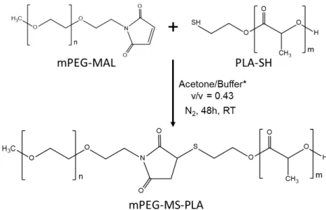
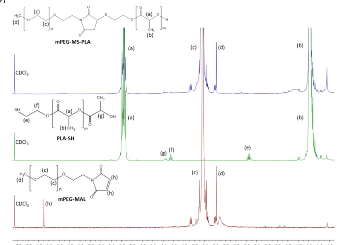
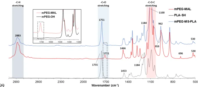
The synthesis of methoxy-poly(ethylene glycol)-maleimide-thiol-poly(L-lactide) (mPEG-MS-PLA) diblock copolymer was performed by a highly selective Michael-type coupling between the thiol moiety of PLA-SH and the double bond of the N-substituted maleimide group in mPEG-MAL, resulting in a succinimidyl thioether adduct that linked the hydrophilic and hydrophobic polymers (Figure 1A). The reaction was performed under mild conditions by comprising a mixture of acetone and phosphate buffer with 5 mM EDTA. The crude products were then purified by dialysis and freeze dried to obtain a fine white powder. The resulting mPEG-MS-PLA diblock copolymers (Figure S1) were then characterized by 1H NMR and ATR-FTIR to access the synthesis of the amphiphilic copolymer. The obtained proton NMR and FTIR spectra exhibited the characteristic peaks of both hydrophilic and hydrophobic blocks, thus providing evidence for chemical conjugation of the maleimide and thiol moieties (Figures S2 and S3).
Following successful copolymer synthesis, Naringin-loaded polymeric micelles were then self-assembled via nanoprecipitation by mixing the flavanone and the amphiphilic copolymer in the organic phase. To promote micelles assembly the drug-copolymer mixture was added drop-wise added into a determined amount of aqueous solution (V = 5 mL) at room temperature (RT) (Figure 1B). The physicochemical properties of the resulting Naringin-loaded and blank (control) micelles were characterized by DLS. As demonstrated in Figures 1C and 1D the formulated blank micelles (75.15 ± 1.06 nm) were smaller than those loaded with Naringin (84.48 ± 2.44 nm). This increase could be attributed to the relatively 101 102 103 104 105 106 107 108 109 110 111 112 113 114 115 116 117 118 119 120 121 122 123 124 125
amphiphilic features of the flavanone glycoside Naringin chemical structure (Figure 1A). This could explain the small increase in hydrodynamic size of drug-loaded micelles over blank micelles and is a valuable information since, to the best of our knowledge, this is the first time that Naringin has been encapsulated in polymeric micellar systems. We hypothesize that Naringin’s bulkier non-planar structure comprised by hydrophobic (aglycone, Naringenin) and hydrophilic (glycoside, Neohesperidose) domains could potentially decrease the hydrophobic interactions with PLA micelles core. This hypothesis is supported by Sanver on experimental modelling of flavonoid membrane interactions, which suggests that flavonoids such as Naringin may partition in the lipid/water interface.[16] This is an important factor that also supports the use of nanosized delivery systems for the intracellular delivery of this drug, which otherwise can only be partitioned in cells membrane. Moreover, Yan and his team studied Naringin/-cyclodextrin (-CD) inclusion complexes, where computational and experimental simulations indicated that while the benzene ring of Naringin was embedded within the hydrophobic (-CD) cavity, the glycoside domain was on plane with the relatively hydrophilic outer wide rim.[17] It is important to mention that the capacity for PEG to form hydrogen bonds with flavanone glycosides such as Naringin is limited,[18] whereas the PLA repeating lactide units containing carbonyls (-C=O) may provide more opportunities for hydrogen-bonding with the flavonoid hydroxyls (-OH).[19] Therefore, the amphiphilic character of Naringin could play a role in the interactions governing the hydrophobic micelle core. These insights are extremely important to optimize nanocarriers properties when the encapsulation of flavonoids such as Naringin, or others, is envisioned.
Apart from this, it is also important to note that the obtained Naringin-loaded micelles size (84.48 ± 2.44 nm) is suitable for potential parenteral delivery to bone tissue in the future since nanoparticles with diameters over 150 - 200 nm are reported to accumulate significantly in mononuclear phagocytic system (MPS) organs.[20] Furthermore, current knowledge of the 126 127 128 129 130 131 132 133 134 135 136 137 138 139 140 141 142 143 144 145 146 147 148 149 150
physiological barriers for bone nanotherapeutics delivery indicate that maintaining a nanocarrier size between 60 - 100 nm might be valuable for improving both para- and transcellular uptake into the marrow stroma.[21] From this standpoint, the nanomicelles here produced are also promising for systemic passive delivery to skeletal sites.
Regarding mPEG-MS-PLA micelles zeta potential, both blank (-13.2 ± 0.8 mV) and Naringin-loaded (-12.7 ± 0.6 mV) formulations displayed negative potential. These results are consistent with other studies employing PEG-PLA micelles in the literature.[22,23] The obtained surface charge values are interesting for parenteral delivery, since slightly negative-charged nanoparticles have longer circulation times and are less prone to opsonization by the MPS.[20] 2.2. Nanomicelles Morphological Characterization
The morphology of mPEG-MS-PLA core-shell micelles was characterized by scanning transmission electron microscopy (STEM). The obtained micrographs indicate that both blank and Naringin-loaded micelles have a well-defined spherical morphology (Figures 1D and 1E). The shape of nanocarriers is known to play a key role in their interaction with cell membrane at the nano-biointerface thus influencing the cellular uptake kinetics.[24] Spherical particles have shown a significant cellular uptake efficacy in comparison to rods, particularly for sub-100 nm nanoparticles.[25]
2.3. Nanomicelles Colloidal Stability Evaluation
The colloidal stability of Naringin-loaded mPEG-MS-PLA micelles in aqueous solutions was investigated via DLS by monitoring changes in their physicochemical properties, namely particle size, PDI and zeta potential, up to 14 days (Figure S4). Overall, the obtained results indicate that micelles are stable after a two-week storage period at 4 ºC in both deionized water and PBS buffer at physiological pH = 7.4. The produced micelles maintained a relatively constant particle size, PDI and zeta potential throughout the storage period, corroborating the stability of the maleimide-alkylthiol linkage in these conditions. Regarding 151 152 153 154 155 156 157 158 159 160 161 162 163 164 165 166 167 168 169 170 171 172 173 174 175
the micelles dispersed in PBS, they presented a slight increase in the final PDI. In conclusion, the stability studies highlight a high colloidal stability of mPEG-MS-PLA nanomicelles. 2.4. Drug Encapsulation Efficiency and In vitro Drug Release
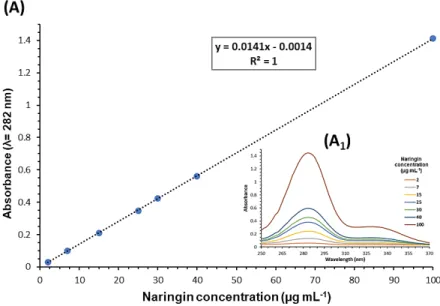
Naringin encapsulation efficacy in micelles was determined by UV-VIS analysis of the characteristic flavanone peak at λ= 282 nm. The amount of encapsulated Naringin was extrapolated from Naringin calibration curve in water (concentration range from 2 to 100 g mL-1, Figure S5) The obtained data shows that mPEG-MS-PLA micelles achieved a high encapsulation efficiency of Naringin (87.2 ± 4.6 %).
It is important to emphasize that to date, the delivery of Naringin to skeletal sites was only attempted by impregnation in implantable scaffolds, porous composites or in surface-coatings. [26–29] These works focused on the development of bone graft materials either to enhance the repair of osteoporotic bone defects, or to provide a multifunctional orthopedic coating with inherent antimicrobial and pro-osteogenic properties. Outside of the scope of skeletal delivery, one of the few examples focused on Naringin delivery by nanocarriers is the recent study performed by Feng and colleagues, in which Naringin established an inclusion complex with water-soluble ternary nanoparticles (particle size: 212 nm, PDI: 0.252).[30] These carriers consisted of amylose, -linoleic acid and -lactoglobulin, and the aim of the study was to improve Naringin bioavailability using a food grade carrier, as well as study the physicochemical properties of this inclusion complex. Moreover, Naringin encapsulation efficiency within these nanocarriers was 78.7 ± 4.2% as determined via high-performance liquid chromatography (HPLC). Considering this, the mPEG-MS-PLA micelles herein produced achieved a similar high encapsulation efficiency. Such is paramount to reduce the dose of nanocarriers that is required to be administered to stem cells in order to promote their osteogenic differentiation as proposed.
176 177 178 179 180 181 182 183 184 185 186 187 188 189 190 191 192 193 194 195 196 197 198 199
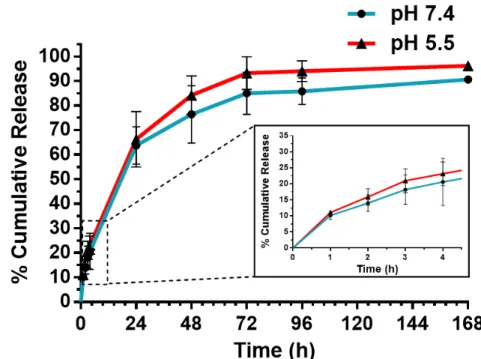
Following encapsulation, the in vitro release profile of Naringin from mPEG-MS-PLA micelles was investigated by the dialysis method under sink conditions. (Figure 2). This study was performed in PBS buffer at different pH to simulate either physiological conditions (pH = 7.4) or the acidic environment within the lysosomal/endosomal intracellular compartments (pH = 5.5).[31] The latter was considered important since mPEG-PLA micelles are reported to be internalized via dynamin- and caveolin-dependent but also clathrin-independent endocytosis pathways which results in the formation of endo-lysosomal vesicles.[32]
The observed spatiotemporal drug release profile was similar at both acidic and physiological conditions (Figure 2). Interestingly, the release profile of Naringin from mPEG-MS-PLA micelles appears to follow a biphasic release: (i) a higher rate of release during the first hours (ca. 22% released drug within 4 h), and then (ii) a slower and sustained release over the following days (Figure 2B). The obtained release profile is comparable to the recent work by Yu and colleagues, where the flavanone was loaded into a mineralized collagen coating, containing or not metal-organic frameworks (MOFs).[28] In this study, the authors observed a Naringin release at 16 h of ca. 65% from Collagen/MOFs and 85% for Collagen coating, respectively. Interestingly, the mPEG-MS-PLA micelles from this work only achieved 65% of released drug after 24 h, suggesting that these nanomicelles provide a more sustained release in comparison with the reported Collagen/MOFs coatings. The authors underline the importance of both preventing an initial burst release and assuring a long-term sustained release of Naringin for enhancing its therapeutic potential. One of the main advantages of the nanocarrier class here explored, is that polymeric micelles have the ability to tune the release profile via testing different polymeric architectures, such as shell or core crosslinking, or by imparting stimuli-responsiveness to the polymeric nanocarrier framework. [33,34] 200 201 202 203 204 205 206 207 208 209 210 211 212 213 214 215 216 217 218 219 220 221 222 223 224
2.5. Nanomicelles Cellular Uptake
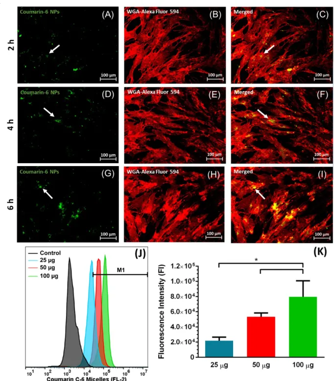
Nanocarriers cellular uptake kinetics in hASCs were initially evaluated by confocal laser scanning microscopy (CLSM) (Figures 3A-I). For this purpose, fluorescent Coumarin-6 was encapsulated within mPEG-MS-PLA micelles and the loaded nanocarriers (particle size: 63.62 ± 1.62 nm, PDI: 0.176 ± 0.003, ζ-potential: -11.8 mV ± 0.7 mV, Figure S6) were then administered to hASCs. As shown in Figure 3A-C, nanomicelles were localized in hASCs intracellular milieu within 2 h after incubation. Such findings were in agreement with three dimensional (3D) CLSM imaging of nanomicelles uptake within hASCs (Figure S7A and B). Moreover, micelles internalization and trafficking into into lyso/endosomal compartments was also confirmed by co-localization studies (Figure S7C to E). Therefore, the obtained results show that the formulated micellar carriers have potential to be internalized by these cells and can be used for stem cell-based therapies. Concerning this, micelles appear to localize to the perinuclear region but not adsorbed on cells surface, which is consistent with intracellular trafficking studies of nanocarriers in the literature.[32,35] Such nanocarriers internalization is crucial for assuring Naringin bioavailability in the intracellular compartment.
In addition to CLSM data, the cellular uptake of Coumarin-6 loaded micelles in hASCs was evaluated by flow cytometry (FCM) (Figures 3J and 3K). Different nanomicelles dosages (25, 50 and 100 g) were incubated for 4 h prior to analysis. FCM data revealed that micelles cellular uptake increased with nanomicelles dose in the tested range, exhibiting a 1.8-and 3.1-fold increase in fluorescence intensity for 50 1.8-and 100 g respectively compared to the lowest dose. Once again, these findings highlight the internalization capacity of the produced mPEG-MS-PLA micelles in these cells, thus showing promise for intracellular delivery of bioactive molecules with ability to guide their differentiation to specific lineages.
2.6. Cellular Viability and Proliferation Assays 225 226 227 228 229 230 231 232 233 234 235 236 237 238 239 240 241 242 243 244 245 246 247 248 249
Although mPEG-PLA diblock copolymers are considered highly biocompatible, investigating a possible cytotoxic response due to drug dosage is a requirement.[22] Naringin flavanone has already been described as a relatively non-toxic compound in various cell lines (e.g. MC3T3-E1, human osteoblast, UMR-106, bone marrow stromal cells), when administered in the range of 1 to 200 g mL-1.[6] However, it is important to emphasize that Naringin may have variable effects in different cell types.[8,12] Therefore, evaluating Naringin effect in hASCs viability is important and a pre-requisite for osteogenesis induction studies. The initial administration of free Naringin concentrations ranging from 5 to 50 g mL-1 showed no significant changes in cell viability across all studied time points (Figure S8A), highlighting its non-toxic features. Moreover, the obtained results show that both blank and Naringin-loaded polymeric nanocarriers maintained their biocompatible profile (Figure S8B and S8C).
Naringin has been described to significantly enhance cell proliferation of osteoprogenitor cells (e.g. MC3T3-E1 and human/rat BM-MSCs) or with an osteoblastic phenotype (e.g. osteoblast-like UMR-106 and MG-63 cells or human osteoblasts).[8,12,27,36–39] Overall, these studies investigated Naringin-induced cell proliferation by performing metabolic assays such as MTT and CCK-8. However, these assays may overestimate cell proliferation when compared to those based on DNA-binding fluorophores.[40] Hence quantifying DNA content with specific fluorophores is an important complement to these metabolic assays. Moreover, flavonoids are reported to dose-dependently reduce tetrazolium salts such as MTT even in the absence of cells, which may significantly influence the obtained results in metabolic assays.[41] In fact, some flavonoids inhibit cell growth but show enhanced MTT reduction. As such, to confirm Naringin proliferative properties, the double-stranded DNA (dsDNA) content was quantified via PicoGreen® in the same samples of hASCs used for cytotoxicity assays. By using this strategy, a correlation between metabolic activity and DNA content could be 250 251 252 253 254 255 256 257 258 259 260 261 262 263 264 265 266 267 268 269 270 271 272 273 274
extrapolated. As shown in Figure S9, free Naringin increased the proliferation of hASCs relatively to control groups on all time points and across the studied concentrations. Interestingly, the obtained data seems to suggest that the effect of Naringin on the rate of proliferation increases over time.
It should be emphasized that this study is one of the few exploring the osteogenic potential of Naringin in hASCs. MSCs extracted from human adipose tissue represent a valuable source of cells for regenerative therapies. Besides their human origin, they can be easily harvested from adult adipose tissue, possess low antigenicity and comparatively to hBM-MSCs, exhibit faster proliferation rates and increased genetic stability in prolonged culture periods.[42,43] 2.7. Naringin pro-osteogenic activity in hASCs
2.7.1. Naringin-induced Stimulation of ALP Activity
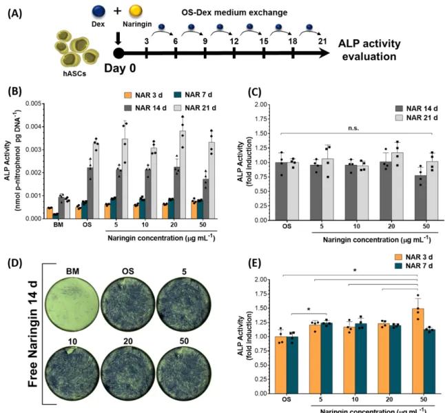
Regarding studies on the pro-osteogenic potential of free Naringin, different literature reports indicate different choices for the osteogenic inductive medium. Most of these reports vary between cells grown in osteoinductive (i) [ascorbic acid + -glycerophosphate] – rOS; or (ii) [ascorbic acid + -glycerophosphate + Dex] – OS-Dex; or (iii) basal culture medium (BM) conditions. As aforementioned, this can lead to different outcomes in the osteogenic differentiation potential of stem cells. Therefore, initially the osteogenic capacity of free Naringin in hASCs was evaluated under in different culture media (Table 1, Experimental Section, Cell Culture subsection). For the first experiment, hASCs were incubated once (t=0h) with different concentrations of free Naringin in OS-Dex medium over 21 days (Figure 4A, administration regime schematics). Throughout the assay, cell culture medium was then exchanged every 3 to 4 days with fresh OS-Dex medium. The alkaline phosphatase (ALP) activity was then quantified by p-nitrophenylphosphate hydrolysis and normalized to dsDNA content by using the PicoGreen® assay.
275 276 277 278 279 280 281 282 283 284 285 286 287 288 289 290 291 292 293 294 295 296 297 298
The obtained results presented in Figure 4B show an increase in ALP activity along time after incubation in OS-Dex control group and with different Naringin doses. The ALP activity is the highest at the final time point of the experiment, i.e., 21 days of incubation, which might suggest that continuously exchanging the osteogenic medium with a renewed Dex dose (100 nM) might lead to continous stimulation of hASCs populations. Alternatively, according to the obtained results, the use of Naringin as an initial stimulation factor to promote osteogenesis appears to be limited to the earlier time points, i.e., 3 and 7 days of incubation. Indeed, as shown in Figure 4C, at 14 and 21 days of incubation, no significant differences in ALP activity can be observed among different Naringin doses and the respective OS-Dex control group. Likewise, these findings are supported by Figure 4D, which shows the ALP-dependent staining of hASCs at 14 days via BCIP/NBT substrate. As can be observed there are no significant differences between osteogenic medium ALP-stained cells and those incubated with various Naringin dosages.
However, for the earlier time points, i.e., 3 and 7 days of incubation, there is a significant osteogenic effect of Naringin over the OS-Dex control groups, as shown in Figure 4E. At 3 days, the highest Naringin dose (50 g mL-1) achieved a 0.5-fold improved ALP activity over the OS-Dex control group and a 0.3-fold ALP increase across all studied doses, from 5 to 20 g mL-1. At 7 days, no significant difference across all Naringin doses could be observed indicating that lower doses are equally beneficial. Moreover, in comparison with OS-Dex control group, an increase in ALP activity (ca. 0.2-fold) could be observed across all Naringin doses at 7 days (Figure 4E).
In summary, a significant pro-osteogenic effect between different Naringin doses was observed at 3 days, while the osteogenic effect between Naringin and OS-Dex control groups was observed at 3 and 7 days. These findings suggest that perhaps constant long-term stimulation of hASCs with Dex, with only an initial stimulatory dose of Naringin (Figure 4A), 299 300 301 302 303 304 305 306 307 308 309 310 311 312 313 314 315 316 317 318 319 320 321 322 323
might dillute this flavonoid effect on the promotion of ALP levels over the course of the experiment.
2.7.2. Naringin-induced BMP-2 expression
In addition to the evaluation of long-term ALP-stimulatory activity mediated by Naringin, the expression levels of other osteogenesis-related markers were evaluated to further investigate the contribution of Naringin in differentiating these stem cells into the osteoblastic lineage.
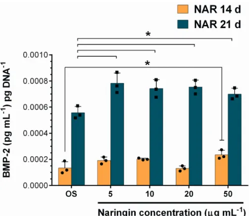
Naringin has been previously described to promote the secretion of BMPs which play a key role in modulating osteogenic differentiation pathways and coordinating bone formation. [12] The quantification of BMP-2 in the culture medium from the previous in vitro assays was performed via ELISA at 14 and 21 days of incubation. The results obtained in Figure 5 highlight the contribution of Naringin over the OS-Dex group in improving the secretion of BMP-2. At 14 days, a dose-dependent stimulation of BMP-2 could be observed, with the highest Naringin dose of 50 g mL-1 significantly improving BMP-2 levels over OS-Dex and 20 g mL-1 dose groups. In addition, the cumulative BMP-2 levels at 21 days show a 0.4-fold improvement over those obtained in OS-Dex control group. In parallel, cumulative BMP-2 expression at 21 days was also augmented upon an initial dose of Naringin-loaded nanomicelles (Figure S11). Here, the highest dose significantly improved BMP-2 levels over osteogenic control group and lower doses (5 and 10 µg/mL). Overall, these results underline that a single initial dose of Naringin was sufficient to elicit an increased BMP-2 production over control-treated hASCs at 21 days. Furthermore, they showcase the importance of evaluating different osteogenic markers, other than ALP activity, because these can convey important findings regarding Naringin effect in hASCs.
324 325 326 327 328 329 330 331 332 333 334 335 336 337 338 339 340 341 342 343 344 345 346
It is worth noting that this increased BMP-2 production did not lead to significant differences in ALP activity after 14 or 21 days of OS-Dex stimulation. These findings are in agreement with a previous study by Cruz and colleagues, where they found that exogenous administration of recombinant BMP-2 to hASCs did not increase ALP levels.[44] The enhanced secretion of BMP-2 by Naringin in hASCs is an important finding, in particular because this bone morphogenetic protein is capable of inducing the differentiation of multipotent stem cell lines into an osteoblastic-phenotype.[45] Moreover, Naringin appears to have a synergistic effect with BMP-2 in the total osteogenic differentiation of MC3T3-E1 pre-osteoblasts.[13] So far, across the literature there are few studies describing Naringin-induced BMP-2 secretion. One study reported this effect in murine primary fetal osteoblasts, i.e., differentiated cells with an already defined osteoblastic phenotype.[12] Alternatively, a second work studied this effect in human degenerative disc nucleous pulposus cells.[46] Therefore, the above findings provide evidence of this effect for the first time in hASCs and its importance is supported by the role of BMP-2 in the osteogenic commitment of stem cells.
2.7.3. Naringin-induced Osteopontin expression
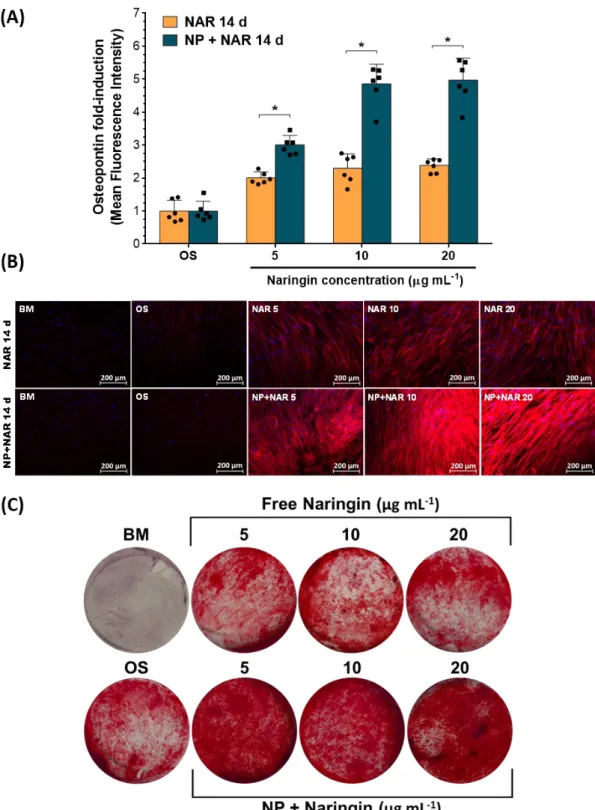
Naringin has been previously described to increase osteopontin (OPN) expression in both osteoprogenitor (e.g. hBM-MSCs, MC3T3-E1) and osteoblastic cell lines (e.g. human and murine primary fetal osteoblasts), reflecting the osteogenic potential of this flavanone for bone tissue engineering applications due to the role of OPN in promoting biomechanical osteointegration.[12,37] To evaluate this potential, hASCs were initially incubated with different concentrations of free Naringin and also Naringin-loaded nanomicelles in OS-Dex containing medium over 14 days. Throughout the assay, cell culture medium was exchanged every 3 to 4 days with fresh OS-Dex medium (Figure 4A, dose regime schematics). The expression of OPN was then evaluated by immunocytochemistry.
347 348 349 350 351 352 353 354 355 356 357 358 359 360 361 362 363 364 365 366 367 368 369 370
The obtained results shown in Figures 6A and 6B emphasize the remarkable capacity of Naringin to induce OPN expression in hASCs. In fact, a single stimulatory dose of Naringin was able to significantly enhance OPN expression after 14 days of culture (Figure 6A). Regarding stimulation with free drug, all tested doses (5, 10, 20 g mL-1) were able to significantly enhance OPN expression by aproximately 1-fold when compared to control groups. Notably, incubation with Naringin-loaded nanomicelles elicited a 2-fold increase at 5 g mL-1 and a 5-fold increase in OPN expression at the higher doses, accordingly 10 and 20 g mL-1 versus control groups. Moreover, the dose-dependent effect of Naringin-loaded micelles was evidenced since the highest doses (10 and 20 g mL-1) improved OPN expression by 1.7-fold in comparison to that obtained at 5 g mL-1 dosage. In addition, it is important to highlight that the controlled delivery of Naringin via nanomicelles led to a higher OPN secretion when compared to free drug at the same doses.
These findings are in agreement with various literature reports investigating the effect of Naringin in OPN expression on other cell lines. For instance, Wu and colleagues investigated the in vitro potential of Naringin in promoting OPN secretion over three different cell lines: MC3T3-E1, human and murine primary fetal osteoblasts.[12] These researchers observed via ELISA assays that after a 3-day incubation period with 3 M of Naringin (i.e., 1.74 g mL-1), a 3-fold increase in extracellular OPN expression over control across all cells was obtained. Alternatively, Yin and his team, evaluated the Naringin-induced pro-osteogenic effect in human periodontal dental ligament stem cells.[47] The obtained results in this study showed a Naringin-mediated 0.4 to 0.7-fold OPN expression induction in comparison to basal medium control, and in a dose-dependent manner.
Another important aspect that should be mentioned in the context of hASCs lineage differentiation, is the relation between OPN secretion and adipogenesis. Concerning this, Chen and colleagues investigated the key role of the interaction between OPN and integrin 371 372 373 374 375 376 377 378 379 380 381 382 383 384 385 386 387 388 389 390 391 392 393 394 395
V/1 in defining MSCs differentiation.[48] These authors compared the adipogenic and osteogenic differentiation potential of mouse BM-MSCs derived from wild-type and OPN-/ -mice. The OPN blockade skewed mouse BM-MSC differentiation towards the adipocyte lineage in vitro. Therefore, this study suggests that increased OPN expression might be valuable for pursuing osteogenic commitment of MSCs. Besides significantly increasing OPN expression, Naringin also modulates other pathways (e.g. inhibition of PPAR and activation of Notch signaling) that all play a role in promoting osteogenesis over adipogenesis.[7,8]
2.7.4. Naringin-induced Mineralization
In the literature, Naringin has been described to promote mineralization of osteoprogenitor cells in a dose-dependent manner, which is ultimately the goal of bone tissue engineering applications.[8,28]
To evaluate this effect in hASCs, the cells were incubated with different concentrations of free Naringin and also Naringin-loaded nanomicelles in OS-Dex containing medium over 21 days, similarly to previous assays. The deposition of calcium nodes in the mineralized cell monolayer was visualized by Alizarin Red S staining.
The obtained results shown in Figure 6C highlight the potential of a single stimulatory dose of Naringin to induce mineralization in a dose-dependent manner after 21 days. These findings are supported by the work of Yu and colleagues who observed a dose-dependent calcium node deposition from 1, 10 to 50 g mL-1 of Naringin in rat BM-MSCs.[8] Herein, the dose-dependent effect of Naringin is clearly visible upon Naringin-loaded micelles administration. In comparison to free drug, the controlled delivery of Naringin markedly improved mineralization of the cell matrix. Mineralization data is paramount for pursuing further applications in the future, especially considering the origin of the used stem cells (adipose-derived) and the effect of Dex in their pro-adipogenic/pro-osteogenic differentiation 396 397 398 399 400 401 402 403 404 405 406 407 408 409 410 411 412 413 414 415 416 417 418 419
duality. Indeed, a study by Ghali and co-workers found that addition of Dex at as low as 100 nM in osteogenic medium strongly induced adipogenesis, even exhibiting a higher lipid droplet accumulation than 500 nM of Dex in adipogenic induction medium.[3] Taking into consideration that this was the concentration of Dex used during these experiments, the above findings provide evidence for significant hASCs mineralization elicited by Naringin, with evident matrix osteogenic calcification. Moreover, the well documented Naringin inhibition of adipogenic regulator PPAR, a nuclear receptor that induces adipogenic comittment, could help explain the improved mineralization of Naringin-loaded nanomicelles groups [49].
2.8. Effect of Naringin dose regime in ALP Activity
Collectivelly, the previous experiments investigating different osteogenic markers reveal the pro-osteogenic potential of Naringin, either in free form or when delivered by micellar carriers as demonstrated by OPN expression and matrix calcium deposition data. Yet, in the previous assays, Naringin was used in combination with osteogenic medium containing Dex, which appeared to dilute the flavonoid effect on the stimulation of ALP activity. In fact, in OS-Dex experiments quantifying ALP activity, differences among Naringin doses were not significant at 7 days.
Therefore, different dose regimes of free Naringin and drug-loaded micelles in reduced osteogenic medium (rOS – ascorbic acid + β-glycerophosphate, not containing Dex) were studied in order to further investigate Naringin pro-osteogenic effect in hASCs as well as its ALP stimulatory activties (Figure 7A) in more in vivo-like conditions without Dex administration. It was hypothesized that, in rOS induction medium, the addition of a second dosage of free Naringin or Naringin-loaded micelles could elicit different results on the ALP activity of hASCs. Such are important findings for the development of stem cell-based therapies based on the most effective Naringin regime for enhancing osteogenic differentiation. 420 421 422 423 424 425 426 427 428 429 430 431 432 433 434 435 436 437 438 439 440 441 442 443 444
Hence, further experiments were designed to evaluate the effect of adding a second dose of free Naringin or Naringin-loaded micelles after 3 days of incubation. The obtained results shown in Figure 7B highlight the superior performance of Naringin-loaded mPEG-MS-PLA nanomicelles over free drug incubation, at 3 days. Naringin-loaded micelles significantly improved ALP activity levels (ca. 0.3 fold over the OS control group), across all doses of free Naringin studied.
Regarding the 7 day assays, different performances according to the administration regimen could be observed for free drug and drug-loaded micelles. For the free drug assays, a single dose of Naringin in rOS promoted ALP activity in a dose-dependent manner, with the highest dose (50 g mL-1) showing a 0.15 to 0.20 fold improval over 5 g mL-1 and the rOS control group (Figure 7C). Also, a single dose of Naringin led to a 0.2 to 0.3 fold increase in ALP activity when compared to the dual administration regimen, specifically 10, 20 and 50 g mL-1 concentrations. In the two-dose nanocarrier administration approach, all Naringin concentrations were capable of significantly improving the ALP activity by 0.3 to 0.4 fold over the rOS control group, which was not observed for the single-dose strategy (Figure 7D).
Overall, different performances according to the dose regime could be observed for free drug and drug-loaded micelles. In the free drug assay, a second dose seemed prejudicial for ALP stimulation, whereas a second dose of Naringin-loaded nanocarriers significantly increased the ALP activity over rOS control group. Cytotoxicity inherent to a second dose administration was not proposed here because dsDNA quantifications of both assays showed no significant changes between the two dose regimens (Figure S10). The above findings are supported by the work of Guo and colleagues, where they investigated the double directional estrogenic effect of Naringin.[50] The authors concluded that Naringin showed estrogenic agonist activity at low concentrations, but acted as estrogenic antagonist at high concentrations. The interaction of Naringin with estrogen receptor and its role in osteogenesis 445 446 447 448 449 450 451 452 453 454 455 456 457 458 459 460 461 462 463 464 465 466 467 468 469
is well described in the literature, and the obtained results above may suggest that intracellular delivery of Naringin might overcome this limitting effect.[50,51]
Figure 7E provides an overall comparison between all the tested dose regimen groups, with both free drug and drug-loaded groups. For one-dose regimen, free Naringin and Naringin-loaded groups exhibited similar enhancements of ALP stimulation at 7 days. Overall, for two dose regimes, Naringin-loaded micelles were significantly superior across all concentrations studied. In addition, the obtained results show that dual nanocarrier administration requires lower Naringin doses to effectively improve ALP activity versus one-dose regimens.
In light of previous results, the superior performance of the dual nanocarrier regime was also evaluated in later timepoints, i.e. 14 and 21 days of culture, and compared against free drug administration (Figure 7F and 7G). At 14 days, the obtained results once again highlight the superior osteopromoting potential of Naringin-loaded nanomicelles over free Naringin treatment modalities (Figure 7F). Indeed, except for the lowest concentration (5 µg/mL), all nanomicelle doses have significantly augmented the ALP-promoting effect of Naringin over the corresponding free drug administrations. Remarkably, the highest dose of Naringin-lodaded nanomicelles achieved a 0.9-fold increase in ALP activity versus the osteogenic control (rOS) and a 0.7-fold increase when compared to the highest free drug dose. Moreover, a dose-dependent increase in ALP activity is particularly evident for the nanomicelle administration regime. Herein, it can be observed that lower doses of nanomicelles (10 µg/mL) can achieve comparable improvements to the highest doses of free Naringin studied (50 µg/mL), which once again highlights the remarkable potential of nanomicelle-delivered Naringin for accelerating osteodifferentiation in hASCs. At 21 days, however, the majority of hASCs have undergone extensive osteodifferentiation and no significant differences can be seen among osteogenic conditions for both free drug and nanomicelles groups (Figure 7G). 470 471 472 473 474 475 476 477 478 479 480 481 482 483 484 485 486 487 488 489 490 491 492 493 494
The obtained results indicate the potential of the formulated nanocarriers to deliver Naringin to hASCs and significantly enhance its pro-osteogenic effect over free drug administration, especially at accelerating cell osteodifferentiation and requiring lower doses. 3. Conclusion
Throughout this study, the potential of a naturally available drug to guide cells differentiation towards an osteoblastic phenotype was explored. The inclusion of Naringin in micellar nanocarriers resulted in a high encapsulation efficiency and led to a sustained drug release in both physiological and endo-lysosomal conditions. Moreover, these nanocarriers were readily internalized by hASCs as demonstrated by microscopy imaging. The controlled delivery of Naringin generally elicited a more pronounced ALP expression over free drug administration. In addition, a single initial stimulatory dose of Naringin-loaded nanocarriers significantly increased OPN expression over free drug after 14 days. Moreover, Naringin delivery via nanomicelles significantly improved hASCs matrix mineralization over free drug at 21 days. Taken together, the findings of this study provide evidence that the inclusion of Naringin in biocompatible nanocarriers effectively promotes the osteogenic differentiation of hASCs in vitro. Such findings can ultimately provide new design directions for future studies either in stem cell-based therapies or as a novel bone nanomedicine therapeutic agent.
In the foreseeable future, the design of Naringin micellar delivery system could be enhanced by inclusion of stimuli-responsive moieties into nanocarriers polymeric backbone in order to fine tune the delivery process upon specific stimuli. In fact, there are several interesting biophysiological cues within the bone tissue and alterations found in skeletal disorders that could potentially be exploited for stimuli-responsive delivery of bioactive molecules. The latter is particularly interesting since during the onset and progression of different bone disorders many of skeletal microenvironment hallmarks and cellular functions become profoundly deregulated. Each of these disease-specific features represent unique 495 496 497 498 499 500 501 502 503 504 505 506 507 508 509 510 511 512 513 514 515 516 517 518 519
opportunities for nanocarrier-mediated stimuli-responsive release of therapeutics. This spatiotemporally controlled drug release can result in higher stability in vivo and improved biodistribution of the drug at skeletal sites upon parenteral administration. The combination of bioinspired therapeutics with design advances in nanocarriers performance, will undoubtedly pave the future in providing suitable candidates for future commercialization and realistic clinical application.
4. Experimental Section
Materials: Methoxy-poly(ethylene glycol)-maleimide (mPEG-MAL) (Mn 5000, > 90 %
purity), thiol-poly(L-lactide) (PLA-SH) (Mn 5000, PDI < 1.2); 4-Nitrophenyl phosphate disodium salt hexahydrate (4NPhP), 4-Nitrophenol 10 mM (4NPh), phosphate buffered saline (PBS), formaldehyde, Coumarin-6, Naringin ( > 95 % purity), β-glycerophosphate disodium salt hydrate, Alizarin Red S, Triton X-100 and Amicon® Ultra-4 mL (3000 MWCO) were all purchased from Laborspirit (Lisbon, Portugal). Spectra/Por 1 dialysis tubing (6000 - 8000 Da MWCO) and Float-A-Lyzer G2 (3500 - 5000 Da MWCO) dialysis cassettes were purchased from Reagente 5 (Oporto, Portugal). Human adipose-derived mesenchymal stem cells (hASCs, Homo Sapiens, ATCC® PCS-500-011™) were purchased from LGC Standards S.L.U. (Barcelona, Spain). Minimum Essential Medium -modification (-MEM), fetal bovine serum (FBS, E.U. approved, South American origin), antibiotic, TrypLE™ Xpress Enzyme with phenol red (1X), Quant-iT™ PicoGreen® dsDNA Assay Kit, AlamarBlue™, 1-Step™ NBT/BCIP Substrate Solution, and Wheat Germ Agglutinin (WGA) Alexa Fluor® 594 conjugate dye and DAPI were purchased from Alfagene (Lisbon, Portugal). Dexamethasone (Dex, 96% purity) and Dulbecco’s PBS were purchased from Thermo Fisher Scientific (Oeiras, Portugal). L-ascorbic acid 2-phosphate magnesium salt was purchased from VWR (Lisbon, Portugal). 520 521 522 523 524 525 526 527 528 529 530 531 532 533 534 535 536 537 538 539 540 541 542 543
Synthesis of mPEG-MS-PLA Copolymer: mPEG-MS-PLA copolymer was synthesized via
a Michael-type addition reaction between mPEG-MAL and PLA-SH (Figure S1).[52] Briefly, mPEG-MAL (1.5 mPEG/PLA molar ratio, 0.04 mmol) and PLA-SH (0.0267 mmol) were dissolved in a mixture of acetone (v/v = 0.43) and sodium phosphate buffer (pH = 7.2, 100 mM, v/v = 0.57) containing EDTA (5 mM) and mixed under inert conditions (N2) for 2 days at RT. Afterwards, the resulting crude mixture was completely dried in a rotary evaporator (Buchi, Rotavapor® R-300), dissolved in dichloromethane and then precipitated in cold diethyl ether. The recovered precipitate was then dialyzed (6000 - 8000 Da MWCO) against deionized water for 72 h, before being frozen at -80 ºC and freeze-dried for storage.
Spectroscopy Characterization of mPEG-MS-PLA Copolymer: The successful synthesis of
mPEG-MS-PLA was characterized by different spectroscopy techniques. Proton nuclear magnetic resonance (1H NMR) spectra were recorded on a Bruker Advance III 400 MHz spectrometer. Prior to spectra acquisition, samples were dissolved in CDCl3 and transferred to 300 MHz NMR glass tubes (Sigma-Aldrich, Sintra, Portugal). NMR spectra were acquired with 18 s relaxation delay, 256 scans and 32 dummy scans. Data processing was performed in the MestReNova v6.0.2 software, and spectra were normalized according to the established CDCl3 solvent peak at 7.26 ppm. Fourier Transformed Infrared spectra were collected on dried powder samples with attenuated total reflectance (ATR-FTIR) by using a Bruker Tensor 27 spectrometer. The spectra of all samples were recorded at a 4 cm-1 resolution with a total of 256 scans in the spectral width of 4000 to 350 cm-1. Spectral data was processed in OPUS software.
Preparation of mPEG-MS-PLA Nanomicelles: Self-assembly of Naringin-loaded or blank
mPEG-MS-PLA nanomicelles was performed by nanoprecipitation. First, the copolymer (5 mg) was dissolved in 1 mL of freshly prepared acetone solution containing Naringin (10 % w/w of copolymer) under ultrasound sonication for 5 min. Then, the solution was added 544 545 546 547 548 549 550 551 552 553 554 555 556 557 558 559 560 561 562 563 564 565 566 567 568
dropwise into a 10 mL round-bottom flask containing 5 mL deionized water and stirred for 90 min at 400 rpm, RT. Afterwards, acetone was evaporated under a rotary evaporator during 8 min (37 ºC, 50 mbar). The resultant solution was dialyzed (3500 -5000 Da MWCO) against deionized water for 1 h to remove free Naringin. Blank micelles were prepared following the above-mentioned procedure without dialysis.
Physicochemical Characterization of Nanomicelles: The average hydrodynamic particle
radius (Hr), size distribution (PDI) and -potential of the different micelles (1 mg mL-1) were characterized by dynamic light scattering (DLS) with a Zetasizer Nano ZS equipment (Malvern Instruments Ltd., Malvern, UK). All measurements were carried out in triplicate at 25 ºC and with a 173º backscatter angle. Nanomicelles morphology was observed by scanning transmission electron microscopy (STEM). Samples were prepared by drop-wise addition of 10 L of freshly prepared micelles (0.2 mg mL-1) on a carbon-film copper grid and left to dry overnight, at RT. STEM micrographs were acquired in a Hitachi SU-70 STEM microscope, operated at an accelerating voltage of 20.0 kV.
Evaluation of Nanomicelles Colloidal Stability: Nanomicelles colloidal stability was
investigated by monitoring changes in size, PDI and -potential along time upon storage in solution. For stability studies Naringin-loaded nanocarrier formulations (n=3), were prepared by nanoprecipitation and maintained in deionized water or PBS (pH = 7.4) at 4 ºC. Nanomicelles and their physicochemical properties were then analyzed by DLS at different time intervals: 0, 1, 7 and 14 days.
Drug Loading: Naringin encapsulation efficiency was determined by ultraviolet-visible
(UV-VIS) absorbance of the flavanone peak of Naringin (λ= 282 nm) corresponding to the benzoyl moiety. This peak is followed by a another region of smaller intensity to higher wavelengths (300 – 400 nm), associated with the cinnamoyl moiety and is characteristic of 569 570 571 572 573 574 575 576 577 578 579 580 581 582 583 584 585 586 587 588 589 590 591 592
flavanones such as Naringin (Figure S5A1).[53] Briefly, after free drug removal,
Naringin-loaded nanomicelles solution (0.2 mg mL-1) was analyzed by UV-VIS at 282 nm. Blank nanomicelles (0.2 mg mL-1) established the blank for UV-VIS quantification. The UV-VIS absorbance was measured in a microplate reader equipped with a tungsten halogen lamp (Synergy HTX Biotek, Izasa Scientific, Carnaxide, Portugal) by using a quartz microplate (Hella transparent 96-wellplate, VWR, Lisbon, Portugal). A standard calibration curve of Naringin in deionized water was used to determine the drug encapsulation efficacy (Figure S5). Encapsulation efficiency was calculated using the below formula:
Encapsulation Efficiency (%):
Amount of Naringin within themicelles
¿
¿(¿ ¿Theoretical amount of Naringin added)×100
In vitro Drug Release Profile: The in vitro Naringin release profile was investigated in
PBS at various pH conditions to mimic different physiological scenarios: pH 7.4 (physiological pH) and pH 5.5 (endo-lysosomal pH). Briefly, 2 mL of freshly prepared Naringin-loaded micelles (1 mg mL-1) were transferred to a dialysis tubing (3500 - 5000 Da MWCO) and submerged in 15 mL of PBS at 37 ºC at a constant stirring rate (600 rpm). At defined time intervals, 1 mL samples were withdrawn from the dialysate and replaced with the same volume of fresh PBS. Samples were withdrawn at different time points until the plateau phase was achieved. For this study, the standard calibration curve of the drug in water was used (Figure S5).
Cell Culture: Cells were manipulated within a Class II biological safety cabinet and
maintained as per established guidelines in a humidified 5 % CO2 atmosphere incubator with 95 % O2 and at 37 ºC. [54] hASCs were routinely cultured in -MEM supplemented with 10 % (v/v) heat-inactivated FBS and 1 % of antibiotic mixture (basal growth medium, BM) and media was exchanged every 5 days. Cells were subcultured before reaching confluence. All 593 594 595 596 597 598 599 600 601 602 603 604 605 606 607 608 609 610 611 612 613 614 615 616
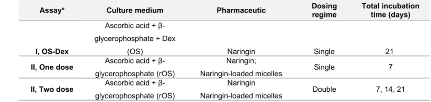
plastic adherent well plates were made of tissue culture treated polystyrene (Sarstedt, Sintra, Portugal). For cellular experiments investigating osteogenic differentiation, ascorbic acid, β-glycerophosphate and Dex stock solutions were prepared in dPBS and frozen at -20ºC. Different osteogenic media were prepared from such aliquots. Osteogenic supplements were added to BM in the following concentrations: (i) reduced osteogenic differentiation medium (rOS) - 50 g mL-1 ascorbic acid and 10 mM β-glycerophosphate; and (ii) OS-Dex – rOS and 100 nM Dex. For preparation of free Naringin concentrations, the drug was initially dissolved in sterile dimethylsulfoxide (50 mg mL-1) and subsequently diluted in the respective assay media (basal or osteogenic) and to a final 0.1 % (v/v) dimethylsulfoxide content across all concentrations. Osteogenic assays involving alkaline phosphatase (ALP) activity and BMP-2 quantifications, as well as OPN and Alizarin Red S visualization followed the below described conditions (Table 1).
Table 1. Overview of the conditions used in osteogenic differentiation assays.
Assay* Culture medium Pharmaceutic Dosingregime Total incubationtime (days)
I, OS-Dex
Ascorbic acid + β-glycerophosphate + Dex
(OS) Naringin Single 21
II, One dose Ascorbic acid +
β-glycerophosphate (rOS)
Naringin;
Naringin-loaded micelles Single 7
II, Two dose Ascorbic acid +
β-glycerophosphate (rOS)
Naringin
Naringin-loaded micelles Double 7, 14, 21
*Across all experiments, medium was exchanged every 3 days containing the respective osteogenic supplements listed above, except for the Two dose assay, where medium with a second dose of pharmaceutic was added after 3 days. Please see the corresponding dose regimen schematics in each result panel.
In vitro Cellular Uptake Studies: Nanomicelles cellular uptake kinetics in hASCs were
evaluated via fluorescence microscopy. Briefly, hASCs were seeded in -Slide 8-well chambers (ibidi GmbH) overnight at a density of 8.0 and 25.0 x 10³ cells well-1, respectively, in free-antibiotic BM. Then, cells were incubated with 250 g well-1 of Coumarin-6-loaded nanocarriers for different time intervals (2, 4 and 6 h). After each time point, cells were fixed 617 618 619 620 621 622 623 624 625 626 627 628 629 630 631 632 633 634 635 636 637 638
in 4% formaldehyde. After 6 h, fixed cells were washed three times with dPBS and then the cells cytoplasm was labelled with 50 L of WGA Alexa Fluor® 594 conjugate dye (0.2 mg mL-1) by incubating for 5 min in the dark. Afterwards, cells were rinsed three times in dPBS and maintained in dPBS until fluorescence microscopy analysis. CLSM and fluorescence microscopy (Axio Imager 2, Zeiss) were used to follow the kinetics of intracellular internalization of the nanocarriers. Image processing was performed using ZEN v2.3 blue edition software (Carl Zeiss Microscopy GmbH). Alternatively, the quantitative measurement of Coumarin-6 fluorescence intensity was conducted using a flow cytometer. hASCs were seeded in 24-well plates overnight at a density of 50 and 40 x 103 cells well-1 (n=4) respectively, in free-antibiotic BM. Then, cells were incubated with the 96-well plate equivalent of 25, 50 and 100 g of Coumarin-6- nanocarriers per well. Nanocarriers were previously concentrated by centrifuging for 30 min in Amicon® ultra centrifugal units (3000 Da MWCO) at 16600 g. After 4 h incubation, cells were washed twice with dPBS, trypsinized (500 L, 5 min incubation at 37ºC), neutralized with equal volume of dPBS and collected by centrifugation at 500 g for 5 min. Then, the supernatant was aspirated and cells were suspended in 200 L of dPBS prior to fluorescent intensity measurements.
Cytotoxicity and Cellular Proliferation Assays: Assessment of hASCs cell metabolism was
performed by using the AlamarBlue® Cell Viability assay. Briefly, hASCs were seeded at a density of 3.5 x 10³ cells well-1 (n=5) in a 96-well plate overnight in BM. Then, cells were incubated with BM containing free Naringin or Naringin-loaded micelles at a final drug concentration of 0 (K-, negative control), 5, 10, 20 and 50 g mL-1 of Naringin. For blank and Naringin-loaded micelles cytotoxicity evaluation, nanocarriers were concentrated in BM and diluted according to the different tested concentrations. For blank micelles experiments, cells were incubated with BM containing nanocarrier dosages of 25, 50, 75, 100, 200 g nanocarriers well-1. For these assays, and different Naringin concentrations of 0 (K-, control), 639 640 641 642 643 644 645 646 647 648 649 650 651 652 653 654 655 656 657 658 659 660 661 662 663
5, 10, 20 and 50 g mL-1 were used. During the assays, all cells were incubated for 24, 48 and 72 h. After each timepoint, the medium was exchanged to BM containing AlamarBlue according to manufacturer’s instructions. Following an overnight incubation period, the media was then transferred to a black clear bottom 96-well plate for analysis. AlamarBlue fluorescence was detected at an excitation/emission of λex = 540 nm/ λem = 600 nm by using a multi-mode microplate reader. All conditions were normalized to the control group (BM) set at 100 % viability. Then, samples from the previous cytotoxicity assays (regarding free drug groups) were rinsed with dPBS, incubated with 200 L of 2 % Triton X-100 in deionized water and sonicated for 7 min before plates were frozen at -20 ºC. This freeze-thaw cycle was repeated one more time before determining lysates dsDNA content with the PicoGreen kit according to the manufacturer’s instructions. Cell dsDNA content was then determined after incubation for 5 min in the dark at RT. The samples were excited at λ = 485 nm and fluorescence intensity was measured at λ = 520 nm in a microplate reader.
ALP Activity Measurement Assays: The ability of Naringin to induce osteogenic
differentiation of hASCs was evaluated in OS-Dex or rOS. hASCs were seeded overnight in a 48-well plate at a density of 3.5 x 10³ cells well-1 (n=4) in BM. Then, medium was replaced with BM and OS-Dex containing free Naringin or Naringin-loaded polymeric nanomicelles at a concentration of 0 (control), 5, 10, 20 and 50 g mL-1 of Naringin, accordingly. All cells were incubated for 3, 7, 14 and 21 days and the respective differentiation medium was exchanged every 3 to 4 days. After each time point, cells were washed twice with dPBS and incubated with 300 L of 2 % Triton X-100 solution in an ultrasound bath for 7 min (37 Hz, sweep field, 60% potency) before being frozen at -20 ºC. This freeze-thaw cycle was repeated one more time before determining ALP activity in the lysates by using 4NPhP ALP-mediated hydrolysis to quantify 4NPh release. For this quantification, 25 L of each lysate sample were added to 75 L of a freshly prepared 4NPhP solution (2 mg mL-1) in 1 M diethanolamine 664 665 666 667 668 669 670 671 672 673 674 675 676 677 678 679 680 681 682 683 684 685 686 687 688
(DEA) buffer (pH 9.8, with 0.5 mM MgCl2). The samples were incubated in the dark at 37ºC for 1 h. Enzyme activity was then quantified by UV-VIS analysis at λ = 405 nm. A standard curve of 4NPh was used as reference (0, 15 30, 50, 75, 95 M in DEA buffer). ALP activity was normalized according to the total DNA content in cell lysate determined by a PicoGreen® DNA Quantification kit according to the aforementioned protocol, and ALP was expressed as nanomole of p-nitrophenol pg DNA-1.
ALP Staining: To visualize the ALP activity in the cell monolayer, cells were stained with
1-Step™ NBT/BCIP Substrate Solution. Briefly, hASCs were seeded overnight in 48-well plates at a density of 3.5 x 10³ cells well-1 (n=3) in BM. Then, cells were treated with BM and OS-Dex containing free Naringin or Naringin-loaded polymeric nanomicelles at a concentration of 0 (K-, control), 5, 10, 20 and 50 g mL-1 of Naringin. Cells were incubated for 14 days and differentiation medium was exchanged every 3 to 4 days. After each timepoint, cells were fixed as aforementioned and rinsed in dPBS before adding 200 L of NBT/BCIP and incubating for 1 h at 37 ºC. Stained cells were imaged with a Stemi 508 Stereo Microscope (Zeiss, Taper, Sintra, Portugal).
ELISA Immunoassay Quantification of BMP-2 Secretion: BMP-2 secretion from hASCs
was determined via ELISA kit for human BMP-2 according to the manufacturer’s instructions. For this assay, culture medium from the ALP activity experiments with OS-Dex at 14 and 21 days was collected and frozen at -20ºC. Then, BMP-2 levels were quantified by UV-VIS analysis at λ = 450 nm with the absorbance measured at λ = 550 nm serving as correction factor by using a microplate reader. BMP-2 levels were expressed in pg mL-1 of BMP-2 normalized to dsDNA content.
Osteopontin Immunostaining: Osteopontin (OPN) expression was visualized via
fluorescence imaging of the extracellular layer formed during in vitro culture. Initially, 689 690 691 692 693 694 695 696 697 698 699 700 701 702 703 704 705 706 707 708 709 710 711 712
hASCs were seeded in TCPS coverslips overnight at a density of 3.5 x 10³ cells well-1 in BM. After, cells were treated with BM and OS-Dex containing free Naringin or Naringin-loaded polymeric nanomicelles at different concentrations. Cells were incubated for 14 days and differentiation medium was exchanged every 3 to 4 days. After each timepoint, cells were fixed as aforementioned and rinsed in dPBS. For immunostaining, cells were first incubated with 0.5 % Triton-X100 in dPBS for 5 min, at RT. Afterwards, cells were rinsed in dPBS and incubated with a 5 % FBS/dPBS blocking solution for 45 min, at RT. Then, cells were washed in dPBS and incubated with 20 L of a mouse anti-human OPN antibody solution (dilution 1:100 in 5% FBS/dPBS; Biolegend, Taper, Sintra, Portugal) overnight at 4°C, in the dark. Cells were then washed with dPBS and incubated with 20 L of an anti-mouse Alexa Fluor 647 conjugate dye solution (dilution 1:100 in 5 % FBS/dPBS) for 1 h, at RT, in the dark. Finally, cells were rinsed three times in dPBS before incubating for 5 min with 20 L of a DAPI solution for nuclei staining (dilution 1:500, original aliquot at 5 mg mL-1 in H2O), in the dark, at RT. Cells fluorescence microscopy analysis was performed in a Axio Imager M2 widefield microscope (Carl Zeiss Microscopy GmbH). Image processing was performed by using ZEN v2.3 blue edition. For OPN quantification, 6 random coverslip regions were imaged by using the 10x/0.25 NA Plan-Achromat objective.
Alizarin Red S Mineralization Assay: In order to detect mineral accumulation in osteogenic
cells, staining with Alizarin Red S dye was performed. For Alizarin Red S staining, hASCs were seeded overnight in 48-well TCPS coverslips at a density of 3.5 x 10³ cells well-1 (n=3) in BM. Cells were treated with BM and OS-Dex containing free Naringin or Naringin-loaded polymeric nanomicelles at different concentrations. Cells were incubated for 21 days and differentiation medium was exchanged every 3 to 4 days. Afterwards, cells were fixed and washed as aforementioned, before incubation with 300 L of Alizarin Red S (40 mM, pH = 4.2), for 1 h in the dark at RT. After incubation, the staining solution was removed and cells 713 714 715 716 717 718 719 720 721 722 723 724 725 726 727 728 729 730 731 732 733 734 735 736 737
were rinsed three times with deionized water. Stained monolayers were imaged with a Stemi 508 Stereo Microscope.
Statistical Analysis: The data obtained is expressed as the mean ± standard deviation (s.d.).
One-way ANOVA was used to determine the significant differences among groups, followed by a Newman-Keuls multiple comparison post-hoc test for pairwise comparison. Two-way ANOVA was used for statistical analysis between two groups, where p < 0.05 was considered significant. Significant differences were analyzed by GraphPad Prism version 6.01.
Supporting Information
Supporting Information is available from the Wiley Online Library or from the author. Acknowledgments
The authors would like to acknowledge the support of the European Research Council grant agreement ERC-2014-ADG-669858 for project ATLAS and under the support of the project CICECO-Aveiro Institute of Materials, POCI-01-0145-FEDER-007679 (FCT Ref. UID /CTM /50011/2013), financed by national funds through the FCT/MEC and when appropriate co-financed by FEDER under the PT2020 Partnership Agreement. The authors also acknowledge the financial support by the Portuguese Foundation for Science and Technology (FCT) through a Post-doctoral grant (SFRH/BPD/119983/2016, Vítor Gaspar).
Received: ((will be filled in by the editorial staff)) Revised: ((will be filled in by the editorial staff)) Published online: ((will be filled in by the editorial staff)) References
[1] A. Uccelli, L. Moretta, V. Pistoia, Nat. Rev. Immunol. 2008, 8, 726.
[2] L. Frese, P. E. Dijkman, S. P. Hoerstrup, Transfus. Med. Hemotherapy 2016, 43, 268. [3] O. Ghali, O. Broux, G. Falgayrac, N. Haren, J. P. T. M. van Leeuwen, G. Penel, P. 738 739 740 741 742 743 744 745 746 747 748 749 750 751 752 753 754 755 756 757 758 759 760 761 762 763
Hardouin, C. Chauveau, BMC Cell Biol. 2015, 16, 9.
[4] R. Dimitriou, E. Jones, D. McGonagle, P. V Giannoudis, BMC Med. 2011, 9, 66. [5] I. El Bialy, W. Jiskoot, M. Reza Nejadnik, Pharm. Res. 2017, 34, 1152.
[6] P. Lavrador, V. M. Gaspar, J. F. Mano, Drug Discov. Today 2018, In Press. [7] J. Fan, J. Li, Q. Fan, Mol. Med. Rep. 2015, 12, 4759.
[8] G. Yu, G. Zheng, B. Chang, Q. Hu, F. Lin, D. Liu, C. Wu, S. Du, X. Li, Stem Cells Int. 2016, 2016, 1.
[9] T. Osathanon, K. Subbalekha, P. Sastravaha, P. Pavasant, Cell Biol. Int. 2012, 36, 1161.
[10] C. Hempen, E. Weiss, C. F. Hess, Support. Care Cancer 2002, 10, 322.
[11] K. Rao, M. Imran, T. Jabri, I. Ali, S. Perveen, Shafiullah, S. Ahmed, M. R. Shah,
Carbohydr. Polym. 2017, 174, 243.
[12] J. Bin Wu, Y. C. Fong, H. Y. Tsai, Y. F. Chen, M. Tsuzuki, C. H. Tang, Eur. J.
Pharmacol. 2008, 588, 333.
[13] X. Gaoli, L. Yi, W. Lili, S. Qiutao, H. Guang, G. Zhiyuan, West China J. Stomatol. 2017, 35, 275.
[14] T. Walle, Free Radic. Biol. Med. 2004, 36, 829.
[15] A. Cassidy, A.-M. Minihane, Am. J. Clin. Nutr. 2017, 105, 10.
[16] D. Sanver, PhD Thesis, Experimental Modelling of Flavonoid Membrane Interactions, The University of Leeds, March, 2017.
[17] H.-H. Yan, J.-Q. Zhang, S.-H. Ren, X.-G. Xie, R. Huang, Y. Jin, J. Lin, J. Incl.
Phenom. Macrocycl. Chem. 2017, 88, 15.
[18] F. I. Kanaze, E. Kokkalou, I. Niopas, M. Georgarakis, A. Stergiou, D. Bikiaris, J. Appl.
Polym. Sci. 2006, 102, 460.
[19] F. Trotta, E. Drioli, C. Baggiani, D. Lacopo, J. Memb. Sci. 2002, 201, 77. [20] E. Blanco, H. Shen, M. Ferrari, Nat. Biotechnol. 2015, 33, 941.
764 765 766 767 768 769 770 771 772 773 774 775 776 777 778 779 780 781 782 783 784 785 786 787 788 789
[21] P. Lavrador, V. M. Gaspar, J. F. Mano, J. Control. Release 2018, 273, 51.
[22] J. G. Marques, V. M. Gaspar, D. Markl, E. C. Costa, E. Gallardo, I. J. Correia, Pharm.
Res. 2014, 31, 2516.
[23] A. Vila, H. Gill, O. McCallion, M. J. Alonso, J. Control. Release 2004, 98, 231.
[24] A. E. Nel, L. Mädler, D. Velegol, T. Xia, E. M. V. Hoek, P. Somasundaran, F. Klaessig, V. Castranova, M. Thompson, Nat. Mater. 2009, 8, 543.
[25] A. Albanese, P. S. Tang, W. C. W. Chan, Annu. Rev. Biomed. Eng. 2012, 14, 1.
[26] Y. Ji, L. Wang, D. C. Watts, H. Qiu, T. You, F. Deng, X. Wu, Dent. Mater. 2014, 30, 1263.
[27] K.-Y. Chen, K. Lin, Y. Chen, C. Yao, Evidence-Based Complement. Altern. Med. 2013, 2013, 1.
[28] M. Yu, D. You, J. Zhuang, S. Lin, L. Dong, S. Weng, B. Zhang, K. Cheng, W. Weng, H. Wang, ACS Appl. Mater. Interfaces 2017, 9, 19698.
[29] Z. Guo, D. Bo, P. He, H. Li, G. Wu, Z. Li, C. Zhou, Q. Li, J. Mater. Chem. B 2017, 5, 7701.
[30] T. Feng, K. Wang, F. Liu, R. Ye, X. Zhu, H. Zhuang, Z. Xu, Int. J. Biol. Macromol. 2017, 99, 365.
[31] D. Schmaljohann, Adv. Drug Deliv. Rev. 2006, 58, 1655.
[32] Z. Zhang, X. Xiong, J. Wan, L. Xiao, L. Gan, Y. Feng, H. Xu, X. Yang, Biomaterials 2012, 33, 7233.
[33] U. Kedar, P. Phutane, S. Shidhaye, V. Kadam, Nanomedicine Nanotechnology, Biol.
Med. 2010, 6, 714.
[34] Q. Zhang, N. Re Ko, J. Kwon Oh, Chem. Commun. 2012, 48, 7542. [35] X. Sun, F. Li, Y. Wang, W. Liang, Pharmazie 2010, 65, 737. [36] L. Li, Z. Zeng, G. Cai, Phytomedicine 2011, 18, 985.
[37] Peng-Zhang, K. Dai, S. Yan, W. Yan, Chao-Zhang, D. Chen, Bo-Xu, Z. Xu, Eur. J. 790 791 792 793 794 795 796 797 798 799 800 801 802 803 804 805 806 807 808 809 810 811 812 813 814 815