Novembro, 2015
Francisco Ribeiro Ferreira
Licenciatura em Ciências de Engenharia Biomédica
Chitosan Nanoparticles as Drug Delivery Systems
Dissertação para obtenção do Grau de Mestre em
Engenharia Biomédica
Orientador: Prof.Doutor João Paulo Borges,Professor
Auxiliar,DCM-FCT/UNL
Co-orientador: Doutora Paula Pereira Soares, DCM-FCT/UNL
Júri:
Presidente: Doutora Carla Maria Quintão Pereira, Professora Auxiliar,FCT/UNL
Arguentes: Doutora Célia Maria Reis Henriques, Professora Auxiliar, FCT/UNL
Chitosan Nanoparticles as Drug Delivery Systems
iii
Francisco Ribeiro Ferreira
Licenciatura em Ciências de Engenharia Biomédica
Chitosan Nanoparticles as Drug Delivery Systems
Dissertação para obtenção do Grau de Mestre em
Engenharia Biomédica
Orientador: Prof. Doutor João Paulo Borges, Professor Auxiliar,
DCM-FCT/UNL
Chitosan Nanoparticles as Drug Delivery Systems
v
Chitosan Nanoparticles as Drug Delivery SystemsCopyright © Francisco Ribeiro Ferreira, Faculdade de Ciências e Tecnologia, Universidade Nova de Lisboa.
Chitosan Nanoparticles as Drug Delivery Systems
vii
“
Every person must work for his own improvement, and at the same time he must share a
general responsibility for all humanity
”
Marie Curie
“A person who never made a mistake never tried anything new”
Chitosan Nanoparticles as Drug Delivery Systems
ix
Acknowledgments
First of all I want to thank to all my friends that I met in these years and that share with me the excellent experience that was study in FCT-UNL. Without them, all the classes and study seasons wouldn’t be the same. An especial thank to the ones that lived with me in Frausto da Silva residence. It wouldn’t be possible to complete my graduation without all of them.
I would like to thank all the support given by the members of the Biomaterials Group, Professors, Post-Doc, PhD and Master’s students that share with me all the knowledge and made my thesis experience much more interesting.
A special thanks to my “chefinha” Paula Soares for all the patience and knowledge that she shared with me. She was always available to help me.
I would like to express my gratitude to Prof. João Paulo Borges, my supervisor in this adventure, for the opportunity of being part of the Biomaterials Group and to give me the chance to share with me his knowledge in the biomaterials area.
I want to thank to Prof. Jorge Silva for all the knowledge, help and for providing the opportunity to use his laboratory for the in vitro cell viability and proliferation studies.
For the encouragement and motivation to finish my thesis, I want to thank to my work colleagues and managers.
Chitosan Nanoparticles as Drug Delivery Systems
xi
Abstract
The goal of the present work is to synthesize chitosan-coated superparamagnetic iron oxide
nanoparticles (CS SPIONs) for doxorubicin (DOX) delivery for cancer theranostics. The CS SPIONs will be loaded with the anticancer drug DOX because it is largely used clinically for different cancers types. In this work chitosan nanoparticles (CS NPs) and iron oxide nanoparticles were synthetized by ionic gelation and thermal decomposition techniques, respectively. Chitosan depolymerization was performed to be used different molecular weights (474 – 39 kDa) to produce CS NPs with different diameters. Magnetic stirring and pH influence were also studied. Dynamic Light Scattering (DLS) measurements indicate that was obtained different nanoparticles diameters, approximately the lowest diameters were around 100 nm and 9 nm for CS NPS and iron NPs respectively. Then, CS SPIONs were formed. Synthetized nanoparticles were characterized by (DLS), UV-Visible (UV-Vis), Fourier Transform Infrared Spectroscopy (FTIR) and Transmission Electrons Microscopy (TEM). Superconducting Quantum Interference Device (SQUID) and magnetic hyperthermia studies indicate that this nanoparticles show a superparamagnetic behavior and the ability to generate heat. These characteristics are essential to be possible to use these nanoparticles in biomedical applications such as contrast agents for MRI, magnetic drug delivery, cancer diagnostics and treatment. The DOX delivery studies indicate that the drug release depends on pH and in the first 10-20 hours the majority of drug is released. Finally, the in vitro cell viability and proliferation studies were conducted using the Vero cell line. These studies indicate that the CS SPIONs synthetized in the present work are non-toxic up to the CS SPIONs concentration of 1.25 mg/ml. Considering all the studies conducted in this work, it can be concluded that the nanoparticles synthetized possess the necessary characteristics to be used in biomedical applications.Keywords:
Chitosan Nanoparticles as Drug Delivery Systems
xiii
Resumo
O objectivo do presente trabalho é sintetizar nanopartículas de óxido de ferro revestidas com quitosano (CS SPIONs) para libertação de doxorubicina (DOX) para teragnóstico de cancro. Estas nanopartículas irão encapsular este farmáco anticancerígeno pois é frequentemente utilizado para diferentes tipos de cancro. Neste trabalho foram produzidas nanopartículas de quitosano (CS NPs) e de óxido de ferro pela técnica de gelação iónica e decomposição térmica respetivamente. Procedeu-se à despolimerização do quitosano para serem usados diferentes pesos moleculares (474 a 39 kDa) de forma a se poder produzir nanopartículas com diferentes diâmetros. A influência da agitação magnética e do pH foram também estudados. As medições de DLS indicam que foram obtidos diferentes diâmetros para as nanopartículas, sendo os diâmetros mais pequenos de aproximadamente 100 e 9 nm para as nanopartículas de quitosano e de óxido de ferro respetivamente. Posteriormente, foram sintetizadas CS SPIONs. As nanoparticulas sintetizadas foram caracterizadas por DLS, UV-Vis, FTIR e TEM. Os estudos magnéticos de hipertermia e SQUID indicam que as nanopartículas apresentam um comportamento de superparamagnetismo e capacidade de gerar calor. Estas características são essenciais para que as nanopartículas possam ser utilizadas em aplicações biomédicas como agentes de contraste para MRI, libertação magnética de fármaco, diagnóstico e tratamento de cancro. Os estudos de libertação da DOX mostram que a libertação depende do pH e a mesma é feita na sua maioria nas primeiras 10-20 horas. Finalmente, os estudos in vitro de viabilidade e proliferação celular, realizados usando a linha celular Vero, mostram que as CS SPIONs produzidas não são citotóxicas até uma concentração de CS SPIONs de 1,25 mg/ml. Considerando todos os estudos realizados neste trabalho pode-se concluir que as nanopartículas produzidas possuem as características necessárias para serem utilizadas em aplicações biomédicas.
Palavras-chave:
Chitosan Nanoparticles as Drug Delivery Systems
xv
Contents
Acknowledgments ... ix
Abstract ... xi
Resumo ... xiii
List of Tables ... xvii
List of Figures ... xix
Abbreviations and Acronyms ... xxi
1.
INTRODUCTION ... 1
1.1.
Motivation and Objectives ... 1
1.2.
Thesis Overview ... 1
2.
NANOTECNOLOGY, NANOMATERIALS, AND NANOPARTICLES ... 3
2.1.
Superparamagnetic Iron Oxide Nanoparticles - SPIONS ... 3
2.1.1.
Synthesis of SPIONs ... 4
2.2.
Chitosan Nanoparticles
–
CS NPs ... 6
2.2.1.
Synthesis of CS NPs ... 7
2.2.2.
Interaction between CS NPS and cells ... 8
2.3.
CS NPS as Delivery Systems for Doxorubicin ... 9
3.
MATERIALS AND METHODS ... 11
3.1.
Chitosan Depolymerization ... 11
3.1.1.
Chitosan Molecular Weight Determination ... 11
3.2.
Chitosan Nanoparticles Synthesis ... 13
3.3.
Iron Oxide Nanoparticles Synthesis ... 13
3.3.1.
Iron Content Determination ... 13
3.4.
Chitosan Coated SPIONs ... 14
3.5.
Doxorubicin Encapsulation and Release ... 14
3.5.1.
Doxorubicin Content Determination ... 14
3.5.2.
Evaluation of Doxorubicin Encapsulation ... 14
3.5.3.
Evaluation of in vitro Doxorubicin Release ... 14
3.6.
Characterization of Nanoparticles ... 15
3.6.1.
Transmission Electron Microscopy- TEM ... 15
xvi
3.6.3.
Superconducting Quantum Interference Device- SQUID ... 16
3.6.4.
X-Ray Diffraction- XRD ... 16
3.6.5.
Fourier Transform Infrared Spectroscopy- FTIR ... 16
3.6.6.
Hyperthermia measurements ... 16
3.7.
Cell Line and Culture Conditions ... 17
3.7.1.
Cytotoxicity Experiments ... 17
4.
RESULTS AND DISCUSSION ... 19
4.1.
Chitosan Characterization ... 19
4.1.1.
Chitosan Molecular Weight Determination ... 19
4.1.2
Chitosan chemical Characterization ... 20
4.2.
Chitosan Nanoparticles Characterization ... 21
4.3.
Iron Oxide Nanoparticles Characterization ... 25
4.3.1.
Magnetic Properties ... 27
4.4.
Chitosan coated Iron Oxide Nanoparticles ... 30
4.5.
Doxorubicin Encapsulation and Release ... 35
4.5.1.
Evaluation of Doxorubicin Encapsulation ... 36
4.5.2.
Evaluation of in vitro Doxorubicin Release ... 37
4.6.
Cytotoxicity studies ... 42
5.
CONCLUSIONS ... 45
Chitosan Nanoparticles as Drug Delivery Systems
xvii
List of Tables
Table 2.1- SPIONs approved to be used clinically in USA, Europe and their applications ... 4
Table 3.1- Amount of NaNO
2corresponding to the different CS/NaNO
2ratio to obtain different
chitosan molecular weight samples. ... 11
Table 4.1- Chitosan molecular weight values and their standard deviation and depolymerization
efficiency. ... 20
Table 4.2- Chitosan molecular weight parameters to obtain smaller chitosan nanoparticles
diameters ... 23
Table 4.3- Chitosan nanoparticles diameter and standard deviation for different
Turrax time
periods ... 23
Table 4.4- SAR and ILD values for each Iron concentration of Fe
3O
4nanoparticles synthetized
Chitosan Nanoparticles as Drug Delivery Systems
xix
List of Figures
Figure 2.1- Chemical structure of chitosan ... 6
Figure 2.2- Chemical structure of Doxorubicin. ... 9
Figure 2.3- Adapted illustration of the modification mechanism of SPIONs with chitosan,
followed by doxorubicin loading [1]. ... 10
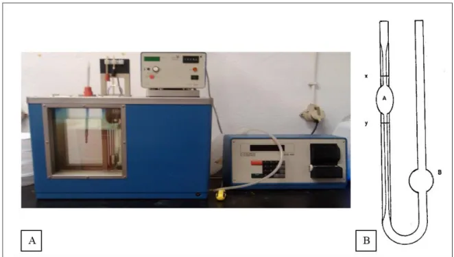
Figure 3.1 A, B- Viscosimetry apparatus used to determinate the chitosan molecular weight (A)
and scheme of the capillary (B). ... 12
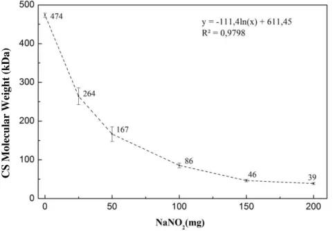
Figure 4.1- Chitosan molecular weight values calculated by viscosimetry using different
amounts of NaNO
2. ... 19
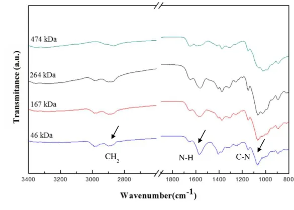
Figure 4.2- FTIR spectra for different chitosan molecular weight between 3400 and 800 cm
-1.
... 21
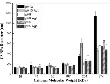
Figure 4.3- Chitosan nanoparticles diameter (nm) for different molecular weight, pH and
magnetic stirring (Agt.). ... 22
Figure 4.4- Chitosan nanoparticles diameter (nm) for different molecular weight using
Turrax
equipment. ... 24
Figure 4.5- FTIR spectra for chitosan nanoparticles, TPP and chitosan between 3400 and 600
cm
-1. ... 25
Figure 4.6- XDR spectrum with the characteristic peaks of iron nanoparticles. ... 26
Figure 4.7- Iron Oxide nanoparticles TEM image and respective particle size distribution. .. 26
Figure 4.8- Hyperthermia curves of Fe
3O
4nanoparticles synthetized by thermal decomposition
with different iron concentrations. ... 27
Figure 4.9- Left: magnification of the hysteresis loops of Fe
3O
4measured at 10 K and 315 K.
The below right inset correspond to the magnification of hysteresis loops at the same
temperatures with a different magnetic field scale. Right: temperature dependence of the
zero-field cooled/zero-field cooled (ZFC-FC) magnetization of the magnetite nanoparticles measured by
SQUID under an applied field of 100 Oe. ... 29
Figure 4.10- Chitosan coated iron oxide nanoparticles (CS SPIONs) diameters. Each chitosan
molecular weight is represent in one graph where is represented the influence of pH 5, 9, and
13 with and without agitation (Agt.) as well as different CS/SPIONS ratios (0.5-1; 1-1; 2-1).
... 31
Figure 4.11- FTIR spectra for chitosan 474 kDa coated iron oxide nanoparticles (a); chitosan
39 kDa coated iron oxide nanoparticles (b); chitosan 474 kDa (c) and Iron oxide (d) between
4000 and 600 cm
-1... 32
Figure 4.12- Hyperthermia curves of chitosan coated Fe
3O
4nanoparticles with different
chitosan molecular weights (39 and 474 kDa) and TPP volumes (0.5, 1 and 1.5 ml). ... 33
Figure 4.13- Magnification of the hysteresis loops of chitosan coated Fe
3O
4measured at 315 K.
xx
Figure 4.15- Doxorubicin release time study (0-100 h) for room temperature (25ºC) with pH
4.5 inside and PBS solution outside the dialysis membrane. ... 38
Figure 4.16- Doxorubicin release time study (0-150 h) for room temperature (25ºC) with PBS
solution inside and outside the dialysis membrane. ... 38
Figure 4.17- Doxorubicin release time study (0-100 h) for body temperature (37ºC) with PBS
solution inside and pH 4.5 outside the dialysis membrane. ... 39
Figure 4.18- Doxorubicin release time study (0-100 h) for body temperature (37ºC) with pH 4.5
inside and PBS solution outside the dialysis membrane ... 40
Figure 4.19- Doxorubicin release time study (0-100 h) for body temperature (37ºC) with PBS
solution inside and outside the dialysis membrane ... 41
Figure 4.20- Cellular viability of Fe
3O
4nanoparticles on Vero cell line in the range of
0.003-0.4 mg Fe
3O
4NPs/ml... 42
Figure 4.21- Cellular viability of freeze dried (FD) and non-freeze dried Chitosan nanoparticles
(39 and 474 kDa) on Vero cell line in the range of 0.039 - 5 mg CS NPs/ml. ... 43
Figure 4.22- Cellular viability of chitosan coated Fe
3O
4nanoparticles comparing to CS NPs on
Chitosan Nanoparticles as Drug Delivery Systems
xxi
Abbreviations and Acronyms
ASTM: American Society for Testing Materials
CS: Chitosan
CS Mv: Chitosan Molecular Weight
CS NPs: Chitosan nanoparticles
DLS: Dynamic Light Scattering
DMEM: Dulbecco’s Modified Eagle Medium
DMSO: Dimethyl Sulfoxide
DOX: Doxorubicin
FC: Field-cooled
FTIR: Fourier Transform Infrared Spectroscopy
ISO: International Organization of Standardization
MS: Saturation magnetization
MRI: Magnetic Resonance Image
NP: Nanoparticle
PBS: Phosphate Buffered Saline
SCENIHR: Scientific Committee on Emerging and Newly Identified Health Risks
SPIONs: Superparamagnetic iron oxide nanoparticles
SQUID: Superconducting Quantum Interference Device
SSA: Specific Surface Area
TEM: Transmission Electrons Microscopy
TPP: Tripolyphosphate
TREG: Triethlylene Glycol
UV-Vis: UV-Visible
XRD: X-Ray Diffraction
1
1. INTRODUCTION
1.1. Motivation and Objectives
Chitosan-coated iron oxide nanoparticles have been widely investigated because of its potential biomedical applications such as in magnetic resonance image (MRI), magnetic drug delivery, cancer diagnostics and treatment.
The big challenge in cancer therapy is minimizing side effects and maximizing efficacy. To solve the reducing sensitivity of tumor cells to cytotoxic drugs [1], different biodegradable materials are being used in the cancer therapeutic systems.
In order to improve patient care and quality of life, it is necessary reduce off-target toxicities by directing anticancer drugs (e.g. doxorubicin) to intracellular targets of tumor cells [2]. It is probably possible by using chitosan-coated SPIONs approaches to drug delivery because magnetic nanoparticles seem to be the most promising materials [1].
In order to achieve a carrier that is biocompatible and biodegradable, the goal of this thesis is to synthesize chitosan-coated superparamagnetic iron oxide nanoparticles for doxorubicin delivery for cancer theranostics. The chitosan-coated SPIONs will be loaded with the anticancer drug doxorubicin (DOX) because it is largely used clinically for different cancers types.
1.2. Thesis Overview
This thesis is divided in 4 parts: Introduction, Materials and Methods, Results and Discussion, and Conclusions.
Chitosan Nanoparticles as Drug Delivery Systems
3
2. NANOTECNOLOGY, NANOMATERIALS, AND NANOPARTICLES
Nanotechnology is an interdisciplinary area that involves chemistry, physics, biology, engineering sciences and technology. Nanotechnology allows the development of several biomedical applications such as diagnostic devices, contrast agents for magnetic resonance imaging (MRI), and drug delivery systems, for instance, anticancer drugs, proteins, and genes [3, 4].
Current nanoparticle (NP) definition is complex and not consensual because only takes into account the particle size. For instance, ISO, International Organization of Standardization, states that NP is a particle with all three dimensions from 1 to 100 nm, while ASTM, American Society for Testing Materials, and SCENIHR, Scientific Committee on Emerging and Newly Identified Health Risks, defend that NP is a particle with only two (or three) dimensions from 1 to 100nm [4-6]. On the other hand, some authors consider that nanoscale is not only from 1 to 100 nm, but from 1 to 999 nm, therefore they defend that NP is a particle with its dimensions in a size range from 1 to 999 nm [4].
However, none of those definitions take into account how properties may change with the size. In order to achieve a better definition, the European Commission considers that the size should not be the only criteria to categorize nanomaterials and NPs, so defined the other criteria to categorize them such as size distribution, specific surface area (SSA), surface modifications, and other physical properties [4-6].
2.1. Superparamagnetic Iron Oxide Nanoparticles - SPIONS
In recent years, superparamagnetic iron oxide nanoparticles (SPIONs) have been widely studied because of its potential biomedical applications such as contrast agents for MRI, magnetic drug delivery, cancer diagnostics and treatment [7, 8]. Currently, as is shown in table 2.1, several nanoparticles are already used clinically.
In addition to those, SPIONs offer other applications such as magnetic guidance and magnetic fluid hyperthermia [8]. Hyperthermia is a type of cancer treatment in which body tissue is exposed to
high temperatures (40ºC-45°C) to damage and kill cancer cells, without using drugs or radiation, when
4
Table 2.1- SPIONs approved to be used clinically in USA, Europe and their applicationsUSA Europe Applications
Ferumoxides
Feridex®
[11, 12]
Endorem®
[13-15]
MR imaging to detect and characterize
focal liver lesions
Ferucarbotran
Resovist™
[12, 16]
Resovist™
Liver imaging and diagnostics of
benign and malignant lesions
Combidex®
[15, 17, 18]
Sinerem™
[19, 20]
Detect metastatic lymph nodes in MRI
and liver imaging
Ferumoxytol
[21-23]
Feraheme
[24]
Rienso®
[25]
Prostate cancer; functional brain
activation; iron-deficiency anemia in
adults with chronic kidney disease
Despite these advantages the medical use of SPIONs possess some challenges, such as the disappearance from the bloodflow in a few minutes after their intravenous injection, the lack of surface functional groups and the need to obtain hydrodynamic stability under biological conditions such as temperature and ionic concentration [8]. A solution for those problems could be masking SPIONs’ surface charges and coating the particles with hydrophilic polymers.
2.1.1.
Synthesis of SPIONs
There are numerous methods to synthesize superparamagnetic iron oxide nanoparticles such as micro emulsion, chemical precipitation and high temperature reactions [26].
(i) Micro emulsion
Micro emulsion is related to a mixture of the following three components: an oil phase, an aqueous phase and the third component called surfactant. The dispersed droplets, obtained by this technique, possess a diameter between 10-300 nm [27].
Chitosan Nanoparticles as Drug Delivery Systems
5
into an aqueous phase, called micelles, or water in the oily phase, known as reversed micelles. Surfactants can be obtained from synthetic or natural resources and can be simple molecules such as sodium or potassium salt of the carboxylic acids, or polymers with various molecular weights. Several functional groups can be used as head groups such as carboxylates, sulphates, phosphates, sulfonates, quaternary amines and polyethers.This method produces monodispersed iron oxide nanoparticles, presenting a high control of its distribution size [28], smaller and uniform when compared to others methods [29]. Although these advantages, some disadvantages are important to enunciate, such as organic solvents toxicity which affects the usage of these nanoparticles in biomedical applications. Besides that, the amount of oil required and the difficulty of surfactant molecules removal from the nanoparticles makes this method difficult to dimension [28].
(ii) Chemical Precipitation
Chemical precipitation is a simple and efficient chemical pathway to produce SPIONs. This method allows us to produce large quantities of SPIONs. These nanoparticles are prepared by co-precipitating Fe (III) and Fe (II) in an aqueous medium. The reaction can be written as:
Fe2+ + 2Fe3+ + 8OH-→ Fe
3O4 + 4H2O Eq. 1
Physical and chemical properties of SPIONs can be affected by ions oxidation. To avoid oxidation, this reaction must be done under nitrogen environment.
Although chemical precipitation is a simple and efficient method, it exhibits some disadvantages such as the difficulty to control particle size and size distribution. Therefore, several studies were made to adjust different factors that affect the size and size distribution such as ionic strength, pH, temperature, and Fe3+/Fe2+ ratio [26].
(iii) High Temperatures Reactions
Thermal decomposition method is a technique to produce high-quality superparamagnetic and monodisperse magnetite nanoparticles by the decomposition of the iron acetylacetonate, Fe(acac)3, in a high-boiling temperature solvent with surfactants such as oleic acid and oleylamine [30].
6
With this method we can directly synthesize water-soluble SPIONs [30, 31]. SPIONs produced by this technique show a hydrophilic coating, acceptable size uniformity, and good crystallinity, thus offering powerful potential for biomedical applications such as the ability of TREG-coated SPIONs to act as superparamagnetic contrast agents in MRI or as agents for magnetic hyperthermia [31].These SPIONs are biocompatible up to an iron concentration of 80µg/ml with SiHa, B16F10 and mouse primary fibroblast cells. The significant temperature rise (RF radiation with 20 MHz frequency and 100 W power) from 31-49ºC in 275 seconds for a sample with iron concentration of 0.5 mg/ml and from 35-70ºC in 150 seconds for 1 mg/ml, corroborates their applicability for magnetic hyperthermia application [30].
2.2. Chitosan Nanoparticles
–
CS NPs
Chitosan is a natural polysaccharide composed of D-glucosamine and N-acetyl-D-glucosamine units linked by β-(1,4)-glycosidic bonds. It is obtained by partial deacetylation of chitin, the second most abundant polymer in nature. Chitin and chitosan differ in the molar fraction of the deacetylated units (DD in figure 2.1). The DD is responsible for the different physic-chemical properties of these polymers. Chitosan is the polymer with a DD higher than 0.5 (typically 0.7-0.8) soluble in acidic solutions. Chitin is almost insoluble in most organic solvents and because of its low solubility, chitin has limited applications. On the contrary chitosan is soluble in acidic solutions due to protonation of the amine groups (-NH2) of the D-glucosamine unit, where CS is converted to a cationic polyelectrolyte, and possess a great affinity to form films, gels, and fibers used in different applications [32, 33].
Figure 2.1- Chemical structure of chitosan
Chitosan Nanoparticles as Drug Delivery Systems
7
Chitosan nanoparticles (CS NPs) possess interesting biomedical applications. They can be used as carriers in controlled drug delivery of:(i) doxorubicin - an anticancer drug used for the treatment of several tumors [34, 35];
(ii) 5-fluorouracil (5-FU) and leucovorin (LV) - drugs used in the treatment of colon cancer [36]; (iii) avidin and biotin – drugs used for hepatic carcinoma treatment [37], among others.
CS NPs have been also studied in: gene delivery systems [38, 39]; siRNA delivery [40]; release of vitamin C [41]; delivery of pDNA against hepatitis B through nasal mucosa [42]; protein delivery systems [43], such as insulin [44] or BSA [45] delivery; release of polyphenolic antioxidants , for instance, catechins in gastrointestinal track (GIT) [46-48]; and as supports for enzyme immobilization [49]. CS NPs could also inhibit the growth of several microorganisms and possess , in some cases, higher antibacterial activity than CS itself [50].
2.2.1.
Synthesis of CS NPs
There are several techniques to synthesize CS NPs such as micro emulsion, electrospray, emulsification solvent diffusion, and ionic gelation [51].
(i) Micro emulsion
Micro emulsion method was first developed by Maitra and his coworkers [52]. This technique consists in the conjugation of the functional amine group of CS with aldehyde groups of the cross-linking agent, such as glutaraldehyde. In this method, CS nanoparticles are formed by adding, under stirring, chitosan solution with glutaraldehyde to a mixture that contains a surfactant dissolved in N-hexane. The organic solvent and the excess surfactant are removed, and the nanoparticles suspension is dialyzed and finally lyophilized.
This method allows control the particle size by using different amounts of cross-linked agent glutaraldehyde. However, micro emulsion exhibits some disadvantages such as the use of organic solvents, lengthy preparation process, and it requires a complex washing step [52, 53].
(ii) Electrospray
8
narrow size distribution, simplicity, and fast preparation. However, there are a few studies using this technique to produce nanoparticles [54].(iii) Emulsification Solvent Diffusion
Originally developed by Niwa and his team with PLGA, El-Shabouri (2002) used this technique to prepare chitosan nanoparticles. This method consists in an oil-in-water (o/w) emulsion obtained by injecting an organic solvent into a chitosan solution with a stabilizing agent, such as polomaxer, under mechanical stirring. Then, a large volume of water is added to the emulsion to overcome organic solvent miscibility in water. Nanoparticles are formed because of the diffusion of organic solvent into water that promotes polymer precipitation.
Emulsion solvent diffusion shows some disadvantages such as the use of organic solvents and high shear forces used to prepare nanoparticles [52].
(iv) Ionic Gelation
Ionic gelation technique was first reported by Calvo et al. (1997). This method consists in the addition of tripolyphosphate (TPP), a crosslinking agent, into the aqueous phase containing chitosan, thus leading to the formation of CS NPs [39]. In this work it was used ionic gelation technique with TPP because it is simple, mild, less toxic and suitable for scaling up [51]. Furthermore, it is possible to control the size of the CS NPs by adjusting the following parameters: CS and TPP concentration, CS/TPP weight ratio and volume ratio, pH value [51].
The formation of CS NPs by ionic gelation occurs in two steps. The first one is the addition of aqueous TPP solution to the previously dissolved CS in acidic solution. The second one is the spontaneously inter and intracrosslinking - gelation - between the protonated amine group on the CS molecules and TPP anions.
In order to achieve smaller NPs with narrower size distribution (low polydispersity) it is necessary a rapid mixing, which promotes a fast and uniform dispersion of the TPP anions within the CS chains [51].
2.2.2.
Interaction between CS NPS and cells
Chitosan Nanoparticles as Drug Delivery Systems
9
The effects of CS nanoparticles on the cells membrane are more intense than CS itself in some cases, but similar in others. Possibly, the effects are similar because both CS and CS nanoparticles are positive charged and they interact with negatively charged live cells by electrostatic forces by the same mechanism. On the other hand, the transformation of CS molecules into CS nanoparticles promotes the interaction because it changes the linear configuration of CS molecules, in microscale, to the condensed configuration of CS nanoparticles in nanoscale [55].CS is considered non-cytotoxic and CS nanoparticles also exhibit no cytotoxicity although they accumulated more inside of the cells. There was no significant difference in cytotoxicity between both [55].
2.3. CS NPS as Delivery Systems for Doxorubicin
Doxorubicin is part of the family of anthracycline antibiotics. It is formed by an amino-sugar daunosamine, linked to the C7 of a tetracyclic aglycone, doxorubicinone [35, 56]. The chemical structure of doxorubicin is shown in Figure 2.2.
Figure 2.2- Chemical structure of Doxorubicin.
Doxorubicin is generally used in the treatment of several cancers such as acute leukemia, lymphomas, soft-tissue and osteogenic sarcomas, pediatric malignancies and adult solid tumors as breast and lung carcinomas. It is also used with other drugs such as methotrexate, cisplatin, ifosfamide, vincristine and etoposide [35].
However, only a small amount of DOX reaches the tumor target site because about 40% are excreted by liver metabolism. Furthermore, doxorubicin induces cardiac toxicity [34, 35, 56], but not in the same way to the different patients, for some started within 1 year of DOX therapy, while for others it occurred 15 years after the end of the treatment [35].
10
tumor sites by a phenomenon known as “Enhanced Permeability and Retention” (EPR) effect [58]. However, its efficacy is not consensual due to low response rates percentage in clinical trials and due to toxic dermatological reaction occurred in about 50 % of patients treated with Doxil® [35].A solution to protect patients from DOX side effects is by using a drug delivery system compound by CS NPs. This way, and attending to CS properties, nontoxicity, biocompatibility and biodegradability, it is possible to encapsulate and delivery DOX with reduced side effects [35]. Figure 2.3 shows the mechanism of SPIONs encapsulation by CS.
Chitosan Nanoparticles as Drug Delivery Systems
11
3. MATERIALS AND METHODS
3.1. Chitosan Depolymerization
To depolymerize the commercial chitosan (Cognis), 500 ml of a solution of Chitosan 1% (w/v) in acetic acid (Panreac) 1% (v/v) was prepared. In order to obtain different chitosan molecular weights, different amounts of NaNO2 (Sigma-Aldrich), shown in Table 3.1., dissolved in 10 ml of distilled water were mixed with chitosan and let this final solution under mechanic stirring for 1h at 1000 rpm. After 1h, a 4M NaOH (Merck) solution was added to the mixture until a pH of 9-10 was reached. The formed precipitate was centrifuge (Heraeus Multifuge X1R Centrifuge) for 10 min at 10000 rpm and washed with distilled water several times. Then, the precipitate was freeze dried for 24h (VaCo2 Zirbus Technology).
Table 3.1- Amount of NaNO2 corresponding to the different CS/NaNO2 ratio to obtain different chitosan molecular weight samples.
Ratio
CS/NaNO
2NaNO
2(mg)
200-1
25
100-1
50
50-1
100
33-1
150
25-1
200
3.1.1.
Chitosan Molecular Weight Determination
12
Figure 3.1 A, B- Viscosimetry apparatus used to determinate the chitosan molecular weight (A) andscheme of the capillary (B).
The polymer molecular weight is related to the intrinsic viscosity [ƞ] by the Mark-Houwink-Sakurada (MHS) equation:
[ƞ] = 𝐾𝑀𝑣𝑎 Eq. 2
Where 𝑀𝑣 is the viscosity-average molecular weight, and K and a are the constants for the solute-solvent system [59]. The values of MHS equation constants, a and K, were calculated using the following equations:
𝑎 = 0.6202 +(0.4806 + 𝑥)0.699𝑥 Eq. 3
𝑙𝑜𝑔𝐾. 10−5= −5.7676. 𝑎 + 5.9232 Eq. 4
Where 𝑥 is [DA/pH. µ], with DA, chitosan degree of acetylation, pH of chitosan solution in a solvent with ionic strength of µ.[60]
This technique requires a solvent, 0.2M HAc/0.1M NaAc (Scharlau), to dilute the chitosan samples with different concentrations. Using equation (3) and (4), the values of constants a and K were calculated to be 0.81 and 16.8 x 10⁵ dL.g-1 [59]. For each sample the viscosity measurements were made in triplicate.
Chitosan Nanoparticles as Drug Delivery Systems
13
3.2. Chitosan Nanoparticles Synthesis
Chitosan nanoparticles were prepared by ionic gelation technique using tripolyphosphate (TPP) (Sigma-Aldrich) [51]. For all the chitosan samples a 0.4% (w/v) chitosan solution with 1% (v/v) acetic acid was prepared and 0.25% (w/v) TPP solution of pH 5, 9, and 13.
To produce nanoparticles equal volumes of 0,25% (w/v) TPP and 0.4% (w/v) chitosan solutions were used, which were mixed during 5 minutes using Turrax (IKA T10 Basic).
3.3. Iron Oxide Nanoparticles Synthesis
Water-soluble Fe3O4 nanoparticles were prepared by thermal decomposition of Fe(acac)3 (Alfa Aesar) in TREG (Sigma-Aldrich) at high temperature without using surfactant. Typically, 2 mmol of Fe(acac)3 were dissolved in 20 ml of hydrophilic TREG media and then magnetically stirred (100 rpm) under a flow of nitrogen. The solution was dehydrated at 200ºC for 1h, and then quickly heated to 350 ºC and kept at this temperature for 2h.
The black solution was cooled to room temperature overnight by removing the heat source. The nanoparticles were precipitated by addition of 20 ml of ethyl acetate (Sigma-Aldrich) and then isolated by centrifugation (Heraeus Multifuge X1R Centrifuge) for 20 min at 10000 rpm.
This washing process was repeated and then the washed particles were dispersed in water to get a stable aqueous ferrofluid suspension [30].
3.3.1.
Iron Content Determination
14
3.4. Chitosan Coated SPIONs
SPION nanoparticles suspensions were added to a 0.4% chitosan solution with 1% (v/v) acetic acid in a ratio of 0.5:1, 1:1, and 2:1 (v/v). To this solution was added 1 ml of TPP of pH 5, 9, and 13. Each sample was repeated 6 times, 3 of them were left in magnetic stirring for 24h at room temperature and the other 3 were left without stirring for 24h at room temperature.
3.5. Doxorubicin Encapsulation and Release
3.5.1.
Doxorubicin Content Determination
The absorbance of the samples containing DOX was measured at 480 nm using a UV-VIS spectrophotometer (T90+ UV/VIS Spectrometer PG Instruments Ltd). The concentration of Doxorubicin was calculated using the following obtained calibration curve:
𝑦 = 0.0178𝑥 − 0.0011
Eq. 5Where 𝑦 is the Doxorubicin concentration and 𝑥 is the absorbance of the solution measured at 480 nm [62].
3.5.2.
Evaluation of Doxorubicin Encapsulation
Different volumes of TPP 0.25% pH5 (500µl, 1000µl and 1500µl) were added to 500µl of 3:1 (w/w) CS-DOX solution using Turrax (IKA T10 Basic) and then centrifuged at 10000 rpm for 5min. Then the samples were washed with distilled water and centrifuged again at 10000 rpm for 5min. Supernatant DOX concentrations were calculated by UV-VIS spectroscopy (T90+ UV/VIS Spectrometer PG Instruments Ltd) with an emission wavelength at 480 mm. Dilutions of samples and calibration curves were performed in distilled water , and all measurements were performed in triplicate. Encapsulation Efficiency (6) and Drug Loading (7) were determined according to the following equations:
𝐸𝑛𝑐𝑎𝑝𝑠𝑢𝑙𝑎𝑡𝑖𝑜𝑛 𝑒𝑓𝑓𝑖𝑐𝑖𝑒𝑛𝑐𝑦 =𝑡𝑜𝑡𝑎𝑙 𝐷𝑂𝑋 − 𝑓𝑟𝑒𝑒 𝐷𝑂𝑋𝑡𝑜𝑡𝑎𝑙 𝐷𝑂𝑋 Eq. 6
𝐷𝑟𝑢𝑔 𝐿𝑜𝑎𝑑𝑖𝑛𝑔(%𝑤/𝑤) = 𝑀𝑎𝑠𝑠 𝑜𝑓 𝑑𝑟𝑢𝑔 𝑙𝑜𝑎𝑑𝑒𝑑 𝑖𝑛 𝑁𝑃𝑠 ∗ 100𝑀𝑎𝑠𝑠 𝑜𝑓 𝑛𝑎𝑛𝑜𝑝𝑎𝑟𝑡𝑖𝑐𝑙𝑒𝑠 Eq. 7
3.5.3.
Evaluation of
in vitro
Doxorubicin Release
Chitosan Nanoparticles as Drug Delivery Systems
15
PBS 1x pH 7.4 or HAc 2M/NaAc 1M. One milliliter of nanoparticles suspension was placed inside a dialysis tube. The dialysis was performed against 10 ml of PBS 1x pH 7.4 or HAc 2M/NaAc 1M either at 25ºC or 37ºC.To measure the Doxorubicin release, 3 ml of the release medium were taken from the dialysis apparatus, and replaced with fresh medium at pre-defined times. The amount of doxorubicin released was measured by UV-VIS spectroscopy against free DOX.
All measurements were performed in triplicate.
3.6. Characterization of Nanoparticles
Size and morphology of the nanoparticles were determined using transmission electron microscopy (TEM) and dynamic light scattering (DLS) while crystal structure was identified by X-ray diffraction (XRD). Surface charge and surface coating of the nanoparticles were analyzed by Fourier transform infrared spectroscopy (FTIR). Magnetic characterization of the nanoparticles was performed by SQUID magnetometry (S700X Cryogenic, Ltd.).
3.6.1.
Transmission Electron Microscopy- TEM
The particle size and morphology of the nanoparticles were determined by transmission electron microscopy imaging (TEM, Hitachi H-8100 II) at Instituto Superior Técnico in Lisbon. This TEM offers 200 kV acceleration voltage with high brightness LaB6 thermionic electron source and phase contrast resolution of better than 0.27nm (point) and 0.14nm (line).
The samples were prepared by diluting the suspension with ultrapure water to a 1:100 ratio. Afterwards one drop of the solutions was evaporated on a carbon coated substrate. The specimen holder is equipped with two samples and was loaded to the observation chamber. Chitosan and iron oxide nanoparticles produced by thermal decomposition were investigated. The particle size distributions of the samples were obtained statistically by measuring the diameter of 100 nanoparticles using TEM images.
3.6.2.
Dynamic Light Scattering- DLS
Chitosan nanoparticles and chitosan coated SPIONs hydrodynamic diameters were measured by means of dynamic light scattering (DLS) equipment (AvidNano) using the
bladecell
®at 20ºC. For
16
It was also studied the effect of Turrax on nanoparticles’ diameter. In the preparation of the samples, it was used Turrax for 5 min to homogenize the samples.3.6.3.
Superconducting Quantum Interference Device- SQUID
Magnetic measurements were performed by 7T SQUID magnetometer (S700X Cryogenic, Ltd.)
at Instituto Superior Técnico in Lisbon. The zero-field-cooled (ZFC) and field-cooled (FC)
measurements were performed by cooling the sample to 5 K at zero fields or in the presence of an external field of 100 Oe, respectively. All the magnetic measurements were carried out within a temperature range 5-320 K. Isothermal magnetization curves were obtained for fields up to 5 T for temperatures of 10 and 320 K. Saturation magnetization (MS) of chitosan coated magnetite particles were determined.
3.6.4.
X-Ray Diffraction- XRD
The crystalline phases of the materials were verified using powder X-ray diffraction (XRD, PANAlyticalX’Pert PRO) using a Cu-Kα radiation (λ= 1.54060Å). Dry nanoparticles were obtained by freeze drying (Vaco2, Zirbus) a part of the suspension overnight. To obtain the XRD pattern of the particles 2θ values were taken form 15° to 80°, with a step size of 0.033. The Scherrer`s equation was used to measure the average crystallite size.
3.6.5.
Fourier Transform Infrared Spectroscopy- FTIR
FTIR spectra of the samples were obtained using a Nicolet 6700 – Thermo Electron Corporation Total Reflectance-Fourier Transform Infrared spectrometer (ATR-FTIR). FTIR allows the detection of functional groups and the characterization of covalent bonding information. By this method, chemical changes of chitosan, iron oxide, chitosan coated magnetite nanoparticles, and chitosan coated magnetite nanoparticles loaded with doxorubicin were investigated.
3.6.6.
Hyperthermia measurements
Chitosan Nanoparticles as Drug Delivery Systems
17
𝑇(𝑡) = (𝑇0− 𝑇𝑒𝑞)𝑒−1𝜏𝑡+ 𝑇𝑒𝑞 Eq. 8
In the equation 𝑇0 is the initial temperature of the sample, 𝑇𝑒𝑞 is the equilibrium temperature of the sample; T the maximum temperature reached by the sample and τ is characteristic time of heating.
3.7. Cell Line and Culture Conditions
3.7.1.
Cytotoxicity Experiments
Day 1: unfreeze the cells
The first step to unfreeze the cells was to remove a sample with 1ml, around 2 million cells, from -80ºC and put them in a 37ºC water bath to accelerate the unfreeze process.
To a T25 bottle with 5ml of cellular solution (DMEM) with serum was added the previous unfreeze sample with 1ml of cells. This solution was allowed in an incubator with constant temperature (37ºC). Some hours later, the cellular solution was replace in order to remove the toxic DMSO (unfreeze agent).
Day 2: Cell seeding
The solution of the previously unfreeze cells was removed and washed with PBS 1x solution (5ml) without Ca and Mg. Then, the PBS solution was removed and 500 µl of tripsin was added at 37ºC in the incubator for 5 minutes. The sample was gently agitated and a 5ml mixture of DMEM solution and Glutomax 1x (Gibco) was added. For each well it was pipetted 100 µl of the previously solution (cells were counted before) and was left in incubator at 37ºC for 24 hours.
Day 3: Add the NPs synthetized into the NPs cells
The last step was to add the lyophilized and sonicated chitosan, magnetite and chitosan coated iron oxide nanoparticles into the cells previously seed. For each well, the DMEM solution was removed and NPs solution was added. To the first line of wells, a 5 mg/ml NPs solution in DMEM was added and then a 1:2 dilution was performed to have different concentrations of NPs. For each concentration it was made 4 replicas. The plate with the mixture of cells and NPs was left in the stove at 37ºC for 24 hours.
Day 4: Cellular Viability determination
Chitosan Nanoparticles as Drug Delivery Systems
19
4. RESULTS AND DISCUSSION
4.1. Chitosan Characterization
The main goal of the present work was to produce chitosan nanoparticles that could be used in drug delivery systems. Before the production of these nanoparticles it was necessary to obtain and characterize different molecular weight samples of commercial chitosan. In the present section 4.1 is described the results obtained for the different chitosan molecular weight samples and their characterization.
4.1.1.
Chitosan Molecular Weight Determination
Viscosimetry measurements used to determinate the chitosan molecular weight (CS Mv) are shown in this section. The main goal of this procedure was to decrease the CS Mv in order to study the influence of Mvin the CS NPs’ diameters and their properties. In order to decrease the molecular weight of chitosan was used different amounts of an oxidant agent - NaNO2 - keeping all others conditions unchanged.
Figure 4.1- Chitosan molecular weight values calculated by viscosimetry using different amounts of NaNO2.
As can be seen in Figure 4.1, the commercial CS Mv was 474 kDa, determined without addiction of NaNO2. Different CS samples with Mv between 264 - 39 kDa by the depolymerization technique were obtained, using different amounts of NaNO2 (25-200 mg).
C
S
Mol
ecul
ar
Wei
ght (
k
D
a
20
Table 4.1- Chitosan molecular weight values and their standard deviation and depolymerizationefficiency.
NaNO
2(mg)
CS
(g)
MW
(kDa)
S.D
Efficiency
(%)
0
5
474
5
25
264
22
99.0
50
167
19
99.6
100
86
6
98.0
150
46
3
85.4
200
39
2
92.6
Table 4.1 shows chitosan molecular weight values obtained through depolymerization of 474 kDa commercial chitosan. As seen before the depolymerization was obtained by an oxidation reaction of the commercial chitosan using NaNO2 as an oxidant agent. Figure 4.1 and table 4.1 show the decrease of the chitosan molecular weight with increase of NaNO2. The degree of efficiency for the 5
depolymerizations is quite high, representing an efficiency above 85% for all chitosan’s molecular weights.
It can be concluded, from the results shown in Figure 4.1 and Table 4.1, that the depolymerization technique is an efficient procedure to decrease the CS molecular weight.
4.1.2 Chitosan chemical Characterization
FTIR measurements were performed in order to examine the chemical structure of chitosan for different degrees of depolymerization. In Figure 4.2 FTIR spectra are presented for the four molecular weights of chitosan obtained by the depolymerization of commercial chitosan. Some characteristic bands, arising from the presence of amine and methylene groups on chitosan, are observed. Bands at 2924 cm-1 and 2852 cm-1 are assigned to the CH
2 antisymmetric and symmetric stretching modes, respectively. Due to the presence of amine group, a band at ~1600 cm-1 is assigned to the bending mode of N-H and other at ~ 1080-1360 cm-1 is assigned to C-N stretching modes.
Chitosan Nanoparticles as Drug Delivery Systems
21
Figure 4.2- FTIR spectra for different chitosan molecular weight between 3400 and 800 cm-1.4.2. Chitosan Nanoparticles Characterization
After the characterization of the different molecular weight chitosan samples the next goal of the present work was to produce chitosan nanoparticles by ionic gelation technique. To produce them it was necessary to study the influence of some parameters in the chitosan nanoparticles diameter, such as chitosan molecular weight, pH, magnetic stirring and the use of Turrax equipment. The characterization of chitosan nanoparticles is also described in section 4.2 as well as the results obtained by the study of the mentioned parameters.
4.2.1.
Chitosan Nanoparticles Diameter
DLS measurements were made in order to analyze chitosan nanoparticles diameters. For each chitosan molecular weight obtained in the previous section 4.1, the CS NPS diameter was analyzed under different conditions of pH (pH5, 9 and 13) and agitation (with and without magnetic stirring (Agt.)). The results of this study are shown in Figure 4.3 and Table 4.2.
22
Figure 4.3- Chitosan nanoparticles diameter (nm) for different molecular weight, pH and magneticstirring (Agt.).
The samples with lower molecular weight, 39 – 167 kDa and considering the standard deviation, exhibit a diameter below 200 nm for the different pH and agitation used and mentioned above. This behavior is not observed for the 264 and 474 kDa samples. Chitosan nanoparticles produced using these chitosan molecular weights present higher diameters, particularly for pH 13 with and without agitation for 264 and 474 kDa respectively.
Figure 4.3 shows that the chitosan nanoparticles diameters increaese with increasing chitosan molecular weight. Although there are not significant differences between pH 5, 9, and 13 with the exception of pH 13 in the higher chitosan molecular weight (264 and 474 kDa). The magnetic stirring does not display a homogeneous result for all CS Mw. In some cases the particles are smaller with magnetic stirring (39 and 46 kDa for pH 5, 9 and 13) whereas on other Mw it does not show any influence.
Chitosan Nanoparticles as Drug Delivery Systems
23
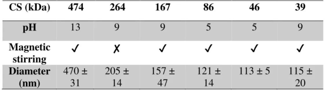
Table 4.2- Chitosan molecular weight parameters to obtain smaller chitosan nanoparticles diametersCS (kDa)
474
264
167
86
46
39
pH
13
9
9
5
5
9
Magnetic
stirring
✔
✘
✔
✔
✔
✔
Diameter
(nm)
470 ±
31
205 ±
14
157 ±
47
121 ±
14
113 ± 5
115 ±
20
As can be seen in Figure 4.3 and Table 4.2 the influence of magnetic stirring is not very strong. To complement this study another procedure and equipment was used to prepare chitosan nanoparticles and to measure its diameters – Turrax (IKA T10 Basic).
It was used a sample of 46 kDa chitosan pH 5 (one of the parameters conjugation to obtain smaller diameters, as shown in Table 4.2) to study the influence of Turrax in the chitosan nanoparticles diameter and to be possible to compare the magnetic stirring with Turrax.
Table 4.3- Chitosan nanoparticles diameter and standard deviation for different Turrax time periods
Time
(min)
Diameter
(nm)
S.D.
5
115
9
10
150
10
15
154
8
20
138
14
It was performed a time study in order to understand for how long the Turrax needs to be used to obtain the smaller diameter in CS NPS. Time periods between 5 to 20 min were used. For the lower period, 5 min, a diameter around 115 nm was obtained and for the higher period, 20 min, a diameter around 138 was obtained. From Table 4.3 it can be seen that the lower diameter is obtained when Turrax is used for only 5 minutes.
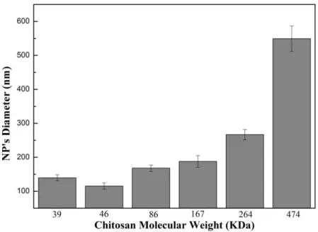
24
Figure 4.4- Chitosan nanoparticles diameter (nm) for different molecular weight using Turrax equipment.The samples with chitosan molecular weight between 39 and 167 kDa exhibit a diameter below 200 nm. This value is not the same for the higher chitosan molecular weight samples. Chitosan nanoparticles produced using these chitosan molecular weights present higher diameters, around 300 and 550 nm for 264 and 474 kDa respectively.
Figure 4.4 shows that chitosan nanoparticles diameters increase with increasing chitosan molecular weight as it was seen in Figure 4.3. Note that although the diameters are similar to those obtained previously, the standard deviation is much smaller compared to the ones shown in Figure 4.3. Another relevant aspect is that the Turrax is faster than magnetic stirring. Therefore nanoparticles with the same diameter can be obtained with only 5 minutes instead of 24 hours of agitation.
4.2.2.
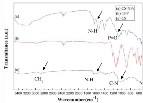
CS NPs chemical characterization
Chitosan Nanoparticles as Drug Delivery Systems
25
Some characteristic bands, arising from the presence of amine and methylene groups on chitosan, are observed. Bands at 2924 cm-1 and 2852 cm-1 are assigned to the CH2 antisymmetric and symmetric stretching modes, respectively. Due to the presence of amine group, a band at ~1600 cm-1 is assigned to the bending mode of N-H and other at ~ 1080-1360 cm-1 is assigned to C-N stretching modes [63].
In chitosan nanoparticles the band at 3438 cm-1 has a shift to 3320 cm-1 and becomes wider with increased relative intensity indicating an enhancement of hydrogen bonding. In nanoparticles the peaks for N-H bending vibration of amine І at 1600 cm-1 and the amide ІІ carbonyl stretch at 1650 cm-1 shifted to 1540 cm-1 and 1630 cm-1, respectively. The cross-linked chitosan also show a P=O peak at 1170 cm -1. These results have been attributed to the linkage between phosphoric and ammonium ion. So we
conclude that the tripolyphosphoric groups of TPP are linked with ammonium groups of chitosan. The inter- and intra-molecular actions are enhanced in chitosan nanoparticles [63].
4.3. Iron Oxide Nanoparticles Characterization
Considering that the chitosan nanoparticles have been produced and characterized as stated in the previous sections, another important goal of the present study includes the production of iron oxide nanoparticles by thermal decomposition technique as well as the study of their diameter and magnetic properties.
Several techniques were used to characterize the iron oxide nanoparticles synthesized. One of those techniques was phase identification (Structural characterization) performed by X-ray diffraction (XRD).
Figure 4.5- FTIR spectra for chitosan nanoparticles, TPP and chitosan between 3400 and 600 cm-1. N-H
P=O
CH2 N-H
26
Figure 4.6- XDR spectrum with the characteristic peaks of iron nanoparticles.It can be clearly identified, from Figure 4.6, the characteristic diffraction peaks of crystalline cubic magnetite structure in particular to 2𝜃 = 30.12, 35.43, 43.22, 57.19 and 62.80 [64]. Narrower peaks represent the samples’ crystalline structure. The XRD pattern of this sample was assigned to the phase of bulk magnetite.
The average crystallite size, 𝜏 , was calculated to be 13.54 nm using the Scherrer’s equation 𝜏 =
𝐾𝜆
𝛽𝑐𝑜𝑠𝜃 , where 𝐾 is the grain shape factor (𝐾 = 0.94); λ is the incident X-ray wavelength (λ = 0.15406 nm); 𝛽 denotes the full width at half-maximum (in radians) of the highest intensity peak (𝛽= 0.02532 rad), and 𝜃 is the corresponding diffraction angle (2𝜃 = 35.43 𝜃=17.715) corresponding to (311).
Size, shape and particle size distribution of the iron oxide nanoparticles were determined by TEM image analysis represented in Figure 4.7.
Chitosan Nanoparticles as Drug Delivery Systems
27
Figure 4.7 shows iron oxide nanoparticles dispersed in water. These nanoparticles present a roughly spherical morphology and were not bonded. The particle size was determined by statistical analysis of the dimensions of at least 100 particles yielding an average particle diameter of 10.3 ± 2.0 nm. This value agree with the value obtained from XRD data 𝜏 = 13.54 nm. The small difference observed in the particles diameter could be due to reduced accuracy of XRD technique comparatively to TEM. Reduced accuracy are related to high surface area that originates defects in the particles surface, high interfacial tension between particles that correspond to an expansion of diffraction and due to the fact that nanoparticles could be crystalline, amorphous or quasi crystalline [65].4.3.1.
Magnetic Properties
To characterize the magnetic properties of Fe3O4 nanoparticles, hyperthermia and SQUID measurements were performed.
Hyperthermia studies were performed in order to evaluate the ability of iron oxide nanoparticles on generating heat. Several concentrations of iron were tested as shown in Figure 4.8.
Figure 4.8- Hyperthermia curves of Fe3O4 nanoparticles synthetized by thermal decomposition with different iron concentrations.
28
present higher generated heat ability, as can be seen in the sample of 3mg/ml (b) and 6 mg/ml (a). The 6 mg/ml sample presents a temperature increase around 22.5ºC.In summary, Fe3O4 nanoparticles synthesized by Thermal Decomposition technique possess the ability to generate heat. It can be seen that the heating ability of these nanoparticles rises with increasing of iron concentration.
Specific Absorption Ratio (SAR) is used to characterize the heating efficiency that a magnetic material possess by measuring the energy absorption when that material is expose to an alternative magnetic field. It is defined as the power absorbed per mass unit and it units are watts per gram (W/g). SAR is calculated by the following equation:
𝑆𝐴𝑅 (𝑊 𝑔⁄ ) = (𝜕𝑇𝜕𝑡)
𝑚𝑎𝑥𝑥
𝐶𝑁𝑃 𝑥 𝑚𝐹𝑒 𝑥 𝐶𝑙𝑚𝑙
𝑚𝐹𝑒
Eq. 9
where (𝜕𝑇
𝜕𝑡)𝑚𝑎𝑥is the maximum gradient of the temperature curve of the colloid submitted to a hyperthermia test, 𝐶𝑁𝑃 is the specific heat of the nanoparticles, 𝐶𝑙 is the specific heat of the liquid, 𝑚𝑙 is the fluid mass, and 𝑚𝐹𝑒 is the iron mass in the colloid [66].
SAR values of the same colloid measured under different experimental conditions (external magnetic field intensity - H - and frequency - f - of the applied magnetic field) will be different, making impossible to directly compare results obtained in different laboratories or equipment. For this reason intrinsic loss Power (ILP) has been proposed because it is independence of the field used and the equipment [66]. It is defined as:
𝐼𝐿𝑃 (nH𝑚Kg ) =2 𝑆𝐴𝑅𝑓𝐻2 Eq. 10
Table 4.4- SAR and ILD values for each Iron concentration of Fe3O4 nanoparticles synthetized by Thermal Decomposition.
[Fe]
(mg/ml)
SAR
(w/g)
(nH.m
ILP
2/kg)
Chitosan Nanoparticles as Drug Delivery Systems
29
Table 4.4 shows the SAR values obtained by the maximum of (𝜕𝑇𝜕𝑡) which occurs in the beginningof the hyperthermia measurements, when adiabatic conditions were guaranteed. It can be seen that SAR and iron concentration values are proportional. This means that higher iron concentrations correspond to higher heating efficiencies of the Fe3O4 nanoparticles. The same occurs to ILP values, iron concentrations values are proportional to ILP values and that correspond to higher heating efficiencies of the Fe3O4 nanoparticles.
In order to evaluate the superparamagnetic properties of Fe3O4 nanoparticles produced, SQUID measurements were performed. The results obtained with this study are shown in Figure 4.9.
Figure 4.9- Left: magnification of the hysteresis loops of Fe3O4 measured at 10 K and 315 K. The below right inset correspond to the magnification of hysteresis loops at the same temperatures with a different
magnetic field scale. Right: temperature dependence of the zero-field cooled/field cooled (ZFC-FC) magnetization of the magnetite nanoparticles measured by SQUID under an applied field of 100 Oe.
Figure 4.9-Left shows a magnification of the hysteresis loops of Fe3O4 measured at 10 K and 315 K. The below right inset correspond to the magnification of hysteresis loops at the same temperatures with a different magnetic field scale. It is evident the absence of coercivity and remanence at 315 K. This indicates the superparamagnetic behavior of Fe3O4 nanoparticles at this temperature.
30
Figure 4.9-Right shows the temperature dependence of the zero-field cooled/field cooled (ZFC-FC) magnetization of the magnetite nanoparticles measured by SQUID under an applied field of 100 Oe. The maximum of ZFC curve is reached about 165 K which means that above that temperature, called blocking temperature (TB), the sample is superparamagnetic and below is ferromagnetic. At room temperature (315 K) the sample is superparamagnetic.4.4. Chitosan coated Iron Oxide Nanoparticles
On previous sections it was described the chitosan and Fe3O4 nanoparticles synthesis by ionic gelation and thermal decomposition respectively and its characterization by a battery of techniques.
The next step, that was the main goal of the present study, was to join the two synthesized nanoparticles in order to form the chitosan coated iron oxide nanoparticles. In the present section is described all characterization techniques that were used to characterize them.
4.4.1.
Chitosan coated Iron Oxide Nanoparticles Diameter
One of the characterization techniques used was DLS measurements to obtained chitosan coated iron oxide nanoparticles diameter.
In Figure 4.10 it can be seen the diameter values obtained for all the parameters studied. For each chitosan molecular weight was studied the influence of pH (5, 9, and 13), agitation (with and without agitation) as well as different CS/SPIONS ratios (0.5-1; 1-1; 2-1).
Chitosan Nanoparticles as Drug Delivery Systems
31
Figure 4.10- Chitosan coated iron oxide nanoparticles (CS SPIONs) diameters. Each chitosan molecular weight isrepresent in one graph where is represented the influence of pH 5, 9, and 13 with and without agitation (Agt.) as well
as different CS/SPIONS ratios (0.5-1; 1-1; 2-1).
32
Table 4.5 - Chitosan molecular weight, pH, agitation, and Cs/SPIONs Ratio parameters to obtain smallerchitosan coated iron oxide nanoparticles diameters
S ( kDa)
474
264
167
86
46
39
pH
13
5
9
13
13
13
Agitation
✔
✔
✔
✔
✘
✔
Cs/SPIONs
Ratio
1-1
2-1
0,5-1
1-1
2-1
2-1
Diameter(nm)
232±1
210±5
213±34 144±6
68±4
146±3
Considering Figure 4.10 and Table 4.5 analysis, pH 13 seems to be the best pH to be used and magnetic stirring gives smaller diameters. On the other hand, CS/SPIONs ratio depends on which chitosan molecular weight is being used. The smaller diameter - 68±4 - was obtained with CS 46 kDa, pH13 without magnetic stirring and CS/SPIONs ratio 2-1. Should be noted that the standard deviation are relatively large in some samples which must be taking into account in the analysis made in this section.
4.4.2.
CS coated SPIONs Chemical Characterization
In order to examine the structure of chitosan coated iron oxide nanoparticles produced, FTIR measurements were performed between 4000 and 600 cm-1. FTIR spectra are presented in Figure 4.11 for commercial chitosan coated iron oxide nanoparticles (a), 39 kDa chitosan coated iron oxide nanoparticles (b) as well as commercial chitosan (c) and iron oxide (d) separately.
Figure 4.11- FTIR spectra for chitosan 474 kDa coated iron oxide nanoparticles (a); chitosan 39 kDa coated iron oxide nanoparticles (b); chitosan 474 kDa (c) and Iron oxide (d) between 4000 and 600 cm-1
Fe-O Fe-O
Fe-O NH
2
NH2
NH2
O-H
Chitosan Nanoparticles as Drug Delivery Systems
33
To confirm the chemical composition of synthesized nanoparticles, FTIR spectra were obtained. The presence of Fe3O4 core could be identified by the strong stretching absorption band at 579 cm-1, which corresponded to the Fe–O bond. The band located in the 583 cm-1 region is found in bare and chitosan-coated nanoparticle’s spectra, confirming that the products contain magnetite. The bands around 1615±15 cm-1, assigned to the NH2 group bend scissoring, are present in both chitosan and chitosan-coated nanoparticle’s spectra, proving that magnetite nanoparticles were successfully coated by chitosan polymer. In the IR spectrum of chitosan the band at 1628 cm-1 is assigned to NH
2 group bend scissoring, the band at 1422 cm-1 to OH bending of primary alcoholic group, and 1156 cm-1 to C– N stretch in chitosan. In the spectrum of chitosan coated iron oxide nanoparticles, the 1628 cm-1 band of NH2 group bend scissoring in chitosan, shifted to 1618 cm-1 and a new sharp band at 583 cm-1 has appeared. All characteristic bands of chitosan and iron oxide were present in the spectrum of chitosan coated iron oxide nanoparticles. Results indicated that iron oxide nanoparticles were successfully coated with chitosan and these results are compatible with results in the literature [67].
4.4.3.
Magnetic Properties
To characterize the magnetic properties of chitosan coated Fe3O4 nanoparticles, as it was done for the Fe3O4 nanoparticles, hyperthermia and SQUID measurements were performed.
Hyperthermia studies were performed in order to evaluate the ability that the chitosan coated iron oxide nanoparticles possess to generate heat. Several CS/TPP ratios were tested for 39 kDa and 474 kDa chitosan as can be observed in Figure 4.12.


![Figure 2.3- Adapted illustration of the modification mechanism of SPIONs with chitosan, followed by doxorubicin loading [1].](https://thumb-eu.123doks.com/thumbv2/123dok_br/16547645.737009/32.892.159.736.335.635/figure-adapted-illustration-modification-mechanism-chitosan-followed-doxorubicin.webp)