Volume 2013, Article ID 469195,4pages http://dx.doi.org/10.1155/2013/469195
Case Report
Isolated Bulbar Conjunctival Kaposi’s Sarcoma as a Primary
Presentation of AIDS: A Case Report
Sofia Maia, Miguel Gomes, Luís Oliveira, and Paulo Torres
Departamento de Oftalmologia, Hospital de Santo Ant´onio, Centro Hospitalar do Porto, Largo Professor Abel Salazar 4099-001 Porto, PortugalCorrespondence should be addressed to Sofia Maia; [email protected] Received 11 April 2013; Accepted 17 June 2013
Academic Editors: A. A. Bialasiewicz, H. Perry, and S. Schwartz
Copyright © 2013 Sofia Maia et al. This is an open access article distributed under the Creative Commons Attribution License, which permits unrestricted use, distribution, and reproduction in any medium, provided the original work is properly cited. Kaposi’s sarcoma (KS) is a malignant vascular tumor, caused by the human herpesvirus 8. It is one of the commonest tumors in human immunodeficiency virus (HIV) patients and not uncommonly the first manifestation of acquired immunodeficiency syndrome (AIDS). Case. We present a case of an isolated bulbar conjunctival KS on a 43-year-old HIV positive male, with no other lesions. Excision and cryotherapy were performed, and the patient remains free of lesions to date. Conclusion. Isolated bulbar conjunctival KP is an unusual site for its initial presentation and must be kept in mind in HIV positive patients.
1. Introduction
Ocular manifestations of acquired immunodeficiency syn-drome (AIDS) are varied, affecting almost all structures of the eye. The posterior segment is mainly affected; however, less frequently ocular adnexa, anterior segment, orbit, and neuroophthalmic lesions can also occur. Human immunod-eficiency virus- (HIV-) associated disease occurs in 50–90% of patients in the course of their illness [1].
Kaposi’s sarcoma (KS) is the most common lesion of the anterior segment in these patients [2]. It is an AIDS defining illness and affects about 30% of the patients with HIV [2]. It may indicate worsening of the underlying dis-order appearing in patients with CD4 count <500 cels/𝜇 [1].
KS is a malignant neoplasm of the vascular endothelium that can affect the eyelids, the lacrimal sac or gland, the orbit, and bulbar or palpebral conjunctiva. Isolated KS of the conjunctiva was first reported in 1967 [3]. If it affects the conjunctiva, it appears as a bright red fleshy to violaceous nodular mass, commonly seen in the fornix [2,4].
We report a clinical case of a patient with an isolated conjunctival KS and its treatment.
2. Case Report
A 43-year-old HIV positive male since August 2010 presented with a three-month history (since December 2010) of a growing mass in the bulbar conjunctiva of the right eye. The patient had previously undergone right eye cataract extraction with insertion of intraocular lens in 1992 in another medical institution.
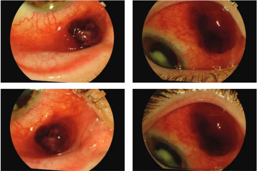
On ophthalmic examination, the best corrected visual acuity was 20/25 on his right eye. The biomicroscopy revealed two reddish nodular lesions localized in the superior and inferior nasal bulbar conjunctiva, loosely connected to the underlying tissue (Figure1). Fundoscopy was normal. The left eye showed no abnormalities. Screening and confirmatory tests were positive for HIV with CD4+ lymphocyte cell count of 154 cells/mm3.
An excisional biopsy and cryotherapy on the conjunctival edges were done on January 2011. Histologic report of the biopsy was consistent with the diagnosis of KS with con-junctival edges free of neoplasm (Figure2). CD31 and CD34 immunohistochemical stains were positive (Figure3). CD31 is an endothelial cell marker, CD34 is an important adhesion molecule required for T cells, and both are markers for KS.
2 Case Reports in Ophthalmological Medicine
Figure 1: Preoperative biomicroscopy.
(a) (b)
Figure 2: Histopathological finding (a) hematoxylin and eosin stain, (b) peroxide acid-Schiff stain.
(a) (b)
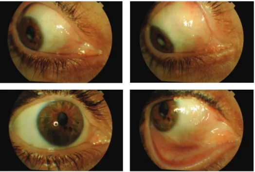
Figure 4: Postoperative biomicroscopy.
The patient initiated standard antiretroviral therapy in January 2011 and remains in followup in the Infectious Diseases Department. Two years later the patient remains with no signs of recurrence (Figure4).
3. Discussion
Improved survival as a result of antiretroviral therapy has led to an increase in systemic and ocular complications. KS is the most common tumor arising in HIV-infected patients. Ophthalmic presentation often occurs in the context of systemic KS, and isolated conjunctival lesions are rare [5–7]. KS may affect many structures of the eye. Involvement of the conjunctiva appears as a flat, reddish lesion, most often localized in the inferior conjunctiva and fornix. It may be misdiagnosed with chronic subconjunctival haemorrhage.
The differential diagnosis includes granuloma pyo-genicum, cavernous haemangioma, foreign-body granuloma, malignant melanoma, metastatic tumor, and chronic subcon-junctival haemorrhage [7].
Dugel et al. [8] have classified adnexal KS clinically into three stages. In stages 1 and 2, the tumors are patchy and flat (less than 3 mm high), and in stage 3, the tumors are nodular and elevated (more than 3 mm high).
Biopsy with histopathological and immunohistochem-istry report has an essential role in the diagnosis.
The type of treatment depends on the size and location of the tumor. KS responds well to treatment by
(1) surgical excision: in extensive, large tumors, loosely connected with healthy tissue;
(2) radiotherapy: for more extensive lesions; (3) cryotheraphy: for early flat tumors; (4) local injection of cytostatics [9,10].
This case describes an isolated conjunctival Kaposi’s sar-coma as primary presentation of AIDS, which is an unusual location for KS [11].
Complete surgical excision with cryotheraphy on con-junctival edges proved to be effective in our patient, with no evidence of clinical recurrence.
This case emphasizes the importance of considering KS as a possible diagnosis in HIV positive patients. Also, KS can be the first AIDS-defining illness in patients with decreased immunological response.
References
[1] M. G. Wani, “Eye complications of acquired immune deficiency syndrome (AIDS),” South Sudan Medical Journal, vol. 2, no. 2, 2009.
[2] C. Muccioli and R. B. J´unior, “Manifestac¸˜oes oftalml´ogicas em doentes com AIDS,” Revista Diagn´ostico & Tratamento, vol. 6, no. 3, pp. 27–30, 2001.
[3] A. Mortada, “Conjunctival regressing Kaposi sarcoma,” British
Journal of Ophthalmology, vol. 51, no. 4, pp. 275–280, 1967.
[4] R. S. Bhatia, “Ophthalmic manifestations of AIDS, short review article,” Journal, Indian Academy of Clinical Medicine, vol. 3, no. 1, pp. 85–88, 2002.
[5] U. R. Kurumety and J. M. Lustbader, “Kaposi’s sarcoma of the bulbar conjunctiva as an initial clinical manifestation of acquired immunodeficiency syndrome,” Archives of
Ophthal-mology, vol. 113, no. 8, p. 978, 1995.
[6] N. Murray, P. McCluskey, D. Wakefield, and P. Beaumont, “Isolated bulbar conjunctival Kaposi’s sarcoma,” Australian and
New Zealand Journal of Ophthalmology, vol. 22, no. 1, pp. 81–82,
1994.
[7] M. D. Bavishi, A. Ashraf, and L. Lee, “AIDS-associated Kaposi’s Sarcoma of the conjunctiva in a woman,” International Journal
4 Case Reports in Ophthalmological Medicine
[8] P. U. Dugel, P. S. Gill, G. T. Frangieh, and N. A. Rao, “Ocular adnexal Kaposi’s sarcoma in acquired immunodeficiency syn-drome,” American Journal of Ophthalmology, vol. 110, no. 5, pp. 500–503, 1990.
[9] M. Nijhoff, “Kaposi’s sarcoma of the conjunctiva and CMV-retinitis in AIDS,” Documenta Ophthalmologica, vol. 65, pp. 77– 85, 1986.
[10] P. U. Dugel, P. S. Gill, G. T. Frangieh, and N. A. Rao, “Treatment of ocular adnexal Kaposi’s sarcoma in acquired immune defi-ciency syndrome,” Ophthalmology, vol. 99, no. 7, pp. 1127–1132, 1992.
[11] L. Pantanowitz and B. J. Dezube, “Kaposi sarcoma in unusual locations,” BMC Cancer, vol. 8, article 190, 2008.
Submit your manuscripts at
http://www.hindawi.com
Stem Cells
International
Hindawi Publishing Corporationhttp://www.hindawi.com Volume 2014
Hindawi Publishing Corporation
http://www.hindawi.com Volume 2014
INFLAMMATION
Hindawi Publishing Corporation
http://www.hindawi.com Volume 2014
Behavioural
Neurology
Endocrinology
International Journal ofHindawi Publishing Corporation
http://www.hindawi.com Volume 2014 Hindawi Publishing Corporation
http://www.hindawi.com Volume 2014
Disease Markers
Hindawi Publishing Corporation
http://www.hindawi.com Volume 2014
BioMed
Research International
Oncology
Journal of Hindawi Publishing Corporationhttp://www.hindawi.com Volume 2014
Hindawi Publishing Corporation
http://www.hindawi.com Volume 2014 Oxidative Medicine and Cellular Longevity Hindawi Publishing Corporation
http://www.hindawi.com Volume 2014
PPAR Research
The Scientific
World Journal
Hindawi Publishing Corporation
http://www.hindawi.com Volume 2014
Immunology Research
Hindawi Publishing Corporation
http://www.hindawi.com Volume 2014
Journal of
Obesity
Journal ofHindawi Publishing Corporation
http://www.hindawi.com Volume 2014
Hindawi Publishing Corporation
http://www.hindawi.com Volume 2014 Computational and Mathematical Methods in Medicine
Ophthalmology
Journal ofHindawi Publishing Corporation
http://www.hindawi.com Volume 2014
Diabetes Research
Journal ofHindawi Publishing Corporation
http://www.hindawi.com Volume 2014
Hindawi Publishing Corporation
http://www.hindawi.com Volume 2014 Research and Treatment
AIDS
Hindawi Publishing Corporation
http://www.hindawi.com Volume 2014
Gastroenterology Research and Practice
Hindawi Publishing Corporation
http://www.hindawi.com Volume 2014