INTRODUCTION
Familial amyloid polyneuropathy (FAP) is an autosomal dominant heredi-tary disease characterized by the extra-cellular deposition of amyloid fibrils in the connective tissue, affecting the pe-ripheral nervous system in particular (1,2). The onset of clinical symptoms generally occurs before age 40, with a progressive and severe sensory and auto-nomic neuropathy leading to death in about 10–20 years.
FAP amyloidoses are related to single transthyretin (TTR) amino acid substitu-tions in which the mutated protein leads to extracellular amyloid fibril deposition. Although more than 80 transthyretin mutations associated with TTR
amyloi-dosis have been described (3), the most common variant has a valine substituted by a methionine at position 30 (V30M) (4). TTR is a 55-kDa homotetrameric pro-tein synthesized mainly in the liver, eye, and choroid plexus, and its main func-tion is the transport of thyroxin (T4) and vitamin A (retinol) associated with the retinol binding protein.
Why mutated TTR deposits in the form of amyloid is unknown, but x-ray crystallographic studies of TTR mutants related to aggressive forms of FAP show conformational changes in this protein (5). These changes may lead to tetramer dissociation into a nonnative TTR monomer with low conformational sta-bility, which results in partially unfolded
monomeric species with a strong ten-dency to aggregate (6). Pathogenic events associated with TTR deposition in FAP patients have been investigated by analy-ses of nerve and salivary gland tissues from FAP patients and asymptomatic V30M carriers; cytotoxicity begins in a presymptomatic stage of the disease, with nonfibrillar TTR aggregates trigger-ing oxidative damage, inflammatory re-sponses, induction of the nuclear tran-scription factor kB (NFkB) pathway, and activation of caspase-3 before amyloid fibrillar deposition (7,8). Increased levels of the endoplasmic reticulum (ER) stress sensor BiP were found to correlate with the extracellular TTR deposition ob-served in salivary gland tissue from FAP patients (9).
Early detection of pathological lesions through the use of biomarkers may aid in correct clinical management of pa-tients and possible delay of morbidity. In this regard, animal models of TTR
Amyloidotic Polyneuropathy
Bárbara Macedo,
1,2Ana Rita Batista,
1José Barbas do Amaral,
3and Maria João Saraiva
1,2Address correspondence and reprint requests to Maria João Saraiva, Molecular
Neurobi-ology, Instituto de Biologia Molecular e Celular, R.Campo Alegre, 823. 4150-180 Porto, Portugal. Phone: 351-226074900, Fax: 351-226099157, E-mail: mjsaraiv@ibmc.up.pt. Submitted July 5, 2007; Accepted for publication September 14, 2007.
1Molecular Neurobiology, Instituto de Biologia Molecular e Celular, Porto; 2Instituto de Ciências Biomédicas de Abel Salazar, University of Porto, Portugal; 3Estomatology, Maxillofacial Surgery, Hospital Geral de Santo António, Porto, Portugal
The identification of specific biomarkers provides opportunities to develop early diagnostic parameters, monitor disease pro-gression, and test drug efficiency in clinical trials. We previously demonstrated that in familial amyloidotic polyneuropathy (FAP) related to the abnormal extracellular tissue deposition of mutant transthyretin (TTR), inflammatory and apoptotic pathways are triggered in the presymptomatic stages of the disease, when nonfibrillar TTR deposits are present. In the present work, to better define biomarkers for future assessment of prophylactic and therapeutic drugs in the treatment of FAP, we extended the search for oxidative stress and apoptotic biomarkers to clinical samples and animal models presenting nonfibrillar and fibrillar TTR. We found that lipid peroxidation measured by hydroxynonenal, oxidative DNA damage measured by 8-hydroxy-2′-deoxyguanosine, and cellular redox homeostasis measured by glutaredoxin 1 were consistently increased in biopsy specimens from FAP patients and in tissues from transgenic mouse models presenting nonfibrillar TTR deposition. Death-receptor Fas, caspase-8, and Bax were also found to be increased, indicative of the involvement of death receptors in the observed apoptosis process. Removal of TTR deposition by an immunization protocol resulted in significant decreases of the selected markers we describe, corroborating the relationship between TTR deposition, oxidative stress, and apoptosis. Taken together, our results provide a robust biomarker pro-file for initial experimental animal studies and clinical trials to assess the application of the selected markers in therapies aimed at removal and/or inhibition of TTR polymerization.
Online address: http://www.molmed.org doi: 10.2119/2007-00068.Macedo
deposition with defined sets of correla-tive biomarkers are essential tools to test and guide the application of pro-phylactic and therapeutic drugs before clinical testing.
Mice transgenic for human TTR V30M in a null background (hTTR Met30) serve as animal models for TTR deposition. Nonfibrillar deposition begins when the mice are three months old, and the de-posits evolve to amyloid fibrils when the mice are nine months old, with particu-lar involvement of the gastrointestinal tract and skin (10). hTTR Met 30 mice have already been used to demonstrate that TTR deposits can be removed by an immunization protocol with a TTR vari-ant expressing a cryptic epitope, TTR Y78F (11).
By crossing hTTR Met 30 mice to mice with a heat shock transcription factor 1 (HSF1) null background, a novel trans-genic mouse model was generated, hTTR Met30/HSF1-KO. This new mouse model shows TTR deposition in the pe-ripheral [dorsal root ganglia (DRG) and nerve] and autonomic nervous systems, a deposition pattern not observed in hTTR Met30 models (12).
We sought to define new, reliable ox-idative stress and apoptotic biomarkers associated with TTR deposition in human clinical samples and in transgenic mouse models before and after deposi-tion removal by immunizadeposi-tion. MATERIALS AND METHODS Human Samples
Labial minor salivary gland biopsy specimens were obtained from V30M FAP patients before they underwent liver transplantation (13), the only available treatment for this disorder. Control sali-vary gland tissue samples were from non-FAP volunteer individuals who had no evidence of infection. The collection of biopsy material was approved by the ethics committee of Hospital Geral de Santo António, Porto, Portugal, and all specimen donors gave informed consent.
For immunohistochemical (IHC) anal-ysis, salivary gland samples were
col-lected into 4% paraformaldehyde in PBS. The general characterization of salivary gland tissue consisted of TTR immuno-histochemistry and analysis of the pres-ence of amyloid deposits by Congo red (CR) staining. Salivary gland tissues with TTR deposition and with amyloid de-posits were classified as FAP.
Transgenic Mice
Transgenic mice bearing the human TTR V30M in a TTR null background (hTTR Met300) and transgenic mice ex-pressing the human TTR V30M crossed into an HSF1 null background (hTTR Met30/HSF1-KO) were used for the ex-periments (10,12). In this case, we ana-lyzed animals heterozygous for HSF1, labeled as hTTR Met30/(+/–) HSF1-KO. Animals were killed after administra-tion of ketamine/xylazine anesthesia. DRG and stomachs were dissected and fixed in 4% paraformaldehyde, embed-ded in paraffin, and cut into 5-μm sec-tions. The animals were housed in path-ogen-free conditions in a controlled temperature room maintained with 12-hour light/dark periods. Water and food were freely available. All animal experiments were carried out in accor-dance with the European Communities Council Directive.
Immunohistochemistry
Sections (5-μm thick) were deparaffi-nated in histoclear (National Diagnostics, Atlanta, GA, USA) and dehydrated in a descent alcohol series. Endogenous per-oxidase activity was inhibited with 3% hydrogen peroxide/100% methanol, and sections were blocked in 4% fetal bovine serum and 1% bovine serum albumin in PBS. Primary antibodies used were: rab-bit polyclonal anti–thioredoxin 1 (Trx1) (1:500; Chemicon, Temecula, CA, USA), rabbit polyclonal anti–glutaredoxin 1 (Grx1) (1:500; Abcam, Cambridge, UK), goat polyclonal anti–8-hydroxy-2′-deoxyguanosine (8-OHdG) (1:1000 with proteinase K epitope retrieval; Chemicon), goat polyclonal anti–4-hydroxynonenal (HNE) (1:500; Abcam), mouse mono-clonal for active caspase-8 (1:200; Cell
Signaling, Danvers, MA, USA), rabbit polyclonal for active caspase-3 (1:50; Cell Signaling), rabbit polyclonal for active caspase-9 (1:20; Abcam), rabbit poly-clonal anti-Fas (antihuman CD95) (1:100; Santa Cruz Biotechnology, Santa Cruz, CA, USA), rabbit polyclonal anti–Fas L (antihuman CD95 ligand) (1:50; Santa Cruz Biotechnology), rabbit polyclonal anti-Bax (1:50; Santa Cruz), rabbit poly-clonal anti-Bcl2(1:100; Santa Cruz Biotechnology), and rabbit polyclonal anti–Apaf-1 (apoptotic protease-activating factor 1) (1:100 with citrate buffer epitope retrieval; Abcam), which were diluted in blocking solution and in-cubated overnight at 4ºC. Antigen visual-ization was performed with the biotin-extravidin-peroxidase kit (Sigma, St. Louis, MO, USA), using 3-amino-9-ethyl carbazole (Sigma) as substrate. On paral-lel control sections, the primary antibody was replaced by blocking buffer and by nonimmune immunoglobulins corre-sponding to the species used as primary antibodies, namely rabbit and goat; staining was absent in all the cases.
Semiquantitative analysis of IHC im-ages was done with the Universal Imag-ing system (NIH), which performs auto-mated particle analysis in a measured area, i.e., the area occupied by pixels cor-responding to the IHC substrate color is counted and normalized relative to the total area. Each slide was analyzed in five different selected areas. Results shown represent the percentage occupied area ± SD.
Initially, tissues of transgenic mice were selected for TTR deposition by IHC analysis using a rabbit polyclonal anti-TTR (1:1000; Dako, Carpinteria, CA, USA). TTR-positive–CR-negative tissues were classified as (+/–) and TTR-positive–CR-negative tissues as (–/–). Expression of several markers was com-pared in hTTR Met30 mice with TTR deposition in the stomach and age-matched mice (six months old) without TTR deposition in the stomach. The same type of evaluation was performed for stomach and DRG deposition in hTTR Met30/HSF1-KO mice.
Statistical Analysis
Group values, expressed as the mean ± SD, were compared by Student t test, and
P values <0.05 were considered significant. RESULTS
Tissue Markers for Oxidative Stress Several markers of oxidative damage have been widely studied in different pathologies, such as Alzheimer disease (14), and include 8-OHdG, HNE, glutare-doxins such as Grx1, and thioreglutare-doxins such as Trx1. We investigated the expres-sion of HNE, 8-OHdG, Grx1, and Trx1 in biopsy specimens from human salivary glands and in tissues from two FAP ani-mal model strains.
IHC analyses for the lipid peroxidation biomarker HNE were performed in sali-vary gland tissue biopsies from V30M FAP individuals (n = 5) and non-FAP controls (n = 4). In FAP-patient salivary gland tissue, HNE was increased by ap-proximately 50% compared with controls (Figure 1A); the immunostaining was ex-clusively cytoplasmic and was present in epithelial cells in salivary gland excre-tory ducts, at the same location of TTR fibrillar amyloid deposits (13). This result is in accordance with a previous report of the involvement of lipid peroxidation in colon biopsies of FAP patients (15). In addition, we investigated a possible cor-relation between nonfibrillar TTR depo-sition and HNE levels in the transgenic
mice strains under study. In the stomach and DRG in both hTTR Met30 and hTTR Met30/(+/–)HSF1-KO transgenic mice, HNE were significantly increased in mice with TTR deposition compared to mice without TTR deposition (Figure 1A). Quantification of IHC images of HNE staining revealed that HNE was in-creased approximately 70% in the stom-ach and 80% in the DRG (Figure 1A), correlating with nonfibrillar TTR deposi-tion. These results validate the use of hTTR Met30 and hTTR Met30/ (+/–)HSF1-KO mice models as tools for lipid peroxidation studies in FAP.
Although 8-OHdG DNA oxidation has been extensively investigated in other neurodegenerative pathologies, this
Figure 1. (A) Top panels: Representative 4-hydroxy-2-nonenal (HNE) staining of salivary glands from healthy individuals (left, n = 6) and FAP patients (right, n = 6). Middle panels: Representative HNE staining in stomach tissue of hTTR Met30 mice without TTR deposition (–/–, left, n = 5) and with TTR deposition (+/–, right, n = 5) and in stomach tissue of hTTR Met30/(+/–)HSF1-KO mice, without TTR deposition (–/–, left, n = 5) and with TTR deposition (+/–, right, n = 5); Lower panel: Representative HNE staining in DRG of hTTR Met30/(+/–)HSF1-KO mice without TTR deposition (–/–, left, n = 4) and with TTR deposition (+/–, right; n = 4). 20× magnification. Quantitation of IHC images of HNE staining are represented as percentage of occupied area ± SD (*P < 0.05; **P < 0.01, and # P < 0.005). (B) Top panels: Representative 8-OHdG staining of salivary glands from healthy individuals (left, n = 6) and FAP patients (right, n = 6). Middle panels: Representative 8-OHdG staining in stomach tissue of hTTR Met30 mice without TTR deposition (–/–, left, n = 5) and with TTR deposition (+/–, right, n = 5) and in stomach tissue of hTTR Met30/(+/–)HSF1-KO, without TTR deposition (–/–, left, n = 6) and with TTR deposition (+/–, right, n = 6); Lower panel: Representative 8-OHdG staining in DRG tissue of hTTR Met30/(+/–)HSF1-KO mice without TTR deposition (–/–, left, n = 4) and with TTR deposition (+/–, right; n = 5). 20_ magnification. Quantitation of IHC images of 8-OHdG staining are represented as percentage of occupied area ± SD (*P < 0.05 and #P < 0.005).
study is the first to assess 8-OHdG DNA oxidation in FAP patients. Immunostain-ing of FAP salivary gland tissue samples revealed 50% more oxidized DNA than in control samples (Figure 1B). The in-crease in DNA oxidation was even more dramatic in the transgenic mice strains under study. Stomach tissue from trans-genic hTTR Met30 mice with TTR aggre-gates showed approximately 75% more
8-OHdG than tissues without TTR depo-sition (Figure 1B), and in hTTR Met30/ (+/–)HSF1-KO mice we confirmed ap-proximately the same level of increase in DRG (Figure 1B). Thus, we validated the use of these animal models as tools to in-vestigate oxidative stress involving DNA damage.
Finally, IHC studies on salivary glands revealed that Grx1 is up-regulated in
FAP individuals with TTR deposition compared with normal individuals (Fig-ure 2); in contrast, Trx1 levels did not dif-fer in FAP and control individuals (Fig-ure 2). Grx1 resides mainly in the cytosol, although it may translocate into the nucleus in response to certain stim-uli. For example, in the brains of patients with Alzheimer disease, control samples revealed Grx1 was located mainly in the cytosolic compartment, contrary to the nuclear staining in Alzheimer disease neurons (16). In our study nuclear translocation of Grx1 was evident in FAP salivary glands (indicated by arrows in Figure 2).
We then investigated Trx1 and Grx1 endogenous antioxidant systems in hTTR transgenic mice. In both hTTR Met30 and hTTR Met30/(+/–)HSF1-KO mice, in-creased Grx1 was associated with TTR deposition in the stomach (Figure 2) but Trx1 was not (not shown). In the periph-eral nervous system of hTTR Met30/ (+/–)HSF1-KO mice, represented by DRG tissue samples, Grx1 and Trx1 were both up-regulated in samples with TTR deposition (Figure 2).
Tissue Markers for Apoptosis
We investigated the Fas death receptor in FAP salivary glands and in FAP trans-genic mouse models and observed that although there were no differences in FasL expression (Figure 3), FAP salivary glands expressed higher amounts of the receptor protein, probably related to in-creased activation of this apoptosis-initiating cascade (Figure 3). We next evaluated the cleavage of caspase sub-strates in FAP salivary glands. In FAP salivary glands, both the initiator cas-pase-8 as well as the effector caspase-3 were cleaved more extensively compared with controls (Figure 3), presenting as much as 70% increase in activation. However, no differences were found for caspase-9 activation (not shown).
We then investigated the role of Bcl-2 family proteins in FAP biopsies. We did not find differences in Bcl-2 or Apaf-1 levels between FAP and control salivary gland tissue (not shown), concordant
Figure 2.Top 2 panels: Representative Grx1 and Trx1 staining of salivary glands from healthy individuals (left, n = 6) and FAP patients (right, n = 6); nuclear staining is indicated by the arrow. Middle panel: Representative Grx1 staining in stomach tissue of hTTR Met30/ (+/–)hsf mice without TTR deposition (–/–, left, n = 5) and with TTR deposition (+/–, right, n = 5). Lower 2 panels: Representative Grx1 and Trx1 staining in DRG tissue of hTTR Met30/ (+/–)HSF1-KO mice without TTR deposition (–/–, left, n = 4) and with TTR deposition (+/–, right; n = 5); nuclear staining is indicated by the arrow. 20× and 40× magnification (inset). Quantitation of IHC images of Grx1 and Trx1 staining are represented as percentage of occupied area ± SD (*P < 0.05).
with the results on caspase-9, which did not show evidence for activation. Because Bax and Bak are proapoptotic proteins that initiate mitochondrial dysfunction but also localize to the ER (17) we next assessed Bax levels in salivary gland tis-sue samples. A slight, but not significant, increase was detected in FAP glands com-pared with controls (Figure 3).
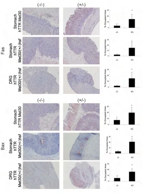
We further investigated a possible cor-relation between nonfibrillar TTR depo-sition and the above-defined markers of apoptosis in the mice transgenic for human TTR V30M and the novel hTTR Met30/(+/–)HSF1-KO mice. A dramatic increase in the Fas death receptor signal was observed in tissues with TTR depo-sition in the transgenic mouse strains under study. Transgenic hTTR Met30 mice presented an increase of approxi-mately 75% in Fas in the stomach of TTR-positive animals (Figure 4), and the same differences were observed in the stomach and DRG of hTTR Met30/ (+/–)HSF1-KO mice (Figure 4) in TTR-positive compared with deposit-free tis-sues. As in human salivary gland tissue, FasL was expressed at the same levels in mouse tissues with or without TTR dep-osition (data not shown).
We found increased levels of Bax (Fig-ure 4) associated with TTR deposition in the stomach of both transgenic mice strains as well as in DRG from hTTR Met30/(+/–)HSF1-KO mice (Figure 4).
Based on these results, we believe that the apoptotic pathway mediated by the Fas receptor is definitely involved in neurotoxicity observed in FAP salivary gland tissue and in the animal models under study that present TTR deposition, and in FAP in general.
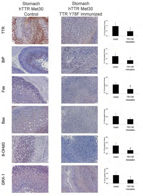
Oxidative Stress and Apoptotic Markers after TTR Removal
IHC analyses of tissues from trans-genic hTTR Met30 mice immunized with Y78F and presenting with TTR removal revealed a decrease in some of the mark-ers described above. We tested one marker of oxidative stress, 8-OHdG, a radical-damaged guanine nucleotide rep-resentative of oxidation at the DNA
level; two markers of apoptosis, the death receptor Fas and the pro-apoptotic Bax; the ER chaperone BiP; and the en-dogenous antioxidant protein Grx1.
IHC analysis of the gastrointestinal tract of TTR Y78F immunized mice (n = 6) revealed decreased levels of these markers in the stomach compared with nonimmunized age-matched controls (n = 6). The decrease was approximately 50% for Fas and BiP and was even more dramatic, approximately 70%, for the ox-idative stress markers 8-OHdG and Grx1. Although Bax levels were lower in
the tissues of immunized mice, the dif-ference was not significant (Figure 5).
These results correlate with the de-crease of TTR in the stomach of TTR Y78F immunized animals reported by Terazaki et al. (11), further validating the use of 8-OHdG, Grx1, Fas, and BiP for follow-up of therapeutic approaches in FAP patients.
DISCUSSION
We generated a set of biomarkers as-sociated with FAP pathology by the use of salivary gland tissue from FAP
pa-Figure 3. Representative FasL, Fas, active caspase-8, active caspase-3, and Bax staining of
salivary glands from healthy individuals (left, n = 6) and FAP patients (right, n = 6). 20× mag-nification. Quantitation of IHC images of FasL, Fas, active caspase-8, active caspase-3, and Bax staining are represented as percentage of occupied area ± SD (*P < 0.05).
tients with amyloid deposition and unique tissues, stomach and DRG from 2 different animal models with nonfibril-lar TTR deposition, that resemble the asymptomatic phase of the disease. A
major novel finding from our study was the detection of specific oxidative and apoptotic markers not only in amyloid-laden tissues, but also before amyloid deposition, in tissues presenting
nonfib-rillar deposition. The biomarkers we found to be associated with deposition did not deviate considerably from those found in other neurodegenerative disor-ders; for instance, in Alzheimer disease abnormal oxidative metabolites, includ-ing HNE and cholesterol-derived alde-hydes, have been reported to modify Aβ and accelerate the early stages of amyloidogenesis (18). The biomarker 8-OHdG, a modified base that occurs in DNA due to attack by hydroxyl radicals formed as byproducts and intermediates of aerobic metabolism and during oxida-tive stress, has been increasingly used as a sensitive, stable, and integral marker of oxidative damage in cellular DNA (19). In a neuroblastoma cell line, Aβ fib-rils caused transient oxidation of both Grx1 and Trx1 (16). Cytosolic mam-malian Trx1 has numerous functions in defense against oxidative stress, control of growth, and apoptosis. Grx1 catalyses glutathione disulfide oxireductions, overlapping the functions of thioredox-ins and using electrons from NADPH via glutathione reductases (20). Despite the importance of Grx1 and Trx1 as reg-ulators of oxidative stress and apoptosis, their role in FAP is completely un-known. We believe that overexpression of Grx1 may have a protective role, act-ing to detoxify FAP tissues as it does the frontal cortex and hippocampal neurons of the brain in Alzheimer disease (16). Trx1 was not up-regulated in the ana-lyzed tissues with TTR deposition, ex-cept for DRG, probably because salivary glands and stomach mucosa are in con-tact with multiple stimuli and irritations. The Trx1 system is probably activated in a constant way by contact with external stimuli, whereas Grx1 better protects against cellular injury. In addition to measurement of the levels of these pro-teins, their reduced and oxidized states should also be investigated, because un-derstanding of the downstream cascades that Grx1 and Trx1 trigger will elucidate the roles of Trx1 and Grx1 in TTR amy-loidosis and may lead to the develop-ment of therapeutic interventions to modulate these markers.
Figure 4. Representative Fas (upper panels) and Bax (bottom panels) staining in stomach and DRG tissue of TTR transgenic mice. Upper panels: Stomach tissue of hTTR Met30 mice without TTR deposition (–/–, left, n = 5) and with TTR deposition (+/–, right, n = 5), of hTTR Met30/(+/–)HSF1-KO, without TTR deposition (–/–, left, n = 6) and with TTR deposition (+/–, right, n = 6) and DRG tissue of hTTR Met30/(+/–)HSF1-KO mice without TTR deposition (–/–, left, n = 6) and with TTR deposition (+/–, right; n = 6). Bottom panels: the same organiza-tion as above. 20× magnification. Quantitation of IHC images of Fas and Bax staining are represented as percentage of occupied area ± SD (*P < 0.05).
Apoptotic features are present exten-sively in the most common neurodegen-erative diseases. Previous studies have evaluated Aβ-induced apoptotic cell death in cerebral endothelial cells by in-vestigation of increased oxidative stress, caspase activation, mitochondrial dys-function, and DNA damage (21).
The evidence we obtained for Fas and caspase-8 activation in tissues with TTR deposition suggest activation of death-receptor pathways not involving the mi-tochondria (22,23); a possible explana-tion for caspase-8 activaexplana-tion is the induction of Fas-associated death domain–independent pathways deriving from prolonged activation of extracellu-lar signal-regulated kinases 1 and 2 (ERK1/2) (24) and activation of ERK1/2, which was shown in FAP salivary gland tissue and nerves and in tissues from TTR transgenic mice presenting TTR deposition (25). Further suggestion for a secondary role of mitochondrial involve-ment was the lack of evidence in the present study for changes in Bcl2 and Apaf-1 in tissues with TTR deposition (22,26). Additional studies regarding morphological and functional alterations in mitochondria, namely the measure-ment of mitochondrial membrane poten-tial, should clarify this issue as tested in another model system (27). Moreover, the trophic signals relayed from TTR to mitochondria remain elusive, with a possible role of intracellular Ca2+ re-maining to be investigated. The ER contributes to pathways in FAP, a char-acteristic that differs from other neu-rodegenerative protein misfolding disor-ders and is suggestive of different pathways involved in apoptosis, possi-bly related to the fact that in FAP protein aggregation occurs extracellularly rather than intracellularly. Recent data showed activation of the classical unfolded-pro-tein response pathways in tissues not specialized in TTR synthesis but present-ing extracellular TTR aggregate and fib-ril deposition. We have previously demonstrated cytotoxicity by Ca2+ efflux from the ER in cell cultures incubated with TTR oligomers, evidence for ER
stress associated with extracellular aggregates (9).
We have demonstrated that the bio-markers of oxidative stress and apopto-sis we investigated are correlated with TTR deposition, as corroborated by the results in tissues from which deposition
was removed by immunization. These results confirm the usefulness of this set of biomarkers in the follow-up of thera-peutic approaches and highlight the im-portance of pursuing FAP treatments based on antioxidative and antiapoptotic therapies.
Figure 5. Representative immunohistochemistry of TTR, BiP, Fas, Bax, 8-OHdG and Grx1 in
stomach tissue of TTR Y78F immunized transgenic mice 9-10 months old (right; n = 6) and nonimmunized age-matched controls (left; n = 6). 20× magnification; Quantification of IHC images of TTR, BiP, Fas, Bax, 8-OHdG, and Grx1 staining are represented as percent-age of occupied area ± SD (*P < 0.05, #P < 0.005).
ACKNOWLEDGMENTS
This work was supported by grants from the POCTI and POCI programs of Fun-dação para a Ciência e Tecnologia–FCT, the Gulbenkian Foundation, Portugal, and fel-lowships SFRH/BD/9243/2002 (to BM) and POCI/SAU-OBS/56929/2004 (to ARB) from the Fundação para a Ciência e Tec-nologia, Portugal. We thank Tânia Ribeiro and Rossana Correia for tissue processing. REFERENCES
1. Andrade C.(1952) A peculiar form of peripheral neuropathy; familiar atypical generalized amy-loidosis with special involvement of peripheral nerves. Brain 75:408-27.
2. Coimbra A, Andrade C. (1971) Familial amyloid polyneuropathy, an electron microscope study of the peripheral nerve in five cases. Brain 94:199-212.
3. Saraiva MJ. (2001) Transthyretin mutations in hy-perthyroxinemia and amyloid diseases. Hum.
Mutat. 17:493-503.
4. Saraiva MJ, Birken S, Costa PP, Goodman DS. (1984) Amyloid fibril protein in familial amy-loidotic polyneuropathy, Portuguese type: defini-tion of molecular abnormality in transthyretin (prealbumin). J. Clin. Invest. 74:104-19.
5. Sebastião MP, Saraiva MJ, Damas AM. (1998) The crystal structure of amyloidogenic Leu55.Pro transthyretin variant reveals a possible pathway for transthyretin polymerization into amyloid fibrils. J. Biol. Chem. 273:24715-22.
6. Quintas A, Vaz DC, Cardoso I, Saraiva MJ, Brito RM. (2001) Tetramer dissociation and monomer partial unfolding precedes protofibril formation in amyloidogenic transthyretin variants. J. Biol.
Chem. 276:27207-13.
7. Sousa MM, Cardoso I, Fernandes R, Guimarães A, Saraiva MJ. (2001) Deposition of transthyretin in early stages of familial amyloidotic polyneu-ropathy: evidence for toxicity of nonfibrillar ag-gregates. Am. J. Pathol. 159:1993-2000. 8. Sousa MM, Du Yan S, Fernandes R, Guimaraes
A, Stern D, Saraiva MJ. (2001) Familial amyloid polyneuropathy: receptor for advanced glycation end products-dependent triggering of neuronal inflammatory and apoptotic pathways. J.
Neu-rosci. 21:7576-86.
9. Teixeira PF, Cerca F, Santos SD, Saraiva MJ. (2006) Endoplasmic reticulum stress associated with extracellular aggregates: evidence from transthyretin deposition in familial amyloid polyneuropathy. J. Biol. Chem. 281:21998-2003. 10. Sousa MM, Fernandes R, Palha JA, Taboada A,
Vieira P, Saraiva MJ. (2002) Evidence for early cy-totoxic aggregates in transgenic mice for human transthyretin Leu55Pro. Am. J. Pathol. 161:1935-48. 11. Terazaki H, Ando Y, Fernandes R, Yamamura K,
Maeda S, Saraiva MJ. (2006) Immunization in fa-milial amyloidotic polyneuropathy:
counteract-ing deposition by immunization with a Y78F TTR mutant. Lab. Invest. 86:23-31.
12. Santos SD, Saraiva MJ. (2005) Proceedings of the Society for Neuroscience 35th annual meeting. Program No. 670.9, Abstract Viewer and Itiner-ary Planner, Society for Neuroscience, Washing-ton, D. C. November 12–16.
13. Sousa MM, do Amaral JB, Guimaraes A, Saraiva MJ. (2005) Up-regulation of the extracellular ma-trix remodeling genes, biglycan, neutrophil gelatinase-associated lipocalin, and matrix metal-loproteinase-9 in familial amyloid polyneuropa-thy. FASEB J. 19:124-6.
14. Mattson MP. (2004) Pathways toward and away from Alzheimer’s disease. Nature 430:631-9. 15. Ando Y, Nyhlin N, Suhr O, et al. (1997)
Oxida-tive stress is found in amyloid deposits in sys-temic amyloidosis. Biochem. Biophys. Res.
Com-mun. 232:497-502.
16. Akterin S, Cowburn RF, Miranda-Vizuete A, et al. (2006) Involvement of glutaredoxin-1 and thioredoxin-1 in beta-amyloid toxicity and Alz-heimer’s disease. Cell Death Differ. 13:1454-1465. 17. Zong WX, Li C, Hatzivassiliou G, Lindsten T, Yu
QC, Yuan J, Thompson CB. (2003) Bax and Bak can localize to the endoplasmic reticulum to initi-ate apoptosis. J. Cell. Biol. 162:59-69.
18. Bieschke J, Zhang Q, Powers ET, Lerner RA, Kelly JW. (2005) Oxidative metabolites accelerate Alzheimer’s amyloidogenesis by a two-step mechanism, eliminating the requirement for nu-cleation. Biochemistry 44:4977-83.
19. Leanderson P, Tagesson C. (1992) Rapid and sen-sitive detection of hydroxyl radicals formed by activated neutrophils in the presence of chelated iron, hydroxylation of deoxyguanosine to 8-hy-droxydeoxyguanosine. Agents Actions 36:50-7. 20. Arner ES, Holmgren A. (2000) Physiological
functions of thioredoxin and thioredoxin reduc-tase. Eur. J. Biochem. 267:6102-9.
21. Xu J, Chen S, Ku G, et al. Amyloid beta peptide-induced cerebral endothelial cell death involves mitochondrial dysfunction and caspase activa-tion. J. Cereb. Blood Flow Metab. 21:702-10. 22. Ashkenazi A. (2002) Targeting death and decoy
receptors of the tumor-necrosis factor superfam-ily. Nat. Rev. Cancer 2:420-30.
23. Strasser A, O’Connor L, Dixit VM. (2000) Apop-tosis signaling. Ann. Rev. Biochem. 69:217-45. 24. Cagnol S, Van Obberghen-Schilling E, Chambard
JC. (2006) Prolonged activation of ERK1,2 in-duces FADD-independent caspase 8 activation and cell death. Apoptosis 11:337-46.
25. Monteiro FA, Sousa MM, Cardoso I, do Amaral JB, Guimaraes A, Saraiva MJ. (2006) Activation of ERK1/2 MAP kinases in familial amyloidotic polyneuropathy. J. Neurochem. 97:151-61. 26. Cain K. (2003) Chemical-induced apoptosis:
for-mation of the Apaf-1 apoptosome. Drug Metab.
Rev. 35:337-63.
27. Angelin A, Tiepolo T, Sabatelli P, et al. (2007) Mi-tochondrial dysfunction in the pathogenesis of Ullrich congenital muscular dystrophy and pro-spective therapy with cyclosporins. Proc. Natl.