UNIVERSIDADE DA BEIRA INTERIOR
Ciências da Saúde
UTILIZAÇÃO DE UM MODELO DE TRANSFUSÃO DE
ERITRÓCITOS IN VITRO PARA MONITORIZAR A
DIVISÃO E EXPRESSÃO DE CD28 EM LINFÓCITOS T
ATIVADOS
Carolina Alves Padrão
Dissertação para obtenção do Grau de mestre em
Medicina
(Mestrado integrado)
Orientador: Prof. Doutor Fernando A. Arosa
Agradecimentos
Quero agradecer à Dra. Teresa Caeiro e funcionários do Centro do Sangue e
Transfusão de Coimbra (CSST-Coimbra) por fornecer concentrados leuco-plaquetários ou BC (buffy-coats) utilizados neste estudo, assim como ao Dr. Jorge Martinez, responsável pelo Serviço de Imunohemoterapia do Centro Hospitalar Cova da Beira (CHCB), e seus
colaboradores, por fornecer flebotomias de doentes com sobrecarga de ferro e pela prontidão em disponibilizar os BC chegados de Coimbra.
Ao meu orientador professor doutor Fernando A. Arosa por ter aceitado o desafio de orientar uma aluna de medicina para realizar um trabalho de investigação laboratorial e ter mostrado sempre disponibilidade para todas as questões que iam surgindo.
À professora doutora Elsa Cardoso pela disponibilidade, simpatia e apoio no laboratório.
Ao André Esgalhado pelo auxílio, paciência e apoio dado na parte laboratorial. Ao Dmytro Vasyanovych por toda a paciência e compreensão que demonstrou e pelo apoio na edição das imagens.
Aos meus pais, pela compreensão e apoio. Por acreditarem em mim e me instruírem a querer sempre mais. Ao meu irmão por fazer parte da minha vida.
Resumo
Introdução: As transfusões de eritrócitos estão associadas a efeitos imunomoduladores e
inflamatórios. Apesar dos leucócitos residuais presentes nas unidades de sangue transfusional terem sido implicados nestes efeitos, ensaios clínicos com leucorredução mostram efeitos controversos. Estudos prévios realizados pelo nosso grupo demostraram que os eritrócitos, quer autólogos quer heterólogos, possuem bioatividades que favorecem o crescimento e a sobrevivência de linfócitos T activados in vitro, permitindo-lhes entrar em ciclos de divisão consecutivos. Contudo, esses estudos não caracterizaram a expressão de CD28, um receptor importante para a activação de linfócitos T naïve que deixa de ser expresso pela maior parte dos linfócitos T CD8+ com a idade, e em condições inflamatórias.Objetivo: A finalidade deste
estudo foi utilizar um modelo in vitro de transfusão de eritrócitos para analisar a proliferação e expressão de CD28 nos linfócitos T CD4+ e CD8+. Métodos: Foram isoladas células
mononucleares de sangue periférico de dadores saudáveis e de pacientes flebotomizados após centrifugação sobre um gradiente de densidade (Lymphoprep), marcadas com
carboxyfluorescein diacetate succinimidyl ester (CFSE), um corante fluorescente que permite
a monotorização da divisão celular, e cultivadas durante 6 dias com fitohemaglutinina (PHA), um mitogénio policlonal de linfócitos T, na ausência ou presença de eritrócitos numa proporção 50RBC:1PBMC. As células foram colhidas e marcadas com anticorpos conjugados com fluorocromos contra os receptores CD4, CD8 e CD28, e depois adquiridas em um citómetro de fluxo para determinação da proliferação e a expressão de CD28. Resultados: Os linfócitos T proliferaram mais vigorosamente em culturas com eritrócitos. A proliferação esteve associada com a perda do receptor CD28, parecendo mais evidente nos linfócitos T CD8+. Isto resultou num aumento da percentagem de linfócitos CD8+CD28− no fim do período
de cultura. Nas culturas com PHA há uma tendência para o aumento na expressão do CD28 nos linfócitos CD4+. Conclusão e Discussão: Este estudo preliminar sugere que os eritrócitos
induzem a formação de linfócitos CD8+CD28− através da perda de expressão do receptor CD28.
Considerando que os linfócitos T CD8+CD28− contem quer células imunossupressoras quer
inflamatórias, estes resultados podem ser relevantes no contexto de patologias humanas onde as transfusões são necessárias.
Palavras-chave
Resumo alargado
As transfusões de eritrócitos apesar de salvarem muitas vidas estão associadas a efeitos imunomoduladores e inflamatórios. Esses efeitos podem ser manifestados pelo aumento da susceptibilidade a infecções, prolongamento do tempo de internamento, insuficiência respiratória e mortalidade. Mas nem tudo é mau, pois também podem aumentar a sobrevivência de transplantes. Apesar de os leucócitos residuais terem sido implicados nestes efeitos, ensaios clínicos com leucorredução mostraram efeitos controversos reforçando a possível relevância dos eritrócitos na imunomodulação. Os concentrados de eritrócitos com mais tempo de armazenamento estão sujeitos a sofrer lesões, diminuindo a sua qualidade; no entanto, as manifestações na clinica ainda são duvidosas. Estudos prévios in vitro demostraram que os eritrócitos autólogos e heterólogos possuem bioatividades sobre os linfócitos T activados que vão desde o aumento do crescimento e sobrevivência até o favorecimento da entrada em ciclos consecutivos de divisão. Contudo, estes estudos não caracterizaram a expressão de CD28, um receptor co-estimulador de linfócitos T que deixa de ser expresso na maior parte dos linfócitos T CD8+ durante o envelhecimento e em doenças
inflamatórias crónicas, levando à acumulação de linfócitos T CD8+CD28− polifuncionais que
possuem propriedades imunossupressoras e citotóxicas. A finalidade deste estudo foi utilizar este modelo in vitro de transfusões de eritrócitos autólogos para analisar o seu efeito na proliferação e expressão de CD28 em linfócitos T CD4+ e CD8+ activados.
Para o efeito, foram isoladas células mononucleares de sangue periférico (PBMC das siglas em inglês) de 8 dadores saudáveis e de 3 pacientes flebotomizados por sobrecarga de ferro, após centrifugação sobre um gradiente de densidade (Lymphoprep). Os PBMC foram marcados com carboxyfluorescein diacetate succinimidyl ester (CFSE), um corante fluorescente que atravessa livremente a membrana plasmática das células ficando posteriormente retido no interior destas após clivagem da ligação ester por esterases e subsequente ligação covalente a proteínas. A perda de fluorescência em cada ciclo celular, como resultado da formação de células filhas com a metade de CFSE que a célula mãe, permite monitorizar a divisão celular ao longo do tempo. Os PBMC foram depois colocados em placas de cultura de 6 poços durante 6 dias com fitohemaglutinina (PHA das siglas em inglês), um mitogénio policlonal de linfócitos T que apenas funciona na presença de monócitos existentes nos PBMC, na ausência ou presença de eritrócitos. As células foram colhidas ao fim do período de cultura e marcadas com anticorpos monoclonais conjugados com fluorocromos contra os receptores CD4, CD8 e CD28. Seguidamente foram adquiridas num citómetro de fluxo para determinação da proliferação e expressão do CD28.
Os resultados confirmaram os dados previamente obtidos pelo nosso grupo, mostrando que os linfócitos T proliferam mais vigorosamente quando na cultura in vitro são adicionados eritrócitos. Verificou-se uma maior expressão de CD28 nos linfócitos T CD4+
análise de expressão de CD28 em linfócitos CD4+ e CD8+ revelou dois resultados novos
interessantes. Em primeiro lugar, ao longo da divisão as células CD4+ em culturas com PHA
têm tendência para aumentar a expressão do CD28, enquanto que nas células cultivadas com PHA+eritrócitos ocorre o inverso, havendo uma diminuição da expressão do CD28. Por outro lado, observou-se que em algumas culturas com PHA os linfócitos CD8+ tinham tendência a
perderem a expressão do receptor CD28 com os ciclos de divisão, um efeito que foi observado na grande maioria das culturas com eritrócitos, resultando no aumento da percentagem de linfócitos CD8+CD28− no fim do período de cultura.
Este estudo preliminar, que tenta reproduzir o efeito biológico de uma transfusão sanguínea em linfócitos T activados, sugere que os eritrócitos podem induzir uma população de linfócitos T CD8+CD28− através da indução da perda de expressão do receptor CD28.
Considerando que os linfócitos T CD8+CD28− T podem produzir citocinas inflamatórias (por ex.,
IFN-,TNF-α) e/ou supressoras (por ex., TGF-, IL-10), estes resultados podem ser relevantes no contexto de patologias humanas onde as transfusões são necessárias.
Abstract
Introduction: Blood transfusions are associated with transfusion related inflammatory
and immunomodulatory effects. Although residual leukocytes present in the transfused red blood cell units have been implicated in these effects, clinical trials of leukoreduction have shown conflicting results. Our previous in vitro studies with human T cells have demonstrated that red blood cells (RBC), either autologous or heterologous, have cell growth and survival bioactivities that allow activated T cells to enter consecutive cycles of cell division. However, these studies did not characterize expression of CD28, an important receptor for naïve T cells that is lost by most CD8+ T cells during aging and chronic inflammation. Objective: The
purpose of this study was to use our in vitro model of blood transfusion to analyze the extent of proliferation and the expression of CD28 on CD4+ and CD8+ T cells. Methods: Peripheral
blood mononuclear cells were isolated from blood samples of regular blood donors and iron overloaded phlebotomized patients after centrifugation over Lymphoprep, labeled with carboxyfluorescein diacetate succinimidyl ester, a fluorescent dye that allows monitoring of cell division, and cultured for 6 days with the polyclonal T cell mitogen phytohaemagglutinin (PHA) in the absence or presence of red blood cells at a 50:1 RBC to PBMC ratio. Afterwards, cells were harvested, labeled with fluorochrome-conjugated antibodies against CD4, CD8 and CD28, acquired in a flow cytometer, and proliferation and expression of CD28 determined.
Results: T cells proliferated more vigorously when RBC were present in the culture. Dividing
CD8+ T cells showed a higher loss of CD28 than CD4+ T cells, namely in cultures with red blood
cells. As a result, the percentage of CD8+CD28− T cells increased at the end of the culture
period. Finally, a tendency to downregulate CD28 expression with each cell division is seen in cultures with PHA+RBC. In cultures with PHA alone a tendency to increase the values of CD28 is observed in CD4+. Conclusions & Discussion: This preliminary study suggests that red blood
cells induce a population of CD8+CD28− T cells by promoting CD28 downregulation. Taking into
consideration that CD8+CD28− T cells contain both immunosuppressive and inflammatory
lymphocytes, these results may be relevant in the context of human pathologies where blood transfusions are needed.
Key-words
Índice
Agradecimentos ...II Resumo ... III Palavras-chave ... III Resumo alargado ... IV Abstract... VI Key-words ... VI Índice ... VII List of abbreviations ... VIIIIntroduction ... 1
Materials and Methods ... 3
Subjects ... 3
Reagents and monoclonal antibodies ... 3
Cells ... 3
CFSE labeling ... 4
Culture conditions ... 5
Flow cytometry analysis ... 5
Determination of auto-fluorescence ... 6
Statistical analysis (IBM SPSS) ... 6
Results ... 7
Effect of RBC on T cell growth and proliferation ... 7
Effect of T cell proliferation on CD28 expression ... 9
Accumulation of CD8CD28 cells after culture with RBC ... 11
CD28 expression changes with cell division ... 11
FL1 auto-florescence analysis ... 14 Discussion ... 15 Conclusion ... 18 Future prospectives ... 18 Bibliography ... 19 Supplemental data ... 22
List of abbreviations
BC – Buffy Coats
CFSE - Carboxyfluorescein Succinimidyl Ester
CPD-Anticoagulant Citrate Phosphate Dextrose Solution FBS - Fetal Bovine Serum
Hb - Hemoglobin
MFI - Mean Fluorescence Intensity NS - No Significant
PB - Phlebotomies
PBMC - Peripheral Blood Mononuclear Cells PBS - Phosphate Buffered Saline
PHA - Phytohaemagglutinin
PSA – Penicillin/Streptomycin/Amphotericin B RBC - Red Blood Cells
RPMI - Roswell Park Memorial Institute
SAG- Mannitol- Saline Adenine Glucose -Mannitol SEM - Standard Error of the Mean
Introduction
Red blood cells (RBC) transfusions can be lifesaving in cases of severe anemia or blood loss. Yet, recent data suggest that excessive transfusions may harm, rather than help, critically ill patients. Since the seminal work of Opelz and Terasaki showing that blood transfusions improved kidney-graft survival (1), there has been a wealth of data showing that transfusional blood units of packed RBC are associated with transfusion related immunomodulation (TRIM), including immunosuppressive and immunostimulatory effects (2,3). Some studies have also reported an association between RBC transfusions and an increase in length of hospitalization, post-operative infections, lung injury, tissue hypoxia, bleeding/thrombosis, and multiple organ failure, which may all relate to a TRIM effect (2–5). Other reports have suggested that transfusion of blood that has been stored for longer periods of time may not be as beneficial as transfusion of blood stored for shorter periods of time in terms of oxygenation, TRIM, and inflammatory complications, although the translation into the clinic is controversial (6–8). In this respect, it is important to note that stored RBC have been poorly characterized and their constituents may change as function of preparation, manipulations and storage conditions. In Portugal transfusional blood units are leukoreduced and stored in CPD SAG-Mannitol bags between 2-6ºC for a maximum of 42 days before transfusion.
Residual leukocytes present in the RBC units have been implicated in the detrimental effects. However, clinical trials of leukoreduction have shown conflicting results, and point to RBC as strong candidates in mediating the TRIM effect (9,10). Thus, even though the classical view of RBC as inert cells contributed to neglect research aiming to identify and characterize the biological factors in stored RBC units as a function of storage time, and to evaluate their effects on host immune cells, several mechanisms were proposed to explain the potential detrimental effects of RBC transfusion, namely morphologic changes and shedding of microvesicles, both resulting from the so-called storage lesion (11-13). Thus, RBC are emerging as a cell with the capacity to modulate immune cells which in turn may impact physiological processes regulated by T cell secreted cytokines. Indeed, the accumulated evidence suggest that RBC are able to regulate a variety of physiological processes, including vascular contractility (14), platelet aggregation (15), neutrophil apoptosis (16), lymphocyte rolling in the endothelium (17), secretion of IL-12, a key player in immune responses, by dendritic cells (18), and T cell growth and survival (19).
Regarding T cells, our group has provided compiling evidence regarding the bioactivities of RBC on activated human T cells in vitro, with implications for the use of immunosuppressive drugs and development of leukemia (20-25). These studies prompted further studies by other groups studying the in vitro effect of new and old RBC on activated T cells in vitro (26,27), thus boosting interest in this overlooked area of research. However, in our previous studies we did not address the study of the expression of the CD28 receptor, a
stimulatory cell surface receptor which provides co-stimulatory signals for T cells and whose loss is accompanied by phenotypic changes that alter the immune function of the T cells ranging from suppressive to cytotoxic. This physiological process that occurs with aging and in a number of chronic inflammatory disorders has important implications for healthy aging (28– 31).
This project aimed at characterizing the effect of RBC on the expression of CD28 by human CD4+ and CD8+ T cells activated in vitro and correlate CD28 loss with the extent of cell
Materials and Methods
Subjects
Buffy coats (BC) from eight healthy regular blood donors randomly selected (5 males and 3 females; mean age 43,5 years; range 26-63 years) from Centro do Sangue e da
Transplantação de Coimbra (CST-C) were included in the study. In addition, phlebotomies
(PB) from two hemochromatosis patients and one patient with secondary iron overload (males, mean age 64.3 years, range 58-71 years) from Centro Hospitalar Cova da Beira (CHCB) were also included in the study. Exclusion criteria were the presence of infectious diseases. All blood samples were between 1-2 days old when used. This study was approved by the Ethics Committee of CHCB and is part of a joint research project between CICS-UBI and the Service of Immunohemotherapy from CHCB (project nº 105/2013). Informed consent was obtained from all subjects following the guidelines of local institutions.
Reagents and monoclonal antibodies
Phytohaemagglutinin (PHA, from Phaseolus vulgaris), bovine serum albumin (BSA), Tris base, ammonium chloride, RPMI-1640 medium, and an antibiotic-antimycotic solution containing Penicillin/Streptomycin/Amphotericin (PSA), were obtained from Sigma-Aldrich (Madrid, Spain). Heat-inactivated fetal bovine serum (FBS) was from Biochrom (Berlin, Germany). Lymphoprep was from STEMCELL Technologies (Grenoble, France). Sodium azide was obtained from Amresco (Solon, USA). Cell Trace Carboxyfluorescein diacetate succinimidyl ester (CFSE) Cell Proliferation Kit was from Molecular Probes (Amsterdam, The Netherlands). Fluorochrome-conjugated monoclonal antibodies against CD4, CD8 and CD28 were purchased from Immunotools (Heidelberg, Germany) and BD Biosciences (San Diego, CA, USA).
Cells
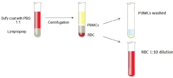
Peripheral blood mononuclear cells (PBMC) were obtained from the buffy coats of healthy donors and from the blood bags of therapeutic phlebotomies after centrifugation at 1000xg for 30 minutes over Lymphoprep (Figure 1). PBMC were washed with 1x phosphate buffered saline (PBS) followed by centrifugation at 700xg for 10 minutes. Contaminating RBC in the PBMC preparations were lysed in pre-heated RBC lysis solution (10mM Tris base, 150mM ammonium chloride, pH 7.2) for 10 minutes at 37ºC. After RBC lysis, PBMC were washed once again with PBS and centrifuged at 500xg for 10 minutes. RBC were collected from the pellet region after Lymphoprep centrifugation and diluted 1:10 in culture medium (Figure 1).
Figure 1. Diagrammatic representation of the process of isolation of PBMC and RBC from blood samples described above (BC and PB).
CFSE labeling
Carboxyfluorescein diacetate succinimidyl ester (CFSE) is a fluorescent dye that passively diffuses into cells and forms dye-protein adducts which are retained by the cells until they enter mitosis, where the dye-protein adducts are split between daughter cells when they enter cell division (Figure 2). For CFSE labeling PBMC were resuspended in PBS at a concentration of 10x106 cells/mL and incubated with 5µM of CFSE for 10 minutes at 37°C,
with occasional mixing. After labeling, PBMC were washed twice with PBS/20% heat inactivated FBS and resuspended in RPMI culture media supplemented with 2.5% FBS and 1% PSA (complete RPMI culture media). CFSE labeling efficiency was analyzed by flow cytometry immediately after label procedure and further confirmed by the analysis of fluorescence from unstimulated PBMC at the end of cultures.
Culture conditions
PBMC (1.5x10⁶ cells) were cultured in 6-well plates in a final volume of 5ml of
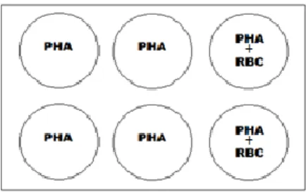
complete RPMI culture media and stimulated with 5μg/mL of PHA in the absence or presence of 400μL of the autologous RBC solution referenced above, which gives approximately a 50:1 proportion (50 RBC to 1 PBMC). In some experiments, cultures of non-stimulated PBMC (no PHA added) were also performed. Figure 3 shows a schematic representation of the experimental set-up. Culture plates were kept during 6 days in a humidified incubator at 37ºC and 5% CO2.
Figure 3. Scheme of the set-up of the in vitro cultures
Flow cytometry analysis
After the 6-day culture, cells were harvested and washed with PBS. In the cultures with RBC, cells were treated with RBC lysis solution for 10 minutes at 37ºC twice in order to remove RBC. Previous studies by members of the research group have shown that this treatment did not alter the activation parameters studied when compared with non-treated cells (20). Then, cells were incubated in round-bottomed plates at 4ºC during 40 minutes with staining solution (PBS, 0.2% BSA, 0.1% Sodium azide) containing a mixture of CD28-PE, CD8-PerCP and CD4-APC monoclonal antibodies. Due to the impossibility to include a CD3 antibody to identify T cells because the four fluorescence channels were already occupied, all analyses of CD28 expression and CFSE labeling was done on CD4+ and CD8+ cells. However, we are
confident that the CD8+ cells analyzed did not include CD8+CD3− NK cells since these cells do
not proliferate in response to PHA or PHA+RBC stimulation (20-23). Afterwards, cells were washed with staining solution, acquired in a BD Accuri™ C6 flow cytometer and cell proliferation and CD28 expression analysed using BD Accuri™ C6 software. Only experiments where the PBMC proliferated in response to PHA stimulation were used. T cell activation and division were determined by two methods: 1) Changes in determination of cell size and complexity according to FCS/SSC parameters (blasts) and 2) Sequential halving of CFSE-fluorescence intensity. The level of CD28 expression on CD4+ and CD8+ cells was determined
by the mean fluorescence intensity (MFI) units. The percentage of CD28+ and CD28— cells
within CD4+ and CD8+ cells was also calculated. A description of the determination of these
Figure 4. Scheme showing how proliferation and CD28 receptor expression were determined. Left dot-plot shows a representative experiment were T cell transformation induced by PHA stimulation can be seen. Some activated T cells do not increase in size (small cells) while some augment their size as a result of the mitogenic stimulation (blasts cells). The right dot-plot shows CFSE fluorescence (x-axis)
versus CD28receptor expression (y-axis). The vertical line divides the T cells that did not proliferate
(non proliferating, CFSE+) from the ones that proliferated (proliferating, CFSE-). The horizontal line defines the cells that express CD28 (upper right (UR), upper left (UL) and the cells that lose CD28 expression (lower right (LR), lower left (LL). The arrows heads on the proliferating cells indicate the number of cell divisions.
Determination of auto-fluorescence
PBMC from ten blood samples (8 FB all males mean age 57.67 and 2 BC one female age 50 and one male age 49) were cultured for 6 and 12 days in the conditions mentioned above. After the culture period, cells were harvested, RBC lysed, and directly acquired in a BD Accuri™ C6 flow cytometer. Auto-fluorescence detected in the FL1 channel was recorded and analyzed.
Statistical analysis (IBM SPSS)
The data base was created in Microsoft Excel 2010 and IBM SPSS statistics 23, and all statistical analysis and graphics were done using the same programs. Student’s independent samples T-test was used to determine the significance of the differences in CD28 expression and CFSE fluorescence between each culture condition. Mann-Whitney test was used to determine the significance of the differences between buffy coat and phlebotomy samples. Normality was verified by Shapiro-Wilk (n<30). Pearson’s correlation was used to assess the correlation between the expression of CD28 and cell cycle division. Statistical significance was defined as p<0.05.
Results
Effect of RBC on T cell growth and proliferation
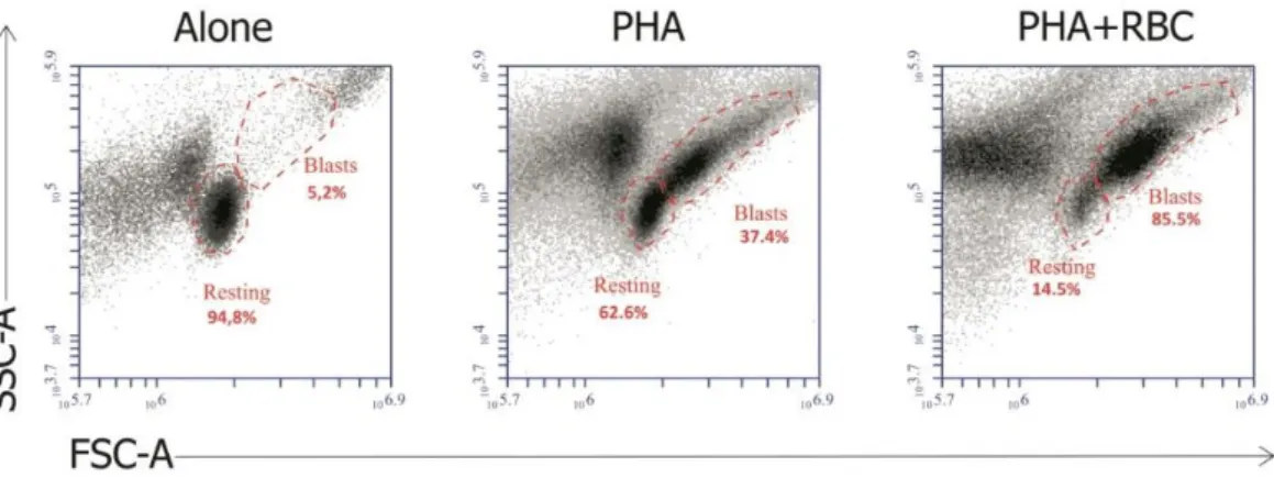
First we wanted to validate our in vitro model, where we previously demonstrated that the presence of RBC on cultures of PHA-activated PBMC markedly increased T cell growth and proliferation (20-23). To that end, we compared the morphologic characteristics of PBMC in the 3 culture conditions (No stimulus, PHA and PHA+RBC) using forward scatter (FSC) and side scatter (SSC) characteristics. These parameters allow to monitor the percentage of activated T cells that increase in size and complexity (cell growth), becoming blast cells, as well as the percentage of T cells that do not respond to the stimulus and remain as small cells (see materials and methods) As illustrated in Figure 5, activation with PHA (middle dot-plot) induced a percentage of T cells present in the PBMC preparations to increase in size and complexity, indicative of T cell blast transformation, in comparison with non-activated PBMC (left dot-plot). This increase was much more evident when RBC were present in the culture (right dot-plot), as illustrated by a marked increase in the percentage of T cell blasts (from 37% to 85% in this particular experiment). The few blast cells observed in unstimulated cultures represent basal stimulation during the 6-day culture period. Therefore, our in vitro system permitted us to obtain different levels of T cell blast transformation.
Figure 5. The presence of RBC in cultures of PBMC activated with PHA increases T cell growth. Dot-plots show resting cells and blast cells in unstimulated PBMC cultures (Alone), or PBMC cultures stimulated with PHA in the absence (PHA) and presence (PHA+RBC) of RBC after 6 days of culture and acquired in a flow cytometer according to FSC and SSC characteristics. The region of resting and blast cells in each culture condition are indicated with respective relative percentage. Results from one representative experiment is shown.
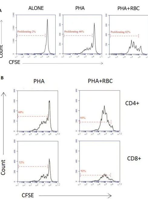
Next, we wanted to ascertain whether the increase in T cell blast transformation was paralleled by an increase in the T cell proliferation. For that, we compared the loss of CFSE fluorescence in each culture condition. Activation of PBMC with PHA induced a loss of CFSE fluorescence that allowed us to determine the percentage of cells that entered cell division and how many times they divided. As shown in Figure 6, while in unstimulated cultures there
was not CFSE fluorescence loss (Figure 6A, left histogram), stimulation with PHA induced a fraction of cells to enter cell division as determined by the loss (sequential halving) of CFSE fluorescence (Figure 6A, middle histogram). The presence of RBC in the PHA-stimulated cultures markedly augmented this percentage as well as the number of cell divisions (Figure 6A, right histogram). Cell labeling with anti-CD4 and anti-CD8 antibodies to identify the two major T cell populations showed that both CD4+ and CD8+ cells proliferated under these
conditions (Figure 6B)
Figure 6. The presence of RBC in cultures of PBMC activated with PHA increases the extent of T cell proliferation. PBMC were labeled with CFSE and cultured for 6 days in the presence of PHA or PHA+RBC, as indicated in the Material and Methods. Then, PBMC were harvested and labeled with anti-CD28-PE, anti-CD8-PerCP and anti-CD4-APC fluorochrome-conjugated monoclonal antibodies and acquired in a
flow cytometer. Cells were acquired in a flow cytometer; gates were created around PMBC, CD4+ and
CD8+ cells. The percentages of proliferating/dividing cells are indicated. (A) Histograms show CFSE
fluorescence halving in unstimulated PBMC cultures and PBMC cultures activated with PHA and
PHA+RBC. (B) Histograms shown CFSE fluorescence halving on gated CD4+ and CD8+ T cells in PBMC
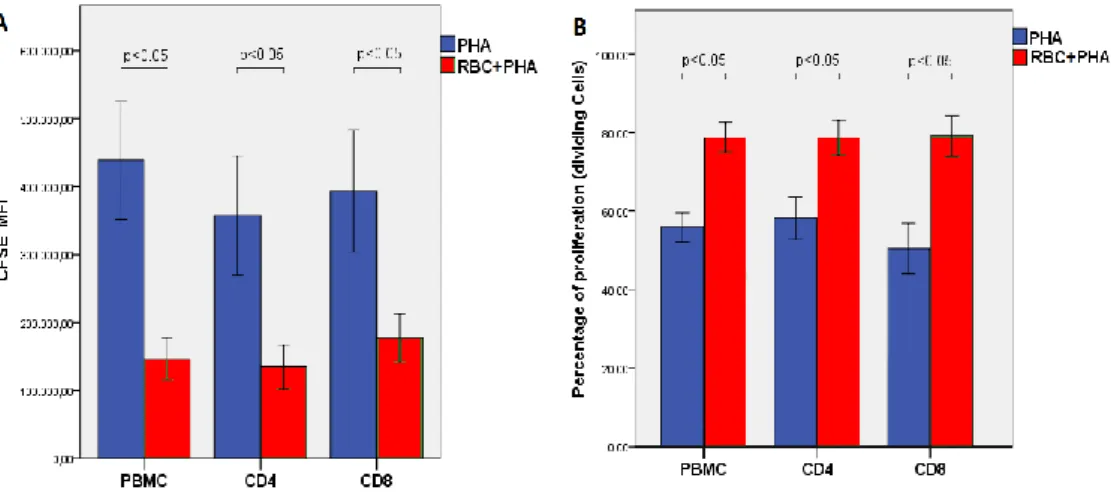
On average, the presence of RBC in cultures stimulated with PHA significantly increased T cell growth and proliferation as determined both by a decrease in the mean fluorescence intensity of CFSE (Figure 7A), indicative of higher proliferation levels, and by an increase in the percentage of proliferating/dividing cells (Figure 7B). These differences were observed both in CD4+ and CD8+ T cells. No differences were observed between PBMC
obtained from healthy regular donors and iron overload patients (data not shown).
Figure 7. Summary of T cell proliferation parameters in the two activating culture conditions. (A) CFSE
mean fluorescence intensity (MFI) values (meanSEM) in cultures stimulated with PHA (blue) and
PHA+RBC (red) in all PBMC, CD4+ and CD8+ T cells (CFSE MFI values was on average 736000 in
non-proliferating cells). (B) Percentage of dividing cells (meanSEM, n=11) in cultures with PHA (blue) and
PHA+RBC (red) in all PBMC, CD4+ and CD8+ T cells.
Effect of T cell proliferation on CD28 expression
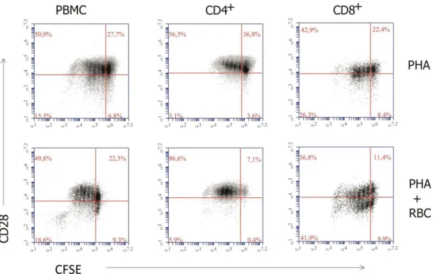
Next, we wanted to examine the expression of CD28 on the proliferating T cells. As shown in Figure 8, when all PBMC were analyzed, PHA-activation induced some proliferating T cells to downregulate CD28 expression (Figure 8, left dot-plots). When the CD4+ and CD8+
populations were analyzed separately, some differences were observed. Thus, CD4+ cells
showed no significant changes in the expression of CD28 with the division cycles, and some even showed an increase in CD28 expression when cultured only with PHA (Figure 8, middle dot-plots). In marked contrast, CD28 expression by CD8+ cells was decreased generating CD28−
cells during the division cycles, an effect that was more striking in cultures with RBC (Figure 8, right dot-plots, compare upper and lower left quadrants). Noteworthy, in some experiments CD28 was also downmodulated in CD8+ cells that did not enter cell division,
namely in cultures with RBC (compare upper right and lower right quadrants in the lower right dot-plot of Figure 8). No differences were observed between PBMC obtained from healthy regular donors and iron overload patients (data not shown).
Figure 8. Relationship between T cell proliferation and CD28 downmodulation. PBMC were labeled with CFSE and cultured for 6 days in the presence of PHA or PHA+RBC, as indicated in the Material and Methods. Then, PBMC were harvested and labeled with anti-CD28-PE, anti-CD8-PerCP and anti-CD4-APC fluorochrome-conjugated monoclonal antibodies and acquired in a flow cytometer. The graphs show
CFSE fluorescence halving versus CD28 expression in total PBMC and gated CD4+ and CD8+ cells. The
percentage of cells in each quadrant is indicated. One representative experiment of 11 performed.
Interestingly, analysis of the overall expression levels of CD28 in all CD4+ and CD8+
cells after the culture period showed that CD4+ expressed higher levels of CD28 than CD8+
cells (3-fold)(Figure 9).
Figure 9. Mean florescence intensity of CD28 in PBMC, CD4+ and CD8+ cells. PBMC were
stimulated and processed as indicated in the legend of Figure 8 and acquired in a flow cytometer. The
graph shows CD28 MFI values (meanSEM, n=11) in cultures stimulated with PHA (blue) and PHA+RBC
(red) of all PMBC, and gated CD4+ and CD8+ cells. Statistically significant differences between conditions
Accumulation of CD8
CD28
cells after culture with RBC
In view of the downmodulation of the CD28 receptor, namely in proliferating CD8+cells (see Figure 8), we wanted to ascertain if this effect resulted in an increase in CD8CD28
cells at the end of the culture period. Indeed, quantification of the percentage of CD8CD28 cells in the lower left quadrants (containing exclusively cells that proliferated) showed that in cultures with PHA+RBC the percentage of CD8CD28 cells increased on average from 19% to 36% (p<0.05). In contrast, the increase observed in CD4CD28 cells was minimal (Figure 10).
Figure 10. The presence of RBC increases the percentage of CD8CD28 cells in cultures of PHA-activated
cells. PBMC were stimulated and processed as indicated in the legend of Figure 8, and acquired in a flow
cytometer. The results show the percentage (meanSEM, n=7) of proliferating CD4CD28 and CD8CD28
cells in each culture condition obtained from the percentage of lower left quadrants of Figure 8. Statistically significant differences between conditions (p values) are indicated.
CD28 expression changes with cell division
In view of these results we went to analyze the expression of CD28 expression in each cell division cycle. To do that, we determined the values of CD28 expression (as determined by the MFI values) in each division cycle considering only CD28 expressing cells, meaning cells that expressed CD28 before starting to divide, as shown below.
Figure 11. Population under analysis is indicated by the dashed red rectangles. Each rectangle demarcates one division.
When CD4+ cells obtained from PHA-stimulated cultures were analyzed, a statistically
significant positive correlation between CD28 expression and cell proliferation was observed (Figure 12). The increase in CD28 expression in PHA-stimulated CD4+ cells was observed in 5
out of 7 separate samples (Suppl. data). In contrast, in CD4+ cells obtained from
PHA+RBC-stimulated cultures a statistically significant negative correlation between CD28 expression and cell proliferation was found (Figure 12). The decrease in CD28 expression in PHA+RBC-stimulated CD4+ cells was observed in 4 out of 7 experiments (Suppl. data). Interestingly, in
cultures stimulated in the presence of RBC a statistically significant increase (p<0.05) in the expression of CD28 by CD4+ cells that did not enter cell division was observed (compare
division cycle 0 between PHA and PHA+RBC conditions in Figure 12).
Figure 12. Correlation between cell division and CD28 expression in CD4+ cells. PBMC were stimulated
and processed as indicated in the legend of Figure 8. CD28 expression values in each division cycle are
shown (meanSEM, n=7). Correlation coefficient and statistical significance (Pearson’s) are indicated.
In contrast to CD4+ cells, no significant changes in CD28 expression were observed in
CD8+ cells obtained from PHA-stimulated cultures (Figure 13 and Suppl. data). However, in
PBMC cultures stimulated with PHA+RBC a statistically significant negative correlation between CD28 expression and cell proliferation was also observed (Figure 13). The decrease in CD28 expression in PHA+RBC-stimulated CD8+ cells was observed in 6 out of 7 separate
experiments (Suppl. data). Similarly to the observation with CD4+ cells, in cultures stimulated
in the presence of RBC an increase in the expression of CD28 by CD8+ cells that did not enter
cell division was observed (compare division cycle 0 between PHA and PHA+RBC conditions in Figure 13).
Figure 13. Correlation between cell division and CD28 expression in CD8+ cells. Correlation between cell
division and CD28 expression in CD4+ cells. PBMC were stimulated and processed as indicated in the
legend of Figure 8. CD28 expression values in each division cycle are shown (meanSEM, n=7).
FL1 auto-florescence analysis
Finally, and based in previous studies in or lab suggesting that T cells activated in the presence of RBC appeared to show higher levels of auto-florescence, we performed a series of experiments to determine if the presence of RBC in the PBMC cultures influenced the level of auto-fluorescence in FL-1. In order to do that, PBMC were cultured for 6 and 12 days without stimulus or with PHA, in the absence or presence of RBC. After the culture period, PBMC were directly acquired in a flow cytometer and auto-fluorescence in the FL-1 detector on the lymphocyte region recorded. The results showed that auto fluorescence levels increased when RBC were present in the cultures but only when cells were activated. This increase was observed both 6 and 12 days after stimulation and was statistically significant. No significantly differences between day 6 and day 12 were verified (Figure 14).
Figure 14: RBC increase auto-fluorescence of PHA-activated PBMC. PBMC were cultured without and with PHA in the absence or presence of RBC and cultured for 6 and 12 days. Then, PBMC were harvested, acquired in a flow cytometer and fluorescence in the FL-1 channel determined. The results
show auto florescence levels (FL-1 MFI values) (meanSEM, n=10) in the different culture conditions, at
day 6 and day 12. Statistically significant differences between conditions (p values) are indicated. Results are representative of 10 experiments.
Discussion
The main objective of this project was to extend previous studies showing that RBC increase cell proliferation and survival of activated human T cells in vitro by evaluating the effect of RBC on the expression of CD28 by dividing human T cells, as an in vitro model of transfusion. Previous studies by our group have shown that human T cells activated in vitro by a variety of stimuli, including TCR-mediated (e.g., anti-CD3/CD28, PHA) and TCR-independent (e.g., IL-2, IL-15, phorbol esters, etc.) and cultured in the presence of either autologous or heterologous RBC increase their proliferation and survival (20-24). However, the expression of CD28 was not addressed.
This study has corroborated the already known effect of fresh autologous RBC in stimulating the exit from a resting state, inducing high levels of proliferation and promoting cell survival (19). Regarding CD28, we have shown four interesting results. First, the overall expression of CD28 when all cells obtained from the 6-day period culture were considered was much higher in CD4+ than in CD8+ cells (Figure 9). Second, analysis of CD28 along the cell
division process, by using CFSE fluorescence halving, revealed that the presence of RBC in cultures of PHA-activated PBMC induced a gradual decreased in CD28 expression with each cell division cycle, both in CD4+ and CD8+ cells (Figures 12 and 13). Third, the presence of RBC
in cultures of PHA-stimulated PBMC allowed CD8+CD28 cells to enter several cycles of cell
division (see Figure 8, lower left quadrant of the lower right dot-plot). Fourth, we have shown that the presence of RBC in PHA-activated cultures, but not in unstimulated ones, markedly increased the auto-fluorescence of lymphocytes (Figure 14).
Although it is already known that CD4+ T cells express higher levels of CD28 than CD8+
T cells (28-31), the upregulation of CD28 expression in the CD4+ cells that did not divide when
RBC were present in the cultures has not been reported before. One possible explanation is that RBC are delivering signals to CD4+ cells that lead to CD28 up-regulation, thus conferring
them a more “pro-immunogenic” phenotype ready to respond to antigenic signals via their TCR. This could be important in CD4+ T cell priming and differentiation into effector T cells
subsets, namely Th1 cells and follicular helper CD4+ Tfh cells (32).
On the other hand, while in cultures without RBC the expression of CD28 by CD4+ cells
increased progressively with the cycles of cell division (Figure 12, left graph), in the presence of RBC the opposite effect was observed, that is, a proliferation-dependent decrease in CD28 expression (Figure 12, right graph). These results suggest that the CD28 upregulation observed in CD4+ cells during the proliferation process could represent a physiological response to
TCR-mediated stimulation, and the presence of RBC may interfere with this process by sharply increasing CD28 expression during the early phases of activation, which may allow more
cycles of cell division. Whether RBC transfusions may have a similar impact on already activated CD4+ T cells present on lymphoid organs, such as the spleen, thus potentiating CD4+
T cell responses, is an interesting issue that needs additional investigations.
In our study we found that CD8+ cells expressed less CD28 than CD4+ cells (Figure 9)
and stimulation with PHA did not induced any significant change on the level of expression of CD28 during the proliferation process (Figure 13, left graph). However, stimulation in the presence of RBC had the same effect as the observed in CD4+ cells, that is, a sharp increase in
CD28 expression after stimulation (Figure 13, compare CD28 expression values at 0 cell division cycles between culture conditions) followed by a gradual decrease in CD28 expression as CD8+ cells entered subsequent cycles of cell division. Unlike CD4+ cells, this CD28 decrease
resulted in the accumulation of a population of CD8+CD28 cells at the end of the culture
(Figure 10). Analysis of proliferation parameters in this population also showed that CD8+ cells
that have lost CD28 expression (i.e., CD8+CD28 cells) are able to proliferate (see Figure 8,
lower left quadrant of the lower right dot-plot).
These results with CD8+ cells are stimulating findings because CD8+CD28 T cells are
lymphocytes traditionally associated with a limited antigen induced proliferation and a shorter replicative lifespan (29-31). However, recent studies are revealing that CD8+CD28 T
cells are a highly heterogeneous population that contain polyfunctional lymphocytes capable to exert both effector (e.g., cytotoxic, inflammatory) as well as regulatory (e.g., immune suppression, inflammation suppression) activities (33). Indeed, it is now consensual that loss of CD28 is accompanied by up-regulation and de novo expression of surface receptors that regulate CD8+ T cell function, including NK cell receptors, chemokine receptors and other
regulator molecules (29-31,33). In the elderly, the suppressive effect of CD8+CD28 T cells has
been associated with reduced responses to vaccines, pathogens and cancer (29,30). However, recent studies have suggested that CD8+CD28 T cells develop as a compensatory mechanism
for the loss of naïve CD8+ T cells that occurs with aging, and may be beneficial for healthy
aging (31). Paradoxically, CD8+CD28 T cells can be also found among young patients with
chronic immune-mediated diseases in an age-disproportionate manner (31). Our results, have provided preliminary evidence that RBC participate in the generation of CD8+CD28 T cells.
Their role in the context of RBC transfusions needs to be investigated.
Finally, we also showed that the presence of RBC in cultures of activated PBMC resulted in an increased lymphocyte auto-florescence. Several intracellular compounds are thought to emit natural fluorescence, for example porphyrins. Thus, these results raise the possibility that activated T cells may incorporate compounds provided by the RBC present in the culture (e.g., heme) which then auto-fluoresce upon incidence of the 488nm laser light of the flow cytometer. These preliminary results warrant more studies in order to assess this possibility.
Although it is difficult to extrapolate our in vitro results into the clinic (i.e., in cases where packed RBC are transfunded into injured people or anemic patients due to a variety of underlying pathologies), this in vitro model of RBC transfusion could be a starting point to understand some of the described effects associated with RBC transfusions, both beneficial (e.g., acceptance of allografts) and deleterious (e.g., post-operative infections, lung injury, tissue hypoxia, bleeding/thrombosis, etc.). Therefore, whether the impact that RBC have on activated T cells in vitro, namely downmodulation of CD28, also occurs after RBC transfusions is something that remains to be elucidated. Nevertheless, in combination with recent studies, our results suggest that the effect of RBC on the immunological system may depend on whether they are fresh or old, with implications for the clinic (27). Additional studies comparing the bioactivities of fresh versus old packed RBC will certainly disclose some of the molecular mechanisms. Finally, it should be stressed that we have consistently shown that RBC alone, either autologous or heterologous, are not mitogenic for T cells in vitro, and therefore the effects that we observe are not due to an allogenic response (19-25). Moreover, the same effect can be seen using highly pure T cells, thus ruling out a possible antigen presentation by monocytes present in the PBMC preparations (22,24).
Limitations of the Study
First, due to the combination of antibodies and fluorochromes, we could not include anti-CD3 antibodies in the study, thus restricting the analysis to CD4+ and CD8+ and not to
CD3+CD4+ or CD3+CD8+,which in theory could have introduced a bias by including some CD8+
NK cells (known to be CD3CD8+). However, we are confident that this possibility is remote
because we, and others, have already shown that activation of PBMC with the T cell mitogen PHA does not activate NK cells or B cells and we have shown that CD8+ cells at the end of the
cultures with RBC are almost exclusively CD3+CD8+ T cells (22,23).
Second, the use of autologous RBC in the in vitro model, which in theory does not reproduce the transfusions of heterologous RBC commonly used in clinical practice. However, it should be mentioned that we have previously shown that heterologous RBC obtained from healthy regular blood donors have the same effect that autologous RBC on T cell proliferation and survival (24). Nevertheless, additional experiments using heterologous RBC should be done to confirm that the effect of RBC on CD28 is the same regardless of the autologous
versus heterologous nature of the sample.
Third, the sample size is small. We are aware of this restraint but several factors contributed to this. First, the unpredictable availability of samples. Second, some experiments were unsuccessful due to impossibility to isolate PBMC, to high levels of cell death, to low levels of proliferation or to culture contamination. Last, but not least, some experiments were lost due to technical problems with the flow cytometer.
Conclusions
The main conclusions of this work are:
RBC augment the proliferation of activated CD4+ and CD8+ T cells.
RBC augment the percentage of CD8+CD28− T cells and allow them to proliferate.
Expression of CD28 in non-dividing CD4+ and CD8+ T cells is increased in cultures
with RBC.
Expression of CD28 decreases with CD4+ and CD8+ T cells proliferation.
In view of these results it is tempting to speculate that RBC transfusions may have immunomodulatory effects in immune system that may be mediated, in part, by CD8+CD28 T
cells. The translation of this work to the clinic is difficult, but these potential effects should be taken into consideration for the well-being of the patient receiving RBC transfusions. In this respect, clinical trials conducted in adults and children have demonstrated that a restrictive transfusion strategy where the threshold for transfusion is a Hb<7g/dl was as effective as a more liberal one (i.e., Hb<9g/dl in adults and Hb<9,5g/dl in children) (34 and reviews there in). This is already acknowledged by renowned medical books. For example, in Harrison’s it is referred to avoid frequent transfusions in patients in the ICU with pneumonia, because it adversely affects the immune host response against respiratory pathogens (35).
The key message to medical doctors is that they should be aware of the immunomodulatory effects of RBC transfusion, and they should ponder well the pros and cons and prescribe it wisely only when the benefits exceed the harms.
Future prospectives
This study could “open a door” to a clinical study with humans receiving allogenic RBC transfusions of different storage ages, comparing T cell proliferation and CD28 expression before and after the transfusion(s) and correlate these with clinical parameters. Other interesting study would compare T cell proliferation and CD28 expression in patients with differences in the hematocrit. In other words, do anemics have impaired T cell proliferation and CD28 expression compared to normal people or people with high hematocrit, such as polycythemia patients? All these questions deserve to be investigated.
Bibliography
1. Opelz G, Terasaki PI. Improvement of Kidney-Graft Survival with Increased Numbers of Blood Transfusions. N Engl J Med. 1978 Oct 12;299(15):799–803.
2. Vamvakas EC, Blajchman MA. Transfusion-related immunomodulation (TRIM): an update.Blood Rev. 2007 Nov;21(6):327-48.
3. Muszynski JA, Spinella PC, Cholette JM, Acker JP, Hall MW, Juffermans NP, Kelly DP, Blumberg N, Nicol K, Liedel J, Doctor A, Remy KE, Tucci M, Lacroix J, Norris PJ; Transfusion-related immunomodulation: review of the literature and implications for pediatric critical illness. Transfusion. 2017 Jan;57(1):195-206 doi: 10.1111/trf.13855. 4. Buddeberg F, Schimmer BB, Spahn DR. Transfusion-transmissible infections and
transfusion-related immunomodulation. Best Pract Res Clin Anaesthesiol. 2008 Sep;22(3):503-17
5. Dasararaju R, Marques MB. Adverse effects of transfusion. Cancer Control. 2015 Jan;22(1):16-25.
6. Zimring JC. Fresh versus old blood: are there differences and do they matter? Hematol Am Soc Hematol Educ Program. 2013;2013:651–5.
7. Prudent M, Tissot J-D, Lion N. In vitro assays and clinical trials in red blood cell aging: Lost in translation. Transfus Apher Sci. 2015 Jun;52(3):270–6.
8. Cohen B, Matot I. Aged erythrocytes: a fine wine or sour grapes? Br J Anaesth. 2013 Dec;111 Suppl 1:i62-70.
9. Lannan KL, Sahler J, Spinelli SL, Phipps RP, Blumberg N. Transfusion immunomodulation--the case for leukoreduced and (perhaps) washed transfusions. Blood Cells Mol Dis. 2013 Jan;50(1):61–8
10. Bianchi M, Vaglio S, Pupella S, Marano G, Facco G, Liumbruno GM, et al. Leucoreduction of blood components: an effective way to increase blood safety? Blood Transfus Trasfus Sangue. 2016 May;14(2):214–27.
11. Bosman GJCGM. Survival of red blood cells after transfusion: processes and consequences. Front Physiol. 2013 Dec 18;4:376. doi: 10.3389/fphys.2013.00376.t
12. D’Alessandro A, Hansen KC, Silliman CC, Moore EE, Kelher M, Banerjee A. Metabolomics of AS-5 RBC supernatants following routine storage. Vox Sang. 2015 Feb;108(2):131–40 13. Almizraq RJ, Seghatchian J, Acker JP. Extracellular vesicles in transfusion-related
immunomodulation and the role of blood component manufacturing. Transfus Apher Sci. 55(3):281–91.
14. Pawloski JR, Hess DT, Stamler JS. Export by red blood cells of nitric oxide bioactivity. Nature. 2001 Feb 1;409(6820):622–6.
15. Vallés J, Santos MT, Aznar J, Martínez M, Moscardó A, Piñón M, et al. Platelet-erythrocyte interactions enhance alpha(IIb)beta(3) integrin receptor activation and P-selectin expression during platelet recruitment: down-regulation by aspirin ex vivo. Blood. 2002 Jun 1;99(11):3978–84.
16. Aoshiba K, Nakajima Y, Yasui S, Tamaoki J, Nagai A. Red blood cells inhibit apoptosis of human neutrophils. Blood. 1999 Jun 1;93(11):4006–10.
17. Melder RJ, Yuan J, Munn LL, Jain RK. Erythrocytes enhance lymphocyte rolling and arrest in vivo. Microvasc Res. 2000 Mar;59(2):316–22.
18. Schäkel K, von Kietzell M, Hänsel A, Ebling A, Schulze L, Haase M, et al. Human 6-sulfo LacNAc-expressing dendritic cells are principal producers of early interleukin-12 and are controlled by erythrocytes. Immunity. 2006 Jun;24(6):767–77.
19. Arosa FA, Pereira CF, Fonseca AM. Red Blood Cells as Modulators of T Cell Growth and Survival. Curr Pharm Des. 2004;10(2):1404-16.
20. Fonseca AM, Porto G, Uchida K, Arosa FA. Red blood cells inhibit activation-induced cell death and oxidative stress in human peripheral blood T lymphocytes. Blood. 2001 May 15;97(10):3152–60.
21. Porto B, Fonseca AM, Godinho I, Arosa FA, Porto G. Human red blood cells have an enhancing effect on the relative expansion of CD8+ T lymphocytes in vitro. Cell Prolif. 2001 Dec;34(6):359-67
22. Fonseca AM, Pereira CF, Porto G, Arosa FA. Red blood cells promote survival and cell cycle progression of human peripheral blood T cells independently of CD58/LFA-3 and heme compounds. Cell Immunol. 2003 Jul;224(1):17-28
23. Fonseca AM, Pereira CF, Porto G, Arosa FA. Red blood cells upregulate cytoprotective proteins and the labile iron pool in dividing human T cells despite a reduction in oxidative stress. Free Radic Biol Med. 2003 Dec 1;35(11):1404-16
24. Antunes RF, Brandão C, Carvalho G, Girão C, Arosa FA. Red blood cells carry out T cell growth and survival bioactivities that are sensitive to cyclosporine A. Cell Mol Life Sci. 2009;66:3387-98.
25. Antunes RF, Brandão C, Maia M, Arosa FA. Red blood cells release factors with growth and survival bioactivities for normal and leukemic T cells. Immunol Cell Biol. 2011;89:111-21. 26. Bernard A, Meier C, Ward M, Browning T, Montgomery A, Kasten M, et al. Packed red blood cells suppress T-cell proliferation through a process involving cell-cell contact. J Trauma. 2010 Aug;69(2):320–9.
27. Long K, Meier C, Ward M, Williams D, Woodward J, Bernard A. Immunologic profiles of red blood cells using in vitro models of transfusion. J Surg Res. 2013 Sep;184(1):567–71. 28. Arosa FA. CD8+CD28- T cells: certainties and uncertainties of a prevalent human T-cell
subset. Immunol Cell Biol. 2002 Feb;80(1):1–13.
29. Weng N-P, Akbar AN, Goronzy J. CD28(-) T cells: their role in the age-associated decline of immune function. Trends Immunol. 2009 Jul;30(7):306–12
30. Mou D, Espinosa J, Lo DJ, Kirk AD. CD28 negative T cells: is their loss our gain? Am J Transplant Off J Am Soc Transplant Am Soc Transpl Surg. 2014 Nov;14(11):2460–6.
31. Michel JJ, Griffin P, Vallejo AN. Functionally Diverse NK-Like T Cells Are Effectors and Predictors of Successful Aging. Front Immunol. 2016;7:530.
32. Linterman MA, Denton AE, Divekar DP, Zvetkova I, Kane L, Ferreira C, et al. CD28 expression is required after T cell priming for helper T cell responses and protective immunity to infection. elife. 2014 Oct 27;3. doi: 10.7554/eLife.03180.
33. Arosa FA, Esgalhado AJ, Padrão CA, Cardoso EM. Divide, Conquer, and Sense: CD8CD28
in Perspective. Front Immunol. 2017 Jan 3;7:665. doi: 10.3389/fimmu.2016.00665. 34. Holst LB. Benefits and harms of red blood cell transfusions in patients with septic shock in
the intensive care unit. Dan Med J. 2016 Feb;63(2). PhD thesis.
35. Kasper, Fauci, Hauser, Longo, Jameson, Loscalzo. Harrison’s principles of internal medicine. In: 19th ed. Mc Gram Hill; 2015. p. 810.
Supplemental data
Graphics 1: Analysis of the correlation between CFSE expression (MFI values, x-axis) and CD28
expression (MFI values, y-axis) in CD4+CD28+ cells
cultured with PHA in each single experiment. Pearson’s correlation (r) and significance (p) are indicated.
Graphics 2: Analysis of the correlation between CFSE expression (MFI values, x-axis) and CD28
expression (MFI values, y-axis) in CD4+CD28+
cells cultured with PHA+RBC. Results in in each single experiment are shown. Pearson’s correlation (r) and significance (p) are indicated.
Graphics 3: Analysis of the correlation between CFSE expression (MFI values, x-axis) and CD28 expression (MFI values, y-axis) in
CD8+CD28+ cells cultured with PHA. Results in
each single experiment are shown. Pearson’s correlation (r) and significance (p) are indicated.
Graphics 4: Analysis of the correlation between CFSE expression (MFI values, x-axis) and CD28 expression (MFI values, y-axis) in
CD8+CD28+ cells cultured with PHA+RBC.
Results in each single experiment are shown. Pearson’s correlation (r) and significance (p) are indicated.