Ciências
Proteomic study of the therapeutic effect of a
DNA vector on cervical cancer cells
Adriana Resende Pinto
Dissertação para obtenção do Grau de Mestre em
Biotecnologia
(2º ciclo de estudos)
Orientador: Professora Doutora Ângela Maria Almeida de Sousa
Co-orientador: Professor Doutor Luís António Paulino Passarinha
Acknowledgments
First, I would like to express gratitude, to my supervisor Doctor Professor Ângela Sousa and my co-supervisor Doctor Professor Luís Passarinha for the opportunity to join your research team, allowing me to expand horizons and grow as a student and future professional and for all the support, dedication, guidance, the encouragement words and scientific knowledge shared with me throughout this journey.
To the University of Beira Interior, in particular to the Health Sciences Research Center (CICS), for providing the necessary facilities for the development of this dissertation.
Special thanks to Margarida Almeida and Dalinda Eusébio for all the help you have given me throughout this year. Thank you for all the encouragement, for despite all the work you have never denied me advice or help with what I needed, for your patience and friendship. Thank you, it was a privilege to work and learn with you.
To Jorge Ferreira, thank you for all the tips for being able to reveal the bastards of my Western-Blot membranes, for the good humor and jokes that always raised the mood in the lab.
To my lab partner, Diana Pereira, easily mistaken as my Siamese, thank you for putting up with me this year, for always know what to say when I felt like sending everything out the window, packing up, and leaving. Thank you for all the hours we spent together for making this year so much fun. Above all, thank you for all your patience and friendship.
To Diana, Raquel, Carapito and Proença, who since 2014 helped to make Covilhã my home, thank you, girls. Thank you for all these wonderful years, for enduring my bad temper (I still don't know how you did it), for making this whole journey so much better, for all the tears and laughter. To Micas, Diana Gomes, Rosa and Flor thank you very much for all your friendship, for all the encouragement and wise words, for all the fun moments I will be forever grateful. To the latest members of the tea club, Sofia, Fafe and Bifas, thank you very much for appearing at CICS and raising our spirits. THANK YOU all for making this last year remarkable!
To my hometown friends, thank for always be there for me even though our journeys have separated and we have all gone to different cities to study, thank you for all your support, night cafes for gossip, for always being present at the good times and for helping me to get through the less good times on my academic journey.
Obrigada por nunca me terem ensinado a lutar por tudo aquilo que sempre quis e nunca me terem deixado desistir nos momentos menos bons desta jornada. Obrigada por terem aturado o meu mau feitio e as minhas explosões quando chegava a casa frustrada com o meu trabalho e começava a disparar em todas as direções. Obrigado por todo o apoio e compreensão.
Resumo Alargado
O cancro é um dos principais problemas de saúde, sendo responsável por mais de 8 milhões de mortes todos os anos, a nível mundial. O cancro do colo do útero é a 3ª malignidade mais frequente, sendo a 4ª causa de morte nas mulheres a nível mundial. A infeção persistente por HPV de alto risco é o fator principal para o desenvolvimento de cancro do colo do útero, dado que as oncoproteínas E6 e E7 são capazes de promover o desenvolvimento cancerígeno através da degradação e inibição das proteínas supressoras de tumor, p53 e pRb, respetivamente. A terapia génica é uma estratégia promissora no tratamento de doenças adquiridas e/ou desordens genéticas, tendo como objetivo a entrega de material genético em células ou tecidos alvo, de forma a induzir um efeito terapêutico. O DNA minicircular (mcDNA) é um novo produto biofarmacêutico, que se caracteriza por ser apenas constituído pela unidade de transcrição eucariota, aumentando a sua segurança e efeito terapêutico, sendo obtido através da recombinação intramolecular do plasmídeo parental. Além disso, tem-se atribuído aos microRNAs (miRs) um papel regulador da carcinogénese, por exemplo, está descrito que o miR-375 possui a capacidade de silenciar as oncoproteínas E6 e E7.
Desta forma, este trabalho tem como objetivo a produção e purificação de vetores de mcDNA que codifiquem para os genes primiR-375 e p53, de forma a silenciar as oncoproteínas E6 e E7 e reestabelecer os níveis das proteínas supressoras de tumor p53 e pRb nas células do cancro do colo do útero.
A purificação dos vetores de mcDNA foi efetuada utilizando colunas de sefarose (Sephacryl S-1000 SF) de cromatografia de exclusão molecular, demonstrando que para o vetor de tamanho mais pequeno, mcDNA-primiR-375, uma coluna de 106 mL permitiu uma recuperação eficiente de frações cromatográficas compostas apenas por mcDNA. No entanto, para os vetores de maior tamanho, mcDNA-p53 e mcDNA-primiR-375+p53, foi necessário explorar os parâmetros que afetam a separação das moléculas das amostras a purificar, optando-se por utilizar uma coluna com o volume de 180 mL, permitindo assim, à semelhança do mcDNA-primiR-375, recuperar frações compostas apenas por mcDNA. Paralelamente foram realizados ensaios de citotoxicidade em fibroblastos humanos e células CaSki, revelando que nenhum dos vetores é tóxico nos fibroblastos humanos e que o mcDNA-primiR-375+p53 apresentou a menor viabilidade celular em células CaSki. Para além disso, os resultados do ensaio de proliferação realizado em células CaSki demonstraram que o número de células viáveis diminui nas células transfectadas com os vetores de DNA, corroborando os ensaios de citotoxicidade realizados nesta mesma linha celular cancerígena. A realização do ensaio de Western-Blot confirmou o restabelecimento dos níveis da proteína supressora de tumor p53 após 48 horas de
Os resultados obtidos nos em ensaios in vitro desenvolvidos neste trabalho, sugerem que o uso de vetores de DNA que codifiquem para mais do que um gene com finalidade terapêutica, como é exemplo o mcDNA-primiR-375+p53, permitiram observar efeitos mais significativos, sugerindo assim que este tipo de vetores podem ter uma ação terapêutica mais eficiente.
Keywords
Abstract
Cancer is a major global health problem, accounting for more than 8 million deaths/year
worldwide. In particular, cervical cancer is the 3rd most common malignancy and 4th cause of
death among women globally, being a persistent High Risk-HPV infection the main factor for cervical cancer development. E6 and E7 HPV oncoproteins are responsible for cancer development by degrading and inhibiting p53 and pRb tumour suppressor proteins, respectively. Moreover, microRNAs (miRs) have been found to regulate tumorigenesis. In fact, miR-375 has the ability to silence HPV E6 and E7 oncoproteins. Gene therapy is a promising strategy to treat acquired diseases and/or genetic disorders, aiming to deliver genetic material into target cells or tissue, expressing it to induce a therapeutic effect. Minicircle DNA (mcDNA) is a new biopharmaceutical product only composed by the eukaryotic transcription unit, improving its safety and therapeutic effect, obtained through intramolecular recombination of the parental plasmid.
Thus, this work aims to produce and purify a mcDNA vector encoding primiR-375 and p53 genes to silence E6 and E7 oncoproteins and re-establish p53 and pRb tumour suppressor levels on cervical cancer cells.
The purification of mcDNA vectors was performed using size exclusion chromatography sepharose columns (Sephacryl S-1000 SF), showing that for the smallest vector, mcDNA-primiR-375, a 106 mL column allowed the efficient recovery of chromatographic fractions composed only with mcDNA. On the other hand, for the biggest mcDNA vectors, it was necessary to exploit the parameters affecting the sample molecules separation, choosing to use a column with 180 mL of volume, allowing, similarly to the mcDNA-primiR-375 purification, recover fraction only composed with mcDNA. Simultaneously, cytotoxicity assays on human fibroblasts and CaSki cells were performed, confirming that none of the vectors were toxic to the fibroblasts and the mcDNA primiR-375+p53 presented the lower cell viability for CaSki cells. In addition, the proliferation assay results performed on CaSki cells show that number of viable cells decreases for the mcDNA vectors transfected cells, corroborating the cytotoxicity assays results for this cell line. Western-Blot confirmed the re-establishment of tumour suppressor proteins levels after 48h of transfection using p53 and mcDNA-primiR-375+p53.
Overall, these results suggest that the use of DNA vectors encoding for more than one therapeutic gene, for example, the mcDNA-primiR-375+p53, allowed to achieve, on in vitro assays, results suggesting a possible faster therapeutic action, thus demonstrating that they may revolutionize their application in targeted therapies.
Keywords
Table of Contents
Chapter I – Introduction --- 1 1.1 Cancer --- 1 1.1.1 Human papillomavirus (HPV) --- 1 1.1.2 Molecular biology --- 2 1.1.3 Infection --- 3 1.1.4 Oncoproteins E6 and E7 --- 5 1.1.5 p53 protein --- 8 1.1.6 pRb protein --- 8 1.2 Cell apoptosis --- 10 1.2.1 Extrinsic pathway --- 11 1.2.2 Intrinsic pathway --- 121.3 DNA based therapy --- 12
1.3.1 DNA vaccines --- 12
1.3.2 Gene therapy --- 13
1.3.2.1 Gene therapy for cervical cancer --- 14
1.3.3 DNA vectors --- 15
1.4 Micro RNA (miRNA) --- 19
1.4.1 miRNA-375 --- 20
1.5 Minicircular DNA (mcDNA) --- 21
1.5.1 Production --- 22
1.6 Purification --- 22
1.6.1 Chromatography --- 23
1.6.1.1 Hydrophobic interaction chromatography --- 23
1.6.1.2 Ion exchange chromatography --- 24
1.6.1.3 Affinity chromatography --- 24
1.6.1.4 Size exclusion chromatography --- 25
Chapter II - Aim --- 27
Chapter III – Materials and Methods --- 29
3.1 Production --- 29
3.1.1 Bacterial growth conditions --- 29
3.2 Extraction --- 29
3.2.1 Kit Qiagen® --- 29
3.2.2 Modified alkaline lysis --- 30
3.3 Purification --- 31
3.3.1 Size exclusion chromatography --- 31
3.3.1.1 Agarose gel electrophoresis --- 31
3.4 in vitro assays --- 31
3.4.1 Cell lines and culture conditions --- 31
3.4.2 Transfection --- 32
3.4.3 Cellular cytotoxicity evaluation assay --- 32
3.4.4 Proliferation assay --- 32
3.4.5 Protein extraction --- 33
3.4.5.1 Protein quantification --- 33
3.5 Western-Blot --- 34
Chapter IV – Results and Discussion --- 37
4.1 Parental plasmid growth curve --- 37
4.2 Extraction --- 38
4.2.1 Kit Qiagen® - manufacturer protocol --- 38
4.2.2 Kit Qiagen® - optimized protocol --- 39
4.2.3 Modified extraction --- 40
4.3 Size exclusion chromatography --- 41
4.3.1 mcDNA-primiR-375 --- 42
4.4.2 Proliferation assay --- 53
4.5 Western-Blot --- 54
Chapter V – Conclusions and future perspectives --- 57
List of Figures
Figure 1. Human Papillomavirus genome representation --- 3
Figure 2. Human papillomavirus life cycle scheme --- 4
Figure 3. E6 and E7 oncoproteins mechanism of action --- 6
Figure 4. Crystal structure of the HPV16 E6/E6AP/p53 ternary complex --- 7
Figure 5. Crystal structure of the pRb --- 9
Figure 6. Extrinsic and intrinsic apoptosis pathways schematic representation --- 10
Figure 7. The biogenesis of miRNA scheme --- 19
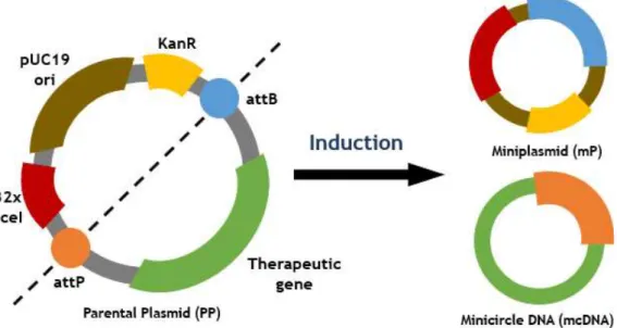
Figure 8. mcDNA production scheme from parental plasmid --- 22
Figure 9. Standard curve with BSA standard protein --- 34
Figure 10. Graphical representation of the growth curve --- 37
Figure 11. Agarose gel electrophoresis of the extraction and purification according to the manufacturer protocol --- 38
Figure 12. Agarose gel electrophoresis of the extraction and purification optimized protocol 39 Figure 13. Agarose gel electrophoresis of the samples obtained after the modified extraction --- 41
Figure 14. Size exclusion chromatography (A) and agarose gel electrophoresis (B) of the mcDNA-primiR-375 vector --- 42
Figure 15. Size exclusion chromatography (A) and agarose gel electrophoresis (B) of the mcDNA-p53 vector --- 44
Figure 16. Size exclusion chromatography chromatogram using a 106 mL column (A) and agarose gel electrophoresis (B) of the mcDNA-primiR-375+p53 vector --- 46
Figure 17. Size exclusion chromatography chromatogram using a 180 mL column (A) and agarose gel electrophoresis (B) of the mcDNA-primiR-375+p53 vector --- 48
Figure 18: Structure of resazurin substrate and the pink fluorescent resofurin product resulting from the reduction in viable cells --- 50
Figure 19: Cell viability percentage in comparison to the negative control --- 51 Figure 20: Cell growth assay in CaSki cells after transfection with mcDNA-p53, primiR-375 and
Figure 21: Western-Blot for the detection of tumour suppressor p53 protein and respective normalization with housekeeping protein β-actin --- 54 Figure 22: Graphical representation of the normalization of p53 protein expression in relation to β-actin (p53/β-actin) relative intensity --- 55 Figure 23: Western-Blot for the detection of tumour suppressor p53 and pRb proteins and respective normalization with housekeeping protein β-actin s --- 56 Figure 24: Western-Blot for the detection of E6 oncoprotein --- 56
List of Tables
Table 1. Clinical assays approved for cervical cancer gene therapy --- 15
Table 2. Viral vectors and its major advantages and disadvantages --- 17
Table 3. Non-viral vectors delivery methods --- 18
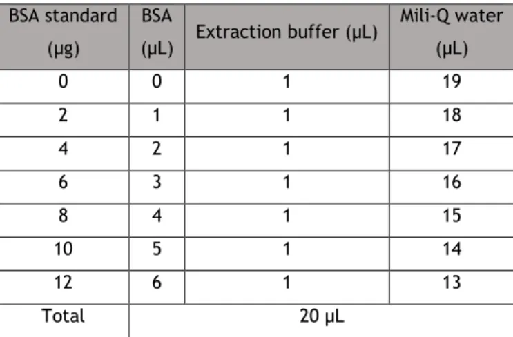
Table 4. Volumes needed to prepare BSA standards --- 33
Table 5. Final mcDNA-primiR-375 and mcDNA-primiR-375+p53 concentration following the optimized protocol and respective band intensities for the mcDNA and impurities. --- 40
Table 6. Representation of the concentration of fractions collected and desalted during the mcDNA-primiR-375 size-exclusion chromatographic assay --- 43
Table 7. Representation of the concentration of fractions collected and desalted during the mcDNA-p53 size-exclusion chromatographic assay --- 45
Table 8. Representation of the concentration of fractions collected and desalted during the mcDNA-primiR-375+p53 size-exclusion chromatographic assay, using a 106 mL column --- 47
Table 9. Representation of the concentration of fractions collected and desalted during the mcDNA-primiR-375+p53 size-exclusion chromatographic assay,using a 180 mL column --- 49
List of Abbreviations
2D-PAGE: Two-dimensional polyacrylamide gel electrophoresis AC: Affinity chromatography
AEC: Anion exchange chromatography AGO: Argonaute
AIF: Apoptosis inducing factor bac-ORI: Bacteria replication origin Bax: Bcl-2-associated X protein Bcl: B-cell lymphoma
bHLH: Basic helix-loop-helix bp: Base pair
BSA: Bovine serum albumin CaSki: Cervical cancer cell line CDK: Cyclin-dependent kinase
CEC: Cation-exchange chromatography Da: Dalton
DD: Death domain
DISC: Death-inducing signalling complex
DMEM-F12: Dulbecco’s Modified Eagle’s Medium/Ham’s F-12 Nutrient Mixture DNA: Deoxyribonucleic acid
EDTA: Ethylenediamine Tetraacetic Acid EGFR: Epidermal Growth Factor Receptor EGTA: Ethylene Glycol Tetraacetic Acid EMA: European Medicines Agency FADD: Fas-Associated Death Domain FBS: Fetal Bovine Serum
FDA: Food and Drug Administration g: G force
gDNA: Genomic DNA
HDL: High-Density Lipoprotein hFIB: Human Fibroblasts cell line
HIC: Hydrophobic Interaction Chromatography HPV: Human Papillomavirus
HR-HPV: High-Risk Human Papillomavirus HSPGs: Heparin Sulphate Proteoglycan IEX: Ion Exchange Chromatography IRF-3: Interferon Regulatory Factor 3 Kb: Kilobase
Kbp: Kilo basepair kDa: Kilo Dalton
LDL: Low-Density Lipoprotein
LR-HPV: Low-risk Human Papillomavirus m/z: Mass-to-charge ratio
MALDI: Matrix Assisted Laser Desorption Ionization Time of Flight MAML1: Mastermind-like protein 1
mcDNA: Minicircular DNA miRNA: Micro RNA
MOMP: Mitochondrion Outer Membrane Permeabilization MOPS: 3-(N-morpholino)propanesulfonic acid
mP: Mini plasmid mRNA: Messenger RNA MW: Molecular Weight
NCBI: National Center for Biotechnology Information NCR: Non-coding region
OD: Optical Density
ORF: Open Reading Frames Ori: Origin of replication PBS: Phosphate buffer solution pDNA: Plasmid DNA
pfu: Plaque-forming unit pI: Isoelectric point
PMSF: Phenylmethylsulfonyl fluoride PP: Parental Plasmid
pre-miRNA: Precursor miRNA
pri-miRNA: Long primary transcripts of miRNA PTM: Post-translational modifications
PVDF: Polyvinylidene difluoride RBC: Carboxyl-terminal domain RF: Radiofrequency
RNA: Ribonucleic acid rpm: Revolutions per minute
RPMI: Roswell Park Memorial Institute sc: Supercoiled
SDS: Sodium Dodecil Sulphate
SDS-PAGE: Sodium Dodecyl Sulphate-Polyacrylamide Gel Electrophoresis SEC: Size Exclusion Chromatography
SiHa: Cervical cancer cell line
Smac: Second mitochondria-derived activator of caspases SNS: Portuguese National Health System
TAE: Tris-acetate-EDTA TB: Terrific Broth TBS: Tris-Buffered Saline
TBS-T: Tris-Buffered Saline 0.1% Tween 20 TGF-β: Transforming Growth Factor Beta
TRAIL: TNF-Related Apoptosis Inducing Ligand Tris: Tris(hydroxymethyl)aminomethane Tween 20: Polysorbate 20
UTR: Untranslated Region
List of Scientific Communications
Oral communication
Adriana Resende Pinto, Diana Carvalho Pereira, Dalinda Eusébio, Ana M. Almeida, João A. Queiroz, Luís A. Passarinha, Ângela Sousa. “Therapeutic effect evaluation of mincircle DNA (mcDNA) vectors”. XIV Annual CICS-UBI Symposium. 4-5 July 2019, University of Beira Interior, Covilhã, Portugal.
Poster communication
Resende-Pinto, A.; Carvalho-Pereira, D.; Eusébio, D.; Almeida, A. M.; Queiroz, J. A.; Passarinha, L. A.; Sousa, Â. “Evaluation of tumour suprressor and HPV oncoprotein expression in cervical cancer cells by using minicircle DNA vectors”. III International Congress HSR - Trends in Aging and Cancer. 14-16 November 2019, Covilhã, Portugal.
Chapter I – Introduction
1.1 Cancer
Cancer is a major global health problem accounting, annually, for more than eight million deaths globally, being a complex multifactorial disease that comprises changes in the
genome, orchestrated by host and environmental interactions1. The hallmarks of cancer are
self-sufficiency in growth signals, the ability for tissue invasion and metastasis, limitless
replicative potential, sustained angiogenesis and evasion of apoptosis1. The tumour
microenvironment, which is composed of various non-malignant cells expressing several regulatory proteins, as well as the extracellular matrix, plays a pivotal role in the initiation
and progression of cancers2.
The process of cancer initiation is irreversible and results from the normal cell exposure to physical, chemical and/or biological agents capable of promoting genetic damages, leading to
genomic instability3, being characterized by the increase in cell plasticity and entropic status,
resulting in clonal expansion of mutated cells4.
The cervical cancer is the third most common malignancy and the fourth cause of death in
the world5. According to data from the National Health System (SNS) in Portugal, cancer
incidence has been growing at a constant rate of 3% per year, mainly associated with the aging of the population. To the date, each year 50000 new cases of cancer are diagnosed, and the mortality is 152.6/100000. Particularly for cervical cancer in Portugal, 1000 new cases per year are diagnosed and, in comparison to other countries in Europe, Portugal has an incidence and mortality rates very favourable. It is important to note that Portugal has a free vaccine against HPV on their national vaccination program since 2008, and the most recent statistical analysis point that 90% of the women population under the age of 27 are vaccinated.
1.1.1 Human papillomavirus (HPV)
Human papillomavirus (HPV) is the etiological agent of common dermatologic and sexually
transmitted diseases6. A persistent infection with HPV is the main risk factor for the
development of cervical cancer7 and even though being discussed as a sexually transmitted
disease7, evidences suggest that the HPV can be vertically transmitted, as viral DNA
sequences were identified in the oral cavity of new-borns, breast milk, amniotic liquid,
The infections caused by HPV, according to their oncogenic potential and prevalence in
cancer can be classified in low-risk HPVs (LR-HPV) and in high-risk HPVs (HR-HPV)7. High-risk
HPVs are the causative agents of cervical carcinoma since, it has been demonstrated that E6 and E7 viral oncoproteins are able to promote cancer development by principally targeting
and inhibiting p53 and pRb tumour suppressor proteins, respectively10.
More than two hundred different HPV types have been identified and classified into five
genera: α, β, µ and ν6. The HPV belonging to the α genus has proven oncogenic potential, a
well-established role in cervical carcinoma11. The International Agency for Research on
Cancer (IARC) classified 12 different HR-HPV types as carcinogenic to humans: 16, 18, 31, 33,
35, 39, 45, 51, 52, 56, 58 and 5912. The most common types of HPV are the 16 and 18 most
frequently found in cervical cancer, in 60% and 15% of the cases, respectively, both
preventable by vaccination 6,7,12. Moreover, HR-HPV are related to other cancers as vulvar,
vaginal, anal, penile and oropharyngeal squamous cell carcinoma7. An HPV phylogenetic tree
has been designed that groups the different HPV types into genera based on the homologous
nucleotide sequence of the major capsid protein, L113.
1.1.2 Molecular biology
Human papillomavirus belongs to the Papilomaviridae family9, capable of infecting epithelial
cells of the skin, oral and genital mucosa6,12. HPVs are small non-enveloped viruses with a
double-stranded circular DNA with a length of about 8 Kb7,9. The viral particles of HPV
(virions) show a conserved icosahedral morphology6,9, a molecular weight of 5x10⁶ Da14 and a
diameter of 50-55 nm9.
The viral genome is divided into three regions (Figure 1):
i. early (E) region, that contains E1, E2, E4, E5, E6 and E7 genes, associated with the
viral replication 6,7,9;
ii. late (L) region, which encodes the major (L1) and minor (L2) capsid proteins6,9,15;
iii. non-coding region (NCR), located between the L1 and E6 containing the open reading
frames (ORFs)9.
Human papillomavirus include genes that express non-structural proteins needed for DNA
replication, transcription or viral assembly and release6. Even tough non-coding, the NCR also
known as long control region (LCR), is a fragment of the HPV genome of approximately 900
bases7, contains most of the regulatory elements involved in viral DNA replication and
Figure 1. Human Papillomavirus genome representation (adapted from Tomassino, 2014)12.
The replication of HPV depends upon the host cell differentiation program7. E1 and E2 are
expressed at the beginning of the infection, bind to the LCR and participate in viral
replication and transcription7. E4 is expressed mainly during later stages of infection,
contributes to the amplification of the viral genome and disrupts the cellular keratin
network, facilitating virus release7. E5 inhibits maturation of endocytic vesicles from early to
late endosomes7. E6 and E7 are the main viral oncoproteins, that as multifunctional proteins,
regulate the cell cycle through their interaction with a set of cellular proteins, being within
the most relevant the tumour suppressor proteins p53 and pRb, respectively16,17. E6 and E7
oncoproteins target numerous cellular proteins and thus promote cell cycle progression,
apoptosis inhibition, DNA damage and evasion of the host immune response18.
1.1.3 Infection
As the HPV is not capable of self-replicate (this virus does not express polymerases), during the amplification process, the early proteins interact with the host cell, inducing the S phase
entry as obtaining mechanism of DNA polymerases19,20. However, this proliferative action can
lead to DNA replication stress9 and, as a consequence, to numerical and structural instability,
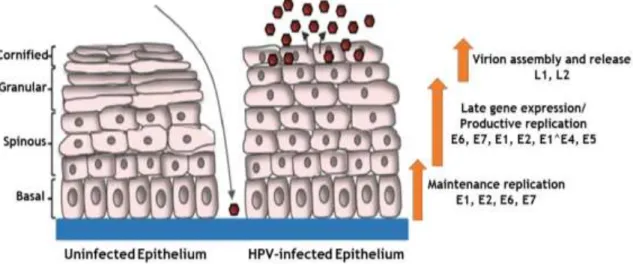
The life cycle of HPV is intimately linked to the differentiation status of the host cell
keratinocyte and characterization by three distinct phases of replication15,20. The HPV
replicative cycle begins in the basal layer of the squamous epithelia, where viral particles arrive through micro wounds and there, they complete their life cycle, taking advantage of
the differentiation program of the host epithelial cells (Figure 2)7,9,20.
Figure 2. Human papillomavirus life cycle scheme (adapted from Moody, 2017)20.
After the virus entry, the HPV genome is replicated as an episome7. At the initial infection in
undifferentiated basal cells, low levels of the E1, E2, E6 and E7 proteins are expressed,
delaying normal keratinocyte differentiation7. E1 is the only viral protein with enzymatic
activity - it is a helicase/ATase which is recruited by E2 at the origin of viral replication,
generating approximately 50 to 100 copies of viral episomes per cell20. These numbers
increase to thousands of DNA viral copies throughout the differentiation of the epithelium21.
From the middle to upper differentiating epithelial layers, E6 and E7 are expressed in high amounts and the viral life cycle is completed by the expression of L1 and L2 proteins in the
uppermost layer of the epithelium7. Only upon epithelial differentiation is the productive
phase of the viral life cycle activated, resulting in the amplification of viral genomes to thousands of viral copies per cell in the suprabasal layers, as well as activation of late gene
expression and virion assembly and release20. Regulation of the viral life cycle in this manner
allows HPV to avoid detection by the immune response as high levels of viral gene expression as well as virion production are restricted to the uppermost layers of the epithelium, which
are not under immune surveillance20.
Upon reached the basal cells, the L1 protein binds to heparin sulphate proteoglycan
(HSPGs)22, promoting a conformational change in the viral capsid9. The conformational change
mechanisms mediated by clathrin and caveolin. Upon lysosomes binding to these endocytic vesicles forming the phagolysosomes, a reduction in pH occurs, leading to the viral capsid
disassembling and, consequently, the viral genome release9,19.
It is important to note that, a complete viral cycle does not represent a risk for cancer
development7, as near 90% of HPV infections, even with HR-HPV, are eliminated within two
years7. A persistent infection with HR-HPV is the main determinant for cervical cancer
development, where the accumulation of damaged DNA, probably due to the interactions of E7 and E6 with their cellular targets, leads to genomic instability and, in most cases, to the
integration of the viral genome into the host genome7. Viral genome integration precedes the
overregulation of E6 and E7 oncogenes, since in most of the cases E2 gene is disrupted, which
causes the overexpression of E6 and E7 oncogenes23.
1.1.4 Oncoproteins E6 and E7
E6 and E7 from HR-HPVs play a key role in carcinogenesis by altering pathways related to the
immune response and cellular transformation12. The expression of E6 and E7 proteins are
associated with the integration of viral DNA into the host genome, malignant transformation
and ultimately progression to cancer6. These proteins deregulate fundamental cellular events
contributing to cancer initiation and progression by altering the cell cycle regulation and telomere maintenance, apoptosis, DNA repair, senescence and differentiation, inducing and facilitating the accumulation of damaged DNA and genomic instability, blocking tumour
suppressor pathways and progression towards malignancy6,12.
Once HPV 16 and 18 are the most frequently detected types in cervical cancer worldwide,
their E6 and E7 proteins have been extensively studied12, lacking enzymatic activities and
exert their functions by associating with a broad spectrum of cellular proteins19. The E6 and
E7 oncoproteins are the primary transforming viral proteins, playing an important role in immune evasion by targeting cytokine expression to alter cell proliferation and interferon
responses6. If the HPV-infected cells are rapidly eliminated by the immune system, despite
the transforming properties of E6 and E7, they do not have sufficient time to accumulate
chromosomal abnormalities and acquire a malignant phenotype12.
However, for the cases that the HPV-infected cells are not eliminated, E6 and E7 constitute attractive therapeutic targets since their inhibition rapidly induces senescence in
HPV-positive cancer cells24. These viral proteins are the key factors that maintain the malignant
phenotype of HPV-positive cancer cells, specifically:
i. E6 and E7 are the only viral genes that are always retained and expressed in
HPV-positive cancer cells24;
iii. inhibition of E6/E7 expression results in rapid induction of cellular senescence, an irreversible proliferative arrest, in HPV-positive cancer cells.
The first indication of the oncogenic properties of E6 and E7 was provided by studies on
cervical cancer-derived cells, such as SiHa and CaSki cells12. In these cell lines, viral DNA was
found to be randomly integrated into the host genome, leading to the disruption of several
viral genes and preservation of E6 and E7, which were actively transcribed12. Many studies
have shown that the frequency of viral DNA integration increases with the severity of the
cervical lesion, indicating that this event is implicated in the progression of the disease12.
Several evidences suggest that HPV is involved in the reprogrammed metabolism of cervical
cancer cells7. Diverse interactions of E6 and E7 with cellular partners have been
demonstrated, and many of those affect cell biological functions, leading to transformation7.
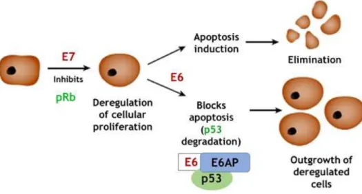
When integrated into the host squamous epithelial cell genome, the high-risk HPV types (16 and 18) will express E6 gene, which will induce the degradation of p53 tumour suppressor
protein6. E7 also expressed early, is an oncoprotein that binds to the tumour suppressor
retinoblastoma protein (pRb) (Figure 3) and allows DNA synthesis6. The continuous expression
of E6 and E7 are required to maintain the malignant phenotype7,12,24. The silencing of both
viral genes in cervical cancer-derived cell lines results in rapid cellular death (Figure 3)12.
Figure 3. E6 and E7 oncoproteins mechanism of action (adapted from Hope-Seyler and co-workers, 2017)24.
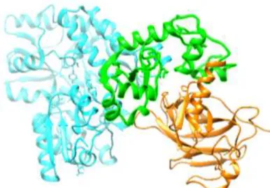
E6 is a basic and cysteine-rich protein of approximately 150 amino acids and 23 kDa (Figure
4)12. This oncoprotein identify some of their target proteins by specifically interacting with
co-characterized properties of this protein is its ability to induce degradation of the p53 tumour
suppressor via the ubiquitin pathway12, through the interaction of the LxxL motif of E6AP26.
E6 binds to a short leucine (L)-rich LxxLL consensus sequence of a 100 kDa cellular protein12
within the cellular ubiquitin ligase E6AP and subsequently the heterodimer E6/E6AP recruits
and degrades p5326. The E6-associated protein (E6AP), which functions as an E3 ubiquitin
protein ligase12, targeting cellular proteins to be ubiquitinated for proteasomal degradation7.
The E6/E6AP complex then binds to the central region of p53 which becomes rapidly
ubiquitinated and targeted to proteasomes12. In this way HR-HPV E6 induces p53 degradation
by forming a complex with E6AP26 and, thus, p53 dependent apoptosis is blocked7. E6
interacts not only with LxxL motifs but also with PDZ domains involved in cell polarity and adhesion, and also possesses a self-association interface required for the p53 degradation
activity25. Neither E6 nor E6AP are separately able to recruit p53, and the precise mode of
assembly of E6, E6AP and p53 is unknown26.
Figure 4. Crystal structure of the HPV16 E6/E6AP/p53 ternary complex (E6AP – blue, p53 – orange, E6 – green) (PDB ID: 4XR8).
E7 is an acidic phosphoprotein of about 100 amino acids and 17 kDa that, likewise to E6, contains zinc-binding motifs being structurally and functionally associated to adenovirus E1A and, grounded on this similarity is divided into three domains: conserved regions 1-3
(CR1-3)12. The association of HR-HPV E7 with pRb resulting in the degradation and functional
inactivation of pRb24 via proteasomal pathways, leading to unscheduled cell cycle
progression24, for example with consequent activation of E2F-regulated transcription12 from
their negative control by pRb and, subsequently, the activation of cell cycle-promoting E2F
target genes24. The genes regulated by E2F1-3 include cyclin A and cyclin E, which are
positive regulatory subunits of cyclin-dependent kinase (CDK) complexes whose accumulation
results in activation of CDKs and cell cycle progression12. The genes regulated by E2F1-3
include cyclin A and cyclin E (both positive regulatory subunit of cyclin-dependent kinase
Furthermore, scientific studies indicate that E6/E7 deregulate the cellular miRNA network,
upon repression of endogenous E6/E7 expression in cervical cancer cell lines26. Many of these
miRNAs have been linked to carcinogenesis, and the expression of several of the most
abundant E6/E7-regulated miRNAs is altered in cervical cancer tissues24. Vice versa, cellular
miRNAs may also be involved in the regulation of HPV E6/E7 gene expression, as it has been
reported for miRNA-37527.
1.1.5 p53 protein
The p53 protein is a stress sensor regulating many cell pathways, such as cell cycle arrest, senescence, apoptosis, metabolic alterations, DNA repair and other mechanisms of tumour
suppression28. The p53 pro-apoptotic tumour suppressor is mutated or functionally altered in
most cancers26. This protein consists of a transactivation domain (amino acids 1-44), a
proline-rich domain (102-292), a tetramerization domain (325-356) and a C-terminal domain
(357-393)29. The prevalence (95%) of p53 mutations in human cancers are missense mutations,
mostly located within the DNA-binding domain (amino acids 102-292) with hot spots at codons
R175, G245, R248, R249, R273 and R28230.
In the majority of human cancers, p53 activity is often impaired by missense mutations
distributed within the exons 4-8 in the TP53 gene, encoding for the DNA-binding domain31. In
most cases, mutated p53 proteins become inactive but acquire stability and accumulate in
cancer cells31. Some mutant p53 proteins, besides losing their oncosuppressor activities,
acquire oncogenic features (gain of functions) that endow the cells with overgrowing and
survival advantages30.
p53 is a transcription factor that regulates the expression of genes encoding regulators of the
cell cycle, DNA repair machinery and apoptosis12. Under cellular stress, such as hypoxia or
DNA damage, p53 triggers cell cycle arrest or apoptosis to guarantee the integrity of cellular
genome12. By blocking the cell cycle, p53 prevents the replication of damaged DNA and allows
the cell to repair the damage before S phase12. Alternatively, if DNA damage is too great and
difficult to repair, p53 can divert the cell into apoptosis, thus preventing the production of
potentially transformed progeny12.
In epithelial tumours induced by high risk mucosal human papillomavirus, including human cervical carcinoma and a growing number of head and neck cancers, p53 is degraded by the
viral oncoprotein E626.
This protein is composed by 928 amino acids and is usually described as having three domains
(Figure 5)32. A central domain was recognized as the minimal region necessary to bind viral
oncoproteins, such as adenovirus E1A, SV40 Tag and human papillomavirus E7, and it was termed the “pocket”, that comprises two subdomains, A and B, each resembling a cyclin fold with three extra helices, cooperating with each other through an extensive non-covalent
interface such that the pocket folds into a single structural unit32. Two additional cyclin folds
constitute the structured amino-terminal domain RBN, which resembles the pocket structure
except for a few subtle differences32. Roughly the last 150 residues of RB form the
carboxyl-terminal domain (RBC), which is inherently disordered32.
Figure 5. Crystal structure of the pRb (PDB ID: 2QDJ).
Abolishing pRb function permits uncontrolled cell cycle progression and leading to tumour
growth promotion32. In its simplest form, pRb functions primarily rely on the inactivation of
transcription factors, such as E2F responsible for targeting genes in the G1 phase of cell cycle, so that when freely available this protein stimulates cellular cycle transition to S
phase33,34. Growth factors signal arouse the assemblage and activation of cyclin D and its
associated cyclin-dependent kinases (CDK4 or CDK6) that firstly phosphorylate pRb in the G1 phase, which releases the respective E2F family member from pRb, leading to the activation
of transcriptional targets to advance the cell cycle34. The abundance of mutations affecting
members of this pathway, and the resulting lack of proliferative control, has led to the idea
that pRb pathway disruption is a hallmark of cancer34.
Upon viral infection, E7 is expressed and binds to pRb, disrupting its complex with E2F and
contributing to continuous cell proliferation33. Furthermore, E7 oncoproteins is also
implicated with the interference of p107 and p130 functions, two proteins that also regulate
cell-cycle proliferation through E2F transcription factor binding33. The ultimate inhibition of
pRb, p107 and p130 by E7 viral protein contributes to the uncontrolled cell proliferation and
1.2 Cell apoptosis
Apoptosis or programmed cell death is a regulator of physiological growth control and tissue homeostasis, as well as controlling functions of the immune system being characterized for presenting typical biochemical and morphological hallmarks, in particular, cell shrinking,
nuclear DNA fragmentation and membrane blebbing35,36,37.
Apoptosis usually maintains the integrity of healthy tissues and organs in response to DNA damage, cellular stress or oncogene expression through the eradication of damaged cells,
thereby blocking tumour growth35. In the recent years, one of the most significant
developments in cancer research is the acknowledgement that cell death mostly by apoptosis is significantly tangled in the regulation of tumour formation and also critically determine
treatment response36.
Apoptosis is a tightly controlled and strictly regulated pathway process that eliminates unwanted or potentially harmful cells, once all tumour cells require inhibition of apoptotic
pathway to survive, that becomes deregulated in tumorigenesis35.
There are two basic pathways that lead to apoptosis: the intrinsic mitochondrial pathway and
Killing of tumour cells by most anticancer strategies currently used in clinical oncology, for example chemotherapy, γ-irradiation, suicide gene therapy or immunotherapy, has been linked to activation of apoptosis signal transduction pathways in cancer cells such as the
intrinsic and/or extrinsic pathway36.
Proteolytic enzymes such as caspases (a family of cysteine proteases that act as common death effector molecules in several forms of cell death) are important effector molecules in apoptosis, once their activation in response to anticancer therapies can be initiated through activation of the extrinsic (receptor) pathway or at the mitochondria by stimulating the
intrinsic pathway36. Caspases are synthetized as inactive proforms and upon activation, they
cleave next to aspartate residues, resulting in amplification of caspase activity through a
protease cascade36.
1.2.1 Extrinsic pathway
The extrinsic pathway of apoptosis is associated with death receptors that include members of tumour necrosis factor (TNF) superfamily, this is TNF receptor-1, Fas death receptor-3, TRAIL receptor-1 and receptor-2 and death receptor-6, Fas ligand (Fas-L), TNF-related
apoptosis inducing ligand (TRAIL) are involved in the extrinsic pathway35,36. Members of the
TNF receptor family share similar, cysteine-rich extracellular domains36.
Death receptors consists of more than 20 proteins with a broad range of biological functions,
including regulation of cell death and survival, differentiation or immune regulation36 and are
composed by 80 amino acids in their cytoplasmatic domain, denominated the death domain
(DD)35, which plays a crucial role in transmitting the death signal from the cell’s surface to
intracellular signalling pathway36.
The DD is essential for the initiation of apoptotic signalling and transduction via transmitting death signal from cell surface to a variety of intracellular targets, where a specific ligand or
an agonist antibody can activate the receptors and trigger the apoptotic signalling35,36.
Trimerization of Fas and TRAIL receptors, the serial recruitment of the adaptor protein Fas-associated DD (FADD), pro-caspase-8, caspase-10 and c-FLIP to Fas DD could ultimately form the death-inducing signalling complex (DISC), culminating in the activation of the caspase-8
and the cell is committed to apoptosis35. Though, in addition to FADD, the TNRF1-associated
death domain (TRADD) is involved in formation of DISC in the case of TNF-R1 activation, initiating a cascade of events, leading to a caspase-8 activation and lastly to apoptosis
1.2.2 Intrinsic pathway
In the mitochondrial pathway of apoptosis, caspase activation is closely linked to permeabilization of the outer mitochondrial membrane by proapoptotic members of the Bcl
family36, once internal stimuli (include growth factor deprivation and oxidants35) promote the
activation of caspases and pro-apototic members of the Bcl-2 family, leading to
mitochondrion outer membrane permeabilization (MOMP) via Bax channels37.
Numerous cytotoxic stimuli and proapoptotic signal-transducing molecules converge on
mitochondria to induce outer mitochondrial membrane permeabilization36. Internal stimuli
promote the activation of caspases and pro-apototic members of the Bcl-2 family, leading to
mitochondrion outer membrane permeabilization (MOMP) via Bax channels37. This
permeabilization is regulated by proteins that regulate bioenergetic metabolite flux and
components of the permeability transition pore36. Upon disruption of the outer membrane, a
set of proteins normally found in the space between the inner and outer mitochondrial membranes is released, including cytochrome c, Smac/DIABLO, Omi/HtrA2, AIF and endonuclease G, that once in the cytosol, these apoptogenic proteins trigger the execution of cell death by promoting caspase activation or by acting as caspase-independent death
effectors36. In its turn, MOMP leads to the release of cytochrome-C, which induces the
formation of the apoptosome, a complex seven caspase-9 units37. Caspase-9 will trigger an
executioner caspase (3 or 7) ensuring the continuity of the process37. The anti-apoptotic
members of the Bcl-2 family (Bcl-2, Bcl-xL, Mcl-1) are able to inhibit apoptosis by quenching
the activity of their pro-apoptotic counterparts37.
1.3 DNA based therapy
The use of DNA biopharmaceuticals is becoming very attractive for biological therapy due to the fact that the therapeutic transgenes of interest are generated by the cell transcriptional
machinery39. In this way, much attention has been drawn recently for new forms of
immunotherapy for infectious diseases and cancer. DNA vaccines still possess the intrinsic advantage of target flexibility, low cost, and physical stability over a wide range conditions
that may be encountered when targeting diseases with global distribution40. Similar to other
gene-based therapies, this technology plays a key role in the final efficacy of DNA vaccines40.
1.3.1 DNA vaccines
be related with a given pathology) . Subsequently inoculation, the treated individual should be capable of producing a strong immune response against the antigen for which genetic information was beforehand delivered, triggered mainly as a result of the transfection of antigen-presenting cells (APCs) and non-APCs that will later express the antigen encoded in
the DNA vaccine33.
Due to the reason that DNA vaccines can have preventive and therapeutic effects, they are currently under research in the treatment of cancer, once numerous cancers show specific antigens that could be used in the development of a DNA vaccine to induce a specific immune
response against those antigens33.
Researchers have mostly considered that the majority of DNA vaccines developed to the date do not present the high-level immunogenicity observed in mouse models, therefore mitigating the lack of DNA vaccines approved for human use, being the reason for new methodologies be currently under study to improve the clinical outcome of DNA vaccines, appearing from the
remarkable progress that has been observed in the past years for this area of studies33.
1.3.2 Gene therapy
Gene therapy is a promising strategy to treat acquired diseases and/or genetic disorders, aiming to deliver genetic material into target cells or tissue and to express it with the
intention to gain a therapeutic effect1, thus regulating cellular processes and responses41.
Relying on the purpose and delivery method, successfully delivered genes could have several destinies: modifying defective host genes, replacing the deficient host genes, inserting into
the host genome or staying in the nucleus without integration into the host genome41.
The European Medicines Agency (EMA) defines that a gene therapy medicinal product is a biological therapeutic product which fulfills the following two characteristics: (a) contains an active substance which consists of a recombinant nucleic acid used in or administered to human beings with a view of regulating, repairing, replacing, adding or deleting a genetic sequence; (b) its therapeutic, prophylactic or diagnostic effect relates directly to the recombinant nucleic acid sequence it contains, or to the product of genetic expression of this sequence. Gene therapy medicinal products shall not include vaccines against infectious diseases.
Gene therapy has the advantage over traditional therapies since it can be administered locally, allowing the delivery of a high therapeutic dose without taking the risk of rising
systemic adverse effects1. The subsequent transgene expression can re-establish normal
cellular processes or induce new cellular responses, therefore realize the aim of therapy41.
However, the main challenge in gene therapy remains in being able to deliver an adequate amount of genetic material into target cells or tissues and to maintain gene expression for a
desired period of time1. In addition, since most gene therapies are of single time application,
in the long run, they can be cost-effective1.
The perfect delivery system should be capable of ensuring a successful gene therapy and must
satisfy the following criteria42:
i. must not interact with vascular endothelial cells and blood components;
ii. be required to be capable of avoiding uptake by the reticuloendothelial system; iii. ought to be small enough to pass through the cell membrane and reach the nucleus. Generally, gene therapy can be categorized into two categories: germline and somatic gene
therapy1. The difference between these two approaches is that in somatic gene therapy
genetic material is introduced in some target cells, but the change is not passed to the next generation, while in germline gene therapy the therapeutic or modified gene will be passed
to the next generation1. This is a major and very important difference since current
legislation only allows gene therapy on somatic cells1.
To improve gene transfer efficiency, specificity, and safety, numerous gene transfer systems, usually called gene vectors have been developed, being divided into two categories: viral and
non-viral gene delivery41.
1.3.2.1
Gene therapy for cervical cancer
Since the effects of E6 and E7 are the loss of function of p53 and pRb, respectively, it seem likely that HPV-associated tumours could be controlled by replacement of wild-type p53 or
pRb proteins in a functional form43. The potential use of p53 or pRb as gene therapy has been
examined for many types of cancers, quite apart from those that are associated with HPV and
has progressed to the stage of multiple human trials43.
In 2012, several clinical assays were in progress using several DNA plasmids, summarized in
Table 144.
Once the HR-HPV principal tumorigenic effects have been related to the expression of E6 and E7 genes, a remarkable effort have been done to develop a suitable gene therapy for blocking
their expression43. The first method for blocking the translation of the RNA to protein was the
use of an antisense RNA, as this molecules can bind to the RNA of the opposite strand, leading
to a duplex, that soon will be degraded, resulting in a reduction of the gene expression43.
Other approaches used for the prevention of gene expression have been exploited . For instance, the E6 protein may have its function blocked by RNA molecules that bind specifically, known as an aptamer, having an investigation group showed that this could cause
apoptosis in HPV-16-positive cancer cells but not HPV-negative cells43.
Table 1. Clinical assays approved for cervical cancer gene therapy (using a naked plasmid DNA as vector)43.
ID
tri
al Title Country Clinical
phase
Genes transferred
US
-1093
Phase III placebo-controlled study of VGX-3100, (HPV 16 E6/E7, HPV 18 E6/E7 DNA vaccine) delivered intramuscular followed by electroporation.
USA Phase II HPV 16 E6/E7 fusion protein HPV 18 E6/E7 fusion protein
US
-0595
pNGVL4a-Sig/E7/HSP70 for the treatment of patients with HPV 16 cervical intraepithelial neoplasia 2/3
USA Phase I and II HPV 16 E7 oncogene US -0916
Dose escalation study to evaluate the safety, tolerability and immunogenicity of HPV DNA plasmid (VGX-3100) + electroporation in adult females with histological diagnosis of grade 2 and 3 cervical intraepithelial neoplasia
USA Phase I HPV 16 E6/E7 oncogenes HPV 18 E6/E7 oncogenes
US
-0984
pNGV4a-CRT/E7 for the treatment of patients with HPV 16 cervical intraepithelial neoplasia 2/3
USA Phase I and II HPV 16 E7 oncogene US -1040
Evaluate the safety, tolerability and immunogenicity of a 4th dose of HPV DNA plasmid (VGX-3100) + electroporation
in adult females previously immunized with VGX-3100
USA Phase I HPV 16 E6/E7 oncogenes HPV 18 E6/E7 oncogenes
1.3.3 DNA vectors
An ideal vector should satisfy several criteria42:
i. must not cause a strong immune response;
ii. should be capable of transporting nucleic acids regardless of their size; iii. need to lead to a sustained and regular expression of its genetic cargo;
iv. ought to deliver the gene to only certain type of cells, especially when the target cells are scattered throughout the body, or when they are part of a heterogeneous population;
vi. must be of easy preparation, inexpensive and commercially available at high concentrations;
vii. need to either remain in episomal position or integrate into a specific region of the genome, but not integrate randomly.
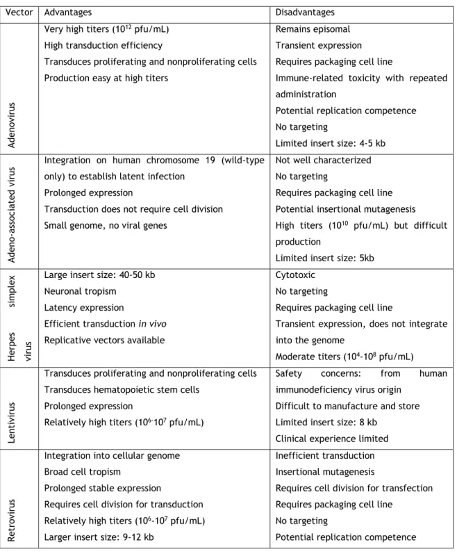
In fact, viruses (Table 2) were the first carriers to be used to deliver and protect the
therapeutic gene, benefiting from the virus life cycle41,42. This type of carrier, known as a
viral vector, is one of the most used vectors in gene therapy, also due to its capacity to carry
the gene efficiently and guarantee long term expression41,42. Though, the risk of a immune
response caused by the use viruses as delivering vectors, its high cost and difficulty relating to their preparation and the limited size of genetic materials that can be inserted into human cells, as well as the associated immunogenicity have constrained the use of these vectors in
gene therapy and led to research into safer and cheaper and safer alternatives41,42.
Therefore, non-viral vectors have appeared, presenting several advantages that include relatively safety, generally causes low immune response, can be easily prepared at low cost and in large quantities, have the ability to transfer large size genes, cause less toxicity and
can be modified with ligands for tissue or cell-specific targeting41,42. In addition, they can
transfer different and large transgenes, being able to be stored for long periods due to their
stability42. However, non-viral methods present limitations of low transfection efficiency and
poor transgene expression41.
The non-viral approaches can be divided into two groups (Table 3): physical approaches and
chemical vectors42. The physical approaches rely on a physical force that weakens the cell
membrane to facilitate the entrance of the gene into the nucleus and include needle
injection, electroporation, gene gun and ultrasound42. The chemical vectors can be prepared
by electrostatic interaction between a polycationic derivatives, which can be lipids or polymers, and the anionic phosphate groups of the DNA to form a particle called polyplex. On the other hand, a lipoplexe is formed when the DNA interacts with a lipid or by encapsulation of DNA within biodegradable spherical structures that lead to micro or nanoparticles
Table 2. Viral vectors and its major advantages and disadvantages (adapted from Ibraheem and co-workers, 2014)44.
Vector Advantages Disadvantages
A
denovi
rus
Very high titers (1012 pfu/mL)
High transduction efficiency
Transduces proliferating and nonproliferating cells Production easy at high titers
Remains episomal Transient expression Requires packaging cell line
Immune-related toxicity with repeated administration
Potential replication competence No targeting
Limited insert size: 4-5 kb
A deno -a ss oc ia ted v irus
Integration on human chromosome 19 (wild-type only) to establish latent infection
Prolonged expression
Transduction does not require cell division Small genome, no viral genes
Not well characterized No targeting
Requires packaging cell line Potential insertional mutagenesis High titers (1010 pfu/mL) but difficult
production
Limited insert size: 5kb
Herpe s si m plex vi rus
Large insert size: 40-50 kb Neuronal tropism
Latency expression
Efficient transduction in vivo Replicative vectors available
Cytotoxic No targeting
Requires packaging cell line
Transient expression, does not integrate into the genome
Moderate titers (104-108 pfu/mL)
Lent
iv
irus
Transduces proliferating and nonproliferating cells Transduces hematopoietic stem cells
Prolonged expression
Relatively high titers (106-107 pfu/mL)
Safety concerns: from human
immunodeficiency virus origin Difficult to manufacture and store Limited insert size: 8 kb
Clinical experience limited
R
et
ro
vi
rus
Integration into cellular genome Broad cell tropism
Prolonged stable expression
Requires cell division for transduction Relatively high titers (106-107 pfu/mL)
Larger insert size: 9-12 kb
Inefficient transduction Insertional mutagenesis
Requires cell division for transfection Requires packaging cell line
No targeting
Table 3. Non-viral vectors delivery methods (adapted from Ibraheem and co-workers)44. Physi ca l m et hods
Electroporation Induce the uptake of injected DNA into the cell, by increasing the permeability of the cell membrane through exposure to a controlled electric field.
Applied to several tissues, such as skin, muscle, liver, and tumour.
Efficacy is affected by the pulse duration, field intensity, cell size, shape, and density.
Safe, efficient and reproducible.
Difficult to employ for transferring DNA into a large area of tissue requires surgery to install the electrode in the internal organs and can cause incurable harm and mutilation in the tissue treated due to the high voltage applied. Gene gun The transgene delivery into the target cell/tissue is carried out by the use of
accelerated particle carriers’ biocompatible heavy metals (gold, tungsten, silver).
The efficiency is determined by the size of the particles employed as DNA-carriers, gas pressure used to accelerate them, and the dosage of transgene blasted into the target.
Effectively employed in genetic vaccination, immune therapy and suicide gene therapy to treat cancer.
Presents high-level gene expression quickly achieved, long-lasting gene expression and can reach numerous organs without injury the surrounding tissues.
Shows a low efficiency when used to transfer the gene onto whole tissue due to low penetration by the metal particles and often needs surgery to apply the approach for deep tissues.
Ultrasound Depends on increasing the permeability of the cell membrane by using ultrasound waves to create pores or acoustic cavitation in the exposed cell membrane.
It is safe, non-invasive and it can reach internal organs without the need for surgery.
The efficiency depends on the frequency, intensity and period of cell exposure to the ultrasound irradiation, DNA concentration, the use of a contrast factor.
Electrostatic interaction
This system uses the electrostatic attraction between the anionic DNA and a cationic lipid or polymer, leading to a positive complex know as lipoplex or polyplex, respectively.
The major problem hindering the use of lipoplex in vivo, besides poor efficiency, is the cytotoxicity resulting from the lipoplex positive charge. Moreover, the use of polyplexes in vivo must overcome many obstacles, such
superparamagnetic iron oxide nanoparticles.
Liposomes are structures formed spontaneously when certain lipids are placed in the aqueous phase, through the self-assembly of these lipids in such a way as to orient their hydrophobic parts away from the water, while the hydrophilic parts are oriented towards the aqueous phase surrounding the hydrophobic ones.
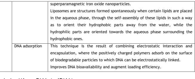
DNA adsorption This technique is the result of combining electrostatic interaction and encapsulation, where the positively charged polymers adsorb on the surface of biodegradable particles to which DNA can be electrostatically linked. Improves DNA bioavailability and augment loading efficiency.
1.4 Micro RNA (miRNA)
miRNA are a class of endogenous, single-strands non-coding small RNAs with 17-25 nucleotides
that are involved in regulating gene expression at the post-transcriptional level 10,45,46.
Figure 7. The biogenesis of miRNA scheme (adapted from Etheridge and co-workers, 2011)47.
Long primary transcripts of miRNA (pri-miRNAs) are mainly transcribed by RNA polymerase II and partially by RNA polymerase III within and outside the nucleus by Drosha and Dicer,
respectively48,49. The pri-miRNA is hundreds of nucleotides long and modified by adding a 5’
cap and a 3’ poly-A tail49. These pri-miRNAs are processed by a RNase III endonuclease Drosha
by another RNase III endonuclease called Dicer to form a mature miRNA duplex with 17-25
nucleotides49. One of the strands (guide strand) of the mature miRNA is incorporated into a
miRNA induced silencing complex that is composed of the Argonaute (AGO) protein family and
the transactivation-responsive RNA binding proteins49. miRNAs mediate the target mRNA
repression or degradation via partial or complete base pairing to the 3’ untranslated region of
the target mRNA49. The other strand (passenger strand) of pre-miRNA is either degraded or
exported from cells by exosomes, microvesicles, high-density lipoprotein (HDL) and low-density lipoprotein (LDL) or RNA binding proteins and then released into the circulation
(Figure 7)47.
The mature miRNAs function by incorporating into RNA-induced complementary sites in the 3’ untranslated region (3’UTRs) of messenger RNAs (mRNAs), which causes translation repression
and, in some cases, the degradation of the target mRNA48. These small nucleic acids (miRNAs)
can be applied to discover unknown cancer origins, identify therapeutic targets and classify cancer subtypes, as well as being correlated with tumorigenesis, cancer progression,
prognosis and treatment response50,51. Depending upon the function of the gene product of
miRNA target, the tissue type, mRNA target and microenvironment49, the end result of any
miRNA action could be tumour promoting (oncomirs) or tumour suppressor51. In addition,
miRNA dysregulation is involved in many critical pathways in cancer development such as those that regulate apoptosis, cell proliferation, epithelial-to-mesenchymal transition and
metastasis49.
1.4.1 miRNA-375
Human miRNA-375 is located on chromosome 2 between CRYBA2 and CCDC108 genes and is
transcribed from its own promoter48. The region upstream of the pre-miRNA-375 sequence is
found highly conserved, implicating the possibility of these sequences for transcription factors
binding and gene expression control48. Many studies have found that miRNA-375 upstream
sequence contains transcription factors binding sites such as TATA box and E-box48. TATA box
has the core DNA sequence 5’-TATAAA-3’ to which transcription factors bind and get involved in the process of transcription by RNA polymerase, while E-box contains a specific DNA sequence, typically CACGTC, which can be recognized and bound by transcription factors usually containing the basic helix-loop-helix (bHLH) motif and mediates transcription
initiation48.
A recently described mechanism that potentially contributes to the activation of E6 and E7 is the downregulation of miRNA-375 in multiple types of cancer and acts as a tumour suppressor