Rute Juliana Ferreira Macedo de Araújo
Tear Film Parameters and Clinical
Performance of Daily Disposable
Contact Lenses
R ut e Juliana F err eir a Macedo de Ar aújo Tear F ilm P arame ter s and Clinical P er formance of Dail y Disposable Cont act LensesUniversidade do Minho
Escola de Ciências
Rute Juliana Ferreira Macedo de Araújo
Dissertação de Mestrado
Mestrado em Optometria Avançada
Tear Film Parameters and Clinical
Performance of Daily Disposable
Contact Lenses
Universidade do Minho
Escola de Ciências
Trabalho realizado sob a orientação do
DECLARAÇÃO
Nome: Rute Juliana Ferreira Macedo de Araújo
Endereço eletrónico: rjfmaraujo@gmail.com
Número do Bilhete de Identidade: 13976757 6ZZ2
Título da Dissertação de Mestrado:
Tear Film Parameters and Clinical Performance of Daily Disposable Contact Lenses Parâmetros Lacrimais e Desempenho Clínico de Lentes de Contacto Descartáveis Diárias
Orientador:
Professor Doutor José Manuel González-Méijome
Ano de conclusão: 2014
Designação do Mestrado: Optometria Avançada
De acordo com a legislação em vigor, não é permitida a reprodução de qualquer parte desta dissertação.
“The most exciting phrase to hear in science, the one that heralds the most discoveries, is not 'Eureka! ' (I found it!) but 'That's funny... '”
AKNOWLEDGEMENTS
Aos meus pais Sameiro e Júlio, e também à minha avó Irene por terem estado sempre presentes e terem sido os meus pilares para tudo. Também à minha madrinha Sameiro pelo exemplo que tem sido para mim e por todos os conselhos e força nos momentos mais difíceis.
Ao meu namorado e amigo José por todo o carinho e ânimo transmitidos em todos os momentos e também a todos os amigos que me acompanharam nesta fase e me ajudaram a descontrair nas fases de maior tensão.
A todos os colegas que fazem parte do Laboratório de Investigação em Optometria Clínica e Experimental (CEORLab). Em especial, àquelas grandes amigas que permitiram que esta dissertação se concretizasse da melhor maneira e me ajudaram em todas as fases do projeto, sempre com boa disposição: Helena e Laura, sem vocês não teria sido a mesma coisa. Laura, outro muito obrigada pela preciosa ajuda em todas as consultas.
Ao Vicente, pelo auxílio no arranque deste projeto e pela constante preocupação e ajuda.
Um especial agradecimento ao meu orientador Professor Doutor José Manuel González-Meijome por toda a disponibilidade, partilha de conhecimentos e experiências em todas as fases de realização da presente dissertação, e pelo apoio constante em todas as situações.
Ao Professor Doutor António Queirós Pereira pelo auxílio no tratamento inicial dos dados.
A todos os voluntários que disponibilizaram grande parte do seu tempo para participar neste projeto, um obrigado pela paciência e simpatia.
ABSTRACT
Contact lenses (CLs) are an alternative to the traditional optical correction that is suffering constant and challenging developments. In fact, eye care professionals have observed changes in the anterior ocular surface since the first CL insertion, with visible signs and reported symptoms. The discomfort caused by CL wear is an important and widely-spoken theme that largely affects the CL wearers. When placed on eye, CLs divide the tear film into two layers: the pre-lens tear film (PLTF) and post-lens tear film (PoLTF). This division causes the well-known tear film destabilization and many other biophysical and biochemical changes that can affect the integrity of the tear film.
Stating this, it is important to know what mechanisms lead to the discomfort and if the symptomatology could be reduced with the adaptation of new soft daily disposable CL materials, as well as assess the differences in tear film, visual quality performances’ and clinical parameters between different lenses. For this assessment, Delefilcon A and Stenfilcon A lenses were used in a randomized, double-masked and contralateral way. After a 5 days trial, the two lenses used proved their effectiveness in reducing ocular symptomatology, which was shown by the reduction in total score of OSDI questionnaire, answered at baseline visit and at the final outcome visit, in both lenses. There were few differences between the two lenses in tear film, optical quality and clinical parameters. The high and low-contrast visual acuities were similar between the lenses, as well as the subjective optical quality and pre-lens NIBUT. Dynamic topography and dynamic wavefront aberrometry proved to be sensitive in the assessment of tear film’s temporal changes, although the second technique has shown some limitations. Clinical parameters measured with slit lamp showed some differences between the two lenses as well as dehydration, with Stenfilcon A having a greater dehydration values than Delefilcon A in both morning and afternoon visits. In average, the comfort assessment during the dispending consultations showed a slightly better performance for Delefilcon A lens, with a significant improved comfort from day 1 to day 3. In a global evaluation, patients have preferred Delefilcon A lens. So, the aim that daily disposable contact lenses can reduce ocular symptomatology was supported by this work.
RESUMO
As lentes de contacto (LC) são uma alternativa à correção ótica tradicional que tem vindo a sofrer bastantes desenvolvimentos. De facto, os profissionais dos cuidados de visão têm vindo a observar mudanças na superfície ocular anterior desde a primeira inserção de uma LC, reportando-se sinais visíveis e sintomas. O desconforto causado pelo uso de LC é um tema importante e muito falado que afeta os usuários. Quando colocada no olho, a LC divide o filme lacrimal em duas camadas: o filme lacrimal pré-lente e o filme lacrimal pós-lente. Esta divisão causa uma maior destabilização do filme lacrimal, bem como outras mudanças biofísicas e bioquímicas que podem afetar a integridade do filme lacrimal.
Assim, torna-se importante saber qual o mecanismo que leva ao desconforto e se a sintomatologia pode ser reduzida com a adaptação de novos materiais de LC descartáveis diárias presentes no mercado, assim como avaliar as diferenças nos desempenhos do filme lacrimal, clínicos e visuais entre diferentes lentes. Para esta avaliação, foram usadas as lentes Delefilcon A e Stenfilcon A num estudo aleatório, duplo-cego e contralateral. Após 5 dias de uso das LC, as duas mostraram ser efetivas em reduzir a sintomatologia ocular, uma vez que houve uma redução significante do valor do OSDI que foi efectuado na visita baseline e no final da última visita de seguimento. À parte desta avaliação, houve poucas diferenças entre as duas lentes. A acuidade visual de alto e baixo contraste foi bastante similar entre as lentes, assim como a qualidade ótica subjetiva e o NIBUT pré-lente. A topografia e a aberrometria dinâmicas mostraram ser sensíveis em detetar as mudanças temporais no filme lacrimal, embora a segunda tenha demonstrado algumas limitações. Foram encontradas algumas diferenças no exame de lâmpada de fenda e na desidratação, com a lente Stenfilcon A a mostrar maiores níveis de desidratação nas visitas da manhã e da tarde. Em média, a avaliação do conforto mostrou uma pequena preferência pela lente Delefilcon A, que mostrou ter um melhor desempenho ao longo dos dias. Globalmente, os pacientes preferiram a Lente Delefilcon A. Assim, a ideia que as LC descartáveis diárias conseguem reduzir a sintomatologia, foi apoiada por este estudo.
INDEX
AKNOWLEDGEMENTS ... v
ABSTRACT ... vii
RESUMO ... ix
ABBREVIATIONS AND ACRONYMS ... xiii
INDEX OF FIGURES... xvi
1. LITERATURE REVIEW ... 21
1.1 Pre-ocular and Pre-lens Tear Film ... 23
1.1.1 Tear Film Characteristics ... 24
1.1.2 Contact Lens Interactions with Tear Film ... 26
1.1.3 Tear Film Stability ... 30
1.1.3.1 Measuring Tear Film Stability ... 31
1.1.4 Tear film and Optical Quality with Contact Lenses. ... 37
1.1.4.1 Wavefront Aberrometry for assessing tear film ... 38
1.2 Discomfort with Contact Lenses ... 41
1.2.1 Dehydration and its relation with discomfort ... 44
1.3 Clinical Performance of Soft Contact Lenses ... 46
1.3.1 Subjective Assessment of Comfort... 46
1.3.2 Biomicroscopy ... 47
2. HYPOTHESIS AND OBJECTIVES OF THE STUDY ... 49
2.1 Problem formulation ... 49
2.2 Hypothesis ... 49
2.3 Objectives ... 49
3. MATERIAL AND METHODS ... 50
3.1 Study design... 50
3.2 Participants and Sample Size ... 50
3.3 Experimental Procedure ... 51
3.3.1 Contact Lenses Used ... 51
3.3.2 Ocular Surface Disease Index (OSDI) ... 52
3.3.4 Clinical Examination Routine ... 53
3.3.5 Visual Acuity ... 55
3.3.6 Subjective Optical Quality ... 56
3.3.7 Non-Invasive Tear Break-Up Time (NIBUT) ... 57
3.3.7.1 Tearscope ... 57 3.3.7.2 Dynamic Topography ... 58 3.3.8 Biomicroscopy ... 63 3.3.9 Ex-Vivo Dehydration ... 64 3.4 Statistical Analysis ... 66 4. RESULTS ... 67
4.1 Sample Characteristics (Baseline visit) ... 67
4.2 Visual Acuity ... 69
4.3 Subjective Optical Quality ... 73
4.4 Tear Film ... 75
4.4.1 Pre-lens NIBUT ... 75
4.4.2 Dynamic Topography ... 78
4.5 Wavefront aberration dynamics. ... 88
4.6 Biomicroscopy ... 91
4.7 Dehydration ... 94
4.8 Questionnaires ... 99
4.8.1 OSDI ... 99
4.8.2 Patient Daily Questionnaire ... 100
5. DISCUSSION ...105
6. CONCLUSIONS ...121
7. FUTURE WORK ...123
ABBREVIATIONS AND ACRONYMS
BUT: Break-up Time
CCLRU: Cornea and Contact Lens Research Unit Grading Scale CL: Contact lens
CLD: Contact Lens Discomfort
CLDEQ: Contact Lens Dry Eye Questionnaire CLIDE: Contact Lenses Induced Dry Eye CLs: Contact lenses
DEQ: Dry Eye Questionnaire
ETDRS: Early Treatment of Diabetic Retinopathy Study EWC: equilibrium water content
HCDVA: High contrast distance visual acuity HCVA: High contrast visual acuity
HOA: High Order Aberrations IBI: Interblink Interval IDEEL: Symptoms of Dryness
LCDVA: Low contrast distance visual acuity LCVA: Low contrast visual acuity
LOA: Low Order Aberrations
LogMAR: Units of measurement of visual acuity by the Logarithm of the Minimum Angle of Resolution NIBUT: Non-Invasive Tear Break-up Time
OSDI: Ocular Surface Disease Index p: Statistical significance
PLTF: Pre-lens tear film PoLTF: Post-lens tear film POTF: Pre-ocular tear film PRO: Patient Reported Outcomes RE: Right Eye
RML: relative mass loss RPG: rigid gas permeable SAI: Surface Asymmetry Index SD: Standard deviation Si-Hy: Silicone Hydrogel SOQ: Subjective Optical Quality SRI: Surface Regularity Index TBUD: Tear Break-up Dynamics
TSAS: Tear Film Stability Analysis System TFOS: Tear Film and Ocular Surface Society TFSQ: Tear Film Surface Quality
VA: Visual Acuity
INDEX OF FIGURES
Figure 1.1. Estimated number of contact lens wearers throughout the world (red bars) and in USA (blue bars) over the years. ... 21 Figure 1.2 Distribution of material classes used in fittings and re-fittings throughout the years. Data from contact lens spectrum annual reports. ... 22 Figure 1.3 Distribution of replacement schedules used in fittings and refits by year. Data from contact lens spectrum annual reports. ... 23 Figure 1.4 Schematic representation of tear film structure in three-layers and its’ respective thickness. (http://www.lea-test.fi/en/eyes/images/pict7b.jpg). ... 25 Figure 1.5 The place of contact lens and the tear film division. Image reproduced from Mann. 4 27
Figure 1.6 Change in corneal wavefront aberrations in different times after a blink (1 to 15 sec after blink). Image reproduced from Montés-Micó et al (2007), 103 who referred that, for this image
“Contour line step, 1µm; pupil diameter, 7mm; Only high order aberrations (3th to 6th) are
shown; Piston prism defocus, and astigmatism have been compensated by canceling the corresponding 1st and 2nd- order Zernike coefficients.” ... 39
Figure 3.1 Cross-sectional illustration of Dailies Total1 water gradient. Image from Contact Lens Spectrum.174 ... 51
Figure 3.2 The masked blisters as they were provided to the subjects. The subjects were instructed to wear the “OD” lenses in the right eye and “OE” lenses in the left eye. ... 54 Figure 3.3 EDTRS chart for HCVA measure (right) and LCVA (left). ... 56 Figure 3.4 View of the observer through the Tearscope during the measurements (A and C) and view of the Tearscope’s grid of (B). ... 57 Figure 3.5 Schematic representation of the diagram used to note the location of the first rupture
Figure 3.7 The analysed topographic indexes. A: SAI; B: SRI. Image from Corneal Topography:
From theory to practice. 177 ... 60
Figure 3.8 Efron Graphic Scales for Bulbar and Limbal Hyperemia ... 63
Figure 3.9 Analytical balance used for lens’ weight measures. ... 65
Figure 4.1 OSDI scores grouped by subjects’ habitual CL modality. ... 68
Figure 4.2 Differences in High Contrast Visual Acuity (LogMAR scale) between Total1 and MyDay lenses in the morning visits (A) and afternoon visits (B). No statistical differences were found between the two lenses during the visits. ... 71
Figure 4.3 Differences in Low Contrast Visual Acuiry (10%) between Total1 and MyDay lenses in the morning visits (A) and afternoon visits (B). No statistical differences were found between the two lenses during the visits. ... 72
Figure 4.4 Subjective optical quality for morning visits (A) and afternoon visits (B). No statistical differences between the 2 lenses during the 3 days. ... 74
Figure 4.5 Pre-lens NIBUT (seconds) for morning visits (A) and afternoon visits (B). No statistical differences between the 2 lenses. ... 76
Figure 4.6 Local of the first tear film disruption in Total1 lens (left) and MyDay lens (right). Results are expressed in percentage of the mean of morning visits and mean of afternoon visits. M: Morning Visits; A: Afternoon Visits. ... 77
Figure 4.7 SRI values for Total1 lens in the morning (A) and afternoon (B) visits. The (C) represents the TSRI that is the maximum-minimum value for each visit. ... 79
Figure 4.8 SRI values for MyDay lens in the morning (A) and afternoon (B) visits. The (C) represents the TSRI that is the maximum-minimum value for each visit. ... 80
Figure 4.9 Differences in SRI (A) and TSRI (B) in morning and afternoon visits between the two lenses. ... 82
Figure 4.11 SAI for MyDay lenses. (A) morning visits; (B) afternoon visits; (C) TSAI value. ... 85 Figure 4.12 Differences in SAI values and TSAI values between the two lenses. ... 86 Figure 4.13 Comparative analysis of wavefront aberration dynamics (3mm-pupil) between Total1 and MyDay lenses and between morning (V5) and afternoon (V6) visits of the third day. A: Vertical coma; B: Horizontal coma; C: Spherical aberration; D: RMS of 3rd to 6th order. Data from 9
patients and throughout 12 seconds (10 steps) after a complete blink. ... 90 Figure 4.14 Relative Mass Loss (RML, %) Total1 (up) and RML MyDay (down). ... 95 Figure 4.15 Comparison between the two lenses: a- morning; b – afternoon; c – mean of the three morning and three afternoon visits. ... 97 Figure 4.16 Dehydration from 2h to 8h (morning and afternoon visits). For this graphic, the baseline values were not considered. ... 98 Figure 4.17 Comparison between OSDI Score from baseline visit and OSDI score in the final visit for Total1 and MyDay lenses separately. * p<0.05 ; ** p<0.001. ... 100
INDEX OF TABLES
Table 1.1 Summary of results of recent studies evaluating the TBUT. ...33 Table 1.2 Summary of results of studies evaluating NIBUT. ...34 Table 1.3 Results of prevalence of CLD from Population-based studies (in a natural population setting, most preferred for epidemiological studies) and Clinical Practice/Hospital-based. ...43 Table 3.1 Scheme of the 6 days of the study. ...55 Table 4.1 Demographic characteristics of the sample. ...67 Table 4.2 Slit lamp examination results (Limbal and bulbar hyperemia, BUT, corneal and conjunctival staining). The results shown are the sum of the different quadrants, except for BUT that is the mean of three measures. ...69 Table 4.3 Monocular and binocular High Contrast Visual Acuity and Low Contrast Visual Acuity (LogMAR scale) for the Total1 and MyDay lenses, measured over 3 days, in the morning and by the afternoon visits. Results are expressed in Mean±SD. ...70 Table 4.4 Subjective Optical Quality (seconds) for Total1 and MyDay lenses. Results are expressed in Mean±SD. ...73 Table 4.5 Monocular Pre-lens NIBUT (seconds) for Total1 and MyDay lenses. Results are shown in Mean±SD. ...75 Table 4.6 Values of Limbal Hyperemia, Bulbar Hyperemia, Corneal Staining and Conjunctival Staining for Total1 and MyDay lenses in morning and afternoon visits. The results are shown in Mean±SD. p-value is shown in italics and statistical significant differences in bold. ...92 Table 4.7 Representation of the differences found between Total1 and MyDay lenses in all visits for the parameters measured in slit lamp. ...93 Table 4.8 Mean BUT values for Total1 and MyDay lenses in all visits. p-value is shown in italics. No statistical significant differences were found...94 Table 4.9 Scores of Daily Questionnaire with a VAS scale. ... 102 Table 4.10 Results of the forced-choice questions of the daily questionnaire. ... 103
1. LITERATURE REVIEW
Contact lenses (CL) are an alternative to the traditional optical correction that is suffering constant and challenging developments, making this a constant hot topic for researchers.
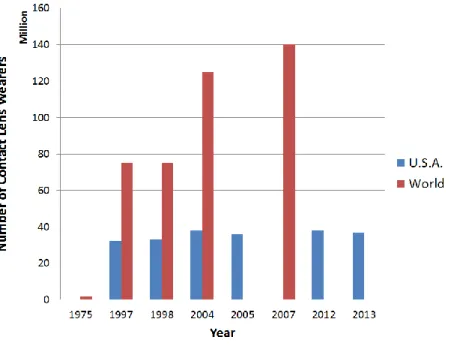
Although Leonardo da Vinci (1452-1519) is traditionally considered the inventor of the first device in contact with the eye, is to Thomas Young (1773-1829) that is attributed the first method of changing the ocular refraction in contact with the eye. Only later, around the year 1888, began to emerge scientific publications about CLs by Adolf Fick, Eugene Kalt and August Muller, showing already some consequences of its use. Almost a century later (1975) and after the appearance of soft contact lens in the 1970s, more than 2 million people worldwide were already using CLs.1 Currently,
more than 140 million people wear CLs throughout the world2 (Figure 1.1), and some perspectives
pointed to an increase to about 202 million for the last year 2010.1
Despite this great numbers, there’s still a dark side: CL wear discontinuation which is a significant problem for the clinicians and for the industry. In fact, patients continue to complain about ocular dryness and related symptoms (such as discomfort), affecting about 35 to 60% of CL users, and contributing to CL drop-out.3 There are many factors that can lead to this situation and all will be
discussed in the introduction of this thesis.
Figure 1.1. Estimated number of contact lens wearers throughout the world (red bars) and in USA (blue bars) over the years.
As is known, the last decades have been very important for CL industry, with the development and emergence of new CL materials in order to enhance wearer’s comfort and to decrease drop-out percentages. After the invention of soft lenses in the 1970s, the emergence of silicone hydrogels (SiHy) in 1998 is undoubtedly the most significant and exciting breakthrough in lens material technology, as seen in Figure 1.2. This lead to an inclusion of a significant proportion of siloxy groups which contains the element silicon directly linked to oxygen and carbon atoms, generating an increase in both oxygen permeability and hydrophobicity, worsening lens wettability.4 This material also attracts
more lipids and lipophilic proteins from tears, causing tear film destabilization.4 Although the original
intent for SiHy was for extended wear because of its enhanced oxygen permeability 5, they quickly
became available for daily wear. Daily disposable CLs have emerged in the mid-1990s and since then they have experienced constant increases (Figure 1.3). These new modality theoretically provides enhanced comfort6, decreased lens deposition and improved ocular health.7 Solomon et al 8 compared
daily disposables to conventional wear and frequent replacement CLs during a 3-year study and concluded that daily disposable was the most trouble-free option of wearing CLs, with fewer symptoms of redness and cloudy vision, fewer surface deposits and complications and better vision.
Figure 1.2 Distribution of material classes used in fittings and re-fittings throughout the years. Data from contact lens spectrum annual reports.
Figure 1.3 Distribution of replacement schedules used in fittings and refits by year. Data from contact lens spectrum annual reports.
1.1 Pre-ocular and Pre-lens Tear Film
To ensure a healthy and comfortable functioning of all ocular surface many things deserve attention. In fact, the ocular surface involves a wider concept with several structures involved. Initially, it was described as an integrated unit comprising the cornea, conjunctiva, lacrimal glands and eyelids.9 This concept was extended by Gipson 10 that said that “ocular surface includes the surface
and glandular ephitelia of the cornea, conjunctiva, lacrimal gland, accessory lacrimal glands, meibomian glands, and their apical (tears) and basal (connective tissue) matrices; the eyelashes with their associated glands of Moll and Zeiss; those components of the eyelids responsible for the blink and the nasolacrimal duct”.
The tear film and their respective glands seem to have an important role in the proper functioning of the ocular surface. The pre-ocular (POTF) or pre-lens tear film (PLTF) are the first structures that light encounters when reaching the eye, making this air-tear interface the first refractive surface responsible for focusing the light rays. That said, it can be concluded that little irregularities in this interface can affect substantially the quality of vision11 (more in section 1.1.4).
The tear film structure and stability will be briefly reviewed in this chapter, as well as the differences between an intact tear film and a tear film disrupted by a CL interphase.
1.1.1 Tear Film Characteristics
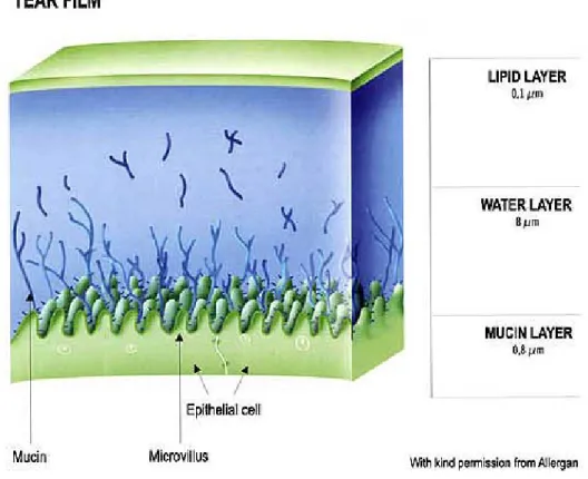
Tear film is an important optical element with vital contributions to proper visual functions. There are two models for the tear film structure. The model traditionally more accepted was enunciated by Wolf in 1946, and has been described as a three-layered liquid film with each layer deriving from a distinct origin with a thickness ranging between 7 and 11µm12 (Figure 1.4), but with differences
between studies and the technique used. In this model, the most superficial layer is the lipid film with a thickness between 0.05µm and 0.2µm (about 0.1µm), representing about 0.02% of the total POTF thickness. This layer inhibits the evaporation of the aqueous components, since it separates the exterior ambient from aqueous layer, delaying the tear break-up time. It consists of several lipids that varies between subjects and is secreted by meibomian glands at the rim of the eyelids. Lipid layer is also determining in some issues related to CL wear, because it can be significantly altered by the CL presence and cause changes in the sensation of dryness and discomfort.13;14
The intermediate and also thickest layer is the aqueous layer, with approximately 7µm, representing about 99.78% of POTF thickness. It is mostly secreted by the main lacrimal gland and can dissolve all the nutritive products, so the tear film can maintain a good function. Its major function is the hydration of ocular surface.
Finally, the mucous layer is in contact with the corneal and conjunctival epithelium. It has about 0.02µm to 0.8µm, representing about 0.2% of all POTF thickness and is secreted by globet cells of the conjunctiva. Among its functions, the decreasing surface tension and the increase in surface energy of the corneal epithelium and conjunctiva can be highlighted, so the TF can be spread over these surfaces. 13
Figure 1.4 Schematic representation of tear film structure in three-layers and its’ respective thickness. (http://www.lea-test.fi/en/eyes/images/pict7b.jpg).
Recent findings in tear film research suggest that there might be no sharp boundaries between the aqueous and mucin layers. Until recently, it was thought that there were insoluble mucins in the first layer, hence this can be considered a separate layer.15;16 However, today’s knowledge seems to ensure
that the mucins secreted are soluble and disperse into the aqueous layer, making the tear film a two-layered structure. 15;16
Despite these findings, tear film layers continue to have a specific and particular function and the overall good functioning of the tear film depends on the contribution of the smooth functioning of all the layers separately, by means of good and balanced quantity and quality of all structures. Knowing the multiple function of POTF can help us to better understand many problems, namely those who are related to the CL wear discontinuation, as CL wearers have more ocular symptoms than non-wearers.17 The POTF has an optical function, maintaining a homogenous surface between the air and
the anterior eye surface, coating small corneal irregularities. This can prevent light scattering and blurred vision.18 POTF also promotes a smooth contact between the conjunctiva and eyelids/ocular
globe, lubricating this surfaces and allowing a tolerable CL use. Drying of the eye can lead to discomfort, epithelial erosions and even ulcerations.19 The antimicrobial protection function can
prevent ocular infections, because of the immunological defense carried out by proteins, antibodies, phagocytic cells and others. Tear film also has a physical protection as the superficial lipids repel dust particles and some types of bacteria.20 The POTF has also a nutritive function, allowing the transition
of oxygen, glycose, minerals, amino acids, vitamins and others, into the corneal epithelium. In other way the cornea won’t receive these nourishing components because of its avascular tissue. There are some metabolic products derived from cornea, such as carbon dioxide and lactate that must be removed from ocular surface. The POTF takes care of eliminating them, and also does limit the passage of contaminant substances from the environment to the ocular surface, acting as a cleaning function. 13
However, all these functions only act in a normal tear film. The absence or lack of tears can cause the augment of debris in ocular surface, with discomfort, decreased quality of vision, weakness of corneal and conjunctival epithelia and increased risk of infection.13
The CL insertion interrupts the normal functioning of the tear film. In the next sub-section, the modifications in tear film caused by CL wear will be discussed.
1.1.2
Contact Lens Interactions with Tear Film
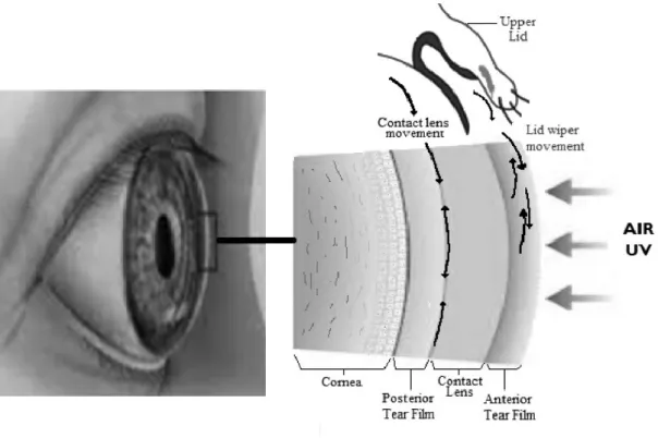
A known cause of tear film destabilization is the CLs’ presence. When placed on the eye, CLs divide the tear film in two layers: the pre-lens tear film (PLTF) and post-lens tear film (PoLTF) (Figure 1.5). The PLTF provides a regular surface to the lens for an adequate interaction with the eyelids and offer a good refraction. This layer consists of a lipid layer and a reduced aqueous layer with approximately 2µm at 3 minutes after lens insertion (measured both with interferometry and ultra-high resolution OCT), and about 6µm right after lens insertion, because of reflex tearing.21
Figure 1.5 The place of contact lens and the tear film division. Image reproduced from Mann. 4
In fact, eye care professionals have observed changes in the anterior ocular surface since the first CL insertion (1888), with visible signs and symptoms.The biophysical and biochemical changes that can affect the integrity of the tear film after CLs’ insertion are many. The biochemical changes require sophisticated laboratory techniques and contemplate changes in biochemistry (lipidome, proteome, mucins and glycocalyx, and others19), changes in cellular content of tears and external components; on
the other hand the biophysical phenomena of lens-tear interactions can be directly observed using clinical techniques and contemplate a series of phenomena listed below.
Blink frequency: evidences that blinking frequency plays an important role in comfort and CL wear date back to 1971 and 1984, that already have shown that subjects with CL-related dry eye have an increased blinking frequency (from 15.5 blinks/minute to 20.3 blinks/minute).23;24 However, the
blinking frequency may be decreased in some tasks essentially related to near vision, increasing the interblink interval (IBI) and exposing the CL surface.19 In fact, the blinking rate is reduced when
reading or using a computer (4 to 8 blinks per minute) 25 and there are more incomplete blinks,
increasing the IBI and exposing the CL surface and conjunctiva, that might result in increased evaporation and a deficient spread of lipid layer over the ocular surface.
Lipid Layer: CLs divide the tear film into two layers and if the aqueous layer at PLTF becomes too thin, the lipid layer will interact directly with CLs’ surface. This can lead to formation of lipid deposits. This is particularly problematic in SiHy lenses because of non-wettable hydrophobic silicone moieties. This can lead to impaired optical quality and non-wettability of lens surface, accelerating TBUT. 26
Tear Film Stability: Tear film is seriously affected with CL use and configures an important part of this thesis. The disruption of lipid layer22;27 and reduced tear film thickness28 are some of the
consequences of CL use. Although tear thinning is significantly faster on the surface of a CL than in corneal surface29, this could not be explained by means of the thinner PLTF. In fact, it has been
proposed that “even when the PLTF and POTF are similar in thickness, the PLTF is still considerably less stable”. 12 More information of tear film stability can be found on the 1.1.3 section of the present
thesis.
Tear Film Evaporation: The normal tear film is lost from the ocular surface by evaporation, absorption and drainage.19 The CL presence increase the rate of tear film evaporation,30 by means of
lipid layer disruption that lead to a more exposure of aqueous layer. These increased evaporation rates can lead to dryness and discomfort symptoms.19 There are some evidences that CLs increase
tear evaporation by 1.2x to 2.6x compared to non-lens wearing, with no relation to lens material or water content.31 However, a recent study conducted by Kojima et al 32 found significant increases in
evaporation rate in hydrogel wearers but not in silicone hydrogel wearers, and a relationship between this evaporation and discomfort.
Tear Film Temperature: The normal tear film temperature is in the order of 32-36oC.33 When a
CL is placed on the eye, the temperature of the PLTF becomes colder34 and the PoLTF becomes
higher, when compared to the non-CL wearing eye.35 High water content materials have lower lens
surface temperature than low water contents.34
Tear Film Thickness: Tear Film Thickness is altered with CL insertion once it divides the tear film into two layers. The PLTF is about 2µm but can be altered by the instillation of eye-drops, although transiently.21 PoLTF maintains its thickness after the instillation of artificial tears.21
Tear Production/Turnover: Technically, it’s difficult to measure tear production. Some studies (the primaries) failed to encounter differences in tear production between CL wearers and
non-CL use.36 There is some evidences on the existing studies that decreased tear meniscus volumes are
related to ocular discomfort at the end of the day.37
Tear Film Profile at the edge of a soft CL: This tear meniscus seems to change with CL insertion. It is smaller in soft CL edge when compared to hard lens edge and augment when artificial tears are instilled.38
Tear Exchange: Tear exchange can be regulated by lens diameter and movement, the blink and tear replenishment rate. It seems to be affect by lens diameter, with lesser exchange the bigger the diameter. 39
Osmolarity: Osmolarity is a clinical and objective measurement that indicates the balance between tear production and their elimination (evaporation, drainage and absorption). A balanced tear production and elimination is important for tear film stability and maintain a normal osmolarity.40 CL
wear changes tear osmolarity, showing a possible role in the reduction of lens movement and increase contact lens adherence.40 The CL insertion leads to a reduction on tear osmolarity because of reflex
tearing with a subsequent increase.41 This increase can occur because of the reduced tear production
(reduced corneal sensitivity) and because of excessive evaporation caused by a disrupted tear film and consequent reduced tear film stability.42
Ferning: Tear ferning is an indicator of tear functionality. Abnormal tear functionality by means of significant increased tear ferning can be seen in contact lens wearers.43
pH: The pH of a normal tear film is about 6.5 – 7.8, and is more acid in CL wearers with decreases of about 0.27 and 0.53 pH units.44 This decrease is attributed to the lens preventing CO2
loss from the eye. 44
Viscosity: The effect of CL wear on tear viscosity is still unknown; however there are reports of differences in this parameter in dry eye disease.45
Surface Tension: The surface tension also plays an important role in tear film stability, with less stability the bigger the surface tension.46 There are no studies contemplating the changes of CL
wear in surface tension.19
All these parameters mentioned are tear biophysical changes. When biophysical and biochemistry are seen together, we can highlight the removal or reduce of some components of the tear film and the augment of the tear film, stimulating the influx of new components or increasing the level of specific existing components.4 This alteration of lacrimal production has two phases. Initially, the CL
causes a hypersecretion of tears because of the mechanical stimulus. In a long-term, the CL wear cause less tear secretion because of the reduced friction between eyelids and ocular surface and the reduction in corneal sensitivity because of the nervous hyper-stimulation caused by the CL (more notice in rigid gas permeable (RPG) lens).13
Among the different components that can affect the lens-tear interactions, we can highlight the properties of the lens: ionicity, water content, moduli, and surface properties, and the characteristics of the individual wearer and the wear schedule.4 In addition, the comonomers, manufacturing process,
rate of deposition of the tear film components and the wearers’ tear film must be taken into account, once all are related to the wetting nature of the lens material.47 Although we can change some of the
mechanisms mentioned above, the patients’ tear film is a non-modifiable factor and it seems to be a major determinant of successful CL wear.48
There are many studies that concluded that CL wear can lead to different and various changes in the structure of ocular adnexa. Nichols et al 49 have concluded that CL wear could damage and change
the structure of the meibomian glands, changing their production. This can lead to alterations in the lipid layer thickness, tear film instability, increased tear osmolality and dehydration of hydrogel lenses.
To mitigate these effects and for a perfect biocompatibility with the eye, the lens should be completely surrounded by tears, having two tear film layers both in front of the eye (PoLTF) and after CL (PLTF), mentioned before.13
1.1.3
Tear Film Stability
Many factors can affect tear film stability. A known and already mentioned factor is the CL presence. Also, tear film stability is not constant throughout the day, with decreased values of TBUT
components, caused by ocular/palpebral surgery.55 Although these known changes, it’s important to
have into account the environmental conditions such as temperature, humidity, air conditioning and pollution, and others.55
Young and Efron56 demonstrated that tear break-up occurred within 3-10s on the front of hydrogel
lenses. Also, they found longer TBUT in high water content CLs, which is consistent with the thicker aqueous layer proportioned by this lenses. Other authors concluded that tears begin to break-up within 2-3s on the front surface of a rigid permeable CL and 5-6s on soft contact lens.28
1.1.3.1
Measuring Tear Film Stability
As we can see by the analysis in the last section, the tear film plays an important role mainly when CL wear is under discussion. Nowadays there are many ways to measure it quantity, stability, and osmolality. In this section the methods for measuring the tear film stability will be reviewed.
Many methods have been developed for measuring tear film stability. In reality, we measure the tear film instability to assess its stability. Some of the principal methods and techniques are listed above.
Tear Break-up Time (TBUT): TBUT was first described by Norm in 1969.57 Since then, TBUT
have been the most frequently test used for evaluate tear film stability.58 In testing TBUT, sodium
fluorescein is instilled into the tear film by means of a sterile strip or a pipette. The patient is instructed to do a complete blink and then avoid blinking for a period of time. The examiner is observing the tear film through a biomicroscope with a cobalt blue light and a wratten #12 yellow filter55, and reports the time (in seconds) between the last complete blink and the appearance of the
first break, dry spot or discontinuity on the tear film. The longer it takes, the more stable the tear film. Tear film is considered abnormal if TBUT is less than 10s59; if TBUT values are between 5-10s they
are considered marginal and if less than 5s is indicative of dry eye syndrome.60 Table 1.1 shows the
The location of the spot of the first break can be also analyzed. In healthy subjects, TBUT occurs most frequently in the inferior or central corneal quadrants and less frequently in the superior quadrant.61;62
Although its’ wide use, TBUT is known by its’ poor reproducibility since the 70s63 In fact, it can be
affected by many factors such as the observer experience, incomplete blinks, illumination techniques,64 and by the characteristics of fluorescein instilled.65 TBUT is dependent on the volume of
fluorescein solution instilled before measurement as well as its’ concentration, pH, and type of fluorescein used. Some studies show an improved repeatability and reproducibility with less volume of fluorescein instilled.66;67 In 1998, Cho et al68 observed that the first measure was always significantly
different from the second and third. Because of this, is recommended to do multiple measures and then take the mean of them.
An application of TBUT, also with the fluorescein instillation, is the tear film break-up dynamics (TBUD). This technique videotapes changes in fluorescein pattern after the first break in the tear film.69
The images are converted in greyscale and analyzed with MATLAB. This allows the assessment of the total area of TBUT and the maximum IBI. Although this technique is highly correlated to patients’ symptoms when compared to TBUT, it also allows the detection of distinct break-up patterns: amorphous (26%), linear (22% - most frequently associated with dry eye), spot (20%), fractured (20%) and wispy (12%).70
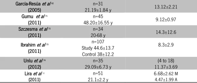
Table 1.1 Summary of results of recent studies evaluating the TBUT.
Author/ Year Sample/ Age TBUT (s)
Mean±SD García-Resúa et al 62 (2005) n=31 21.19±1.84 y 13.12±2.21 Gumu et al 71 (2011) 48.20±16.55 y n=45 9.12±0.97 Szczesma et al 72 (2011) 20-68 y n=34 14.3±12.6 Ibrahim et al 73 (2011) n=107 Study 44.6±13.7 Control 38±12.2 8.3±2.9 Unlu et al 74 (2012) 29.09±6.73 y n=35 11.37±3.69 (4 to 18) Lira et al 51 (2011) n=51 21.1±2.2 y 6.68±2.62 M 4.47±1.99 A
M, Morning; A, Afternoon; S, Seconds;
Non-Invasive Break-Up Time (NIBUT): According to Szczesna and Iskander75 a method is
considered non-invasive if there is no instillation of fluorescein, natural blinking, no contact between the instrument and the eye or adnexa, and the methodology must not alter the normal ocular environment. So, non-invasive techniques basically focus on observing a reflected grid pattern on the ocular surface. NIBUT is the time (in seconds) between the last blink and the appearance of the first irregularity/dry spot on the reflected target.76-78 Thus, NIBUT can be evaluated using the reflected mires
of keratometers, topographers and other approaches, like tearscope (will be explained in: Interferometry of lipid layer) or other custom made techniques based on the same basis. In 1989 Hirji et al79 added a fine grip to the keratometer and concluded that the detection of tear break-up was
easier with this technique and recommended the use of the mean of five measurements for a great reproducibility. Others have joined a hemispherical bowl to the biomicroscope to assess the entire cornea for better measurements of NIBUT.80
Although its inter-observer differences,55 NIBUT is generally longer than TBUT and they are poorly
correlated.81 One explanation could be the tear film destabilization caused by the instillation of
fluorescein for the TBUT measures. Nevertheless, this effect of fluorescein on tear stability is not well known yet.
Table 1.2 Summary of results of studies evaluating NIBUT.
Author/Year Sample/Age NIBUT (sec.) Mean±SD Method
Fonn et al 82 (1999) n=20 AS n=20 S 8.6 AS (0h) 8.7 AS (5h) 9.5 S (0h) 6.5 S (5h) PLTF Slit-lamp biomicroscope Glasson et al (2006) n=11 AS 25-39 y n=9 S 23 – 40 y 21.3±5.7 AS (0h) 13.7±4.3 AS (5h) 13.7±2.8 S (0h) 12.7±4.6 S (6h)
Costum made tearscope + slit lamp
Glasson et al 48
(2003) n=20 AS n=18 S
20±5.6 AS 13.2±3.2 S
Costum made tearscope + slitlamp García-Resúa et al62 (2005) 21.19±1.84 y n=31 AS 17.50±3.06 AS TearScope PCTF Lira et al51 (2011) n=51 NCLW 21.1±2.2 y 6.58±2.62 (Morn.) 5.38±2.53 (Aftern.) PCTF Helmholtz Nichols et al49 (2006) n=161 AS n=199 S 11.03±8.63 AS 8.23±5.67 S Interferometry Guillon et al83 (1997) n=55 NCLW n=184 CLW 16.9±13.5 NCLW 15.3±13.1 CLW PCTF TearScope
Studies using non-invasive techniques have found shorter tear film break-up times (TFBUTs) on SiHy lenses compared with hydrogel lenses.84;85 As seen in Table 1.2, Fonn et al82 conducted a study
which aimed to compare the pre-lens NIBUT values between symptomatic and asymptomatic CL wearers during the day. Before 5h of CL use, they found a decreased NIBUT values, mainly in symptomatic group. Guillon et al found a value of 14.7±12s for symptomatic and 15.7±14s for asymptomatic, with p=0.014.
Interferometry of lipid layer: This technique is used for asses tear lipid layer thickness and more recently for NIBUT, by observing interference patterns generated by the light reflected from the surface of the lipid layer and from the interface between that layer and aqueous layer of the tear film. Recent approaches and modifications led to the development of TearScope Plus, a non-invasive instrument capable of measure NIBUT by means of specular reflection and a flexible grid.86 Its’ cold
light source decreases the possible reflex tearing caused by high intensity light sources.
Topographical analysis systems. Videokeratoscopy: Tear film stability can be evaluated through the analysis of some topographic indices as the surface regularity index (SRI) and surface asymmetry index (SAI)87;88. The topographer can capture several images for some seconds. Thus, the
time that the tear film takes to build-up and reach its’ more regular state can be taken (3 to 10s tear film buil-up time).89
All of these as resulted in a commercially available non-invasive and objective method: the Tear Film Stability Analysis Software (TSAS).90-92 This software automatically capture consecutive corneal
surface images every second for a 10 seconds examination routine and later determine the tear stability by analyzing the changes in corneal topography over time (SAI and SRI).
Other studies used videokeratoscopy to assess the tear film surface quality (TFSQ) of different soft CLs. They are based on the assumption that the quality of the ring reflection is associated to the quality of tear film surface93 and can differentiate between lenses type/material.94 Others have used
the SAI and SRI indexes to characterize the corneal surface of CL wearers and no-wearers but not to assess their tear film stability.95
Confocal microscopy: Despite the high costs, confocal microscopy is a high-resolution, three-dimensional tool that allows the measurement of morphological changes present in TBUT phenomena and better understanding the underlying mechanisms.96 Other variants allow the observation of
Visual Acuity Testing: Tsubota and colleagues don’t found differences between the best corrected VA between normal and dry eye subjects.97 However, dry eye patients continue to report
reduced VA, mainly while reading, driving and watching TV.98 These are visually demanding tasks that
require attention: and it’s known that this lead to a reduced IBI and more incomplete blinks. So, the reported loss of VA can be explained by tear instability caused by deficient blinks.55
Functional Visual Acuity: The concept of Functional Visual Acuity arises because of the anteriorly mentioned problems in dry eye patients referring decreased VA despite normal conventional VA measure. This consists in measuring VA during and after a period of volunteering sustained eye opening, which is more representative of real-life activities. Despite all the critics that this technique has received because of the time that the patient needs to be with the eye open, it’s a widely used method for assessing visual disturbances in dry eye patients.99 In 2005, Ishida et al100 developed a
device that allows continuous monocular VA measurements during 30s without blinking.
Wavefront Aberrometry: Wavefront aberrometry is a non-invasive technique that can assess the tear film stability. The non-uniform tear thickness caused by tear break-up lead to additional corneal and high-order aberrations.101 However, the real contribution of tear-film in these aberrations is
still unknown due of the accommodation microfluctuations.75 More information about the utility of
1.1.4
Tear film and Optical Quality with Contact Lenses.
Despite the famous advances in CL industry over the years, little has been done to improve the quality of vision in soft CL wearers, occasionally inferior to spectacles and RPG. The quality of vision of CL wearers is influenced by several factors that include the aberrations induced by the eye optics and CL optical properties, the interactions between eye and CL (cornea and tear film) and the manufacture process, material, water content and optical design.102 As Montes-Micó said, “…the optical quality of
the human eye is dynamic and is affected by the tear film, in addition to accommodation, age, gaze, lens, vitreous cavity, keratometry, and pupil size.”103
As previously said, little irregularities in the air-tear film interface can affect substantially the quality of vision, as this is the first structure that light encounters when reaching the eye11 and is the most
powerful optical surface once is associated to the largest changes in refractive index (step between air and tear film).104 If the tear film remains uniform in thickness, the cornea/tear combination will have
almost the same power as the cornea alone: if we consider that the tear film thickness can reach the 20μm12, and if the tear film only changes its surface radius (uniform thickness), the maximum power increase will be about 0.10 D (because it can only change up to 20 µm). Considering only this example, where the tear film remains uniformly thick, we can assume as true the common belief that the PCTF has little optical impact. On the other hand, and has been demonstrated by other studies64;80,
the tear film does not remain uniform in thickness between blinks (tear break-up), occurring local variations that will introduce aberrations into the optical system.11;105 Notwithstanding, if the tear breaks
totally, the irregular corneal surface will be exposed, which may increase optical scatter.105 Concluding,
tear film disruption can cause optical changes that contribute to the reduction in retinal image quality and consequently in visual function.106 So, a smooth and regular tear film is important to have
1.1.4.1
Wavefront Aberrometry for assessing tear film
Many techniques such as wavefront sensing, double-pass optical method, videokeratoscopy, interferometry and retroilumination analysis have been developed to quantify the role of the tear film in optical quality. The wavefront aberrometry is the objective technique most used that allows monitoring the through-time evolutions: and has been called the most useful technique to evaluate the optical quality of CL-eye. 107;108 Defocus and astigmatism are low order aberrations (LOA) and are known
as the main responsible for decrease vision. But there also are high order aberrations (HOA), as spherical aberration (0.1±0.1µm for a 6mm-pupil)109 that is the HOA that affects the image quality the
most.110 However, we have to take into account that under the light of previous studies, differences in
spherical aberration between the lens-eye combinations will be the difference in spherical aberration between the lenses themselves.111
In normal eyes, after a trends towards reducing right before a blink (tear build up), there is a gradual increase in optical aberrations a few seconds before (Figure 1.6), which lead to a progressive reduction of the optical quality of the eye (mean increase 21%±8%).104 The aberrations seem to be
lowest approximately after 6 seconds after a blink.103 So, if the IBI is of about 4 seconds is unlikely that
the changes in aberrations will produce detectable effects on vision. These changes in optical aberrations are associated to irregularities in tear film, namely caused by break-up. Nevertheless, the microflutuations of accommodation deserve attention, as well as the age, gaze, lens, vitreous cavity, keratometry, and pupil size which may contribute to changes in dynamics of optical quality of the human eye.103
In their introduction to this topic, Montés-Mico et al102 referred a few studies112;113 that intent to
assess the optical quality of ex vivo CL, stating that these data could not be enough to predict the optical performance of CL when they are in an in vivo ambient. When the CL is placed on the eye, multiple interactions may occur. In fact, the non-CL wear has no diurnal variations in wavefront aberrations when compared to the joint CL + eye, which has some diurnal variations in optical quality.102 They attributed these changes to the optical properties of the CL and to its interactions with
Hong et al 114 demonstrated that wavefront aberrations have significantly higher values in patients
wearing hydrophilic CL or glasses, compared to RPG wearers. These results are justified by the reduction of asymmetrical aberrations and positive spherical aberrations caused by gas permeable CLs. Using a double-pass method, Albarran et al11 have observed a significant reduction in image
quality after tear break-up with and without soft CLs, being these reduction greater when soft CLs were worn.
Dry-eye patients have increased optical aberration values by a 2.5 factor when compared to normal eyes, namely in spherical aberration. These values are caused by tear-film irregularities in ocular surface, with larger vertical coma values than horizontal coma values.101 The mentioned changes in
spherical aberration may be due to the tendency of tear film thin at a different rate in the center of the cornea and at its periphery, with a thinner central tear film inducing more positive spherical aberrations, in both normal and dry eye patients.103 The instillation of artificial tears seems to improve
the optical quality in patients with dry eye.115
Figure 1.6 Change in corneal wavefront aberrations in different times after a blink (1 to 15 sec after blink). Image reproduced from Montés-Micó et al (2007), 103 who referred that, for this image “Contour
line step, 1µm; pupil diameter, 7mm; Only high order aberrations (3th to 6th) are shown; Piston prism
defocus, and astigmatism have been compensated by canceling the corresponding 1st and 2nd- order
Although the utility of these measures is arguably necessary, increasingly subjective measures should be included in research protocols, where the patients have the principal role. It is important to meet patients’ problems and understand if the objective data can be comparable to the patient's comfort and vision (more in sections 1.2 and 1.3).
1.2 Discomfort with Contact Lenses
A comfortable CL wear is not always possible. Is a very common situation subjects without signs or symptoms of dry eye suffering of CL discomfort (CLD). For many years there has been lack of consensus about this term and until a short time ago there was no approved or agreed definition for CLD. In the “International Workshop on Contact Lens Discomfort”, published in 2013, the “Definition and Classification” subcommittee created a definition for this problem: “the contact lens discomfort (CLD) is a condition characterized by episodic or persistent adverse ocular sensations related to lens wear either with or without visual disturbance, resulting from reduced compatibility between the contact lens and the ocular environment, which can lead to decreased wearing time and discontinuation of contact lens wear”.116 As shown in Table 1.6, CLD seems to be more prevalent in
females and cover between 28 and 50% of CL wearers.117;118 Discomfort is the CL related factor which
further lead more CL drop-outs (between 43 and 72%).119
CL wearers have some biophysical changes in common with dry eye disease patients, being some subclinical indicators of dry eye amplified when CL is placed on eye.120;121 This discomfort can be due to
CL inherent factors such as the design (edge, base curve), material (lubricity, water content, wettability), fit and wear (modality and lens interactions) and lens care (care solutions). There are also factors related to the environment, such as factors inherent to the patient (age and gender), modifiable factors (medications), ocular environment (blink, tear stability) and external environment (humidity, air quality).116
The comfortable wearing time is an important clinical consideration. This situation refers to the time that the patient can wear the lens with comfort, being the “comfort” term used for the “no-lens wear” feeling. Most clinicians and scientists use this term to determine if the lens is compatible with the eye. Thus, this includes:
o The ability to wear lens without sensation (lack of awareness); o Maintain visual acuity;
o Have complete tolerance, including the ability to wear lenses as long as desired without problem.
So, this leads to other important term commonly reported by CL wear patients: the end of the day discomfort. This can occur with any lens material type, but is often associated with soft contact lens, both conventional and silicone hydrogel, as they are the CLs more used nowadays.116 There are many
clinical signs to diagnose CLD, though they are poorly correlated with patients’ symptoms.122 They
include assessments of pre-lens tear film48, meibomian glans, hyperemia123, and staining.124 Young et al 125 contended that poor lens wettability and decreased TBUTs and NIBUTs are seen in around 40% of
symptomatic patients.
As said before, the destabilization caused by CL presence is a known factor to take into account; however, there are other factors that deserve interest, as the unknown effect of long-term CL wear and if the effects persist once the lens has been removed for a long time.83 All these changes were already
known in 1986, when McMonnies concluded that dry eye symptoms are more frequent in CL wearers than in non-wearers, stating that the CL wear is a provocative condition for dry eye.123 In an
epidemiologic study conducted in 1996 by Caffery et al 126, a prevalence between 20-30% were found
for dry eye in CL wearers. Two years later, the same authors confirmed that dry eye symptoms were more prevalent in CL wearers, with half of them (50.1%) experienced dry eye symptoms compared with the just 21.7% of non-wearers.127
Although a study have found no differences between hydrogels and Si-Hy lenses in comfort or dryness ratings,128 other found that Si-Hy lenses were more comfortable and led to less dryness
symptoms than hydrogels.129 Others have not encountered differences in comfort scored between lens
types.130 Other study conducted by Efron and colleagues compared the initial comfort (after 5min of
lens wear) of low, medium and high water content CLs and faced a significant negative correlation between lens comfort and lens water content, that is: low water content were more comfortable than high water content lenses.131
A successful CL wear is expected when the patient has a normal ocular surface, normal lid function and when the CL is compatible with eye lids, ocular surface and does not interfere adversely with tear film (excessive evaporation, surface dehydration, etc). 132
Table 1.3 Results of prevalence of CLD from Population-based studies (in a natural population setting, most preferred for epidemiological studies) and Clinical Practice/Hospital-based.
Author/ Year Sample/ Age Prevalence Study type
Doughty et al 117 (1997) n=3285 CLW 10-80 y 50.1% CLD (M + F) Population-based Uchino et al 133 (2011) n=105 CLW ≥40 yrs 28% CLD M 35% CLD F Population-based Uchino et al 118 (2008) n=1298 CLW 15-18 yrs 36.8% CLD M 37.4% CLD F Population-based Brennan et al 134 (1989) n=104 SCLW
24±9 yrs 75% Dryn Clinical Practice
Guillon et al 83
(1997)
n=184 SCLW
31±7 yrs 44% Symp Clinical Practice
Riley et al 135 (2006) n=1092 SCLW 18-42 yrs 28% Dryn 17%discomfort 31% RCWT Clinical Practice Gonzalez-Meijome et al 136 (2007) n=71 SCLW
24.9±5.5 yrs Symptoms often 24% Clinical Practice Young et al 137
(2011) <20 to >61 yrs n=932 SCLW 31% Dryn Clinical Practice CLW, Contact lens wearers; SCLW, Soft Contact lens wearers; M, Male; F, Female; Dryn, Dryness; Symp, Symptomatic; RCWT, Reduced comfortable wearing time.
1.2.1 Dehydration and its relation with discomfort
It was believed that the higher the water content, the more wettable and comfortable the lens were.138 Conversely, quickly it turn into light that high water content CLs suffer more dehydration.139
Dehydration can be assessed with in vitro and ex vivo methods, with in vitro dehydration being limited in predicting on-eye dehydration. In order to equilibrate the hydration in the new environment, CLs begins to dehydrate as soon as they are placed on eye, and continues during the day with greater or lesser intensity.140 In hydrogel lenses, dehydration results in loss of lens mass and volume, affecting
lens size and shape (decreased diameter, steeper base curve, and decreased lens movement after insertion) and affecting their clinical performance.141 Also, the water content changes overtime can lead
to some impact in oxygen transmissibility, namely in hydrogel lenses. This effect has less impact in silicone hydrogels, once their oxygen performance is less dependent in water content.142 Dehydration is
also influenced by several factors such as characteristics of the material, thickness, palpebral aperture, blink rate, tear film quality and environmental conditions.143 Despite this assumption, a study
that investigated the CL dehydration in controlled environmental conditions concluded that lens dehydration was unaffected by extreme environment conditions, stating that the eye may compensate this situation by increasing the blink rate and tear production.144
The TFOS International Workshop on Contact Lens Discomfort also focused on the “Dehydration” topic, concluding that “…considering the body of literature available (…) it is not likely that a causative or associative relation exists between on-eye bulk dehydration of materials and discomfort using the current methods used to capture either dehydration or subjective comfort”.138 As said before, the
major complaints reported by CL wearers are dryness and discomfort, being them the main factors for CL discontinuation.145 This led to a relationship between dehydration and discomfort, mainly at the end
of the day, although the literature is equivocal in this area.146 In the Contact Lens Materials, Design
and Care of the TFOS Workshop, they justified this connection between dehydration and discomfort by “… 1) the potential correlation between lens thickness and desiccation staining, 2) the potential correlation between corneal staining and discomfort, and 3) the increased friction presumably induced by dehydrated, dry lens surfaces.”
In the 80s, the high water content and thin lenses were already perceived as the greatest cause of lens dehydration.147-149 In 1988, Efron and Brennan150 completed these findings, showing that the lenses
of patients who refer less dry symptoms were high water content CLs, and the lenses of those who often experienced dryness had less water content. Other study that intended to correlate CL dehydration and discomfort, dryness and NIBUT in asymptomatic and symptomatic patients failed to correlate dehydration with subjective sensation of comfort and dryness.82 Although Etafilcon A
dehydrated less after 7h of CL wear in asymptomatic 3.9±2.3) when compared to symptomatic (-4.6±2.5), the same does not happened in Omafilcon A, with a greater water content loss in asymptomatic (-1.8±1.2 and -1.6±0.8, respectively). Other studies have failed to show this association.151.144 Although these findings, a significant negative correlation was found in another study
1.3 Clinical Performance of Soft Contact Lenses
Tear and vision related techniques are important, but the contact lens interactions with the eye have also great significance. Ocular and clinical performance can be dependent of material and surface properties of the lens and the design-material interactions with the eye. The clinical performance of SCL can be evaluated by slit lamp examinations and subjective assessments such as anamnesis and questionnaires.
1.3.1 Subjective Assessment of Comfort
The subjective assessment of CLs has demonstrated to be very important as the current clinical tests are not good at predicting CL wearers’ symptoms. Thus, patient-reported outcomes (PRO) are gaining greater importance.153 This comprises the specific questionnaires that can measure and
diagnose dry eye symptoms and can reflect patient’s viewpoints. To determine subject’s dry eye symptoms, many questionnaires are available:
- Dry Eye Questionnaire (DEQ); 154
- Contact Lens Dry Eye Questionnaire (CLDEQ), the only exclusively designed for CL wearers; 154
- Ocular Surface Disease Index (OSDI); 155
- McMonnies Dry Eye Index; 156
- Frequency of Dryness Score, or Subjective Evaluation of Symptoms of Dryness (SESoD);
157
- Impact of Dry Eye on Everyday Life Questionnaire (IDEEL). 158
There are other questionnaires or PRO. A review conducted in 2013 found 121 PRO instruments in ophthalmic area, and constructed a top-8 higher-quality PRO, being in second the OCI (Ocular Comfort Index) that is similar to OSDI, for ocular surface symptoms assessment and its severity in dry eye and ocular surface disease. A study conducted by Michel et al 159 compared the OCI and McMonnies for
authors for use in its original version because it violated the condition of unidimensiolality”. Despite this, OSDI is notable among other similar questionnaires for having undergone psychometric testing and having been accepted by the US FDA as an outcome measure for use in dry eye trials.155 But,
once again, it is criticized because the inconstant step of difficulty between categories and by the no-comparable nature of the difficulty of all questions, which may result in a no-linearly scale related to symptom severity.160
In 2005, Guillon et al 161 applied the McMonnies questionnaire to ascertain the best questions that
could be done to patients to know their symptoms, and concluded that the more predictive question for the detection of dry eye was frequency of ocular dryness instead of scratchiness; burning symptoms and sensitivity to cigarette and make-up products were lesser important.
1.3.2 Biomicroscopy
The slit-lamp evaluation with the proper use of clinical grading scales is important to assess the response of the eye to new CL materials, modalities or through-time changes. The eye can respond to CL wear by different forms. One of them comprises the engorging of limbal vasculature known has limbal redness.162 This is considered a local response and is not affected by hypoxia at the central
cornea, as soft lens wearers show a greater limbal injection than hard lens wearers, which have a vasculature similar to non-wearers.163 This is a situation to be considered, since the chronic vessel
dilation in soft CL wearers can lead to new vessel growth,163 causing neovascularization namely in low
Dk CL wearers.164 More recent studies have found no differences in limbal redness between high Dk/t
Si-Hy wearers and no-lens wear,165 but there are differences between them and low Dk/t Si-Hy
lenses.166
Other common clinical sign is bulbar hyperemia. This answer is also noticed in asymptomatic soft and rigid CL wearers,84 but the changes in this parameter have not been significant over a 10 month
period in soft CL wear.167 These changes can be due to the damage that CL wear causes to the
conjunctival vasculature by direct vaso-occlusion, once there are more conjunctival abnormalities in CL wearers than in non-wearers, with increased vessel diameter and contour over the lens edge.168
Other sign is the conjunctival staining. In CL wearers is often seen 2-mm from the limbus, which is the soft CL edge,169 and is thought to happen because of CL movements and changes in the tear film
at the lens edge.84 Conjunctival staining is most often encountered in CL wearers than in non-wearers,
namely in Si-Hy84 and symptomatic wearers and non-wearers,124 so it can be correlated to some ocular
symptoms.170 These reactions are indicative of conjunctival damage caused by CL edges and by the
evaporation caused by destabilization of the tear film,124 but also include changes in lens parameters,
CL modulus or poor lens fit.171
Corneal staining can occur in CL wearers but also in non-wearers.172 The corneal staining may be
due to several factors, such as mechanical effects (poor lens quality, such as rough edge), inflammatory, exposure, metabolic, toxic, allergic, and infectious.19 Nichols and colleagues have
concluded that there are many factors related to increased corneal staining, such as larger daily wear times, CL deposition, increased tear meniscus height and decreased hydrogel water content, and concluded that the use of Si-Hy lenses improved the corneal staining value.173
A recent study concluded that daily disposable Si-Hy CL have different ocular responses, maybe because of lens materials, surface properties, designs and packaging solutions.166 For example
Etafilcon A exhibited statistically higher limbal redness grades when compared to the other daily-disposable CLs used, and Senofilcon A the higher corneal staining extent and type.166 A study who
hypothesized that daily wear could improve CL related-symptoms in symptomatic CL wearers, found significant improvements in signs of limbal redness, bulbar redness and conjunctival staining at 2 and 4 weeks of daily disposables wear when compared to habitual CLs.6