UNIVERSIDADE FEDERAL DE SÃO CARLOS CENTRO DE CIÊNCIAS BIOLÓGICAS E DA SAÚDE PROGRAMA DE PÓS-GRADUAÇÃO EM FISIOTERAPIA
TESE DE DOUTORADO
Análise da cadeia cinética em atletas arremessadores com e sem
dor no ombro
Lívia Silveira Pogetti
UNIVERSIDADE FEDERAL DE SÃO CARLOS CENTRO DE CIÊNCIAS BIOLÓGICAS E DA SAÚDE PROGRAMA DE PÓS-GRADUAÇÃO EM FISIOTERAPIA
Análise da cadeia cinética em atletas arremessadores com e sem
dor no ombro
Discente: Lívia Silveira Pogetti
Orientadora: Profa. Dra. Paula Rezende Camargo Co-orientadora: Profa. Dra. Theresa Helissa Nakagawa
São Carlos 2017
Dedico esta tese aos meus amados pais,
Joana
e
Carlos
, e a todos os familiares e amigos, que me
AGRADECIMENTOS
Enfim, chegou o momento tão aguardado!
Não poderia iniciar os agradecimentos sem antes agradecer a Deus pela oportunidade que
me deu de chegar até aqui. Agradecer por todos os momentos de alegria, angústia, vitória,
medo, incerteza... Todos vividos intensamente, e que com certeza valeram a pena.
Agradeço à minha orientadora Paula Rezende Camargo pela oportunidade de trabalhar
junto da sua equipe, mesmo sabendo que vinha de outra área. Agradeço pela confiança
depositada no meu trabalho, por todas as oportunidades geradas durante esta jornada, e por
todo o conhecimento ensinado. Sinto-me honrada por ter sido sua orientanda.
À Theresa Helissa Nakagawa agradeço por todo o auxílio, pela prontidão, dedicação, por compartilhar os seus conhecimentos, e principalmente, pela excelente co-orientação. Com
certeza, tudo se tornou mais simples com você. Deixo aqui registrada a minha estima e meu
respeito pelo seu trabalho!
Aos meus pais e irmãos, que estiveram e se fazem presentes em todos os momentos... Apoiando-me e aconselhando-me sempre. Com vocês tudo se torna mais fácil. A vocês
dedico esta conquista!
Aos meus familiares e amigos, agradeço pelos momentos de descontração que me
proporcionaram. Em especial, as tias Rita, Heloísa e Regina Pogetti e Leide Gasparini
À Rosana Souza que sempre me estimulou a sair da “zona de conforto”, e mais do que
isso, pela amizade de sempre e por acreditar em mim mais do que eu mesma. A você,
Rosana, a minha eterna gratidão.
Ao Rafael Salomão que me acompanhou em grande parte desta jornada, pela dedicação, cuidado, compreensão, carinho e amizade. O seu otimismo foi essencial para seguir adiante.
A você e sua família toda a minha gratidão.
À Giovanna Prado Conteçote por me escolher como co-orientadora de iniciação científica, mas, sobretudo pela parceria durante as coletas, assim como nas análises dos
dados. Você foi essencial para que esta conquista!
Aos colegas de laboratório de Avaliação e Intervenção do Complexo do Ombro: Fernanda
Habechian, Melina Haik, Rodrigo Py, Daiana Rosa, Natália Duarte, Larissa Pechincha, Danilo Kamonseki, Elisa Pires e a todos os alunos de iniciação científica que por lá passaram, agradeço por toda a ajuda e amizade durante este período. O convívio com
vocês me ensinou a admirá-los!
Aos demais colegas de departamento, como Iolanda Vilella, Jean Ferreira, Maíra
Lixandrão, Ivana Leão, Carol Castro, Mariana Santos, Carol Corsi, Natasha Toledo e
Adrianinha Santos pelos cafezinhos, confidências e momentos de descontração. Esses momentos foram fundamentais para superar as dificuldades.
A todos os professores do curso de Fisioterapia da Universidade Federal de Uberlândia
(UFU), funcionários e alunos. Em especial, os professores Marcos Kishi e Angelo Piva
Agradeço também a todos aqueles que cruzaram o meu caminho durante este período,
como Bruna Soares, Nayara Tamburús e tantos outros que fizeram, e ainda fazem parte
desta conquista.
Aos professores que aceitaram o convite para compor a minha banca de qualificação,
Catarina Oliveira de Souza, Ana Beatriz de Oliveira e Rodrigo Scattone da Silva, e os
suplentes Gisele Garcia Zanca e Fábio Viadanna Serrão. Assim como, os professores
que aceitaram o convite para compor a minha banca de defesa, Melina Nevoeiro Haik
Guilherme, Lilian Ramiro Felício, Rodrigo Scattone da Silva e Fabio Viadanna Serrão, e os suplentes Gisele Garcia Zanca, Catarina Oliveira de Souza, Ana Beatriz de Oliveira.
À Coordenação de Aperfeiçoamento de Pessoal de Nível Superior (CAPES) e à
Fundação de Amparo à Pesquisa do Estado de São Paulo (FAPESP) pelo apoio financeiro, que foi essencial para que eu conseguisse desenvolver essa pesquisa.
Por fim, agradeço a todos os voluntários que participaram deste trabalho. Em especial, à
equipe de handebol masculino da cidade de Franca-SP, e os demais participantes que se deslocaram até São Carlos. Agradeço a confiança! Sem vocês este trabalho não seria
ORAÇÃO DE AGRADECIMENTO
Agradeço-te Senhor. Pela glória de viver. Pela honra de amar!
Muito obrigada Senhor, pelo que me deste, pelo que me dás! Muito obrigada pelo pão, pelo ar, pela paz!
Muito obrigada pela beleza que meus olhos vêem no altar da natureza! Olhos que fitam o ar, a terra e o mar.
Que acompanha a ave fagueira que corre ligeira pelo céu de anil e se detém na terra verde salpicada de flores em tonalidades mil!
Muito obrigada Senhor, porque eu posso ver meu amor!
Diante de minha visão, pelos cegos, formulo uma oração: Eu sei que depois dessa lida, na outra vida, eles também enxergarão! Obrigada pelos ouvidos meus que foram dados por Deus.
Ouvidos que ouvem o tamborilar da chuva no telheiro, a melodia do vento nos ramos do salgueiro, as lágrimas que choram os olhos do mundo inteiro. Diante de minha capacidade de ouvir pelos surdos, te quero pedir, eu sei que depois desta dor, no teu reino de amor, eles também ouvirão!
Muito obrigada Senhor, pela minha voz! Mas também pela voz que cantam, que ensina, que alfabetiza, que canta uma canção e teu nome profere com sentida emoção!
Diante da minha melodia quero te rogar, pelos que sofrem de afazia, pelos que não cantam de noite e não falam de dia. Eu sei que depois desta dor, no teu reino de amor, eles também cantarão!
Muito obrigada Senhor, pelas minhas mãos!
Mas também pelas mãos que oram, que semeiam, que agasalham... Mãos de amor, mãos de caridade, de solidariedade... Mãos que apertam mãos. Mãos de poesia, de cirurgia, de sinfonia, de psicografias... Mãos que acalentam a velhice, a dor e o desamor. Mãos que acolhem ao seio do corpo, um filho alheio, sem receio...
Pelos meus pés, que me levam a andar sem reclamar. Muito obrigada Senhor, porque posso bailar!
Olho para a terra e vejo amputados, marcados, desesperados, paralisados... Eu posso andar! Oro por eles! Eu sei que depois dessa expiação, na outra reencarnação, eles também
bailarão.
Muito obrigada Senhor, pelo meu lar!
É tão maravilhoso ter um lar... Não importa se este lar é uma mansão, um bangalô, seja lá o que for! O importante é que dentro dele exista amor!
O amor de pai, de mãe, de marido e esposa, de filho, de irmão... De alguém que lhe estenda a mão, mesmo que seja o amor de um cão, pois é tão triste viver na solidão!
Mas se não tiver ninguém para amar, um teto para me acolher, uma cama para me deitar... Mesmo assim, não reclamarei, nem blasfemarei. Simplesmente direi: Obrigada Senhor, porque nasci!
Obrigada Senhor, porque creio em Ti! Pelo teu amor, obrigada Senhor!
Resumo
A dor no ombro é bastante prevalente em atletas arremessadores. A alta incidência pode ser resultado de um déficit da ação integrada da cadeia cinética durante o movimento de arremesso. São escassos os estudos que avaliaram os componentes da cadeia cinética em arremessadores com e sem dor no ombro. Estudos desse tipo são importantes para melhor compreensão dos componentes da cadeia cinética nesta população, a fim de obter melhores protocolos de tratamento e avaliação. Os objetivos da tese foram (1) avaliar o desempenho muscular da cadeia cinética de atletas arremessadores com e sem dor no ombro; (2) avaliar a função do ombro, tronco, pelve e membro inferior nesta população; e (3) analisar a correlação entre estas variáveis e o desempenho muscular nos atletas com dor no ombro. Para avaliação do desempenho muscular da cadeia cinética foram utilizados testes de resistência e de contração isométrica voluntária máxima dos músculos flexores, extensores e flexores laterais do tronco, e pico de torque isocinético dos rotadores laterais e mediais de ombro. As contrações isométricas foram avaliadas com dinamômetro manual (Lafayette Instrument Company, Lafayette, IN). O pico de torque dos rotadores laterais e medias de ombro foi avaliado no modo concêntrico à 90º/s, 180º/s e 240º/s no dinamômetro isocinético Multi-Joint System 3 (Biodex Medical System Inc., NY, USA). A função de ombro foi avaliada por meio dos questionários, nível de satisfação do ombro e amplitude de movimento. A função de quadril, tronco e membros inferiores foi avaliada pelo teste de equilíbrio de excursão em estrela (SEBT) modificado e teste de alinhamento pélvico transverso. De modo geral, os resultados mostraram que os atletas arremessadores com dor no ombro apresentaram menor tempo de resistência dos flexores laterais do tronco e equilíbrio, e pior função de ombro em comparação com os atletas sem dor no ombro. No entanto, a dor no ombro parece não afetar o desenvolvimento do pico de torque isocinético e amplitude de movimento do ombro comparado aos atletas sem dor. Além disso, foi observada uma correlação positiva de fraca a moderada entre o pico de torque isocinético de rotadores laterais e mediais de ombro com resistência e força isométrica dos músculos flexores do tronco. O pico de torque não se correlacionou com o alinhamento da pelve. Os resultados gerais suportam a teoria da cadeia cinética baseada na interdependência dos segmentos para gerar e transferir as forças durante o movimento de arremesso. Além de sugerir que exercícios que trabalhem o controle neuromuscular de membro inferior e força isométrica e resistência, particularmente para os músculos flexores e flexores laterais do tronco, devem ser estimulados nos atletas jogando com dor no ombro como parte do plano de reabilitação.
Abstract
Shoulder pain is quite prevalent in throwing athletes. The high incidence may be the result of a deficit of the integrated action of the kinetic chain during the throwing movement. There is lack of studies that have evaluated the components of the kinetic chain in throwers with and without shoulder pain. Researches in this area are important since it could provide knowledge about the kinetic chain components in this population, in order to obtain better treatment and evaluation protocols. In this way, the purposes of the thesis were (1) to evaluate the muscular performance of the kinetic chain of throwing athletes with and without pain in the shoulder; (2) to evaluate the function of the shoulder, trunk, pelvis and lower limb in this population; and (3) to analyze the correlation between these variables and the muscular performance in athletes with shoulder pain. Endurance time test and maximum voluntary isometric contraction were used to evaluate the trunk flexors, extensors and lateral flexors muscles. Isometric contraction was measured by handheld dynamometer (Lafayette Instrument Company, Lafayette, IN). Isokinetic dynamometer Biodex Multi-Joint System 3 (Biodex Medical System Inc., NY, USA) in the concentric mode was used to evaluated peak torque of external and internal rotation of the shoulder at 90º/s, 180º/s and 240º/s. Questionnaires, shoulder satisfaction level and range of motion were used to measure the shoulder function, while the modified star excursion balance test (SEBT) and transverse pelvic alignment test were used to assess the hip, trunk and lower limb function. In general, the results showed that throwing athletes with shoulder pain presented shorter endurance time of the trunk lateral flexors and balance, and worse shoulder function compared to asymptomatic athletes. However, shoulder pain does not seem to affect the development of shoulder isokinetic peak torque and ROM compared to athletes without shoulder pain. Furthermore, the results showed the weak to moderate positive correlations between internal and external rotation of the arm and endurance and isometric strength of the trunk flexors. Peak torque of the shoulder was not correlated to transverse pelvic alignment. Such results support the kinetic chain theory based on the interdependence of segments to generate and transfer of forces during functional movements. In addition to suggesting that exercises that work neuromuscular control of the lower limb and isometric strength and endurance, particularly for the trunk lateral flexor and flexor muscles should be stimulated in throwers with shoulder pain as part of the rehabilitation plan.
Lista de tabelas
Manuscrito I
Tabela 1. Características demográficas dos atletas. 34 Tabela 2. Dados do tempo de resistência do “core” (s) dos atletas em cada posição. 35 Tabela 3. Dados normalizados do teste modificado de equilíbrio de excursão em
estrela dos atletas. 36
Tabela 4. Dados do pico de torque isocinético do ombro (Nm/kg·m) do atletas em
cada velocidade. 37
Manuscrito II
Tabela 1. Características demográficas dos atletas. 47
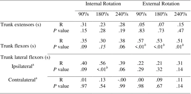
Tabela 2. Correlação entre o tempo de resistência do “core” e pico de torque durante
a rotação isocinética do ombro. 54
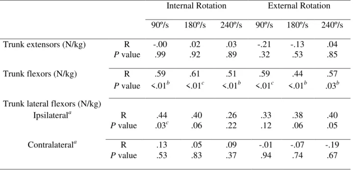
Tabela 3. Correlação entre contração isométrica voluntária máxima e pico de torque
durante a rotação isocinética de ombro. 55
Tabela 4. Correlação entre o alinhamento pélvico transverso e pico de torque
Lista de figuras
Manuscrito I
Figura 1. Participante posicionado para o teste de resistência de (A) extensores, (B)
flexores e (C) flexores laterais. 29
Figura 2. Teste modificado de equilíbrio em excursão de estrela para as direções (A)
anterior, (B) posteromedial e (C) posterolateral em orientação a perna de apoio. 30
Figura 3. Participante posicionado para avaliação, com o braço no final da
amplitude de movimento (90º de rotação lateral de ombro). 32
Manuscrito II
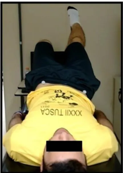
Figura 1. Participante posicionado para o teste de resistência de (A) extensores, (B)
flexores e (C) flexores laterais. 29
Figura 2. Participante posicionado para o teste de contração isométrica voluntária
máxima de (A) extensores, (B) flexores e (C) flexores laterais de tronco. 50
Figura 3. Participante posicionado durante a avaliação do alinhamento pélvico
Sumário
CONTEXTUALIZAÇÃO ... 15
OBJETIVOS DA TESE ... 19
HIPÓTESES DA TESE ... 20
COMPOSIÇÃO DA TESE ... 21
MANUSCRITO I ... 22
RESUMO ... 23
INTRODUÇÃO ... 24
MÉTODOS ... 25
Desenho do estudo e participantes ... 25
Dor, função e nível de satisfação do ombro ... 26
Amplitude de movimento ... 27
Tempo de resistência do “core” ... 27
Teste de equilíbrio de excursão em estrela (SEBT) modificado ... 29
Pico de torque isocinético do ombro ... 31
Análise dos dados ... 32
Análise estatística ... 33
RESULTADOS ... 34
Dor, função, nível de satisfação e amplitude de movimento do ombro ... 34
Tempo de resistência do “core” ... 35
SEBT modificado ... 35
Pico de torque isocinético do ombro ... 36
DISCUSSÃO ... 37
Estabilidade do “core” ... 37
Ombro ... 41
CONCLUSÃO... 42
MANUSCRITO II ... 43
RESUMO ... 44
INTRODUÇÃO ... 45
MÉTODOS ... 46
Desenho do estudo e participantes ... 46
Tempo de resistência do “core” ... 47
Contração isométrica voluntária máxima (CIVM) ... 48
Alinhamento pélvico transverso ... 50
Pico de torque isocinético do ombro ... 52
Processamento de dados ... 53
Análise estatística ... 53
RESULTADOS ... 54
Correlação entre o tempo de resistência do “core” e o pico de torque do ombro ... 54
Correlação entre CIVM dos músculos do tronco e o pico de torque do ombro ... 55
Correlação entre alinhamento pélvico transverso e o pico de torque do ombro ... 55
DISCUSSÃO ... 56
Limitações ... 58
Implicações clínicas ... 58
CONCLUSÃO... 59
CONSIDERAÇÕES FINAIS ... 60
REFERÊNCIAS ... 62
ANEXO I ... 71
APÊNDICE I ... 73
15
CONTEXTUALIZAÇÃO
O arremesso é um gesto esportivo bastante complexo por envolver movimentos de
segmentos adjacentes ao complexo escápulo-umeral (Putnam, 1993). Dividido
tradicionalmente em 6 fases, preparação (windup), passo (stride), armação do braço (arm
cocking), aceleração (acceleration), desaceleração (deceleration), e finalização (follow
through), o movimento de arremesso do beisebol é o mais estudado (Chu et al., 2016) e
servem como base de conhecimento para os demais esportes overhead (Bedi, 2011).
Esportes overhead são os esportes que exigem movimentos acima da cabeça e envolvem
uma ação de alta velocidade para jogar a bola (Cools et al., 2005).
A cadeia cinética é responsável por transmitir forças entre os múltiplos segmentos do
corpo durante as fases do arremesso, incluindo o pé, o qual realiza contato com o chão,
maximizando a força de reação do solo e criando uma base proximal estável para a
mobilidade distal (Sciascia et al., 2012). Assim, a cadeia cinética age de forma sequenciada
e coordenada para a ativação, mobilização e estabilização dos segmentos do corpo,
colocando o segmento distal em uma posição, velocidade e tempo ótimo para produzir a
tarefa desejada (Lintner et al., 2008).
A maior parte da força necessária para propelir a bola é gerada pelo membro inferior
e tronco para transmitir ao membro superior através de contrações musculares sequenciadas
e coordenadas que caracterizam o mecanismo de ação da cadeia cinética fechada.
Subsequentemente essa força é afunilada através do complexo escápulo-umeral e
transferida para o braço (Kibler, 1995; Hirashima et al., 2002; Veeger e Van Der Helm,
2007). De fato, Kibler et al. (1996) demonstraram que a região do quadril/tronco contribue
16 arremesso.
Os músculos do tronco e quadril, coletivamente conhecidos como musculatura do
“core” (centro), são responsáveis pela função de estabilização da coluna e da pelve, e
ajudam a transferir energia para partes distais do corpo durante as atividades esportivas
(Sciascia et al., 2012). Esta estabilidade só é possível devido a sua localização central
(Sciascia e Kibler, 2011). Assim, a ótima estabilidade do “core” é garantida pela força e da
resistência para proporcionar o funcionamento adequado da cadeia cinética, afim de
controlar a posição e o movimento do tronco sobre a pelve e os membros inferiores permite
a ótima produção, transferência e controle da força para os segmentos terminais (Kibler et
al., 2006; Sciascia et al., 2012).
Além da força e da resistência, o controle neuromuscular é outro aspecto importante
para gerar estabilidade do “core” em atividades esportivas (Borghuis et al., 2008). O controle neuromuscular do “core” envolve sua capacidade em responder as perturbações
internas e externas exposta pelo esporte, desencadeando respostas sinérgicas em
antecipação a mudanças posturais repentinas. Assim, contribui para manutenção e
recuperação do equilíbrio postural, permitindo que o corpo permaneça estável mesmo
diante de grandes esforços físicos (Borghuis et al., 2008). Um dos poucos estudos que
buscou a correlação das disfunções no ombro em atletas overheadcom a estabilidade do
“core”, observou que a população com dor no ombro apresentou déficit de equilíbrio no
teste de apoio unipodal quando comparados com atletas sem dor no ombro (Radwan et al.,
2014). Dessa forma, o controle neuromuscular é um importante componente para o
equilíbrio corporal e a sua deficiência pode impactar negativamente no desempenho destes
atletas.
17
corpóreo, como o membro inferior e tronco, os quais são responsáveis pela estabilidade e produção de força, e os menores segmentos localizados no membro superior que produzem mobilidade e aplicam a força sobre a bola (Kibler, Ludewig, et al., 2013). No ombro, a
escápula desempenha o papel de base estável para ação dos músculos do manguito rotador
durante o movimento do braço no arremesso (Sciascia e Kibler, 2011). Como parte da
cadeia cinética, a escápula permite a transferência de energia dos músculos do “core” para
o braço. A posição de extrema rotação lateral e abdução do braço facilita essa transferência de energia. No sistema fechado da cadeia cinética, a alteração em uma área pode gerar alterações em todo o sistema (Sciascia et al., 2012). Dessa forma, a alteração na produção de força nos segmentos proximais da cadeia cinética, provoca um aumento de forças imposta sobre os segmentos distais resultando, frequentemente, em sensação de dor ou lesão anatômica (Putnam, 1993; Kibler, 1995). Assim, é importante que todos os elementos da cadeia cinética estejam íntegros e funcionais para que o movimento de arremesso seja efetivo e minimize o risco de dor ou lesão.
Além disso, estudos têm mostrado que a alteração no movimento da escápula pode
esta associada ao déficit de rotação medial da glenoumeral (Burkhart et al., 2003; Kibler,
Kuhn, et al., 2013; Kibler, Ludewig, et al., 2013; Clarsen et al., 2014). Considerada como
componente chave da biomecânica normal e anormal do arremesso, a rotação glenoumeral
deve ser analisada devido às adaptações osteomioarticulares, que ocorrem durante a fase de
desaceleração do braço. Estas adaptações promovem aumento da rotação lateral e a redução
da rotação medial da glenoumeral e podem ser uma das razões da dor no ombro em atletas
arremessadores (Almeida et al., 2013).
A dor no ombro é o principal problema entre os atletas arremessadores. Segundo
18 ombro. Índices de dor no ombro de 19,6% e 36% foram encontrados nas jogadoras de
handebol do time de elite da Noruega (Myklebust et al., 2013) e Irã (Mohseni-Bandpei et
al., 2012), respectivamente. Além disso, estudos têm reportado que a incidência de dor
aguda ou crônica no ombro está entre 30% e 45% em jogadores de handebol (Pieper, 1998).
Considerando a alta incidência de dor no ombro nos esportes overhead e a escassez de
estudos que tenham avaliado os componentes da cadeia cinética, como o desempenho
muscular do tronco, quadril e ombro, em arremessadores com dor no ombro. Nota-se a
importância de se realizar estudos que possam permitir melhor entendimento dos
componentes da cadeia cinética em atletas arremessadores com e sem dor no ombro a fim
19
OBJETIVOS DA TESE
Os objetivos da tese foram (1) avaliar o desempenho muscular (tempo de resistência
e força isométrica do “core” e torque isocinético dos músculos rotadores laterais e mediais
do ombro) dos componentes da cadeia cinética de atletas arremessadores com e sem dor no
ombro; (2) avaliar a amplitude de movimento de rotação lateral e medial do ombro e sua
capacidade funcional, bem como o desempenho funcional do tronco, pelve e membro
inferior nesta população; e (3) analisar a correlação entre estas variáveis e o desempenho
20
HIPÓTESES DA TESE
As hipóteses desta tese foram que (1) atletas arremessadores com dor apresentariam
pior tempo de resistência e força isométrica do “core” e pico de troque de rotação lateral e
medial de ombro; (2) pior amplitude de movimento de rotação lateral e medial do ombro e
maior incapacidade funcional de ombro; bem como (3) pior desempenho funcional de
pelve, quadril e membro inferior quando comparados com arremessadores sem dor; e (4)
haverá uma correlação entre as variáveis de desempenho muscular e funcional dos
21
COMPOSIÇÃO DA TESE
A tese será apresentada no formato de 2 manuscritos.
Manuscrito 1 – Comparou a estabilidade do “core” e o pico de torque isocinético do
ombro em atletas arremessadores com e sem dor no ombro. Além disso, o estudo comparou
a função do ombro, nível de satisfação com o braço de arremesso e amplitude de
movimento de rotação lateral e medial de ombro. Os resultados apresentados contribuem na
melhor compreensão da influência da cadeia cinética no sobre o desempenho esportivo
destes atletas. Além disso, podem auxiliar no desenvolvimento de protocolos de avaliação e
tratamento de lesões no complexo do ombro para atletas arremessadores.
Manuscrito 2 – Analisou a correlação entre a estabilidade do “core” e o pico de torque isocinético de rotadores laterais e mediais de ombro em atletas arremessadores com
dor no ombro. Os resultados deste estudo podem auxiliar a compreender a relação da
estabilidade do “core” sobre o pico de torque dos músculos rotadores do ombro, assim
22
Manuscrito I
CORE STABILITY, SHOULDER PEAK TORQUE
AND FUNCTION IN THROWING ATHLETES WITH
AND WITHOUT SHOULDER PAIN
Pogetti, LS; Nakagawa, TH; Conteçote, GP; Camargo, PR
Manuscrito submetido no periódico American Journal of Sports Medicine (Fator de
23 ABSTRACT
Background: Shoulder pain in throwing athletes may be linked to core instability.
Purposes: To compare core stability and shoulder isokinetic peak torque in throwing athletes with and without shoulder pain, and to investigate shoulder function in these athletes.
Study Design: Cross-sectional study.
Methods: Thirty healthy and twenty-five throwing athletes with shoulder pain participated in this study. Core stability was evaluated by the modified star excursion balance test (SEBT) and by the endurance time of the trunk flexor, extensor, and lateral flexor muscles. Isokinetic concentric peak torque of internal and external rotation of the shoulder was evaluated at 90º/s, 180º/s and 240º/s. Questionnaires, level of satisfaction with throwing arm and range of motion (ROM) were used to evaluate shoulder function.
Results: There was no significant difference between groups in the endurance time for the
trunk flexors (P=.85) and extensors (P=.95). However, endurance time was shorter for the
trunk lateral flexors performed on the ipsilateral (P=.004) and contralateral sides (P=.04) of
the throwing arm in athletes with shoulder pain. No significant difference was found during SEBT for the anterior direction for both limbs (P>.05), and also for takeoff limb in the
posterolateral direction (P= .14). However, athletes with shoulder pain presented decreased
reach distance for both limbs in the posteromedial direction (P< .05) and for takeoff limb in
the posterolateral direction (P=.04), and smaller composite score for both limbs (P< .05) during SEBT. No significant differences between groups were found in isokinetic peak
torque for external rotation at 90º/s (P=.63), 180°/s (P=.95) and 240°/s (P=.50) as well as
for internal rotation at 90º/s (P =.78), 180°/s (P =.70) and 240°/s (P =1.00). Athletes with
shoulder pain demonstrated worse function of shoulder and a low level of satisfaction with
the throwing arm (P=.000). ROM was not different between groups for shoulder external
(P= .88) and internal rotations (P=.21).
Conclusion: Throwing athletes with shoulder pain have poor core stability and shoulder function.
Clinical Relevance: Exercises for the lateral flexors of the trunk should be stimulated in
the throwing athletes with shoulder painas part of the rehabilitation plan.
24 INTRODUCTION
The prevalence of shoulder pain is enormous in competitive throwing athletes
(Mohseni-Bandpei et al., 2012; Myklebust et al., 2013). The high prevalence of injuries is drawing
attention to biomechanical risk factors on stresses and loads at the upper extremity.
Following this reasoning, many studies have suggested that the shoulder injury may be
linked to core instability due to inefficiency of the neuromuscular system (Kibler et al.,
2006; Sciascia et al., 2012; Radwan et al., 2014; Silfies et al., 2015).
Located in the central region of the trunk, the core (Kibler et al., 2006) muscles can
contribute to around 55% of the kinetic energy and force to the entire throwing motion
(Lintner et al., 2008; Kibler, Ludewig, et al., 2013). According to Kibler (1996), a decrease
of 20% of the kinetic energy delivered from the core to the arm requires an increase of 80%
in mass or 34% in rotational velocity at the shoulder to deliver the same amount of resultant
force to the hand. Consequently, a deficient core seems to increase the risk of injuries in the
upper extremity due to overload in the shoulder joint and, negatively affect the athletic
performance (Kibler et al., 2006; Sciascia et al., 2012).
Recent studies have investigated the relation between core stability and injuries in the
upper limbs of overhead athletes (Garrison et al., 2013; Chaudhari et al., 2014; Endo e
Sakamoto, 2014; Harrington et al., 2014; Oliver, 2014; Radwan et al., 2014). However,
results are still conflicting. Some investigations have related poor core stability to shoulder
injuries and/or pain (Garrison et al., 2013; Chaudhari et al., 2014; Radwan et al., 2014),
while others show no differences in core stability between overhead athletes with and
without shoulder pain (Endo e Sakamoto, 2014; Harrington et al., 2014). More
25 athletes with and without shoulder pain in order to advance with more focused treatments,
evaluation protocols and, consequently, the performance in this population.
The primary purpose of this study was to compare core stability and shoulder
isokinetic peak torque in throwing athletes with and without shoulder pain. A secondary
objective was to investigate shoulder function in these athletes. It was hypothesized that the
throwing athletes with shoulder pain would demonstrate worse core stability, peak torque
and shoulder function.
METHODS
Study Design and Participants
This is a cross-sectional study. Using the GPower software 3.1.9.2 the a priori sample size
was calculated based on the core endurance time of the trunk flexor muscles from the first
10 individuals of each group (with and without shoulder pain). The trunk flexors endurance
time was chosen because these muscles can be highly affected in case of continuous
activation and fatigue during the throwing motion (Kibler et al., 2006). Calculations were
made using α = 0.05, β = 0.80, a within-group standard deviation of 19.90s, and an
expected difference between groups of 19.80s. Based on these criteria, at least 26
individuals per group were required in each group. Fifty-five collegiate throwing athletes
(handball, baseball and softball) participated in study and were divided into two groups:
athletes with shoulder pain (n=25) and athletes without shoulder pain (n=30). Participants
were recruited using flyers posted in university buildings and community public places,
social networks, nearby cities and personal contacts of the investigators. The basic
descriptive characteristics of the participants are presented in Table 1.
Symptomatic athletes had to present signs of shoulder impingement that was
26 tests: Neer (Neer, 1972), Hawkins-Kennedy (Hawkins e Kennedy, 1980), Jobe (Jobe e
Moynes, 1982), painful arc(2010), and resisted shoulder external rotation test (Park et al.,
2005). According to the literature, a combination of shoulder tests may provide better
diagnostic accuracy to confirm shoulder impingement (Michener et al., 2009).
Asymptomatic athletes had to present no history of pain or dysfunction in the shoulder
and/or neck. Only the arm used to throw was evaluated. All players had to be training at
least three times a week, have at least one year of practicing sports, and to be participating
in competitions. Potential participants were excluded if they had of the following: history of
surgery and fracture in the upper and lower limbs or trunk; generalized laxity following the
criterion of Beighton and Horan (Boyle et al., 2003); positive sign for drop arm test
(Lintner et al., 2008); history of pain in the lower limbs and trunk in the last 3 months;
positive sign for cervical compression test (Spallek et al., 2007); history of neurological or
cardio-respiratory disorders as self-reported. This study was approved by the Ethics
Committee of the University (Anexo I). All participants who agreed to participate signed
an informed-consent agreement, which was conducted according to the Helsinki
Declaration (Apêndice I) and followed by a clinical examination questionnaire (Apêndice
II).
Shoulder Pain, Function and Level of Satisfaction
The Brazilian Portuguese version of Athletic Shoulder Outcome Rating Scale (ASORS)
(Leme et al., 2010) and the Shoulder Pain and Disability Index (SPADI) (Martins et al.,
2010) were used to assess shoulder pain and function in all participants. The questionnaires
are valid and reliable for assessing shoulder of athletes (Leme et al., 2010) and pain and
disability (Martins et al., 2010) in individuals with upper-limb symptoms. The ASORS is
27 subjective parameters (pain, strength/endurance, stability, strength, and performance).
Scores range from 0 to 100, with higher scores indicating a better physical condition (Leme
et al., 2010). The SPADI consists of 13 items distributed in the pain domain (five items)
and disability domain (eight items), being each item scored in a Numeric Rating Scale
ranging from 0 to 10 points. The total score of the questionnaire, as well as the score of
each domain separately is converted in percentages ranging from 0 to 100, with the higher
scores indicating a worst condition of shoulder dysfunction (Martins et al., 2010).
The level of satisfaction with the shoulder was scored by a Numeric Scale ranging
from 0 (not satisfied) to 10 (satisfied) points. All individuals had to answer the level of
satisfaction with the shoulder during throwing.
Shoulder Range of Motion
A digital inclinometer (Acumar ™, Lafayette Intrument Company, Lafayette, IN) was used
to measure range of motion (ROM) of the shoulder external and internal rotation.
Individuals were positioned in supine with the shoulder at 90° of abduction and 90° of
elbow flexion (Awan et al., 2002; Thomas et al., 2011). The inclinometer was positioned
on the dorsal and palmar surface of the forearm to assess internal and external rotation,
respectively (Awan et al., 2002; Thomas et al., 2011). Athletes were asked to perform the
maximum range of rotation for the measurement (Awan et al., 2002; Thomas et al., 2011).
They were instructed to avoid compensations with the trunk or contralateral limb during the
movement. A second examiner helped to stabilize the shoulder during measurement of
internal rotation. The averages of two measurements were considered for analysis.
Core Endurance Time
Endurance tests for the trunk flexors and extensors were conducted and measured as
28 test. These tests were evaluated and recorded in seconds. Participants performed one
practice trial that lasted a few seconds to confirm position and then, one test trial was
recorded per randomized positions where the time was measured. The same investigator
visually determined the start and end of all tests, while an assistant investigator recorded
the test by a video camera. The videos were posteriorly analyzed by the two investigators to
confirm the start and end of all tests. A third investigator would be consulted in case of
disagreement, however, this was not necessary. All individuals received standardized
verbal encouragement to keep the positions during the tests. A minimum of 5 minutes was
provided between efforts to facilitate recovery (Mcgill et al., 1999).
Theendurance tests for the trunk extensors and lateral flexors required the athletes to
be positioned with their pelvis over the edge of the treatment table and their limbs
stabilized by two straps (Figures 1A and C) (Mcgill et al., 1999). Before the start, the
athletes were instructed to remove their hands of the chair and cross them across their chest.
The tests ended when participants were unable to keep in the start position (Mcgill et al.,
1999). The lateral flexors endurance test was evaluated on the both sides. The athletes were
instructed to keep the horizontal position as long as possible.
The endurance test for the trunk flexors required the athletes to sit on the treatment
table and place their upper body against a wedge with an angle of 60°. In this position, the
knees and hip were flexed 90°, the arms were crossed across the chest, and toes were
placed under two straps (Figure 1B) (Mcgill et al., 1999). Individuals were instructed to
keep their body position while the supporting wedge was pulled back 10cm to start the test.
29
Figure 1. Participant positioned for (A) extensors endurance, (B) flexors endurance and (C) lateral flexors endurance tests.
Prior to the study, 10 participants were tested on 2 occasions, separated by 3 days to
establish test-retest reliability of endurance time measurements of the core muscles. The
intraclass correlation coefficient (ICC) and standard error of measurement (SEM) were 0.77
and 2.46s for flexion, 0.97 and 4.92s for extension, 0.89 and 4.25s for ipsilateral flexion
and 0.83 and 3.13s for contralateral flexion.
Modified Star Excursion Balance Test (SEBT)
The modified SEBT was used to assess neuromuscular control of the trunk and pelvis. This
test incorporates a single limb stance and requires the individual to use the nonstance limb
to reach maximally to touch a point along the 3 designated lines of the ground: Anterior
(ANT), Posteromedial (PM), Posterolateral (PL) (Plisky et al., 2006; Filipa et al., 2010). To
assess the reaching directions, 3 tape measures were arranged on the ground: 1 anterior
30
(Figure 2) (Coughlan et al., 2012). Both limbs were tested. The stance limb was defined as
the leg ipsilateral to the throwing arm, while the takeoff limb as the leg contralateral to the
throwing arm (Wagner et al., 2011; Chu et al., 2016). The order of limbs and directions to
be assessed was randomized among individuals.
The test was first demonstrated by the first author before the participant completed 4
practice trials in each direction on each limb (Robinson e Grubble, 2008). After the practice
trials were completed, each participant was given a 2-minute rest period and, then
conducted 3 test trials in each direction (Coughlan et al., 2012). The maximal reach
distance was measured by foot touch in the tape measure (Plisky et al., 2006). A trial was
classified as invalid if the participant removed his hands from his hips, did not return to the starting position, applied sufficient weight through the reach foot so as to gain an increase
in reach distance, placed the reach foot on the ground on either side of the line, raised or
moved the stance foot during the test. If an invalid trial occurred, the participant repeated
the trial (Coughlan et al., 2012).
31 Prior to initiation of the study, 10 participants were tested on 2 occasions, separated
by 3 days to establish test-retest reliability of the modified SEBT measurements. The ICC
and SEM for the stance limb was 0.96 and 4.22% for ANT, 0.96 and 4.49% for PM, 0.97
and 4.10% for PL directions. For the takeoff limb, ICC and SEM were 0.97 and 3.60% for
ANT, 0.97 and 3.85% for PM, 0.92 and 7.05% for PL directions.
Isokinetic Shoulder Peak Torque
This evaluation was carried out using the isokinetic dynamometer Biodex Multi-Joint
System 3 (Biodex Medical System Inc., NY, USA) in the concentric mode and recorded at
100 Hz. The dynamometer calibration was checked before every evaluation session.
This test was conducted and measured as described by Zanca et al.(2011). The
participants were evaluated in the seated position while secured with pelvic and diagonal
straps for trunk stabilization. The throwing arm was positioned at 90◦ of shoulder abduction
and 90◦ of elbow flexion (Figure 3). The olecranon was aligned with the dynamometer’s
mechanical axis of rotation. Gravity correction was performed with the arm relaxed at 90◦
of shoulder abduction, in neutral rotation with the forearm parallel to the ground (Zanca,
Oliveira, Saccol e Mattiello-Rosa, 2011).
Peak torque was measured during shoulder internal and external rotation through a
ROM of 90◦, from neutral rotation to 90◦ of external rotation, at three angular velocities in
the following order: 90◦/s, 180◦/s and 240◦/s. For familiarization, the participants
performed 3 submaximal repetitions at each velocity. After 1 min of rest, five maximal
reciprocal repetitions were performed, and the participants received standardized verbal
encouragement to develop maximal strength in all contractions. Two minutes of rest were
allowed between each velocity (Zanca, Oliveira, Saccol e Mattiello-Rosa, 2011). The
32
Figure 3. Participant positioned for isokinetic assessment, with the arm at the end range of motion (90º of shoulder external rotation).
Eight participants were tested on 2 occasions, separated by 3 days to establish
test-retest reliability of isokinetic shoulder peak torque measurements prior to the study. The
ICC and SEM were 0.88 and 3.54Nm for external rotators and 0.86 and 4.40Nm for
internal rotators at 90°/s; 0.89 and 3.26Nm for external rotators and 0.93 and 0.71Nm for
internal rotators at 180º/s; 0.91 and 3.01Nm for external rotators and 0.93 and 5.77Nm for
the internal rotators at 240°/s.
Data Analysis
To express reaching distance as a percentage of limb length, the normalized value was
calculated as reaching distance divided by limb length then multiplied by 100 (Plisky et al.,
2006). Limb length was measured from theanterior superior iliac spine to the center of the
medial malleolus with the individual in supine. Composite reaching distance was the sum
of the 3 reaching directions divided by 3 times limb length, then multiplied by 100 (Plisky
et al., 2006).
Data from the isokinetic tests were exported and processed using MatLab® (version
33
rotation. Although the accuracy for velocity is given as 1◦/s, we used an error of 10% for
the periods of plateau and the preset velocity. In the cases that the preset velocity was not
reached, the subject was excluded of statistical analysis.
Statistical Analysis
Mean and standard deviation were calculated for all data (demographic, ROM, pain scores
and satisfaction level, SEBT, isokinetic shoulder peak torque and core endurance time).
Statistical analysis was performed for all individuals with a complete data set in SPSS
Version 17.0 (SPSS Inc, Chicago, IL). Shapiro-Wilk test was used to verify normality of
the data. Student t test was performed to compare ROM (Internal rotation), SEBT (PL
directions for takeoff limb), isokinetic shoulder peak torque (internal rotation at 90°/s and
240°/s), core endurance time (extension and lateral flexion test performed on the
contralateral side of the throwing arm) between groups with and without shoulder pain.
Mann-Whitney test was used to compare age, body mass, height, ROM (external rotation),
pain scores and function scores (ASORS and SPADI), satisfaction level, SEBT (ANT and
PM directions and composite reach distance for both limbs, and PL direction for stance
limb), isokinetic shoulder peak torque (external rotation at 90°/s, 180°/s and 240°/s, and
internal rotation at 180º/s) and core endurance time (flexion and lateral flexion test
performed on the ipsilateral side of the throwing arm). Mean difference (MD) and 95%
Confidence Interval (CI) was calculated for variables with normal distribution, while the CI
for the variables without normal distribution was calculated using the Hodges-Lehmann
(HL) estimation. The level of significance was set at .05 for all statistical analyses.
Effect sizes for all variables were calculated using the Cohen d coefficient.(Cohen,
1988) An effect size greater than 0.8 was considered large, of approximately 0.5 moderate,
34 RESULTS
Shoulder Pain, Function Evaluation, Satisfaction Level and Range of Motion
The groups showed different characteristics in relation to shoulder pain, function scores and
satisfaction level in the baseline demographics (Table 1). Athletes with shoulder pain
demonstrated a higher scores for the questionnaires ASORS (HL, 20.00; 95% CI, 12 – 28)
and SPADI (HL,-18.845; 95% CI, -28.47 – 5.38), and a low level of satisfaction with the
throwing arm (HL, 2.00; 95% CI, 1.00 – 2.50) when compared to athletes without shoulder
pain. ROM was not different between groups for shoulder external (HL, .75º; 95% CI,
-11.00º – 10.50º) and internal (MD, 3.78º; 95% CI, -2.20º – 9.76º).
TABLE 1
Demographic characteristics of the athletesa
Without shoulder pain (n=30)
With shoulder
pain (n=25) P
Age, y 21.20 ± 3.12 22.04 ± 3.17 .61
Sex 24 man 19 man -
Body Mass, Kg 75.74 ± 15.46 75.55 ± 11.87 .79
Height, cm 168.23 ± 8.34 174.68 ± 9.14 .65
Modalities 2 baseball, 2 softball,
26 handball softball, 21 handball 2 baseball, 2 -
Practice time, y 6.27 ± 3.79 6.60 ± 3.42 .97
Level of satisfaction with throwing arm
(0-10) 8.27 ± 1.38 6.62 ± 1.41 .001
b
Range of Motion, deg
External Rotation 103.60 ± 15.15 102.96 ± 18.50 .68
Internal Rotation 53.70 ± 12.17 49.92 ± 9.42 .30
ASORS (0-100) 94.60 ± 8.63 73.88 ± 16.95 .001b
SPADI, % 0.90 ± 1.90 20.00 ± 17.00 .001b
aData are reported as mean ± SD. ASORS, Athletic Shoulder Outcome Rating Scale;
SPADI, Shoulder Pain and Disability Index.
b
35 Core Endurance Time
There was no significant difference between groups in the endurance time for the
trunk flexors (HL, 1.00s; 95% CI -6.00s – 9.00s) and extensors (MD, .50s; 95% CI, -15.29s
– 16.29s). Endurance time was shorter for the trunk lateral flexors performed on the
ipsilateral (HL, 11.00s; 95% CI, 4.00s – 19.00s) and contralateral sides (MD, 9.98s; 95%
CI, .56 s – 19.42s) sides in athletes with shoulder pain (Table 2).
TABLE 2
Core endurance time (s) data of athletes in each position
Without shoulder
pain (n=30) With shoulder Pain (n=25)
P Effect Size,
Cohen d
Flexors 28.00 ± 26.94 22.56 ±19.27 .85 .23
Extensors 56.90 ±28.63 56.40 ± 29.62 .95 .02
Lateral flexors
Ipsilateral 44.33± 17.25 31.96 ± 14.81 .004 b .76
Contralateral 44.90 ± 19.93 34.92 ±13.70 .04 b .57
aData are reported as mean ± SD.
b
P<.05 (Student t), when both groups were compared.
Modified SEBT
For the ANT direction, no significant difference between groups was found for the stance
(HL, 6.74cm; 95% CI, -.40 cm – 13.73 cm) and takeoff (HL, 5.43cm; 95% CI, -1.54cm –
11.96 cm) limbs. No significant difference also was found reach distance for takeoff limb in
the PL (MD, 7.88cm; 95% CI, -.19cm – 15.94cm). However, athletes with shoulder pain
presented decreased reach distance for stance limb in the PM (HL, 10.39cm; 95% CI,
1.77cm – 18.79cm) and PL (HL, 10.57cm; 95% CI, 3.19cm – 17.82cm) directions and for
takeoff limb in the PM (HL, 9.02cm; 95% CI, 2.69cm – 15.64cm) direction. In addition,
36
takeoff and stance limbs (HL, 22.14%; 95% CI, 4.71% – 37.26% and HL, 25.42%; 95% CI,
7.71% – 43.99%, respectively, Table 3).
TABLE 3
Modified Star Excursion Balance Test normalized data of the athletesa
Without shoulder pain (n=30)
With shoulder pain (n=25)
P Effect Size,
Cohen d Takeoff limb (%)
ANT 78.0 ± 13.0 74.0 ± 16.0 .26 .27
PM 116.0 ± 11.4 105.0 ± 16.0 .02b .79
PL 110.0 ± 15.7 103.0 ± 14.0 .06 .48
Composite 304.0 ± 30.6 281.0 ± 31.0 .03b .75
Stance limb (%)
ANT 77.0 ± 13.0 71.3 ± 14.9 .17 .41
PM 116.0 ± 12.0 104.0 ± 18.0 .008 c .78
PL 111.0 ± 15.0 101.0 ± 14.0 .01 c .69
Composite 303.0 ± 33.0 276.0 ± 36.0 .005 c .78
aData are reported as mean ± SD; ANT, anterior; PM, posteromedial; PL, posterolateral.
b
P < .05 (Mann-Whitney tests), when both groups were compared.
c
P<.05 (Student t), when both groups were compared.
Isokinetic Shoulder Peak Torque
All participants reached the preset velocities in the five repetitions during the concentric
test at 90◦/s and 180º/s. However, four participants without shoulder pain and one
participant with shoulder pain did not reach the isokinetic phase in the concentric tests at
240◦/s, particularly during external rotation. Thus, five participants were excluded from this analysis.
No significant differences between groups were found in isokinetic peak torque for
external rotation at 90º/s (HL, -.01Nm/kg·m; 95% CI -.07Nm/kg·m – .05Nm/kg·m), 180°/s
(HL, -.02Nm/kg·m; 95% CI -.07Nm/kg·m – .05Nm/kg·m) and 240°/s (HL, .02Nm/kg·m;
95% CI -.04Nm/kg·m – .10Nm/kg·m) as well as for internal rotation at 90º/s (MD, .0002
-37
.11Nm/kg·m – .04Nm/kg·m) and 240°/s (MD, .0002Nm/kg·m; 95% CI, -.08Nm/kg·m –
.08Nm/kg·m) (Table 4).
TABLE 4
Isokinetic shoulder peak torque (Nm/kg·m) data of athletes in each velocitya
Without shoulder
pain (n=26) With shoulder pain (n=24) P Effect Size, Cohen d 90º/s
External rotation 0.48 ± 0.15 0.45 ± 0.09 .63 .24
Internal rotation 0.54 ± 0.13 0.55 ± 0.12 .78 .08
180º/s
External rotation 0.47 ± 0.19 0.45 ± 0.10 .95 .13
Internal rotation 0.53 ± 0.15 0.55 ± 0.11 .70 .15
240º/s
External rotation 0.47 ± 0.22 0.41 ± 0.10 .50 .35
Internal rotation 0.53 ± 0.16 0.53 ± 0.11 .99 .00
aData are reported as mean ± SD.
DISCUSSION
The main findings of the current study suggest that throwing athletes with shoulder pain
have decreased core stability and worse shoulder function compared to asymptomatic
athletes. However, shoulder pain does not seem to affect the development of shoulder
isokinetic peak torque and ROM.
Core Stability
Strength, endurance and, sensory-motor control are important to provide core
stability during dynamic activities (Akuthota et al., 2008). This study demonstrated
bilateral decreased endurance time of the lateral flexors of the trunk in athletes with
shoulder pain. However, there was no significant difference for the trunk extensor and
38 An important lateral flexor of the trunk is the latissimus dorsi. There is a hypothesis
that this muscle acts as a transmitter of the force gluteus maximus muscle to the upper
limbs through myofascial connections (Carvalhais et al., 2013). According to Kaur et
al.(2014), the myofascial connection between the hip muscles and lateral flexors of the
trunk increases the activation of the anterior serratus. Thus, weakness of the trunk muscles
may affect the activation of the serratus anterior, resulting in decreased scapular upward
rotation and posterior tilt that may contribute to development of pain and shoulder injury
(Ludewig et al., 1996). As such, the interconnections between the lower extremity and
upper limbs through the lateral flexors of the trunk form the basis for the kinetic chain
theory, and suggest that poor core stability might be associated with shoulder pain in
throwing athletes.
The literature shows conflicting results regarding the test of endurance of the lateral
flexors of the trunk in overhead athletes with injuries in the upper extremity (Tate et al.,
2012; Endo e Sakamoto, 2014; Harrington et al., 2014). A possible reason for these
conflicting results may be differences in the test position. According to Tate et al.(2012),
the side bridge position requires an optimal glenohumeral and scapular control. This
position could initiate the development of pain in the athlete's shoulder that would result in
test termination, and could be mistakenly understood as fatigue of the trunk musculature. In
the current study, positions that primarily assess trunk muscles capacity were chosen in
order to minimize the stresses and strains in the shoulder joint complex (Mcgill et al.,
1999).
The throwing movement requires an important trunk rotation. Previous studies have
shown that improper sequence of the trunk and pelvis rotation required a greater shoulder
39
shoulder to injury (Hirashima et al., 2002; Wagner et al., 2011; Aragon et al., 2012; Oyama
et al., 2014). Besides being lateral flexors of the trunk, the latissimus dorsi and external
oblique muscles also have the function to control excessive anterior pelvic tilt and also
acting eccentrically in the lumbar rotation during the overhead activities (Akuthota e
Nadler, 2004; Leetun et al., 2004). Therefore, we can hypothesize that the athletes with
shoulder pain in the present study possibly perform poor trunk and pelvic rotation during
the throwing, overloading the shoulder joint. However, more studies are necessary to
elucidate this speculation.
Our results highlight the importance of the lateral flexors of the trunk during the
throwing. Studies with baseball players have demonstrated that the rectus abdominis is
mostly activated just before the point of ball release (Hirashima et al., 2002), while that the
paraspinal contralateral to the throwing arm was more active during the cocking phase
(Watkins et al., 1989). On the other hand, there was muscle activation of the trunk lateral
flexors in all levels of pitching (Hirashima et al., 2002). These muscles were considered to
be very effective for the generation of high force and energy in the trunk for the throwing
movement (Hirashima et al., 2002). Therefore, endurance deficit of the lateral flexors of the
trunk seems to highly increase the risk of shoulder pain in throwing athletes.
The balance is another integral component of core stability (Borghuis et al., 2011).
In this investigation, athletes with shoulder pain demonstrated poor dynamic balance in
both stance and takeoff limbs. Previous studies have shown that PL and PM SEBT reach
scores were positively correlated with strength of hip muscles in lacrosse and soccer
athletes(Ambegaonkar et al., 2014) and non-athletes (Kang et al., 2015; Wilson et al.,
40 further backwards and laterally without losing balance. It is important to highlight that for a
good performance of the SEBT, pelvic stabilization is required.
The gluteus medius is one of the main pelvic stabilizers, and the activation of this
muscle was recently shown to be positively correlated with activation of the
scapulothoracic muscles in young baseball pitchers (Oliver et al., 2015). In fact, alterations
in activity of the scapulothoracic muscles were already identified in individuals with
shoulder pain (Phadke et al., 2009). In addition, Radwan et al.(2014) have observed that
overhead athletes with shoulder dysfunction showed poor performance of the single limb
balance test. Although the hip muscles were not assessed in the current study, it is possible
that the throwing athletes with shoulder pain had alterations in the gluteus medius.
In contrast to PM and PL SEBT directions, the ANT reach score was not different
between groups. According to Coughlan et al.(2012), individuals receive visual feedback
from the reach limb during the ANT reach direction as they move and can observe the
scored reach distance on each trial (Coughlan et al., 2012). In the PM and PL directions,
visual awareness is reduced. Therefore, the inability of the individuals to see their scores
places an increased demand on feedforward controls, meaning that individuals should have
a good anticipatory control during the test. In addition, the ankle dorsiflexion has been
reported to be the best single predictor in the ANT reaching (Kang et al., 2015), thus it
doesn’t seem to be closely related to the throwing movement or shoulder function,
explaining the lack of difference between groups.
The relationship between shoulder dysfunction and core stability are not completely
understood. However, there is a theory based upon the hypothesis of deficient core
neuromuscular control. Failure to appropriately and timely activate the core muscles may
41 the overhead activity (Kibler, 1998). This suggests that faulty movement in the core can
lead to trauma to the joint and cause increased risk for shoulder dysfunction and pain.
Shoulder
In this investigation, no significant differences were found in isokinetic concentric peak
torque for external and internal rotation of the shoulder between groups. This finding
refutes the hypothesis that throwing athletes with shoulder pain would demonstrate worse
peak torque compared to asymptomatic ones. Similarly, Zanca et al.(2011) have reported
that handball players with shoulder pain did not show reduced concentric peak torque of
internal and external rotation. A possible reason for the lack of significance in shoulder
peak torque could be the position used to assess the peak torque. The seated position
cancels the influence of the kinetic chain on the sequential force development from the hip
and trunk. Another possibility is that the athletes with shoulder pain were regularly engaged
to their regular routine of sports training. Although the ASORS and SPADI scores, and
level of satisfaction with the throwing arm were significantly lower in the symptomatic
athletes, the level of shoulder pain and function possibly does not affect the ability to
develop torque and reach velocities during the isokinetic evaluations. It is important to
highlight that the velocities in which the athletes were tested in this study are lower than
when they actually are playing (Wilk et al., 2009; Almeida et al., 2013). Therefore, the
lower velocities may not be sensitive enough to detect a difference in torque.
Also, the ROM of the shoulder external and internal rotation was not different
between groups. Although these results suggest that shoulder ROM maybe not be
associated with shoulder pain in these individuals, more investigations are warranted to
evaluate this relationship.
42 The current study has some limitations. Shoulder torque was measured in the seated
position canceling the effect of the kinetic chain. In addition to the low velocities used as
compared to what is really developed during the throwing. Future studies should evaluate
the kinetic chain in a simulated game. Assessment of shoulder stability and proprioception
using a functional testing should be included in further investigations.
CONCLUSION
This study shows that throwing athletes with shoulder pain have poor core stability and
shoulder function as compared to asymptomatic athletes. Shoulder pain does not seem to
affect the development of the shoulder isokinetic peak torque and ROM. These results
suggest that exercises for the lateral flexors of the trunk, particularly those that emphasize
isometric or eccentric trunk twists, should be stimulated in the throwing athletes with
43
Manuscrito II
CORE STABILITY IS ASSOCIATED WITH
ISOKINETIC PEAK TORQUE OF THE SHOULDER
IN THROWERS WITH SHOULDER PAIN
Pogetti, LS; Nakagawa, TH; Conteçote, GP; Camargo, PR
44
Abstract
Context: Alterations in core stability have been associated to shoulder pain in throwers. It is still
not known if core stability is related to the ability to produce torque to internally and externally
rotate the arm in overhead athletes with shoulder pain.
Objective: To verify if there was relationship between core stability and concentric peak torque
during internal and external rotation of the arm in throwers with shoulder pain.
Design: Cross-sectional study
Setting: Research laboratory
Patients or Other Participants: Twenty-five collegiate throwers with shoulder pain.
Intervention(s): The athletes performed endurance and isometric strength tests of the trunk flexor,
extensor, and lateral flexor muscles, and a pelvic alignment test with unilateral knee extension to
evaluate the core stability. Isokinetic concentric peak torque of internal and external rotation of the
shoulder of the dominant (throwing) arm was performed at 90º/s, 180º/s and 240º/s.
Main Outcome Measure(s): Core endurance time, isometric strength, lumbopelvic control, and
isokinetic concentric peak torque of internal and external rotation of the shoulder.
Results: There was weak to moderate positive correlations between internal and external rotation of
the arm (P<.05) and both endurance and strength isometric of the trunk flexors and trunk ipsilateral
flexors of the dominant arm (P<.05). Peak torque of the shoulder was not correlated to any outcome
of the trunk contralateral flexors of the dominant arm, trunk extensors and transverse plane pelvic
alignment (P>.05).
Conclusion: Decreased strength and endurance of the trunk flexors is associated with lower
capacity to generate shoulder rotation torque in throwers with shoulder pain.
45 INTRODUCTION
Overhead throwing is characterized by sequential motions of the segments, progressing
from the most proximal segment to the most distal segment (Putnam, 1993; Sciascia e
Cromwell, 2012).Proper muscle activation helps to generate strength in the core muscles
that are responsible to transfer the strength to the terminal links of the shoulder, elbow, and
hand (Sciascia e Cromwell, 2012).
Core muscles generate 50% to 55% of the total strength and kinetic energy, and
provide a stable base for arm motion in the throwing movement (Burkhart et al., 2003;
Kibler et al., 2006). In a theoretical framework, any breakdown of the kinetic chain seems
to increase the risk of pain or injuries in the shoulder and elbow joints and may adversely
affect the athletic performance (Burkhart et al., 2003; Kibler et al., 2006; Sciascia et al.,
2012; Kibler, Wilkes, et al., 2013). If the core delivers 20% less kinetic energy to the arm,
there may be an increase of 80% in mass or 34% in rotational velocity at the shoulder to
deliver the same amount of resultant force to the hand (Kibler, 1996; Kibler et al., 2006).
Shoulder pain is highly prevalent in overhead athletes (Mohseni-Bandpei et al.,
2012; Myklebust et al., 2013), and alterations in core stability have already been associated
to shoulder pain in these athletes (Garrison et al., 2013; Chaudhari et al., 2014; Radwan et
al., 2014). However, it is still not known if core stability is related to the ability to produce
torque to internally and externally rotate the arm in overhead athletes with shoulder pain.
This study aimed to verify if there was relationship between core stability and
concentric peak torque during internal and external rotation of the arm in throwing athletes
with shoulder pain. It was hypothesized that production of torque during internal and
external rotation of the throwing arm would be positively correlated to strength and