R E S E A R C H
Open Access
New insights into the structural characteristics of
irradiated crotamine
Karina Corleto Oliveira
1*, Patrick Jack Spencer
1, Rui Seabra Ferreira Jr
2and Nanci Nascimento
1Abstract
Background:Since ionizing radiation has the potential to alter the molecular structure and affect the biological properties of biomolecules, it has been successfully employed to attenuate animal toxins. The present study aimed to characterize the structural modifications on irradiated crotamine, a toxin fromCrotalus durissus terrificusvenom, using circular dichroism (CD), fluorescence, Fourier transformed infrared spectroscopy (FTIR), atomic force microscopy (AFM) and differential scanning calorimetry (DSC).
Methods:A combination of size exclusion and ion-exchange chromatography was used to purify the peptide using crude venom. The pure toxin was then submitted to 2 kGy gamma irradiation doses from a cobalt-60 source. Native and irradiated crotamine were analyzed using a fluorescence spectrophotometer. Wavelength was fixed at 295 nm and fluorescence emission scans were collected from 300 to 400 nm. CD and FTIR techniques were used to identify the secondary structure of both samples. DSC analyses were performed at a starting temperature of 20 °C up to a final temperature of 90 °C. AFM provided a 3D profile of the surfaces of both crotamine forms adsorbed on mica. Results:Fluorescence spectroscopy showed that the quantum yield of the irradiated form decreased. CD spectra of native and irradiated crotamine solutions showed differences between the samples in wavelength, indicating that irradiation induced a transition of a small portion of the random coil regions towards anα-helical conformation. FTIR and CD showed that the native and irradiated crotamine spectra were different with regard to secondary structure. The thermodynamic analysis showed that irradiation caused changes in the calorimetric profile and CD showed that temperature-induced changes also occur in the secondary structure. Finally, AFM showed the possible formation of insoluble aggregates.
Conclusions:Our results indicate that irradiation leads to progressive changes in the structure of the toxin, which could explain a decrease in myotoxic activity.
Keywords:Gamma radiation, Structural modifications, Crotamine, Snake venom,Crotalus durissus terrificus
Background
Serum therapy is the only specific and effective treatment against snakebites. In Brazil, it consists of the administra-tion of antivenom produced in immunized horses. How-ever, the high toxicity of snake venom reduces their lifespan and thus hinders the production of antivenom [1–5].
Several methods of snake venom detoxification involve chemical or physical modification of toxins [6–13]. Gamma irradiation is a potential tool for venom detoxification, since the structural changes
caused by this radiation slow down or stop enzym-atic activities of toxic proteins, while retain epitopes that induce efficient immune responses [14–20]. Gamma rays are forms of ionizing radiation that interact with biomolecules in solutions through two processes. The direct process, whereby radiation reaches the target biomolecules, and the indirect process, in which water radiolysis products interact with the target molecules. The indirect process is the main way of interaction and accounts for approximately 80 % of the total effects [21, 22]. Previous studies showed that the most appropriate radiation dose for reducing venom toxicity and * Correspondence:[email protected]
1
Nuclear and Energy Research Institute (IPEN/CNEN-SP), Avenida Professor Lineu Prestes, 2242, São Paulo, SP 05508-000, Brazil
Full list of author information is available at the end of the article
maintaining immunogenicity in order to increase the production of antiserum is 2 kGy [23–27].
Envenomation resulting from Crotalus durissus terri-ficus bites accounts for 14 % of all snake accidents in Brazil and has a high mortality rate [19, 28]. The struc-tural effects of gamma rays on venoms and toxins are not yet fully understood and this study therefore aimed to characterize the effects of radiation on crotamine fromCrotalus durissus terrificusvenom.
Crotamine is a small basic polypeptide myotoxin (pI – 10.3) with a number of isoforms and a molecular weight of 4882 Da. It is composed of 42 amino acid resi-dues without free sulfhydryl groups and reticulated by three disulfide bonds. This toxin induces skeletal muscle spasms in mammals leading to spastic paralysis of per-ipheral origin [29, 30].
In 2013, Coronadoet al. [31] isolated a single isoform and determined the 1.7-Å-resolution crystal structure of crotamine, revealing distinct cationic and hydrophobic surface areas located on opposite sides of the molecule. The topological analysis classified the crystal structure asα1β1α2β2. Residues Lys2-Lys7 form a singleα-helical
turn which flanks a two-stranded antiparallel β-sheet
formed from residues Gly9-Pro13 (β1) and Trp34-Lys38
(β2) located in the core of the molecule. A short α
-helical turn is formed from residues Pro20-Ser23 [31]. Determining protein structure is crucial to understand their mode of action, thus contributing to the develop-ment of their potential as molecular tools or new drugs. Although a number of studies have already shown the biological properties of irradiated proteins, doubts remain on what refers to the structural modifications suffered due to irradiation and the resulting effects on the mode of ac-tion of crotamine [25, 26, 32, 33]. Since the crystal struc-ture of crotamine has already been described, this study aims to investigate the radiation-induced conformational changes suffered by the peptide.
Methods
Venom
Crude lyophilized venom extracted fromCrotalus duris-sus terrificuswas supplied by the Center for the Study of Venoms and Venomous Animals (CEVAP) of São Paulo State University (UNESP) in Botucatu, SP, Brazil.
Protein purification
Crude venom (70 mg) was dissolved in a 200 mM pH 3 ammonium formate buffer, centrifuged at 14,000g for five minutes to remove insoluble material, and then frac-tionated using a Superdex 75 10/300 column (GE Health-care, USA) equilibrated in the same buffer at a flow rate of 0.8 mL/minute. The absorbance of the eluate was moni-tored at 280 nm.
The crotamine fraction was pooled and refractionated using a 1 mL Resource S column (GE Healthcare, USA) equilibrated in 50 mM sodium phosphate buffer, pH 7.8 (buffer A). Buffer B was identical to buffer A, except that it was supplemented with 2.0 M NaCl. After an initial wash (5.0 mL) with 5 % buffer B, the protein was eluted with a linear gradient from 5 to 30 % buffer B. The frac-tions were pooled and then desalted by dialysis against water and lyophilized.
Irradiation
The purified toxin was dissolved in 0.15 M NaCl to a final concentration of 2 mg/mL and submitted to 2 kGy gamma irradiation doses from a cobalt-60 (60Co) source (Gammacell 220, Canada) in the presence of atmos-pheric oxygen, at room temperature and with a dose rate of 1.2 kGy/h.
Fluorescence Quenching
After measuring absorbance at 280 nm of both samples to ensure that irradiation did not cause any alteration in peptide concentration, 100μg/mL aliquots in PBS buffer
(pH 7.2) of the native and irradiated crotamine were ana-lyzed at 25 °C using a Varian Cary Eclipse fluorescence spectrophotometer (Varian Australia Pty Ltd, Australia). The excitation wavelength was fixed at 295 nm and fluores-cence emission scans were collected from 300 to 400 nm.
Circular Dichroism Spectroscopy (CD)
Circular dichroism spectroscopy was used to quantify the secondary structure of the native and irradiated toxin using 500μL of each sample at 100μg/mL, placed in quartz cuvettes with a 1 mm path length using a Jasco J-810 L CD Spectropolarimeter (Japan). Spectra were measured between 190 nm and 260 nm.
Three runs were performed for each sample and the data were processed subtracting the blank spectrum. The temperature was increased in increments of 10 °C from 20 °C to 90 °C throughout the experiment. The al-teration of structural elements was quantified using the K2d software (http://kal-el.ugr.es/k2d/spectra.html).
Fourier Transform Infrared Spectroscopy (FTIR)
Differential Scanning Calorimetry (DSC)
DSC was performed using a Mettler Toledo Differential Scanning Calorimeter 822e after diluting the native and irradiated crotamine samples to 400μg/mL in a 25 mM
pH 7.2 phosphate buffer. The thermodynamic analysis was carried out by gradually heating the samples (1 °C/min) from a starting temperature of 20 °C to a final tem-perature of 90 °C. The signal was registered every 30 s and corrected against the buffer spectrum.
Atomic Force Microscopy (AFM)
AFM provides a 3D profile of the surface on a nanoscale, by measuring forces between a sharp probe (<10 nm) and a surface at a very short distance (0.2-10 nm probe-sample separation). The native and irradiated crotamine were diluted to 0.01, 0.1 and 1μg/mL in phosphate
buf-fer (10 mM KH2PO4, 150 mM KCl, pH 6). A drop of ap-proximately 40 μL of crotamine solution was deposited
on a pre-prepared mica support which was incubated for between 15 and 20 min. Mica surfaces are commonly used for obtaining topographic images of proteins using AFM because they are flat, hydrophilic, and because mica has a high affinity to protein molecules [36]. Each sample was then washed three times in deionized water. This entire process was performed in a laminar flow to avoid contamination of the sample surface. The experi-ment was conducted at room temperature in tapping mode, in which the cantilever oscillates at its resonance frequency or very close to contact.
Results and discussion
Crotamine was isolated using a two-step chromatography procedure: size exclusion followed by ion exchange. The identity and homogeneity of the peptides were confirmed using mass spectrometry which detected three crotamine isoforms due to the low resolution of the ion exchange column used in the second purification step. The molecu-lar mass of the most abundant isoform was 4883.2548 Da (data not shown).
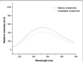
Irradiation of aqueous solutions induces chemical and structural alterations in proteins and peptides related to an attenuation or abolishment of biological activity and interferences in immunological properties [14–20]. It is known that tryptophan fluorescence is strongly influ-enced by its local environment. Thus, changes in protein conformation influence spectrum data. The quenching spectra of the solvent-mediated fluorescence of native and irradiated crotamine can be observed in Fig. 1. The analysis of fluorescence quenching showed that irradi-ated crotamine quantum yield decreased about 25 % in comparison to the native form. This suggests an increase in the solvent accessibility of tryptophan, possibly due to unfolding of the polypeptide chain, since exposure to ionizing radiation is known to alter molecular structure.
Coronado et al.[31] have suggested that solvent expos-ure of hydrophobic residues makes crotamine unusually ‘sticky’. Most hydrophobic residues are located on one side of the molecule and the positively charged residues are clustered on the opposite side. These residues are exposed on the surface of crotamine and form distinct hydrophobic and cationic areas that are positioned roughly on opposite sides of the amphiphilic molecule [31]. The decrease of fluorescence in irradiated toxins can be explained by an increase in the solvent accessibility of the tryptophan, possibly due to unfolding of the poly-peptide since exposure to ionizing radiation changes mo-lecular structure. In the case of crotamine, the decrease was not expressive because most of the hydrophobic resi-dues are already exposed in the native form of the toxin.
Although fluorescence can be a very helpful indicator of molecular change, when it comes to changes in second-ary structure, such measurements are difficult to interpret directly (α-helix and β-sheet). When the structure of
native protein containingα-helical and β-sheet areas is
denatured it undergoes an unfolding process. Often, the fraction of non-covalent bonds inα-helical,β-sheet and
aperiodic conformations may be estimated using highly sensitive measurements, such as circular dichroism (CD) and infrared (IR) spectroscopy. In favorable cases, the spectral effects of conformational changes caused by phys-ical and biologphys-ical phenomena may be observed [37].
Figure 2a shows the CD spectra of native and irradiated crotamine at room temperature. With regard to the native toxin (solid line), the relative proportions of β-sheet and α-helix were 4 % and 48 %, respectively, while, for the
irra-diated peptide (dashed line), theα-helix content was
esti-mated at 8 %, with a corresponding decrease of 4 % of random coil. In theα-helical protein, a negative band near
222 nm is observed due to the strong hydrogen-bonding environment of this conformation. A second transition at 190 nm is split into a negative band near 208 nm and a positive band near 192 nm. The CD spectra of native cro-tamine shows a negative band near 208 nm, indicating the presence ofα-helical conformation.
Pelton and McLean [37] elucidated that short helices can result in reduced bands in the spectra. Coronado et al.[31] illustrated that bothα-helix structures in
crota-mine are formed by few residues, with a single turn inα2
and two turns inα1. The CD spectra ofβ-sheets display a
negative band near 216 nm, a positive band between 195 and 200 nm, and a negative band near 175 nm. However, the position and magnitude of these bands is variable, resulting in less accurate predictions forβ-structure than
for α-helices using CD [37]. Our data indicate that
radiation induced the transition of a small portion of the random coil regions towards an α-helical conformation.
By using a secondary structure prediction tool, we ob-served that a region containing three cysteines (D24-K38) is prone to formingα-helices [38]. It is possible that this
does not occur in the native peptide as these residues form disulfide bonds that stabilize the molecule. Irradi-ation may cause disruption in these reticulIrradi-ation bonds, enabling the formation of a non-native helical domain.
to thermal denaturation lead to changes in CD spectra and it seems that complete denaturation occurred at 90 °C. After reaching the maximum temperature, we re-duced the sample temperature to 20 °C (Fig. 2i) and took a new reading which showed only a partial recovery of structural elements. Irradiated crotamine displayed par-tial denaturation and it is apparent that this form of the toxin was more sensitive to temperature since at 70 °C only random conformation could be observed.
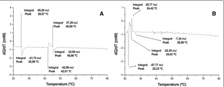
Differential scanning calorimetry (DSC) is extremely useful for characterizing temperature- induced conform-ational changes in proteins, especially with respect to the factors associated with protein stability [39]. The thermodynamic analysis showed that irradiation modified molecule enthalpy. Endothermic transitions decreased while exothermic transitions increased.
Figure 3a shows native crotamine with an upward peak at approximately 25 °C, followed by a downward peak at 45 °C. These data suggest that structure formation and subsequent melting occurred. Furthermore, the results in-dicate another, probably intermediate, state of transition at around 42 °C. It may also be inferred that the toxin be-comes completely denatured at 46 °C.
Figure 3b shows an irregular spectrum with a first component indicating that the irradiated toxin protein melted at 22.23 °C, which reflects lower enthalpy in the irradiated sample. In other words, the energy required to denature the toxin exposed to radiation is much lower, showing that structural stability was significantly affected by the irradiation.
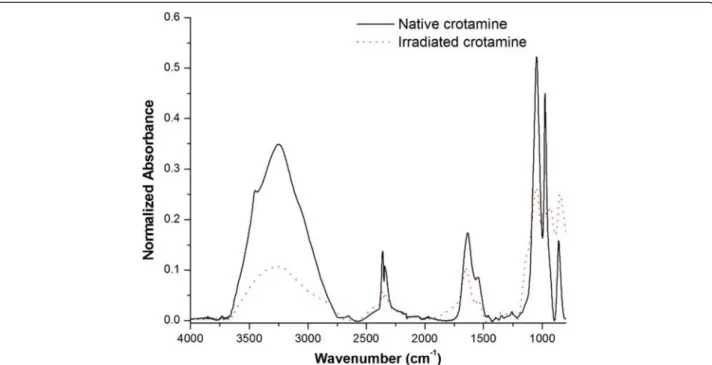
Infrared spectroscopy is a well-established experimen-tal procedure used to analyze the secondary structure of polypeptides and proteins associated with the vibration of repeating structural units, especially the frequency at
which amine bonds absorb infrared radiation. The use of the infrared spectroscopy technique to assess protein secondary structure has undergone a renaissance with the development of Fourier transform infrared spectroscopy. Nine normal modes are allowed for the amide band of proteins in order of decreasing frequency: A, B, and I-VII. The amide band I (80 % C = O stretch, near 1650 cm−1) is the most commonly used to assign secondary structures to proteins [37].
Figure 4 shows the FTIR spectra of crotamine, both in its native (solid line) and in irradiated (dotted line) forms. These spectra show all amide bands of crotamine: amide band I (near 1650 cm−1), II (60 % N-H bend and 40 % C-N stretch, near 1550 cm−1), and III (40 % C-N stretch, 30 % N-H bend, near 1300 cm−1). We observed that the native crotamine curve differs substantially from that of the irradiated sample, with particularly significant differences in the spectral region of the amide band I, indi-cating drastic changes in secondary structure. We also ob-served that the region between 3500 and 3200 cm−1was different in the two forms of crotamine. Although this range is not particularly significant when it comes to the study of the secondary structure, it is important to note that the ~ 3500 cm−1region corresponds to the OH bonds of hydroxyl groups and the ~ 3200 cm−1 region corre-sponds to the NH of the amide grouping links [40]. All proteins are made up of covalently linked amino acid se-quences. These compounds have -OH and -NH groups which have the ability to form a strong network of inter-molecular bonds which gives rise to the tertiary structure of the protein, i.e., its spatial conformation. Thus, the FTIR results suggest that the tertiary structure of crota-mine has changed since absorbance was lower in the curve of the irradiated sample.
Due to the particular importance of the characteristic bands of the amide region I aforementioned, this area is zoomed in Fig. 5. With respect to the irradiated protein, it can be observed that there is a shift in band peaks and an increase in the intensity of some peaks (for example
regions 1, 2 and 3) which could be ascribed to changes in the secondary structure of crotamine. Proteins known to adopt an α-helical conformation have strong amide I
bands between 1650 and 1655 cm−1. The hydrogen-bonding strength ofβ-sheets is more variable due to their
Fig. 4FTIR spectra of the native (solid line) and irradiated (dotted line) forms of crotamine. The curve of the native crotamine differs substantially from that of the irradiated sample, exemplified by a significant discrepancy in the spectral region of the amide I band
flexibility and tendency to twist. Although a strong band between 1612 and 1640 cm−1and a weaker band at about 1685 cm−1are common in
β-sheets, weak bands at
some-what lower frequencies (1665–1670 cm−1) have also been observed [37].
In Fig. 5 the spectra of native crotamine are presented as a smooth curve with peaks along the amide I region, while well-defined peaks appear on the curve of the irradi-ated crotamine. It is evident that the regions most affected by radiation were the spectral regions corresponding to theα-helix (1650 and 1655 cm−1), since major differences
were observed at these wave numbers. It was also ob-served that the curve of the irradiated crotamine shifted approximately 20 cm−1to the left, suggesting lower stabil-ity in this protein, corroborating the results of the DSC.
Given the effects of radiation associated with the ionization of the molecule and its ability to modify the protein structure, these phenomena may be associated with a strong interaction between protein products of lower molecular mass and may demonstrate the formation of peptides, amino acids or even protein aggregates [17, 41]. The presence of these molecular neighbors may modify the interaction of the peptide groups, changing the total energy of the molecule.
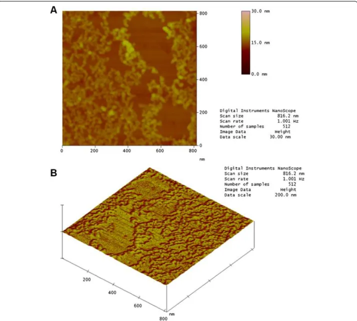
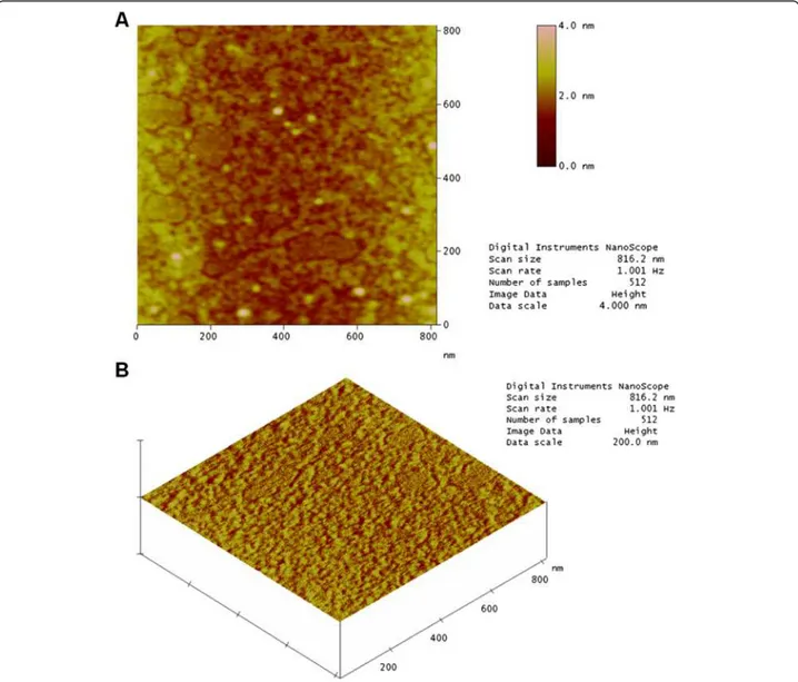
The quality of data from atomic force microscopy (AFM) used for imaging and manipulating biomolecules has improved due to advances in equipment, sample preparation and image acquisition conditions. This is the only instrument that can image samples in aqueous so-lution at the subnanometer scale [42].
Figure 6a shows a topographic image of a mica surface with a sample of native crotamine at a concentration of 0.1 g/mL. It is possible to observe small well-differentiated structures and well-defined rounded “chains”. This is be-cause the mica surface is extremely flat. There are also re-gions where the toxin was not adsorbed. Figure 6b illustrates the results for native crotamine.
The AFM image in Fig. 7a shows that the profile for irradiated crotamine at the same concentration is differ-ent. It is not possible to observe the flat surface of the mica, showing that the irradiated sample covered the entire area, suggesting alterations in the distribution of charged residues on the surface of the molecule. Fur-thermore, the chain structures identified in the native sample were not observed. These results suggest a change in the conformational structure of crotamine.
Exposure to radiation can break the carbon backbone and create smaller fragments that may be reorganized forming aggregates which are invisible to the naked eye. The topographical profile for irradiated toxin was ob-tained in the same way (Fig. 7b).
Conclusions
Based on our findings, we conclude that crotamine suf-fered conformational changes after being exposed to 2 kGy gamma radiation doses from a cobalt-60 source.
The fluorescence analysis allowed us to observe small changes in the tertiary structure of the protein, shown by quenching of the tryptophan fluorophores which are partially exposed in the native molecule.
The results of circular dichroism and infrared spectros-copy showed that there were changes to the secondary
structure of the toxin. Such changes in the secondary structure of proteins may be related to an attenuation or abolishment in its biological activities, such as decreased myotoxic activity. This fact has been demonstrated by pre-vious studies [14–17].
Subjecting the samples to changes in temperature was also used as a mean of comparing the thermal stability of the native and irradiated toxin. Circular dichroism and differential scanning calorimetry showed that irradi-ation induces energy (enthalpy) changes in the peptide, making it more susceptible to thermal denaturation. AFM reinforces these observations, showed by the for-mation of“soluble aggregates”in the irradiated sample.
Taken as a whole, our data indicate that the irradiation of crotamine in solution leads to significant and appar-ently homogeneous conformational changes, with alter-ations of structural elements and a loss of enthalpy, resulting in reduced structural stability. The use of cro-tamine to investigate the impact of gamma ray exposure on peptides provides important insights into the poten-tial of radiation as a tool for attenuating toxins and con-tributing to the design of safer antigens for vaccine and antiserum production.
Abbreviations
CD:Circular dichroism; FTIR: Fourier transformed infrared; AFM: Atomic force microscopy; DSC: Differential scanning calorimetry.
Competing interests
The authors declare that they have no competing interests.
Authors’contributions
KCO performed experimental measurements and analyses of results obtained. Additionaly, she wrote the first draft of the manuscript. PJS analyzed data and contributed to editing and revising of the manuscript. RSFJ provided the crude venom for this study and edited the manuscript. NN contributed to experimental design and revised the manuscript. All authors read and approved the final manuscript.
Acknowledgments
The authors would like to thank the Coordination for the Improvement of Higher Education Personnel (CAPES) for their financial support, Dr. Maria Cecília Salvadori of the Institute of Physics at the University of São Paulo (USP) and Dr. Denise Maria Zezell of the Center for Lasers and Applications at the Nuclear and Energy Research Institute (IPEN–CNEN/SP). Special thanks are also extended to the Center for the Study of Venoms and Venomous Animals (CEVAP) of UNESP. RSFJ is a CNPq DTI fellow researcher (310395/2014-3).
Author details
1Nuclear and Energy Research Institute (IPEN/CNEN-SP), Avenida Professor
Lineu Prestes, 2242, São Paulo, SP 05508-000, Brazil.2Center for the Study of
Venoms and Venomous Animals (CEVAP), São Paulo State University (UNESP
–Univ Estadual Paulista), Botucatu, São Paulo, Brazil.
Received: 28 October 2014 Accepted: 6 May 2015
References
1. Calmette A. Les animaux venimeux et la sérotherapie antivenimeuse. In: Les Venins. Paris: Masson et Cie; 1907.
2. Rosenfeld G. Ação neurotóxica de venenos ofídicos (CrotaluseMicrurus) no sistema nervoso central. Interpretação clínica. Ciênc Cult. 1971;23 Suppl:S1–45.
3. Magalhães RA, Ribeiro MMF, Rezende NA, Amaral CFS. Rabdomióilise secundária a acidente ofídico crotálico (Crotalus durissus terrificus). Rev Inst Med Trop Sao Paulo. 1986;28(4):228–33.
4. Souza FA, Spencer PJ, Rogero JR, Nascimento N, Dal Pai-Silva M, Gallacci M.
60Co gamma irradiation preventsBothrops jararacussuvenom neurotoxicity
and myotoxicity in isolated mouse neuromuscular junction. Toxicon. 2002;40(8):1101–6.
5. Oussedik-Oumehdi H, Laraba-Djebari F. IrradiatedCerastes cerastesvenom as a novel tool for immunotherapy. Immunopharmacol Immunotoxicol. 2008;30(1):37–52.
6. Costa LM, Takeda AK, Barbosa SFC, Berra JAP, Adelina MGF, Soerensen B, et al. Estudo comparativo da resposta imune em cavalos ao veneno de
Crotalus durisssus terrificus,in natura, tratado com formaldeído e submetido à ação térmica. Vac Soros. 1985;1:24–9.
7. Guidolin R, Dias da Silva W, Higashi HG, Caricati CP, Lima MLSR, Morais JF. Hiperimunização de cavalos soroprodutores com venenos botrópico e crotálico tratados por glutaraldeído. Mem Inst Butantan. 1989;51:85–90. 8. Moroz C, Goldblum N, De Vries A. Preparation ofVipera palestinae
antineurotoxin using carboxymethyl-cellulose-bound neurotoxin as antigen. Nature. 1963;200:228–33.
9. Okonogi T, Hattori Z, Ogiso A, Mitsui S. Detoxification by persimmon tannin of snake venoms and bacterial toxins. Toxicon. 1979;17(5):524–7. 10. Relyveld EH, Girard O, Cheyroux M, Asso J, De Rudder J. Nouveau procede
d’inactivation pour la preparation de vaccins. Dev Biol Stand. 1974;33:236–348. 11. Guidolin R, Higashi HG, Nishikawa AK, Yamagushi IK, Lima MLSR, Morais JF,
et al. Venenos botrópicos pré-tratados com inibidores ativos para os sítios enzimáticos em proteases e com substâncias quelantes preservam seu poder imunogênico. Mem Inst Butantan. 1989;51(3):107–15.
12. Heneine IF, Heneine LGD, Daniel JP, Nascimento MCS, Rocha OA. Properties of protein toxins and venoms modified by controlled iodination. Anais Acad Ciênc Est São Paulo. 1988;57(2):55–66.
13. Grasset E. Anavenoms and their use in the preparation of antivenoms sera. Trans R Soc Trop Med Hyg. 1945;38:463–88.
14. Murata Y, Nishikawa AK, Nascimento N, Higashi HG, Dias da Silva W, Rogero JR. Gamma irradiation reduces the toxic activities of Crotalus durissus terrificus venom but does not affect their immunogenic activities. Toxicon. 1992;28:617–28.
15. Nascimento N, Seebart CS, Francis B, Rogero JR, Kaiser II. Influence of ionizing radiation on crotoxin: biochemical and immunological aspects. Toxicon. 1996;34(1):123–31.
16. Clissa PB, Nascimento N, Rogero JR. Toxicity and immunogenicity of
Crotalus durissus terrificusvenom treated with different doses of gamma rays. Toxicon. 1999;37(8):1131–41.
17. Nascimento N, Spencer PJ, Andrade Junior HF, Guarnieri MC, Rogero JR. Effects of gamma radiation on snake venoms. Radiat Phys Chem. 1998;52(1–6):665–9.
18. Ferreira Junior RS, Nascimento N, Martinez JC, Alves JB, Meira DA, Barraviera B. Immunization with native and cobalt 60-irradiatedCrotalus durissus terrificus
venom in swiss mice: assessment of the neutralizing potency of antisera. J Venom Anim Toxins incl Trop Dis. 2005;11(3):299–314 [http://www.scielo.br/ scielo.php?pid=S1678-91992005000300008&script=sci_arttext]
19. Ferreira Junior RS, Nascimento N, Couto R, Alves JB, Meira DA, Barraviera B. Laboratory evaluation of young ovines inoculated with natural or
60Co-irradiatedCrotalus durissus terrificusvenom during
hyperimmunization process. J Venom Anim Toxins incl Trop Dis. 2006;12(4):620–31 [http://www.scielo.br/
scielo.php?script=sci_arttext&pid=S1678-91992006000400009]. 20. Caproni P, Baptista JA, de Almeida TL, Passos LAC, Nascimento N. Study of
irradiated bothropstoxin-1 with60Co gamma rays: immune system behavior.
J Venom Anim Toxins incl Trop Dis. 2009;15(2):216–25 [http://www.scielo.br/ scielo.php?script=sci_arttext&pid=S1678-91992009000200005]
21. Butler J, Hoey BM, Swallow AJ. Radiation chemistry. Annu Rep Prog Chem. 1987;83:129–75.
22. Butler J, Land EJ, Swallow AJ. Chemical mechanisms of the effects of high energy radiation on biological systems. Radiat Phys Chem. 1984;24(3–4):273–82. 23. Moreira EC, Nascimento N, Rosa GJ, Rogero JR, Vassilieff VS. Effects of
gamma irradiation on the behavioral properties of crotoxin. Braz J Med Biol Res. 1997;30(2):245–9.
25. Casare MS, Spencer PJ, Campos LA, Nascimento N. Study of gamma-radiation effects on crotamine and crotoxin. J Radioanal Nucl Ch. 2006;269(3):571–7. 26. Silva MC, Baptista JA, Spencer PJ, Nascimento N. Effects of60Co radiation on
the molecular structure of crotamine. Radiat Phys Chem. 2004;71(1):417–8. 27. Netto DP, Chiacchio SB, Bicudo PL, Alfieri AA, Balarim MRS, Nascimento N.
Hematological changes in sheep inoculated with natural and Cobalt60-irradiatedCrotalus durissus terrificusvenom (Laurenti, 1768).
J Venom Anim Toxins incl Trop Dis. 2004;10(1):34–52 [http://www.scielo.br/ scielo.php?pid=S1678-91992004000100004&script=sci_arttext]
28. Fundação Nacional de Saúde. Manual de diagnóstico e tratamento de acidentes por animais peçonhentos. Brasília: Ministério da Saúde; 1998. p. 131. 29. Laure CJ. Die Primar strutur des crotamins. Hope Seylers z Physiol Chem.
1975;356:213–5.
30. Siqueira AM, Martins NF, de Lima ME, Diniz CR, Cartier A, Brown D, et al. A proposed 3D structure for crotamine based on homology building, molecular simulations and circular dichroism. J Mol Graph Model. 2002;20(5):389–98.
31. Coronado MA, Gabdulkhakov A, Georgieva D, Sankaran B, Murakami MT, Arni RK, et al. Structure of the polypeptide crotamine from the Brazilian rattlesnakeCrotalus durissus terrificus. Acta Crystallogr D Biol Crystallogr. 2013;69(Pt 10):1958–64.
32. Boni-Mitake M, Costa H, Spencer PJ, Vassilieff VS, Rogero JR. Effects of60Co
gamma radiation on crotamine. Braz J Med Biol Res. 2001;34(12):1531–8. 33. Oliveira KC, Silva MN, Gonçalves KO, Spencer PJ, Nascimento N. DSC analysis
of irradiated proteins fromCrotalus durissus terrificus. Rev Bras Pesq Des. 2011;13:155–62.
34. Naumann D. FT-Infrared and FT-Raman spectroscopy in biomedical research. Appl Spectrosc Rev. 2001;36(2–3):239–98.
35. Naumann D. Infrared spectroscopy in microbiology. In: Meyers RA, editor. Encyclopedia of Analytical Chemistry. United Kingdom: John Wiley & Sons, Chichester; 2000. p. 102–31.
36. Ferreira AAP, Yamanaka H. Microscopia de força atômica aplicada em imunoensaios. Quim Nova. 2006;29(1):137–42.
37. Pelton JT, McLean LR. Spectroscopic methods for analysis of protein secondary structure. Anal Biochem. 2000;227(2):167–76.
38. Chou PY, Fasman GD. Prediction of protein conformation. Biochemistry. 1974;13(2):222–45.
39. Sanchez-Ruiz JM. Differential scanning calorimetry of proteins. In: Biswas BB, Roy S, editors. Subcellular biochemistry. Proteins: structure, function and engineering. 24th ed. New York: Plenum Press; 1995.
40. Beekes M, Lasch P, Naumann D. Analytical applications of Fourier transform-infrared (FT-IR) spectroscopy in microbiology and prion research. Vet Microbiol. 2007;123(4):305–19.
41. Cui FZ, Lin YB, Zhang DM, Tian MB. Irradiation effects on secondary structure of protein induced by keV ions. Radiat Phys Chem. 2001;60(1–2):35–8. 42. Engel A, Müller DJ. Observing single biomolecules at work with the atomic
force microscope. Nat Struct Biol. 2000;7(9):715–8.
Submit your next manuscript to BioMed Central and take full advantage of:
• Convenient online submission
• Thorough peer review
• No space constraints or color figure charges
• Immediate publication on acceptance
• Inclusion in PubMed, CAS, Scopus and Google Scholar
• Research which is freely available for redistribution