10.1101/gad.1417706
Access the most recent version at doi:
2006 20: 1283-1293; originally published online Apr 28, 2006;
Genes & Dev.
Neves, Michael Gross, Wanda Viegas and Craig S. Pikaard
Keith Earley, Richard J. Lawrence, Olga Pontes, Rachel Reuther, Angel J. Enciso, Manuela Silva, Nuno
large-scale gene silencing in nucleolar dominance
mediates
Arabidopsis HDA6
Erasure of histone acetylation by
References
http://www.genesdev.org/cgi/content/full/20/10/1283#otherarticles
Article cited in:
http://www.genesdev.org/cgi/content/full/20/10/1283#References
This article cites 35 articles, 12 of which can be accessed free at:
service
Email alerting
click here
top right corner of the article or
Receive free email alerts when new articles cite this article - sign up in the box at the
Notes
http://www.genesdev.org/subscriptions/
go to:
Genes and Development
To subscribe to
Erasure of histone acetylation by
Arabidopsis HDA6
mediates large-scale
gene silencing in nucleolar dominance
Keith Earley,1,5Richard J. Lawrence,1,5Olga Pontes,1,3Rachel Reuther,1 Angel J. Enciso,1 Manuela Silva,3 Nuno Neves,3,4Michael Gross,2Wanda Viegas,3 and Craig S. Pikaard1,61Biology Department,2Department of Chemistry, Washington University, St. Louis, Missouri 63130, USA;3Secção de
Genética, Centro de Botânica Aplicada à Agricultura, Instituto Superior de Agronomia, Tapada da Ajuda, 1349-017 Lisboa, Portugal;4Secção Autónoma de Biotecnologia, Faculdade de Ciências e Tecnologia, Universidade Nova de Lisboa, Monte da
Caparica, 2859-516 Caparica, Portugal
Nucleolar dominance describes the silencing of one parental set of ribosomal RNA (rRNA) genes in a genetic hybrid, an epigenetic phenomenon that occurs on a scale second only to X-chromosome inactivation in mammals. An RNA interference (RNAi) knockdown screen revealed that the predicted Arabidopsis histone deacetylase, HDA6, is required for rRNA gene silencing in nucleolar dominance. In vivo, derepression of silenced rRNA genes upon knockdown of HDA6 is accompanied by nucleolus organizer region (NOR) decondensation, loss of promoter cytosine methylation, and replacement of histone H3 Lys 9 (H3K9) dimethylation with H3K4 trimethylation, H3K9 acetylation, H3K14 acetylation, and histone H4 tetra-acetylation. Consistent with these in vivo results, purified HDA6 deacetylates lysines modified by histone acetyltransferases whose substrates include H3K14, H4K5, and H4K12. HDA6 localizes, in part, to the nucleolus, supporting a model whereby HDA6 erases histone acetylation as a key step in an epigenetic switch mechanism that silences rRNA genes through concerted histone and DNA modifications.
[Keywords: Chromatin; epigenetic; gene silencing]
Received February 6, 2006; revised version accepted March 10, 2006.
Nucleolus organizer regions (NORs) are the genetic loci at which eukaryotic ribosomal RNA (rRNA) genes tran-scribed by RNA polymerase I are clustered in long tan-dem arrays, typically spanning several megabases. Nucleoli form at NORs during interphase as a direct con-sequence of rRNA gene transcription (Grummt 2003; Grummt and Pikaard 2003; Shaw and Doonan 2005). In genetic hybrids of plants or animals, the NORs inherited from one parent are often inactivated, an epigenetic phe-nomenon known as nucleolar dominance (Reeder 1985; Pikaard 2000; Viegas et al. 2002). The molecular basis for nucleolar dominance is the selective silencing of one pa-rental set of rRNA genes via an epigenetic switch mecha-nism that involves both DNA methylation and histone post-translational modifications (Chen and Pikaard 1997; Lawrence et al. 2004). If cytosine methylation is inhibited using 5-aza-2⬘-deoxycytosine (aza-dC) or if his-tone deacetylation is inhibited using Trichostatin A (TSA), the silenced rRNA genes are derepressed. Impor-tantly, treatment with either aza-dC or TSA elicits the same outcome: Coincident with switching from off to
on, rRNA gene promoter cytosines become hypomethyl-ated, promoter-associated histone H3 Lys 9 dimethyl-ation (H3K9me2) is lost, and the euchromatic mark, H3K4me3, is acquired (Lawrence et al. 2004). The fact that inhibiting cytosine methylation induces histone modifications and that inhibiting histone deacetylation causes the loss of cytosine methylation suggests a model whereby DNA methylation and histone deacety-lation are each upstream of one another in a self-rein-forcing pathway (Lawrence et al. 2004).
In Arabidopsis suecica, the allotetraploid hybrid of A.
thaliana and A. arenosa (Fig. 1A), the A. thaliana-de-rived rRNA genes are selectively silenced (Chen et al. 1998). In A. suecica we have exploited RNA interference (RNAi) to knock down predicted chromatin modifying activities and then tested for loss of nucleolar domi-nance. Previously, we knocked down a small
Arabidop-sis gene family (the HDT gene family) of nucleolar pro-teins related to maize HD2 and identified HDT1 as a gene important for nucleolar dominance (Lawrence et al. 2004). HDT family proteins are putative plant-specific histone deacetylases (HDACs) (Lusser et al. 1997; Dangl et al. 2001) that are unusual in that they lack sequence similarity with all other classes of HDACs, which are conserved from plants to humans (Pandey et al. 2002). However, HDT proteins display similarity to FKBP
pro-5These authors contributed equally to this work. 6Corresponding author.
E-MAIL pikaard@biology.wustl.edu; FAX (314) 935-4432.
Article published online ahead of print. Article and publication date are at http://www.genesdev.org/cgi/doi/10.1101/gad.1417706.
teins (Aravind and Koonin 1998), and there are examples of FKBPs that localize to the nucleolus, are required for silencing reporter genes integrated into NORs, that act as histone chaperones for chromatin assembly, or that interact with HDACs (Arevalo-Rodriguez et al. 2000; Yang et al. 2001; Kuzuhara and Horikoshi 2004). These findings raise the possibility that HDT1 may play a role in chromatin assembly and may interact with one or more HDACs, but may not be an HDAC itself. As yet, the TSA-sensitive HDAC(s) required for nucleolar domi-nance remain unidentified.
Here, we report on a systematic knockdown screen of the non-HDT classes of predicted HDACs in
Arabidop-sis, including eight members of the protein superfamily related to yeast Rpd3p. We show that the Rpd3-like pro-tein HDA6 is required for rRNA gene silencing in nucleolar dominance. Previous studies demonstrated roles for the HDA6 gene in transgene and transposable element silencing (Murfett et al. 2001; Aufsatz et al. 2002; Lippman et al. 2003; Probst et al. 2004) and in NOR condensation (Probst et al. 2004). However, no role for HDA6 in rRNA gene silencing was identified in prior studies nor has the biochemical specificity of HDA6 been defined. We show that HDA6 is a TSA-sensitive HDAC capable of removing acetyl groups from multiple lysines of multiple histones. Among the substrates of HDA6 are lysines of histones H3 and H4 whose acetyla-tion is associated with gene activaacetyla-tion. HDA6 is also
required to maintain DNA hypermethylation at the pro-moters of silenced A. thaliana-derived rRNA genes. Col-lectively, these data identify HDA6 as a key component of the epigenetic switch mechanism that silences rRNA genes through concerted changes in histone modifica-tion and cytosine methylamodifica-tion.
Results
Silenced rRNA genes subjected to nucleolar dominance are derepressed inHDA6-RNAi
knockdown lines
To identify specific HDACs that are responsible for rRNA gene silencing, A. suecica plants were trans-formed with transgenes that express double-stranded RNAs matching targeted HDACs (Fig. 1B), thereby in-ducing their RNAi-mediated knockdown. Because A.
thalianaand A. arenosa genes typically share >90% se-quence identity, orthologous mRNAs of both progeni-tors are depleted (Lawrence and Pikaard 2003). There are 16 predicted Arabidopsis HDACs (Pandey et al. 2002). These include the four HDT family members targeted in our previous study (Lawrence et al. 2004), 10 members of the RPD3/HDA1 superfamily, and two genes related to the yeast NAD-dependent HDAC, Sir2p. The latter HDAC is known to suppress recombination among rRNA genes and is required for the silencing of
protein-Figure 1. HDA6 is required for rRNA gene silenc-ing in nucleolar dominance. (A) Diagrammatic rep-resentations of A. thaliana, A. arenosa, and A.
sue-cicachromosome compositions. (B) Organization of transferred DNAs (T-DNAs) containing RNAi-in-ducing transgenes. The T-DNA, delimited by left and right border sequences (LB and RB), contains a selectable herbicide-resistance gene and an inverted repeat of target gene cDNA fragments (500–700 bp), with a chalcone synthase (CHSA) intron spacer, ex-pressed by the Cauliflower Mosaic Virus (CaMV) 35S promoter and terminated by octopine synthase (OCS) 3⬘ sequences. (C) rRNA gene organization sur-rounding internal transcribed spacer 1 (ITS1) with PCR primer (arrows) and HhaI sites indicated. (D) Screening A. suecica RNAi lines targeting predicted Rpd3-like and Sir2-like HDACs. (Lanes 1–5) Con-trols show HhaI-digested RT–PCR products of A.
thaliana (At), A. arenosa (Aa), nontransformed A.
suecica (As), or TSA-treated A. suecica. In the re-maining panels, RNA from five independent RNAi lines targeting each HDAC was tested. (E) S1 nucle-ase protection analysis. Lanes 1 and 2 show A.
thalianaand A. arenosa RNA controls, demonstrat-ing probe specificity. In the remaindemonstrat-ing lanes, RNA of wild-type (lanes 3,4), TSA-treated (lane 5), or HDA6-RNAi lines of A. suecica were probed for A.
thaliana-like or A. arenosa-like rRNA transcripts. (F) HDA6 mRNA levels are knocked down in RNAi lines. RNA from nontransformed or HDA6-RNAi lines was incubated ± RT. Resulting cDNA was am-plified by PCR using primers for HDA6 and a PFK internal control.
coding genes integrated into NORs (Gottlieb and Es-posito 1989; Smith and Boeke 1997).
A systematic knockdown screen of the expressed members of the RPD3 and SIR2-related gene families showed that HDA6 is required for nucleolar dominance (Fig. 1). As a result of a single nucleotide polymorphism that generates a HhaI site (Fig. 1C), A. thaliana and A.
arenosa transcripts that are reverse transcribed into DNA, amplified by PCR (RT–PCR), and then digested with HhaI yield diagnostic banding patterns (Fig. 1D, cf. lanes 1 and 2). In wild-type A. suecica (strain LC1), tran-scripts of the dominant A. arenosa-derived rRNA genes are readily apparent (Aa bands) whereas the underdomi-nant A. thaliana rRNA gene transcripts are detected in only trace amounts (At band) (Fig. 1D, lanes 3,4). How-ever, the silenced A. thaliana rRNA genes are readily derepressed upon treatment with TSA (Fig. 1D, lane 5). Using the RT–PCR assay, we next screened for HDACs whose knockdown could mimic the effects of TSA. As shown in Figure 1D, A. thaliana rRNA transcripts were readily detected in multiple RNAi lines targeting HDA6, but were not detected above wild-type levels in RNAi lines targeting other members of the RPD3/HDA1 super-family (HDA2, HDA5, HDA8, HDA9, HDA14, HDA15,
HDA19), or in lines targeting the two SIR2-like HDACs,
SRT1and SRT2 (the latter is not shown). Northern blot and massively parallel signature sequencing (MPSS) data indicate that, with the exception of SRT2, all of the HDAC genes that were targeted are expressed at some level (see http://www.chromdb.org and http://mpss. udel.edu for more information). It is noteworthy that the derepression of A. thaliana rRNA genes in HDA6-RNAi lines was not accompanied by any discernible visible phenotypes.
Derepression of A. thaliana-derived rRNA genes in HDA6-RNAi lines was confirmed using S1 nuclease pro-tection (Berk and Sharp 1977) with species-specific probes (Fig. 1E). In all of the HDA6-RNAi lines, derepres-sion of underdominant A. thaliana rRNA genes is corre-lated with decreased HDA6 mRNA levels, as determined by RT–PCR and comparison to an internal control, phos-phofructokinase (PFK) (Fig. 1F). Note that A. thaliana rRNA genes are derepressed to a greater extent by TSA than by HDA6 knockdown in the RNAi lines (see Fig. 1E). A likely explanation is that RNAi does not result in an HDA6 null phenotype, but only knocks down HDA6 mRNA levels several-fold (see Fig. 1F). However, we can-not exclude the possibility that other HDACs are par-tially redundant with HDA6.
HDA6 localizes, in part, to the nucleolus
To determine where HDA6 is localized in the cell, we engineered a genomic HDA6 clone, under the control of its own promoter, to express HDA6 fused to the Flag epitope. HDA6-Flag protein was then detected in trans-genic plants using anti-Flag monoclonal antibody (Fig. 2A). HDA6 is not uniformly distributed throughout the nucleus; instead, there are distinct nuclear regions in which HDA6 is concentrated. The most prominent of
these HDA6-positive domains is the nucleolus, which is easily visualized as the region least stained by the fluo-rescent DNA-binding dye, DAPI (Fig. 2A). Native HDA6 was also localized to the nucleolus using an affinity-pu-rified rabbit polyclonal antiserum raised against an HDA6 polypeptide expressed in Escherichia coli (data not shown). Because the nucleolus is the site of rRNA gene transcription, nucleolar localization of HDA6 is consistent with HDA6 playing a role in nucleolar domi-nance via direct modification of rRNA genes. Likewise, HDA6 concentration at other foci in the nucleus is con-sistent with the role of HDA6 in silencing other se-quences (Murfett et al. 2001; Aufsatz et al. 2002; Lipp-man et al. 2003).
RNAi-mediated knockdown of HDA6 induces a heterochromatin-to-euchromatin transition at underdominant NORs
The two silenced A. thaliana-derived NORs and the six dominant A. arenosa NORs can be discriminated in A.
suecica nuclei using fluorescence in situ hybridization (FISH) with species-specific probes (see Fig. 2B for probe location; Pontes et al. 2003). Sequential immunolocal-ization of H3K9me2, an epigenetic mark of heterochro-matin (Richards and Elgin 2002), followed by FISH shows that the two silenced A. thaliana NORs colocalize with two prominent H3K9me2 foci in A. suecica interphase nuclei (Fig. 2C, panels A–C). Other major H3K9me2 foci correspond primarily to pericentromeric heterochroma-tin. Immunolocalization of H3K4me3, a mark of tran-scriptionally active euchromatin (Richards and Elgin 2002), yields a distinctly different pattern, namely a dif-fuse nuclear distribution that is punctuated by dark holes of condensed heterochromatin (Fig. 2C, panels D,J). Strikingly, two of these dark holes correspond to the two silenced A. thaliana NORs (Fig. 2C, panels D–F). Taken together, these results show that A. thaliana-derived NORs in A. suecica are highly condensed and associated with histones displaying the heterochromatin marker H3K9me2 but not the euchromatic marker H3K4me3. In contrast, the dominant A. arenosa-derived NORs in A.
suecica colocalize with both H3K9me2 (Fig. 2C, panels G–I) and H3K4me3 (Fig. 2C, panels J–L), consistent with the interpretation that the dominant A. arenosa-derived NORs include subdomains consisting of active (H3K4me3-associated) or silent (H3K9me2-associated) genes (Lawrence et al. 2004).
Whereas the two A. thaliana NORs are highly con-densed in wild-type A. suecica nuclei, yielding two large FISH signals (see Fig. 2C, panels B,E), three to five dif-fuse, lobed A. thaliana NOR FISH signals are observed in both TSA-treated plants and HDA6-RNAi lines (Fig. 2D, panels B,E,H,K). To interpret these observations, it is im-portant to note that the NOR FISH signals we detect correspond to the portions of the NORs that are com-posed of condensed, inactive rRNA genes assembled in facultative heterochromatin. Whereas a silenced NOR is uniformly condensed to yield a single, large FISH signal, an active NOR includes both heterochromatic regions
and decondensed, active regions (Pontes et al. 2003). The chromatin fibers consisting of active genes are too thin and dispersed to be detected using our methods. How-ever, their existence can be inferred. In Figure 2, the tran-sition from two inactive A. thaliana NORs, observed as two large FISH signals, in wild-type plants to three to five FISH signals, upon TSA treatment or HDA6 knock-down, indicates that internal decondensation has oc-curred within the NORs. Decondensed euchromatic re-gions flanked by blocks of condensed heterochromatin accounts for the increased number of FISH signals. Note also that the FISH signals themselves are qualitatively different in nuclei of TSA-treated and HDA6-RNAi plants, appearing less compact and more diffuse, presum-ably due to numerous local decondensations within the NORs.
In addition to decondensing, A. thaliana rRNA genes in nuclei of TSA-treated plants or HDA6-RNAi lines co-localize primarily with H3K4me3-enriched euchromatin (Fig. 2D, panels F,L) rather than with H3K9me2-enriched chromocenters (Fig. 2D, panels C,I) as is the case in un-treated, nontransgenic plants (cf. Fig. 2C, panel C).
Col-lectively, the data of Figures 1 and 2 suggest that HDA6 localizes within the nucleolus and is required to main-tain the repression of the underdominant A. thaliana-derived rRNA genes in A. suecica. The chromosomal manifestation of this repression is the organization of the NORs into highly condensed facultative heterochro-matin.
HDA6 is a broad-specificity HDAC
To explore the biochemical activity of HDA6, we affin-ity-purified Flag-tagged HDA6 from transgenic A.
thaliana(Fig. 3A). The prominent polypeptide migrating on a SDS-PAGE gel (Fig. 3A, arrow) at the predicted size of ∼55 kD, and absent in nontransgenic (wild-type) plants, was confirmed to be HDA6 by tryptic fingerprint-ing and MALDI-TOF mass spectrometry, yieldfingerprint-ing eight matching peptides representing ∼40% of the HDA6 se-quence (data not shown). We then used recombinant his-tone acetyltransferases (HATs) to acetylate purified
Brassica oleracea (broccoli) core histones (which are identical between Arabidopsis and Brassica for histones H3 and H4), or histone N-terminal peptides (N-peptides),
Figure 2. HDA6 localizes to the nucleo-lus and is required for facultative hetero-chromatin formation and interphase con-densation at underdominant NORs. (A) Immunolocalization of HDA6-Flag using an anti-Flag monoclonal primary antibody (red signal). The tagged protein was engi-neered by modifying a genomic HDA6 clone under control of its own promoter region. Nuclei were counterstained with DAPI (blue). Controls demonstrate the lack of Flag signal in a wild-type (non-transgenic) nucleus or a HDA6-Flag trans-genic nucleus if the primary anti-Flag an-tibody is omitted and only the secondary antibody is used. (B) Organization of rRNA genes at NORs. Genes encoding 18S, 5.8S, and 25S structural rRNA precursors are separated by intergenic spacers. FISH probe and gene promoter (arrow) locations are indicated. (C, panels A–C) Immunolo-calization of H3K9me2 (red signals), FISH localization of A. thaliana rRNA genes (AtNORs; green signals), and the merged image of panels A and B plus a DAPI-stained image (panel C) in meristematic root-tip cell interphase nuclei. (Panels
D–F) H3K4me3 (red) and silent AtNORs. (Panels G–I) H3K9me2 (red) and dominant
A. arenosa NOR (AaNOR) localization. (Panels J–L) H3K4me3 (red) and AaNORs. (D, panels A–C) H3K9me2 (red) and AtNORs (green) in interphase nuclei of meristematic root tip cells of TSA-treated plants. (Panels D–F) H3K4me3 (red) and AtNORs (green) in TSA-treated plants. (Panels G–I) H3K9me2 (red) and AtNORs (green) in an HDA6-RNAi plant. (Panels
J–L) H3K4me3 (red) and AtNORs (green) in an HDA6-RNAi plant.
and asked if Flag-HDA6 could remove the acetyl groups. Three full-length His-tagged A. thaliana HATs, HAG1, HAG2, and HAG5 (Pandey et al. 2002), were expressed in
E. coli and affinity-purified (Fig. 3B). Upon incubation with Brassica histones and 3H-acetyl CoA, we found
that recombinant HAG2 and HAG5 specifically label histone H4 whereas recombinant HAG1 preferentially acetylates histone H3 and histone H2A and/or H2B (Fig. 3C). Purified Flag-HDA6 deacetylates the histones la-beled by the three HATs whereas proteins of wild-type
plants eluted from the anti-Flag resin (see Fig. 3A, left lane) lack HDAC activity (Fig. 3D). The three
Arabidop-sisHATs and HDA6 also displayed activity with histone N-peptides (Figs. 3E, 4; data not shown). For instance, HAG5 acetylates and HDA6 subsequently deacetylates a histone H4 N-peptide (Fig. 3E).
TSA blocks the ability of HDA6 to deacetylate full-length histone H4 (Fig. 3F) or H4 N-peptide (data not shown) acetylated by HAG5. The similar consequences of TSA treatment or HDA6 knockdown on NOR con-densation and rRNA gene silencing (see Figs. 1, 2), to-gether with these biochemical data, suggest that HDA6 can account for the effects of TSA on nucleolar domi-nance.
Determination of histone H3 and H4 acetyl-lysine substrates for HDA6
Determining the specificity of HATs whose acetylation can be removed by HDA6 provides a means for deducing the specificity of HDA6. We employed mass spectrom-etry as well as an independent biochemical assay to iden-tify specific lysines of histone H3 or H4 N-peptides that are acetylated by HAG1, HAG2, or HAG5 in vitro (Fig. 4). In the first approach, acetylated N-peptides were sub-jected to electrospray ionization and quadrupole ion trap mass spectrometry. Ionized peptides appearing as [M + 2H]2+, [M + 3H]3+, and [M + 4H]4+ ions were
de-tected in each of their ESI mass spectra (Fig. 4A–E). HAG2 and HAG5, which label full-length histone H4 (see Fig. 3C,D), each catalyze the monoacetylation of the H4 N-peptide (Fig. 4, cf. B,C and A). Likewise, HAG1, which can label full-length histone H3 (see Fig. 3), also catalyzes the monoacetylation of the H3 N-peptide (Fig. 4, cf. D and E).
To identify the amino acids monoacetylated by HAG1, HAG2, or HAG5, ionized acetylated species resolved in the initial ESI-MS scan (MS1) were selected and
frag-mented to produce daughter ions. Peaks in resulting product-ion spectra (MS/MS experiment) were further fragmented to yield higher-order product-ion spectra (MS3experiments). This approach revealed the sites of
acetylation and indicated that HAG2 acetylates histone H4 on Lys 12 (H4K12), HAG5 acetylates H4K5, and HAG1 acetylates H3K14 (data not shown). These speci-ficities were also inferred from an independent assay in which we asked if pre-existing acetylation of a specific lysine would preclude HAT-catalyzed labeling of the peptide with3H-acetyl CoA (Fig. 4F). HAG1 efficiently
labeled a nonacetylated H3 peptide or an H3 peptide that had a pre-existing acetyl group on K9 (Fig. 4F, top row). However, prior acetylation of H3K14 prevented HAG1-catalyzed labeling with a radioactive acetyl group, con-sistent with H3K14 being the substrate for HAG1. Like-wise, prior acetylation of H4K12 and H4K5 inhibited the labeling of the histone H4 peptide by HAG2 and HAG5, respectively, consistent with the HAG2 and HAG5 specificities that were determined by mass spectrom-etry. Note that HAG1 does not acetylate the H4 peptide, and HAG2 and HAG5 do not acetylate the H3 peptide,
Figure 3. Purified HDA6 has HDAC activity. (A) Affinity pu-rification of Flag-HDA6 expressed in transgenic plants. Extracts of wild-type or Flag-HDA6-overexpressing A. thaliana was in-cubated with anti-Flag resin. A Coomassie blue-stained SDS-PAGE gel of proteins eluted using excess Flag peptide is shown. (B) Coomassie-stained SDS-PAGE gel of His-tagged recombi-nant Arabidopsis HATs HAG1, HAG2, and HAG5 after purifi-cation on nickel-agarose. (C) HAT activity of HAG1, HAG2, and HAG5. Broccoli histones were labeled using3H-acetyl CoA and
the resulting SDS-PAGE gel was Coomassie-stained and subse-quently subjected to fluorography. Histone H3 and H4 bands were definitively identified using mass spectrometry. H2A and H2B variants with overlapping migration patterns precluded de-finitive assignment of H2A and H2B bands. (D) HDA6 deacety-lates full-length histones acetylated by HAG1, HAG2, and HAG5. Histones labeled by the HATs were incubated with equal aliquots of Flag-HDA6 or wild-type protein eluted from anti-Flag resin. The fluorogram and Coomassie-stained histone bands are shown. (E) HDA6 deacetylates histone N-peptides. Anti-Flag resin incubated with extracts of wild-type or Flag-HDA6-expressing plants was washed extensively then incu-bated with HAG5-labeled H4 peptide immobilized on agarose beads.3
H (from3
H-acetyl CoA) released into the reaction buffer was measured by scintillation counting. (F) HDA6 is a TSA-sensitive HDAC. HAG5-labeled broccoli histones were incu-bated with HDA6 ± TSA and then subjected to SDS-PAGE and fluorography.
consistent with the specificities of the HATs on full-length histones. Based on the specificities of HAG1, HAG2, and HAG5 and the fact that HDA6 can remove the acetyl groups added to histones by these HATs, we deduce that the deacetylase activity of HDA6 includes Lys 14 of histone H3 and Lys 5 and Lys 12 of histone H4. Moreover, HDA6 deacetylates histone H2A and/or H2B at undefined amino acids that are acetylated by HAG1 (see Fig. 3D). Collectively, these data indicate that HDA6 is a broad-specificity deacetylase capable of eras-ing acetylation from multiple lysines of multiple his-tones.
In vivo effects of HDA6 knockdown on histone modifications and DNA methylation
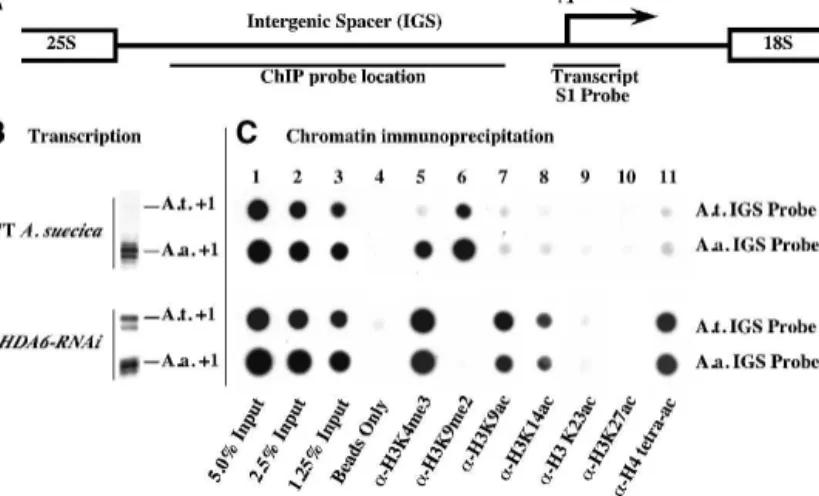
The in vivo consequences of HDA6 knockdown on his-tone modifications at rRNA gene promoters in A.
sue-cica were tested using chromatin immunoprecipitation (ChIP) with antibodies specific for H3K4me3, H3K9me2,
H3K9ac, H3K14ac, H3K23Ac, H3K27ac, or H4ac4 (H4
tetra-acetylated on Lys 5, Lys 8, Lys 12, and Lys 16). Following immunoprecipitation, purified DNA was dot-blotted, and resulting filters were hybridized to species-specific intergenic spacer (IGS) probes (see Fig. 5A).
In wild-type A. suecica, in which A. arenosa rRNA genes are expressed and A. thaliana rRNA genes are si-lenced (Fig. 5B), the A. thaliana genes are preferentially associated with H3K9me2-modified histones (Fig. 5C, column 6, top row), in agreement with cytological ob-servations (see Fig. 2). In contrast, the dominant A.
arenosarRNA genes are associated with both H3K4me3 and H3K9me2 (Fig. 5C, columns 5 and 6, second row), also in agreement with the cytological evidence (see Fig. 2) and consistent with the interpretation that the domi-nant set of genes includes both active and inactive gene subsets. HDA6 knockdown alters the chromatin modi-fication profiles and eliminates the differences between the A. arenosa and A. thaliana genes with respect to transcription (Figs. 1, 5B) and chromatin modification
Figure 4. HAG1, HAG2, and HAG5 monoacetylate histone H3 and H4 N-pep-tides. (A) Mass spectrum of unmodified H4 peptide (M) differentially protonated (H) to generate 2+, 3+, and 4+ charge states. (B,C) Mass spectra of H4 peptide acetylated by HAG2 or HAG5. Unmodified and mono-acetylated peptides (M + Ac) were detected in the 2+, 3+, and 4+ charge states. (D) Mass spectrum of unmodified histone H3 peptide. (E) Mass spectrum of the HAG1-acetylated histone H3 peptide. (F) Deter-mination of HAG1, HAG2, and HAG5 specificities. N-peptides bearing pre-exist-ing, nonradioactive acetyl groups on indi-vidual lysines were incubated with HAG1, HAG2, or HAG5 and3H-acetyl CoA, then
(Fig. 5C, lower two rows). In HDA6-RNAi lines, de-creased association of both parental sets of rRNA genes with H3K9me2 is accompanied by a corresponding in-crease in H3K9 acetylation and H3K4 trimethylation (Fig. 5C, columns 5–7, lower two rows). Notable in-creases in H3K14 acetylation and H4 tetra-acetylation also occur in HDA6-RNAi plants (Fig. 5C, columns 8 and 11), consistent with our biochemical evidence that HDA6 deacetylates H3K14, H4K5, and H4K12. These latter H3 and H4 acetylation marks are detected at much lower levels in wild-type A. suecica, suggesting a dra-matic change in steady-state histone acetylation/ deacetylation activity in HDA6-RNAi lines.
rRNA gene promoter cytosines become demethylated inHDA6-RNAi lines
To examine simultaneously the histone modification and cytosine methylation status of rRNA gene promot-ers, we employed ChIP–chop PCR (Fig. 6; Lawrence et al. 2004). Whereas hypomethylated DNA templates survive McrBC treatment, hypermethylated DNA is “chopped” by McrBC and cannot be amplified by PCR. In wild-type
A. suecica, silenced A. thaliana rRNA gene promoters are immunoprecipitated by H3K9me2 antibodies (Fig. 6, column 6) and have hypermethylated promoters such that McrBC digestion precludes subsequent PCR ampli-fication (Fig. 6, column 7). In contrast, the A. arenosa rRNA gene promoters associate with both H3K9me2 and H3K4me3 (Fig. 6, columns 4–7). Those A. arenosa rRNA gene promoters associated with H3K4me3 are hypo-methylated and resistant to McrBC (Fig. 6, cf. columns 4 and 5) but A. arenosa rRNA gene promoters associated with H3K9me2 are hypermethylated and digested by McrBC (Fig. 6, cf. columns 6 and 7). In HDA6-RNAi lines, neither A. thaliana nor A. arenosa rRNA gene pro-moters associate with H3K9me2 (Fig. 6, lower two rows, columns 6 and 7). Instead, the promoters are found in association with H3K4me3, K3K9ac, and H3K14ac and are resistant to McrBC digestion (Fig. 6, columns 4–5 and 8–11), indicating that cytosine hypomethylation is linked to the occurrence of these specific, euchromatic histone modifications at active rRNA gene promoters.
Discussion
Based primarily on the observation that the DNA meth-ylation inhibitor, aza-dC, and the HDAC inhibitor TSA both cause rRNA gene derepression, promoter demeth-ylation, loss of promoter-associated H3K9me2, and ac-quisition of the euchromatic mark H3K4me3, we previ-ously proposed a model whereby DNA methylation and histone deacetylation are each upstream of one another in a circular, self-reinforcing pathway (Lawrence et al. 2004). Our evidence that HDA6 is a TSA-sensitive HDAC whose knockdown phenocopies the effects of TSA to cause rRNA gene derepression, NOR deconden-sation, and loss of promoter cytosine methylation fits the predictions of this model. Moreover, our latest data indicate that acetylated H3K9, acetylated H3K14, and tetra-acetylated H4 are histone modifications associated with active rRNA gene promoters whose cytosines are hypomethylated, further extending the model (Fig. 7).
Figure 6. HDA6 is required for cytosine hypermethylation at silenced promoters. ChIP–chop PCR was used to evaluate cyto-sine methylation density within A. thaliana- and A. arenosa-derived promoters in wild-type and HDA6-RNAi lines. Ten per-cent of the immunoprecipitated chromatin dot-blotted in Figure 5 was incubated with (lanes 5,7,9,11) or without (lanes 4,6,8,10) McrBC, then PCR was used to amplify A. thaliana or A. arenosa rRNA gene promoter regions. Hypermethylated DNA digested by McrBC is not amplified. (Lanes 1–3) Input controls used 0.1%, 0.05%, or 0.025% of the chromatin subjected to ChIP and show that the assay is semiquantitative.
Figure 5. Transcriptional derepression of rRNA genes in A. suecica HDA6-RNAi lines is accompanied by changes in rRNA gene histone methylation and histone acetylation. (A) Diagram of an rRNA gene intergenic spacer highlighting the transcription initiation site (+1; arrow) and S1 and ChIP probe locations. (B) Pre-rRNA transcripts detected using S1 nuclease protection with
A. thaliana-or A. arenosa-specific probes. (C) ChIP dot-blot analysis. Duplicate samples of wild-type or
HDA6-RNAiplant chromatin, blotted in adjacent rows, were hybridized to A. thaliana- or A. arenosa-specific probes (see diagram). (Columns 1–3) Five percent, 2.5%, or 1.25% of the input chromatin in the ChIP reactions. (Column 4) Protein A beads in the absence of antibod-ies. (Columns 5–11) Chromatin immunoprecipitated with the indicated antibodies.
In vitro, purified HDA6 deacetylates full-length his-tones or histone tail peptides that are acetylated on H3K14, H4K5, and H4K12 by three Arabidopsis HATs whose specificities were previously undefined—namely, HAG1, HAG2, and HAG5. Collectively, the evidence suggests that HDA6 is a broad-specificity HDAC. The in vitro HDAC specificity of HDA6, the in vivo hyperacety-lation of histones at corresponding lysines in HDA6-RNAi lines, and the nucleolar localization of HDA6 are all consistent with the hypothesis that HDA6 directly deacetylates rRNA gene promoter histones to bring about rRNA gene silencing.
It is noteworthy that HDA6 is abundant inside the nucleolus, where the decondensed, active subset of rRNA genes is transcribed, and is not appreciably colo-calized with the highly condensed, heterochromatic por-tions of the NORs located outside the nucleolus. We interpret this as evidence that HDA6 plays a role in switching rRNA genes from an active state to the inac-tive state, which would explain the presence of HDA6 inside the nucleolus where the active genes are located. In contrast, if HDA6 only played a role in maintaining the silent state, we would expect HDA6 to colocalize with the condensed, inactive portions of the NORs pe-ripheral to the nucleolus. HDA6 may play similar roles at transgenes and transposable elements that can switch between active and inactive states, which is a defining characteristic of facultative heterochromatin, thereby driving the steady-state balance toward gene silencing.
It is interesting that HDA6 knockdown derepresses silenced rRNA genes subjected to nucleolar dominance in A. suecica whereas hda6 mutations had no discernible effect on rRNA gene transcription in A. thaliana (Probst et al. 2004). One trivial explanation may be that with ∼1500 essentially identical rRNA genes in diploid A.
thaliana (Copenhaver and Pikaard 1996), an estimated one-third to one-half of which are active (Lawrence et al. 2004), increased rRNA gene transcription is undetect-able against the backdrop of pre-existing active genes. In contrast, sequence differences among the A. thaliana and A. arenosa-derived rRNA genes in A. suecica, com-bined with the nearly complete silencing of the former, makes it easy to detect the derepression of underdomi-nant genes in the hybrid. Alternatively, the number of active rRNA genes may change in A. thaliana hda6 mu-tants without changing the overall level of transcription.
In Saccharomyces cerevisae, the number of active rRNA genes can change more than twofold without changing steady-state rRNA transcript levels owing to compensa-tory changes in the average number of RNA polymerases engaged in transcription on each gene (French et al. 2003). The latter explanation is attractive given that NOR decondensation, increased H3K4 methylation, and subtle changes in DNA methylation at uncharacterized locations within the rDNA were observed in A. thaliana
hda6 mutants (Probst et al. 2004), consistent with our findings using A. suecica HDA6-RNAi lines.
The lack of an effect on nucleolar dominance upon knocking down putative HDAC genes related to yeast
SIR2is noteworthy given that the role of Sir2p in rRNA gene regulation is controversial. Although Sir2p is needed to represss protein coding genes integrated into rRNA genes, Sir2p is tethered to the genes via Net1p, which stimulates RNA polymerase I transcription (Shou et al. 2001). Moreover, silencing of protein-coding genes, which are transcribed by RNA polymerase II, depends on transcription of adjacent rRNA genes by RNA polymer-ase I (Buck et al. 2002; Cioci et al. 2003). Therefore, it is not clear that Sir2p represses yeast rRNA gene transcrip-tion nor is there evidence that Arabidopsis SRT proteins play roles in rRNA gene silencing in nucleolar domi-nance.
Thus far, we have identified two genes that are re-quired for nucleolar dominance in A. suecica: HDT1 and
HDA6. The proteins encoded by both of these genes lo-calize, at least in part, to the nucleolus, consistent with their roles in rRNA gene silencing. As mentioned previ-ously, HDT1 is a putative plant-specific HDAC related to maize HD2 (Lusser et al. 1997). However, HDT1 that is expressed in bacteria or as an epitope-tagged recomi-nant protein in transgenic plants is unable to deacetylate histones using methodologies similar to those employed for HDA6 (K. Earley and C.S. Pikaard, unpubl.). Perhaps we have not yet supplied HDT1 with the proper acety-lated histone substrates in our assays. However, it is in-triguing that HDT1 lacks similarity with other known classes of HDACs but shares limited sequence similarity with FKBP proteins (Aravind and Koonin 1998; Dangl et al. 2001). FKBP proteins have been shown to interact with HDACs, including yeast Rpd3p (Arevalo-Rodriguez et al. 2000; Yang et al. 2001), an HDAC that regulates the accessibility of rRNA genes to the DNA cross-linking
Figure 7. A model for the epigenetic control of rRNA gene on/off states. ChIP–chop PCR data indicate that active and silenced rRNA genes are marked by distinctive DNA and his-tone modifications that are mutually rein-forcing. The model predicts de novo cytosine methylation and histone deacetylation as key events in switching from the transcription-ally permissive to the repressive state. Like-wise, loss of promoter cytosine methylation and histone hyperacetylation are likely key events in switching from the silent to the ac-tive state.
agent psoralen (Sandmeier et al. 2002). HDA6 is a mem-ber of the Rpd3-like protein family in Arabidopsis. Fur-thermore, fission yeast FKBP localizes to the nucleolus, is required for silencing protein-coding genes integrated into the NOR, and acts as a histone chaperone during chromatin assembly in vitro (Kuzuhara and Horikoshi 2004). Thus far, we have been unable to detect a direct interaction between HDT1 and HDA6 in vitro. How-ever, the similar HDT1 and HDA6 knockdown pheno-types with respect to rRNA gene derepression, promoter cytosine methylation, histone modifications, and NOR condensation (Lawrence et al. 2004; this study) implicate both proteins in the same rRNA gene repression path-way. We suspect that functionally conserved FKBP–H-DAC partnerships may play important roles in chroma-tin-mediated regulation of rRNA genes in plants, yeast, and possibly other eukaryotes.
Materials and methods
Plant materials
A. suecica(strain LC1) plants for ChIP experiments were grown on sterile MS medium (Sigma-Aldrich) ± 4 µM TSA (Sigma-Al-drich) for 18 d, 22°C, 24-h photoperiod. Greenhouse-grown plants were used for cytology. Agrobacterium-mediated genetic transformation of A. suecica or A. thaliana was by the floral dip method (Clough and Bent 1998; Lawrence et al. 2004). RNAi vectors are described at http://www.chromdb.org. Genomic A.
thalianaHDA6 was PCR-amplified using Platinum Pfx (Invit-rogen) polymerase and primers 5⬘-CACCCTTTTACTACTT TACTCTCAAGTCAACC-3⬘ and 5⬘-AGACGATGGAGGATT CACG-3⬘ and cloned into pENTR-D TOPO (Invitrogen). The resulting HDA6 insert was recombined into pEarleyGate 302 (Earley et al. 2006) to add a C-terminal Flag tag (amino acids DYKDDDDK) for immunolocalization studies. Construction of a Flag-HDA6 cDNA overexpressed from the CaMV 35S pro-moter was described previously (Earley et al. 2006).
RNA isolation and transcription analysis
S1 nuclease protection using 5⬘-end-labeled probes was as de-scribed previously (Lawrence et al. 2004). Briefly, probes and RNA were hybridized overnight at 50°C and resulting probe– RNA hybrids were digested with 750 U/mL S1 nuclease (Invit-rogen) at 50°C for 45 min. Resulting digestion products were resolved on a sequencing gel, dried onto filter paper, and ex-posed to X-ray film.
Immunological techniques
ChIP and ChIP–chop PCR analyses were as described previously (Lawrence et al. 2004). Briefly, nuclei of seedlings cross-linked in 1% formaldehyde were sonicated, and soluble chromatin was subjected to immunoprecipitation using anti-histone antibodies specific for H3K9me2, H3K4me3, H3K9ac, H3K14ac, or tetra-acetylated H4. Antibodies were purchased from Upstate Cell Signaling solutions, except anti-H3K4me3, purchased from Ab-cam (AbAb-cam Ltd.). Chromatin–antibody complexes were cap-tured on protein A-agarose beads, washed, then eluted with 1% SDS, 0.1 M NaHCO3. DNA–protein cross-links were reversed at
65°C overnight. Purified DNA was dot-blotted onto Genescreen Plus membranes (Perkin-Elmer) and hybridized to labeled
probes. For ChIP–chop PCR, 10% of the immunoprecipitated DNA was digested with 10 U of McrBC (New England Biolabs) and 10% of the digestion reaction (equivalent to 1.0% of the total immunoprecipitated material) was PCR-amplified (28 cycles) using primers flanking an∼400-base-pair (bp) region that includes the rRNA gene promoter. All ChIP and ChIP–chop PCR experiments were repeated in at least three independent experiments.
Immunolocalization was performed in paraformaldehyde-fixed root tip cells according to Houben et al. (1996). For histone immunolocalization experiments, primary antibodies were di-luted 1:2000 in PBS, 1% BSA and detected using Cy3-conjugated secondary antibodies, as described previously (Lawrence et al. 2004). DNA was counterstained with DAPI (4 ⬘,6⬘-diamidino-2-phenylindole hydrochloride) in CITIFLUOR antifade buffer (AF1; Agar Scientific). Epifluorescence microscopy images were recorded using a Zeiss AxioCam digital camera. FISH was per-formed using A. thaliana- or A. arenosa-specific intergenic spacer probes labeled with digoxygenin-dUTP, as described pre-viously (Pontes et al. 2003). For detection of Flag-HDA6, over-night incubation with mouse monoclonal anti-Flag primary an-tibody (Sigma-Aldrich) diluted 1:400 in PBS and 0.5% blocking reagent (Roche) was followed by washing in PBS and incubation with anti-mouse Rhodamine (1:100; Sigma-Aldrich) secondary antibody. Nuclei were counterstained with 2 µg/mL DAPI (Sigma-Aldrich) in Vectashield (Vector Laboratories). Nuclei were examined using a Nikon Eclipse E800i epifluorescence microscope and images collected using a Photometrics Cool-snap ES Mono digital camera. Images were pseudocolored, merged, and processed using Adobe Photoshop (Adobe Sys-tems).
B. oleracea histone isolation
One-hundred grams of broccoli inflorescence were homog-enized in a Waring blender at high speed (three pulses of 15 sec) at 4°C in 200 mL of 20 mM HEPES-KOH (pH 7.4), 10 mM MgCl2, 0.44 M sucrose, 1.25% Ficoll (type 400), 2.5% Dextran
T40, 0.5% Triton X-100, and 0.5 mM DTT, to which protease inhibitors were added just before use (1.0 mM PMSF, 2.5 µg/uL antipain, 0.5 µg/mL bestatin, 0.5 µg/mL leupeptin, 4.0 µg/mL pepstatin A). The homogenate was filtered through two layers of Miracloth (Calbiochem) and subjected to centrifugation at 5000 × g, 4°C, 15 min. Pellets were resuspended in 50 mL of nuclear lysis buffer (50 mM sodium phophate buffer at pH 6.8, 0.6 M NaCl, 1.0 mM DTT, 1.0 mM PMSF). Ten grams of dry Bio-Gel HTP resin (Bio-Rad) were added. The resulting slurry was rocked for 1 h at 4°C, then poured into a column and washed with 10 column volumes of nuclear lysis buffer. Bound histones were eluted in 50 mM sodium phophate buffer (pH 6.8), 2.0 M NaCl, 1 mM DTT, and 1 mM PMSF, and peak protein fractions were pooled, sealed in dialysis tubing (5 kD cutoff), and concentrated∼20-fold to a final concentration of ∼1–2 mg/ mL by immersion in solid sucrose granules. Concentrated samples were dialyzed against 1 L of 10 mM Tris-HCl (pH 8.0), 10% glycerol, 0.1 mM EDTA, 1.0 mM DTT, and 1.0 mM PMSF. Aliquots frozen in liquid nitrogen were stored at −80°C.
HAT expression, purification, and activity assays
GNAT/MYST superfamily HATs HAG1 (gene locus identifier At3g54610), HAG2 (At5g56740), and HAG5 (At5g09740) name designations were according to Pandey et al. (2002). HAG5 has recently been redesignated HAM2 at the chromatin database (http://www.chromdb.org) to reflect the fact that it is a member of the MYST family. ORFs of HAG1, HAG2, and HAG5/HAM2
full-length cDNAs were amplified by PCR using Platinum Pfx (Invitrogen) polymerase and primers HAG1 F (forward) (5⬘-ATG GACTCTCACTCTTCCC-3⬘) and HAG1 R (reverse) (5⬘-CTAT TGAGATTTAGCACCAGATTGG-3⬘), HAG2 F (5⬘-CACCAT GGTTCAGAAGCAGCAAGC-3⬘) and HAG2 R (5⬘-TCAGA CTTTTAGCGTTTGACC-3⬘), and HAG5 F (5⬘-CACCATGG GATCGTCAGCGAATAC-3⬘) and HAG5 R (5⬘-TTAACTCTG GTCCTTGTAAGGTG-3⬘). PCR products were cloned into pENTR-D TOPO (Invitrogen), and inserts were then recom-bined into pDEST17 (Invitrogen). For protein overexpression, pDEST17–HAT constructs in E. coli BL21-AI cells were inocu-lated into 100 mL LB medium at 37°C. When cultures reached an A600of 0.5–1.0, protein expression was induced by addition
of 0.2% (w/v) L-arabinose and incubation, with shaking, for 12– 16 h at 16°C. Cells collected by centrifugation at 5000 × g, 15 min, 4°C, were resuspended in 10 mL of 10 mM Tris (pH 7.6), 500 mM NaCl, 5 mM imidazole, sonicated (four pulses, 30 sec at medium power on ice) to lyse the cells, and centrifuged at 12,000 × g, 15 min, 4°C. Soluble His-tagged HATs were purified by nickel-agarose chromatography resin (Novagen). Affinity-pu-rified HATs, eluted in 10 mM Tris-HCl (pH 8.0), 300 mM NaCl, and 250 mM imidazole, were frozen in liquid nitrogen and stored at −80°C. HAT assays were conducted in 200-µL reac-tions containing 200 ng of purified HAT enzyme, 20 µg of broc-coli histones, and 1 µCi of3
H-acetyl CoA (200 mCi/mmol; Per-kin-Elmer Life Science) in HAT reaction buffer (10 mM Tris-HCl at pH 8.0, 10% glycerol, 0.1 mM EDTA, 1.0 mM DTT, 1.0 mM PMSF). After 1 h at 30°C, reactions were subjected to SDS-PAGE on 4%–12% Tris-Glycine gels (Invitrogen). Gels were soaked in En3
hance (Perkin-Elmer Life Science), vacuum dried onto filter paper, and exposed to Biomax MS film (Kodak). Large-scale, 10-mL histone-labeling reactions for HDAC assays in-cluded 200 µg of purified HAT, 2 mg of broccoli histones, and 10 µCi3
H-acetyl CoA in HAT reaction buffer. Labeled histones (10 mL) were then loaded onto a 500-µL Biorex 70 column, washed with 20 column volumes of 50 mM Tris-HCl (pH 7.5), 2 M NaCl, 10% glycerol, 0.1 mM EDTA, 1 mM DTT, and 1.0 mM PMSF, and eluted with high-salt buffer (50 mM Tris-HCl at pH 7.5, 2 M NaCl, 10% glycerol, 0.1 mM EDTA, 1 mM DTT, 1.0 mM PMSF). Peak fractions were concentrated against solid su-crose to∼1 mg/mL, dialyzed against 1 L of HDA6 reaction buffer (10 mM Tris-HCl at pH 8.0, 150 mM NaCl, 10% glycerol), and stored at −80°C. Biotinylated histone peptides (Upstate) were labeled in 200-µL reactions containing 200 ng of purified HAT enzyme, 20 µg of peptide, and 1 µCi of 3H-acetyl CoA (200
mCi/mmol; Perkin-Elmer Life Science) for 1 h at 30°C. Peptides were subjected to SDS-PAGE on 4%–12% Tris-Glycine gels (In-vitrogen) and exposed to film or captured on streptavidin beads (Upstate) for HDAC assays.
HDAC expression, purification, and assay
Aboveground tissues of 2- to 4-wk-old A. thaliana plants ex-pressing Flag-HDA6 were harvested and ground to a powder in liquid nitrogen. The powder was resuspended in 2 vol (w/v) of Cell Lytic P (Sigma-Aldrich), amended with plant-specific pro-tease inhibitor cocktail (Sigma-Aldrich) diluted 1:100 (v/v) and 1 mM PMSF. Homogenates were filtered through two layers of Miracloth (Calbiochem) and subjected to centrifugation at 6000 × g, 15 min, 4°C. The supernatant was incubated with anti-Flag-conjugated agarose (Sigma-Aldrich) for 1 h at 4°C. The resin was then washed three times with 10 vol of HDAC buffer (10 mM Tris-HCl at pH 8.0, 150 mM NaCl, 10% glycerol) and eluted with HDAC buffer containing Flag peptide (200 µg/mL). Purified HDA6 was then incubated 3 h at 30°C with∼1 µg of
3
H-acetyl-labeled biotinylated peptide captured on streptavidin
agarose in 200-µL reactions. Beads were collected by centrifu-gation, and CPM released into the soluble fraction was deter-mined by scintillation counting. For HDAC assays using B.
oleracea-labeled histones, Flag-HDA6 was incubated in 100-µL reactions including HDAC reaction buffer and 20 µg of labeled histones for 3 h at 30°C. Reactions were subjected to electro-phoresis on 4%–12% gradient Tris-Glycine SDS–polyacryl-amide gels (Invitrogen), and gels were incubated with En3hance
(Perkin-Elmer Life Science) prior to exposure to Biomax MS film (Kodak).
Mass spectrometry
Histone H3 and H4 N-peptides (Upstate Cell Signaling Solu-tions) were acetylated in 100-µL reactions containing 4 µg of peptide, 800 ng of HAT enzyme (HAG1, HAG2, or HAG5), and 10 µM acetyl CoA in HAT reaction buffer, 30°C, 45 min. Tri-fluoroacetic acid (TFA) was then added to a final concentration of 0.1% (v/v), and peptides were purified by reverse-phase chro-matography using ZipTipC18 (Millipore) columns. Peptides
eluted in 10 µL of 50% (v/v) acetonitrile were diluted with 100 µL of 50% (v/v) acetonitrile, 0.1% (v/v) formic acid. Samples were then infused into a Thermo Finnigan LCQ classic, LC quadropole ion trap mass spectrometer at a flow rate of 5 µL/ min and subjected to electrospray ionization. Resulting mass spectra and product-ion mass spectra taken in the MS/MS (MS2)
and MS3modes were acquired in sequence. Acquired spectra
were summed from 20 independent scans. Only peaks display-ing at least 3:1 signal:noise were considered. Peaks whose m/z agreed within 0.1 mass units of theoretical ions predicted by using the Protein Prospector database (http://prospector. ucsf.edu) were considered matches. Some peaks had several po-tential matches.
Acknowledgments
Data for Figures 1, 5, and 6 were generated by Rick Lawrence; cytology was by Olga Pontes and Manuela Silva; data for Figures 3 and 4F were generated by Keith Earley; Rachel Reuther did the mass spectrometry. Nuno Neves, Wanda Viegas, Michael Gross, and Craig Pikaard supervised various aspects of the work. We thank Douglas Chalker (Washington University) for use of his microscope. Olga Pontes was supported by a fellowship (SFRH/BPD/17508/2004) from the Fundação para a Ciência e Tecnologia (Portugal). Pikaard laboratory work was supported primarily by National Institutes of Health grant R01-GM60380. RNAi vector development and HAT specificity experiments were supported by the National Science Foundation under grants 9975930 and 0421619, respectively. Mass spectrometry was supported by NIH grant P41-RR00854. Any opinions, find-ings, and conclusions or recommendations expressed in this material are those of the author(s) and do not necessarily reflect the views of the National Science Foundation or National In-stitutes of Health. Research in the Viegas laboratory was sup-ported by the Fundação para a Ciência e Tecnologia (project POCTI/BCI/38557/2001 to N.N.) and Fundo Comunitário Eu-ropeu FEDER.
REFERENCES
Aravind, L. and Koonin, E.V. 1998. Second family of histone deacetylases. Science 280: 1167a.
Arevalo-Rodriguez, M., Cardenas, M.E., Wu, X., Hanes, S.D., and Heitman, J. 2000. Cyclophilin A and Ess1 interact with and regulate silencing by the Sin3-Rpd3 histone deacetylase.
EMBO J.19: 3739–3749.
Aufsatz, W., Mette, M.F., Van Der Winden, J., Matzke, M., and Matzke, A.J. 2002. HDA6, a putative histone deacetylase needed to enhance DNA methylation induced by double-stranded RNA. EMBO J. 21: 6832–6841.
Berk, A.J. and Sharp, P.A. 1977. Sizing and mapping of early adenovirus mRNAs by gel electrophoresis of S1 endonucle-ase-digested hybrids. Cell 12: 721–732.
Buck, S.W., Sandmeier, J.J., and Smith, J.S. 2002. RNA polymer-ase I propagates unidirectional spreading of rDNA silent chromatin. Cell 111: 1003–1014.
Chen, Z.J. and Pikaard, C.S. 1997. Epigenetic silencing of RNA polymerase I transcription: A role for DNA methylation and histone modification in nucleolar dominance. Genes & Dev.
11: 2124–2136.
Chen, Z.J., Comai, L., and Pikaard, C.S. 1998. Gene dosage and stochastic effects determine the severity and direction of uniparental rRNA gene silencing (nucleolar dominance) in
Arabidopsisallopolyploids. Proc. Natl. Acad. Sci. 95: 14891– 14896.
Cioci, F., Vu, L., Eliason, K., Oakes, M., Siddiqi, I.N., and No-mura, M. 2003. Silencing in yeast rDNA chromatin: Recip-rocal relationship in gene expression between RNA poly-merase I and II. Mol. Cell 12: 135–145.
Clough, S.J. and Bent, A.F. 1998. Floral dip: A simplified method for Agrobacterium-mediated transformation of Arabidopsis
thaliana. Plant J. 16: 735–743.
Copenhaver, G.P. and Pikaard, C.S. 1996. Two-dimensional RFLP analyses reveal megabase-sized clusters of rRNA gene variants in Arabidopsis thaliana, suggesting local spreading of variants as the mode for gene homogenization during con-certed evolution. Plant J. 9: 273–282.
Dangl, M., Brosch, G., Haas, H., Loidl, P., and Lusser, A. 2001. Comparative analysis of HD2 type histone deacetylases in higher plants. Planta 213: 280–285.
Earley, K.W., Haag, J.R., Pontes, O., Opper, K., Juehne, T., Song, K., and Pikaard, C.S. 2006. Gateway-compatible vectors for plant functional genomics and proteomics. Plant J. 45: 616– 629.
French, S.L., Osheim, Y.N., Cioci, F., Nomura, M., and Beyer, A.L. 2003. In exponentially growing Saccharomyces
cerevi-siae cells, rRNA synthesis is determined by the summed RNA polymerase I loading rate rather than by the number of active genes. Mol. Cell. Biol. 23: 1558–1568.
Gottlieb, S. and Esposito, R.E. 1989. A new role for a yeast transcriptional silencer gene, SIR2, in regulation of recom-bination in ribosomal DNA. Cell 56: 771–776.
Grummt, I. 2003. Life on a planet of its own: Regulation of RNA polymerase I transcription in the nucleolus. Genes & Dev.
17: 1691–1702.
Grummt, I. and Pikaard, C.S. 2003. Epigenetic mechanisms con-trolling RNA polymerase I transcription. Nat. Rev. Mol. Cell
Biol.4: 641–649.
Houben, A., Belyaev, N.D., Turner, B.M., and Schubert, I. 1996. Differential immunostaining of plant chromosomes by anti-bodies recognizing acetylated histone H4 variants.
Chromo-some Res.4: 191–194.
Kuzuhara, T. and Horikoshi, M. 2004. A nuclear FK506-binding protein is a histone chaperone regulating rDNA silencing.
Nat. Struct. Mol. Biol.11: 275–283.
Lawrence, R.J. and Pikaard, C.S. 2003. Transgene-induced RNA interference: A strategy for overcoming gene redundancy in polyploids to generate loss-of-function mutations. Plant J.
36: 114–121.
Lawrence, R.J., Earley, K., Pontes, O., Silva, M., Chen, Z.J., Neves, N., Viegas, W., and Pikaard, C.S. 2004. A concerted
DNA methylation/histone methylation switch regulates rRNA gene dosage control and nucleolar dominance. Mol.
Cell13: 599–609.
Lippman, Z., May, B., Yordan, C., Singer, T., and Martienssen, R. 2003. Distinct mechanisms determine transposon inher-itance and methylation via small interfering RNA and his-tone modification. PLoS Biol. 1: E67.
Lusser, A., Brosch, G., Loidl, A., Haas, H., and Loidl, P. 1997. Identification of maize histone deacetylase HD2 as an acidic nucleolar phosphoprotein. Science 277: 88–91.
Murfett, J., Wang, X.J., Hagen, G., and Guilfoyle, T.J. 2001. Iden-tification of Arabidopsis histone deacetylase HDA6 mutants that affect transgene expression. Plant Cell 13: 1047–1061. Pandey, R., Muller, A., Napoli, C.A., Selinger, D.A., Pikaard,
C.S., Richards, E.J., Bender, J., Mount, D.W., and Jorgensen, R.A. 2002. Analysis of histone acetyltransferase and histone deacetylase families of Arabidopsis thaliana suggests func-tional diversification of chromatin modification among mul-ticellular eukaryotes. Nucleic Acids Res. 30: 5036–5055. Pikaard, C.S. 2000. The epigenetics of nucleolar dominance.
Trends Genet.16: 495–500.
Pontes, O., Lawrence, R.J., Neves, N., Silva, M., Lee, J.H., Chen, Z.J., Viegas, W., and Pikaard, C.S. 2003. Natural variation in nucleolar dominance reveals the relationship between nucleolus organizer chromatin topology and rRNA gene transcription in Arabidopsis. Proc. Natl. Acad. Sci. 100: 11418–11423.
Probst, A.V., Fagard, M., Proux, F., Mourrain, P., Boutet, S., Earley, K., Lawrence, R.J., Pikaard, C.S., Murfett, J., Furner, I., et al. 2004. Arabidopsis histone deacetylase HDA6 is re-quired for maintenance of transcriptional gene silencing and determines nuclear organization of rDNA repeats. Plant Cell
16: 1021–1034.
Reeder, R.H. 1985. Mechanisms of nucleolar dominance in ani-mals and plants. J. Cell Biol. 101: 2013–2016.
Richards, E.J. and Elgin, S.C. 2002. Epigenetic codes for hetero-chromatin formation and silencing: Rounding up the usual suspects. Cell 108: 489–500.
Sandmeier, J.J., French, S., Osheim, Y., Cheung, W.L., Gallo, C.M., Beyer, A.L., and Smith, J.S. 2002. RPD3 is required for the inactivation of yeast ribosomal DNA genes in stationary phase. EMBO J. 21: 4959–4968.
Shaw, P. and Doonan, J. 2005. The nucleolus. Playing by differ-ent rules? Cell Cycle 4: 102–105.
Shou, W., Sakamoto, K.M., Keener, J., Morimoto, K.W., Tra-verso, E.E., Azzam, R., Hoppe, G.J., Feldman, R.M., De-Modena, J., Moazed, D., et al. 2001. Net1 stimulates RNA polymerase I transcription and regulates nucleolar structure independently of controlling mitotic exit. Mol. Cell 8: 45–55. Smith, J.S. and Boeke, J.D. 1997. An unusual form of transcrip-tional silencing in yeast ribosomal DNA. Genes & Dev. 11: 241–254.
Viegas, W., Neves, N., Caperta, A., Silva, M., and Morais-Ce-cílio, L. 2002. Nucleolar dominance: A ‘David and Goliath’ chromatin imprinting process. Curr. Genomics 3: 563–576. Yang, W.M., Yao, Y.L., and Seto, E. 2001. The FK506-binding protein 25 functionally associates with histone deacetylases and with transcription factor YY1. EMBO J. 20: 4814–4825.