diet and the stability of human
atherosclerotic plaque
Isabel Gonçalves1,2, Elisavet Andersson Georgiadou3, Sören Mattsson4, Göran Skog5,
Luís Pedro6, José Fernandes e Fernandes6, Nuno Dias7, Gunnar Engström8, Jan Nilsson1 &
Kristina Stenström3
Mediterranean diet has been suggested to explain why coronary heart disease mortality is lower in southern than northern Europe. Dietary habits can be revealed by isotope ratio mass spectrometry (IRMS) measurement of carbon (δ13C) and nitrogen (δ15N) in biological tissues. To study if diet is associated with human plaque stability, atherosclerotic plaques from carotid endarterectomy on 56 patients (21 Portuguese and 35 Swedish) were analysed by IRMS and histology. Plaque components affecting rupture risk were measured. Swedish plaques had more apoptosis, lipids and larger cores, as well as fewer proliferating cells and SMC than the Portuguese, conferring the Swedish a more rupture-prone phenotype. Portuguese plaques contained higher δ13C and δ15N than the Swedish, indicating that Portuguese plaques were more often derived from marine food. Plaque δ13C correlated with SMC and proliferating cells, and inversely with lipids, core size, apoptosis. Plaque δ15N
correlated with SMC and inversely with lipids, core size and apoptosis. This is the first observational study showing that diet is reflected in plaque components associated with its vulnerability. The Portuguese plaques composition is consistent with an increased marine food intake and those plaques are more stable than those from Swedish patients. Marine-derived food is associated with plaque stability.
Coronary heart disease (CHD) mortality differs markedly across Europe and is generally lower in the southern than in the northern and eastern parts of the continent1. Although the underlying causes for this difference remain to be clarified, there is emerging evidence that the Mediterranean diet contributes to the lower CHD mortality in southern Europe2–5. Most acute coronary events are caused by throm-botic occlusion on top of a ruptured atherosclerotic plaque. The risk for plaque rupture is dependent on the structure of the plaque and rupture-prone or vulnerable plaques are characterized by enhanced inflammation, extensive lipid accumulation, large necrotic core, as well as loss of fibrous tissue and of the connective tissue-producing smooth muscle cells6–12.
To what extent dietary habits can influence CHD risk by direct effects on atherosclerotic plaque structure is not known. Using isotope ratio mass spectrometry (IRMS) it is possible to estimate the dietary origin of molecular components incorporated into biological tissues by analysing the composi-tion of certain isotopes13,14. The relative abundance of stable isotopes of carbon (expressed as δ 13C) and
1Cardiovascular Research Group, Department of Clinical Sciences, Malmö, Lund University, Sweden. 2Department
of Cardiology, Skåne University Hospital, Malmö, Sweden. 3Lund University, Department of Physics, Division of
Nuclear Physics, Lund, Sweden. 4Lund University, Department of Clinical Sciences Malmö, Medical Radiation
Physics, Skåne University Hospital, Malmö, Sweden. 5Lund University, Department of Geology, Radiocarbon Dating
Laboratory, Sweden. 6Department of Vascular Surgery, Santa Maria Hospital, Faculty of Medicine, University
of Lisbon, Portugal. 7Departments of Haematology and Vascular Diseases, Skåne University Hospital, Malmö,
Sweden. 8Department of Clinical Sciences, Malmö, Lund University, Sweden. Correspondence and requests for
materials should be addressed to I.G. (email: Isabel.Goncalves@med.lu.se) Received: 07 April 2015
Accepted: 23 September 2015 Published: 22 October 2015
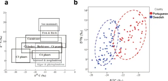
nitrogen (δ 15N) can be used to differentiate between different types of terrestrial and marine-derived food (Fig. 1a).
To understand the role of diet in CHD mortality, we investigated the dietary origin of the nitrogen and carbon molecules present in atherosclerotic plaques obtained from Portuguese and Swedish carotid surgery patients. We also analysed how the dietary origin of the plaque nitrogen and carbon molecules related to structural components associated with plaque stability.
Results
There were no significant differences in clinical characteristics between Portuguese and Swedish patients, except that the use of statins was more common in Swedish patients and the time between the clinical event and operation that was shorter in the Portuguese patients (Table 1).
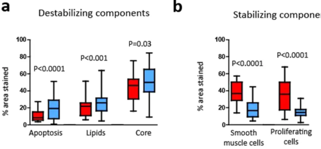
We first analysed the structural components important for plaque vulnerability in both Portuguese and Swedish plaques. Swedish plaques were found to have more apoptotic cells, increased levels of lipids and larger cores than Portuguese plaques (Fig. 2a). The Swedish plaques were also characterized by fewer proliferating cells and less smooth muscle cells (Fig. 2b). Figure 2a depicts destabilizing components ana-lysed, namely apoptosis, lipids and core size. The higher levels present in plaques from Swedish patients
Figure 1. (a) Generalized isotopic trophic diagram for terrestrial and marine food webs34. (b) Stable isotope
(δ 13C and δ 15Ν ) diagram for Portuguese and Swedish plaques.
Swedish patients operated 2005-2011 (n = 35) Portuguese patients operated 2000-2001 (n = 21) P value Age (years) 73.7 (SD 9.7) 69.0 (SD 10.6) NS Gender (males) 23 (66%) 16 (76%) NS Symptoms 27 (77%) 13 (62%) NS
Time between symptoms and operation (days) 29.2 (SD 29.4) 10.0 (SD 10.8) 0.005
Degree of stenosis (%) 87 (SD 9.8) 83 (SD 7.3) NS
Type 2 diabetes 16 (46%) 6 (29%) NS
Hypertension 26 (74%) 20 (95%) NS
Smoking (currently) 12 (34%) 4 (19%) NS
Statin use 22 (63%) 5 (24%) 0.006
Fasting lipoproteins (mmol/L):
Cholesterol 4.4 (SD 1.2) 5.3 (SD 1.4) NS
Low-density lipoprotein (LDL) 2.7 (SD 1.0) 2.9 (SD 1.4) NS
High-density lipoprotein (HDL) 1.1 (SD 0.4) 0.9 (SD 0.4) NS
Triglycerides 1.5 (SD 0.8) 1.5 (SD 0.2) NS
Table 1. Clinical characteristics of the patients that underwent carotid endarterectomy. SD, standard
are shown in blue boxes as compared to the Portuguese in red. Accordingly, using the same colour scheme, in Fig. 2b, the stabilizing plaque components measured are shown, e.g. less smooth muscle cells and proliferating cells in the Swedish plaques.
In Fig. 3 are shown two representative images of carotid plaques from the two countries where larger amounts of lipids making a large lipid core (Fig. 3a, in red) is seen in the Swedish plaques. Moreover, reduced number of smooth muscle cells is observed in the consecutive section (Fig. 3b, in brown). In contrast, a lipid-poor and smooth muscle cell-rich plaque with a thick cap from a Portuguese subject is
Figure 2. Boxplots showing the histological analysis of carotid plaque components (% area) from
Portuguese (in red) and Swedish (in blue) patients: (a) apoptosis (TUNEL, P < 0.0001), lipids (Oil Red O, P < 0.001) and core (P = 0.03), (b) smooth muscle cells (alpha-actin, P < 0.0001) and proliferative cells (PCNA, P < 0.0001).
Figure 3. Representative images of a Swedish (a,b) and a Portuguese (c,d) carotid plaque stained for lipids
(in red, Oil Red O; left panel) and for smooth muscle cells (SMCs, in brown, alpha-actin; right panel). Scale bar 500 μm.
shown in Fig. 3c,d. All of these features are compatible with a more vulnerable phenotype of the Swedish plaques.
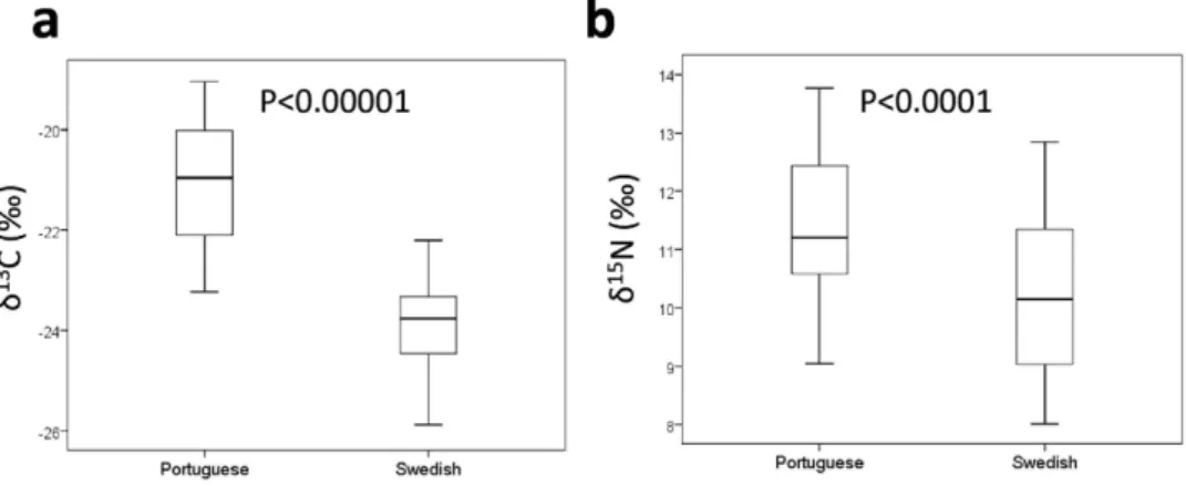
The distribution of δ 13C and δ 15N isotopes in Swedish (in blue) and Portuguese (in red) plaques demonstrated markedly different patterns (Fig. 1b). Comparison of δ 13C and δ 15N levels showed that plaques from Portuguese patients contained significantly higher values of both the δ 13C and δ 15N than plaques from Swedish patients (Fig. 1b and 4). Both δ 13C and δ 15N levels correlated positively with stain-ing for smooth muscle cells while inverse associations were noted for lipids, core size, apoptosis (Table 2). δ 13C was also positively correlated with PCNA staining (Table 2). All associations remained significant after adjustments for age, gender, country of origin, current smoker and diabetes (Table 2).
Discussion
We show for the first time that diet is reflected in plaque components related with its stability. We used mass spectrometry to analyse carbon and nitrogen isotope ratios in human atherosclerotic plaques and expressed the isotopic fractionation in terms of δ 13C and δ 15N. The distribution of these isotopes in living tissues reflects their dietary origin with high levels of both reflecting marine dietary sources, particularly fish (Fig. 1a)15–21. We detected increased levels of both δ 13C and δ 15N in human carotid plaques from Portuguese patients compared to Swedish. Portuguese plaques had a more stable phenotype and δ 13C and δ 15N levels correlated significantly with several factors associated with atherosclerotic plaque stability including the lipid content, core size, smooth muscle cells, as well as rates of cell proliferation and apop-tosis. These associations remained significant when adjusting for country of origin demonstrating that a high intake of seafood was associated with a more stable plaque phenotype in both countries.
There is a wide range of possible speculations for the differences in mortality between southern and northern Europe22, including differences in diet and in plaque stability. The detailed dietary patterns of the two countries studied are well characterized in the reports of Food and Agriculture Organization of the United Nations. The Portuguese, on average, consume more fish products (13.4%) than Swedes (7.8%)23. As shown in Fig. 1a, the Portuguese diet includes more fish (leading to higher δ 13C and δ 15N values) and maize (C4 group, meaning higher δ 13C values), than the Swedish diet, which in general contains a significant amount of potatoes (C3 group, leading to lower values of both δ 13C and δ 15N), as
Figure 4. Boxplots showing (a) δ13C levels (P < 0.00001) and (b) δ15Ν levels in carotid plaques from
Portuguese and Swedish patients (P < 0.0001).
Area of component
Non-adjusted Adjusted*
δ13C δ15N δ13C δ15N
Core − 0.323§ − 0.311§ − 0.186† − 0.272‡
Lipids (Oil Red O) − 0.41§ − 0.256‡ − 0.328§ − 0.186†
Apoptosis (TUNEL) − 0.516§ − 0.374§ − 0.457§ − 0.328§
Macrophages (CD68) NS 0.201† NS NS
Smooth muscle cells (alpha-actin) 0.769§ 0.437§ 0.361§ 0.227‡
Proliferating cells (PCNA) 0.423§ NS − 0.188† NS
Table 2. Correlations between the stable isotope ratios δ13C and δ15N and the different human atherosclerotic plaque components assessed histologically and immunohistochemically. NS,
non-significant. *Adjusted for age, gender, country, current smoker and diabetes. †P < 0.05; ‡P < 0.01; §P < 0.0001.
and AtheroExpress have provided prospective data confirming that presence of vulnerable plaques are associated with an increased risk of future cardiovascular events.
There are some differences between the patient cohorts that could affect our findings. (1) The time of collection (Portuguese plaques 2000–2001 vs. Swedish plaques 2005–2011), (2) the time between the clinical event and surgery (shorter in Portugal than in Sweden) and (3) the use of statins (more common in Swedish patients). Data from the AtheroExpress biobank have shown that atherosclerotic plaques obtained in 2010–2011 had a more stable phenotype than plaques obtained in 2001–200228. Accordingly, it is unlikely that the more stable phenotype of Portuguese plaques in the present study could be explained by the year of surgery. Also the shorter time between the clinical event and surgery for the Portuguese plaques is unlikely to explain their more stable phenotype because there is less time for healing responses after plaque rupture to occur. Finally, statins are known to have stabilizing effects on plaque structure29, thereby one would expect the Swedish plaques to be more stable than the Portuguese.
Our results using samples from Portugal and Sweden should be interpreted with caution, as they cannot be directly extrapolated to other regions of Europe. Large multinational studies ideally evaluat-ing the characteristics of the plaques in asymptomatic patients through Europe are now required. Our study cannot provide exact amounts of dietary elements or even exact types of maritime/seafood dietary sources. The IRMS results also have to be interpreted with some caution, as the δ 13C and δ 15N values do not only reflect the diet, but also depend on the type of tissue17,30. Finally, the histological analysis was only performed in the most stenotic region of the plaques. Plaques are heterogeneous and plaque structure can vary along the vessel wall. The most stenotic region is considered to have most of the representative components31, but variations in other parts of the plaque are possible. Finally the plaques studied corresponded to advanced atherosclerotic disease. No conclusions can be extrapolated to subjects with normal arterial walls or less advanced stages of the disease.
Taken together, these data indicate that the atherosclerotic plaque composition is consistent with the intake of certain dietary atoms. The composition of the Portuguese atherosclerotic plaques is associated with the increased intake of seafood and those plaques are more stable than those from Swedish patients.
This study is pioneer in showing that marine-derived food is associated with plaque stability. Despite the need for further studies, the ultimate clinical implication of this knowledge is to encourage a simple and cheap strategy, as marine dietary intake in prevention of atherosclerosis, to stabilize rupture-prone plaques that ultimately lead to myocardial infarction and stroke.
Methods
Clinical samples. We studied human atherosclerotic plaques obtained by carotid endarterectomy from 56 patients: 21 Portuguese and 35 Swedish. The Swedish patients underwent carotid surgery at the Skåne University Hospital, Malmö, Sweden, during 2005 to 2011 and the Portuguese were operated in 2000–2001 at the Cardiovascular Institute of Lisbon, Lisbon, Portugal. The patients’ characteristics are described in Table 1. All patients were preoperatively assessed by an independent neurologist as having significant stenosis (stenosis > 70% for the plaques associated with symptoms (transient ischemic attacks (TIA), strokes or amaurosis fugax) or > 80% for the asymptomatic). Stenosis grade was measured accord-ing to the velocity criteria assessed by ultrasound32.
All methods were carried out in accordance with the approved guidelines. The study was approved by the local ethical committee (Regional Ethical Review Board in Lund). All patients gave informed consent. Sample processing. All plaques were snap-frozen in liquid nitrogen at endarterectomy. Two consec-utive 1-mm-thick transverse sections of the most stenotic region were sliced from each sample, one for δ 13C/δ 15N analysis and one for histology. The plaque samples for the isotope fractionation analysis were dissected into different regions (fibrous cap, core and interface between the core and the outer cleavage plan of the plaque towards the media).
δ13C and δ15N measurements. The samples were dried and prepared as previously described33. The stable isotope (δ 13C, δ 15N) analysis was performed at IRMS facility at the Environmental Isotope Laboratory (EIL) at University of Waterloo, Ontario, Canada. The required weight of each sample was 0.25-0.30 mg. The ratios of the samples were calibrated against several different standards of known
isotopic composition. The analytical precision obtained for the standards was < 0.3‰ for N and < 0.2‰ for C (1σ )34.
Histological and immunohistochemical analysis. The fragments were cryosectioned in transver-sal 8 μ m sections, fixed with Histochoice (Amresco, Ohio, USA), dipped in 60% isopropanol and in 0.4% Oil Red O in (60%) isopropanol (for 20 min) to stain lipids. For macrophage assessment, primary mon-oclonal antibody mouse anti-human CD68, clone KP1 (DakoCytomation, Glostrup, Denmark), diluted in 10% rabbit serum 1:100, and secondary antibody biotinylated polyclonal rabbit anti-mouse, rabbit F(ab´)2 (DakoCytomation, Glostrup, Denmark), dilution 1:200 in 10% of rabbit serum, were used. For smooth muscle cells (alpha-actin), primary antibody monoclonal mouse anti-human smooth muscle actin clone 1A4 (DakoCytomation, Glostrup, Denmark), diluted in 10% rabbit serum 1:50, and second-ary antibody biotin rabbit anti-mouse Ig (DakoCytomation, Glostrup, Denmark), dilution 1:200 in 10% of rabbit serum, were used. To visualize apoptotic cells in plaques, TUNEL (terminal deoxynucleotidyl transferase dUTP nick-end labelling) In Situ Cell Death detection kit POD (Roche Applied Science, Indianapolis, Ind, USA) was used, according to manufacturer’s instructions. Proliferation was assessed by staining with mouse monoclonal [PC10] anti-human proliferating-cell nuclear antigen (PCNA) prolifer-ation marker (ab29) (1:100, Abcam, Cambridge, UK; overnight incubprolifer-ation at 4 °C). Sections were subse-quently incubated with biotinylated polyclonal rabbit anti-mouse F(ab)2 (E0413,1:200, DakoCytomation, Glostrup, Denmark) for 30 minutes and then with peroxidase-labelled streptavidin (Vectastain ABC-AP kit, Vector Laboratories, Peterborough, UK). Measurements of the area of plaque (% area) for the lipids, macrophages, smooth muscle cells, apoptosis and proliferation, as well as the core region were quantified blindly using Biopix Q 2.1.8 (Gothenburg, Sweden) after scanning with ScanScope Console Version 8.2 (LRI imaging AB, Vista CA, USA).
Statistics. The distribution of δ 13C and δ 15N was approximately normal (skewness < 0.33) and no log transformation was applied. Values are presented as mean (standard deviation, SD). Two-group compar-isons were performed with Chi-square or Mann-Whitney test. Spearman’s rho test was used for correla-tion analysis. Multiple linear regressions, with δ 13C or δ 15N as dependent variables, were used to adjust the relationship between histologic plaque components and δ 13C and δ 15N, for potential confounding factors. Age, gender, current smoker, diabetes, country of origin and histological plaque component were entered into the regression model. We also investigated whether presence of symptoms, time between symptom and operation, statin use, degree of stenosis and date of the operation, could act as confound-ing factors for the relationship between isotope pattern and histological plaque components. However, these factors showed no substantial effect on this relationship and were therefore not included in the final regression model. Differences were considered statistically significant at P < 0.05. SPSS 21 (SPSS Inc., Chicago, Ill, USA) has been used for statistical analysis.
References
1. Allender, S. et al. Mortality. European cardiovascular disease statistics. 2008 edn, 1-112 European Heart Network, (2008) Available at: http://hdl.handle.net/10536/DRO/DU:30020501 (Accessed: 13th October 2009).
2. Keys, A. Mediterranean diet and public health: personal reflections. Am J Clin Nutr 61, 1321S–1323S (1995).
3. Kris-Etherton, P. et al. AHA Science Advisory: Lyon Diet Heart Study. Benefits of a Mediterranean-style, National Cholesterol Education Program/American Heart Association Step I Dietary Pattern on Cardiovascular Disease. Circulation 103, 1823–1825 (2001).
4. Guasch-Ferre, M. et al. Frequency of nut consumption and mortality risk in the PREDIMED nutrition intervention trial. BMC
medicine 11, 164 (2013).
5. Guasch-Ferre, M. et al. Olive oil intake and risk of cardiovascular disease and mortality in the PREDIMED Study. BMC medicine
12, 78 (2014).
6. Howard, D. P. et al. Symptomatic carotid atherosclerotic disease: correlations between plaque composition and ipsilateral stroke risk. Stroke 46, 182–189 (2015).
7. Hansson, G. K. Inflammation, atherosclerosis, and coronary artery disease. N Engl J Med 352, 1685–1695 (2005). 8. Golledge, J., Greenhalgh, R. M. & Davies, A. H. The symptomatic carotid plaque. Stroke 31, 774–781 (2000). 9. Ross, R. Atherosclerosis—an inflammatory disease. N. Engl. J. Med. 340, 115–126 (1999).
10. Falk, E. Why do plaques rupture? Circulation 86, III30–42 (1992).
11. Naghavi, M. et al. From vulnerable plaque to vulnerable patient: a call for new definitions and risk assessment strategies: Part I.
Circulation 108, 1664–1672 (2003).
12. Virmani, R., Burke, A. & Farb, A. Coronary risk factors and plaque morphology in men with coronary disease who died suddenly. Eur Heart J 19, 678–680 (1998).
13. Schoeller, D. A., Minagawa, M., Slater, R. & Kaplan, I. R. Stable isotopes of carbon, nitrogen and hydrogen in the contemporary North American human food web. Ecology of Food and Nutrition 18, 159–170 (1986).
14. Schoeller, D. A. Isotope fractionation: Why aren’t we what we eat?. Journal of Archaeological Science 26, 667–673 (1999). 15. O'Connell, T. C. & Hedges, R. E. Investigations into the effect of diet on modern human hair isotopic values. American journal
of physical anthropology 108, 409–425, (1999).
16. Petzke, K. J., Boeing, H. & Metges, C. C. Choice of dietary protein of vegetarians and omnivores is reflected in their hair protein
13C and 15N abundance. Rapid communications in mass spectrometry: RCM 19, 1392–1400, (2005).
17. Post, D. M. et al. Getting to the fat of the matter: models, methods and assumptions for dealing with lipids in stable isotope analyses. Oecologia 152, 179–189 (2007).
18. O'Brien, D. M. et al. Red blood cell δ15N: a novel biomarker of dietary eicosapentaenoic acid and docosahexaenoic acid intake.
Am J Clin Nutr 89, 913–919 (2009).
19. Georgiadou, E. et al. Bomb-pulse 14C analysis combined with 13C and 15N measurements in blood serum from residents of
29. Crisby, M. et al. Pravastatin Treatment Increases Collagen Content and Decreases Lipid Content, Inflammation, Metalloproteinases, and Cell Death in Human Carotid Plaques: Implications for Plaque Stabilization. Circulation 103, 926–933 (2001).
30. Tieszen, L. L., Boutton, T. W., Tesdahl, K. G. & Slade, N. A. Fractionation and turnover of stable carbon isotopes in animal tissues: implications for δ13C analysis of diet. Oecologia 57, 32–37 (1983).
31. Seeger, J. M., Barratt, E., Lawson, G. A. & Klingman, N. The relationship between carotid plaque composition, plaque morphology, and neurologic symptoms. J Surg Res 58, 330–336 (1995).
32. Hansen, F. et al. Accuracy of duplex sonography before carotid endarterectomy—a comparison with angiography. Eur J Vasc
Endovasc Surg 12, 331–336 (1996).
33. Georgiadou, E. et al. Potential influence of diet on bomb-pulse dating of human plaque samples. Radiocarbon 55, 874–884 (2013).
34. Georgiadou, E. A. Exploring the Possibilities of l4C Bomb-Pulse Dating of Human Tissue Samples, PhD thesis, Lund University,
(15/9/2014).
Acknowledgements
We are grateful to Ana Persson, Lena Sundius and Mihaela Nitulescu, and Dan Csontos at Elevate Scientific for helpful discussion when preparing the manuscript. Sources of Funding: This work was supported by Innovative Medicines Initiative/European Commission’s Seventh Framework Program, Swedish Research Council, Marianne and Marcus Wallenberg, Swedish Heart and Lung Foundations, Swedish Medical Society and Swedish Foundation for Strategic Research.
Author Contributions
I.G. designed the study, performed the analyses and statistics, drafted the manuscript. E.A.G. performed the analyses and statistics. S.M. and G.S. critically revised the manuscript for important intellectual content. L.P. and J.F.F. operated patients, performed analyses and revised the manuscript. N.D. designed the study and performed some of the analyses. G.E. performed the analyses and statistics. J.N. designed the study, critically revised the manuscript for important intellectual content. K.S. designed the study, drafted the manuscript. All authors reviewed the manuscript.
Additional Information
Competing financial interests: The authors declare no competing financial interests.
How to cite this article: Gonçalves, I. et al. Direct association between diet and the stability of human
atherosclerotic plaque. Sci. Rep. 5, 15524; doi: 10.1038/srep15524 (2015).
This work is licensed under a Creative Commons Attribution 4.0 International License. The images or other third party material in this article are included in the article’s Creative Com-mons license, unless indicated otherwise in the credit line; if the material is not included under the Creative Commons license, users will need to obtain permission from the license holder to reproduce the material. To view a copy of this license, visit http://creativecommons.org/licenses/by/4.0/