FACULDADE DE CIÊNCIAS DO MAR E DO AMBIENTE
DECAPOD CRUSTACEAN LARVAE DYNAMICS IN THE WEST
CONTINENTAL COAST OF PORTUGAL: THE REGION
ADJACENT TO AVEIRO COASTAL LAGOON AS MODEL
CÁTIA ALEXANDRA VIEIRA BARTILOTTI
Doutoramento em Ciências do Mar, especialidade de Ecologia Marinha
FARO (2010)
FACULDADE DE CIÊNCIAS DO MAR E DO AMBIENTE
DECAPOD CRUSTACEAN LARVAE DYNAMICS IN THE WEST
CONTINENTAL COAST OF PORTUGAL: THE
REGION ADJACENT TO AVEIRO COASTAL LAGOON AS MODEL
CÁTIA ALEXANDRA VIEIRA BARTILOTTI
Doutoramento em Ciências do Mar, especialidade de Ecologia Marinha
Tese orientada por:
Investigadora Doutora Antonina dos Santos Professora Doutora Margarida Castro
FARO (2010)
iii
“Porque sou do tamanho do que vejo E não do tamanho da minha altura”
v
I would like to express my gratitude to all of those that gave me support through these years of work, even knowing that the words are not enough to recognize all the received assistance. First of all I must thank to FCT for the financial support (SFRH/ BD/ 16695/ 2004), to IPIMAR that gave me the conditions to fulfill my PhD working plan, and to my supervisors Dr. Margarida Castro and Dr. Antonina dos Santos for all the guidance and support. To Dr. Margarida Castro, thank you for all the help at the University, for the statistical analysis and for all the nice talks about such varied themes; to Dr. Antonina dos Santos, thank you for these eight years of lessons, friendship and confidence… As I always say, Gurney is a visionary, so if he had a time machine (as you always say), I am sure that he would like to discuss everything about decapod larvae with a generalist like you!
To Dr Miguel Santos for the constant care. To the “ProRecruit- Shelf processes controlling recruitment to littoral populations in an eastern oceanic boundary: using barnacles and crabs as models” (POCTI/1999/BSE/36663) staff, in particular, Dr. Henrique Queiroga, Dr. Álvaro Peliz and Dr. Patrícia Lourenço.
To the Crustacea friends: Dr. Andrew Rhyne, thank you for all the Lysmata larvae that you gave me (I believe that they will keep me far from Alzheimer); Dr. Juan Ignacio González-Gordillo for the Ilia nucleus larval series and all the confidence deposited in my work; and last but not least, to Dr. Ricardo Calado that is an excellent person but also an excellent scientist always with a new idea to discuss and develop. I also express thanks to all the co-authors of my papers for all the suggestions made that greatly improved the quality of my work and taught me so much.
vi
and taking care of me, thank you for your good mood and thank you for your nice smile; Dr. Sofia Palma and Dr. Alexandra Silva, friends of “marmita” lunch time, living at the next door lab, were the perfect companionship for the larval description pauses; Dr. Susana Garrido, the “perfumed” friend, always looking for digested phyto- and zooplankton in sardines stomachs, shared the smell but also her friendship; Dr. Juan Zwolinski, the tea time friend, who shared the activities of the weekend and also the GAM’s and other statistical methodologies.
To my friends, that always believed: Carla, Daniela, Eugénio, Irina, Joana, Marta, Miguel, Sara, Rui, Vanessa, thank you for your love, your understanding, your support. You make my life happier, simpler and funnier. To all those that I don’t refer but in some way were important to me, thank you!
To my Super-heroes family! Thank you! Aqueles que são meus e que estão comigo sempre, que me amam e protegem incondicionalmente, a eles dedicam o meu trabalho! À Avó, Mãe, Luis e Nino, obrigada por serem os meus anjos! Ao Pai que considero um super-homem a cada dia que passa. Aos tios, primos e bebés, por me mimarem sempre. A todos estes que me ajudaram a percorrer o caminho, muito obrigada! Foi um fim difícil… Aos que não me acompanharam, também agradeço, porque me mostraram que os nossos limites são sempre ultrapassáveis…
vii
FACULDADE: Ciências do Mar e do Ambiente DATA: 12 de Fevereiro de 2010
TÍTULO DA TESE: Dinâmica larvar de crustáceos decápodes na costa ocidental de
Portugal Continental: a região adjacente à Ria de Aveiro como modelo.
RESUMO
O Filo Crustacea é um dos maiores taxa do Reino Animal, e a Ordem Decapoda é a maior com cerca de 50000 espécies, um número que continua a aumentar. A maioria dos decápodes distribui-se no oceano e nas zonas estuarinas adjacentes, no entanto algumas espécies invadiram a água doce, e um pequeno número de espécies ocupou os habitats terrestres. A diversidade de espécies de decápodes reflecte a diversidade de estilos de vida, e sendo na sua maioria bentónicos, estes animais podem viver nos habitats tropicais, mangais, recifes de coral, regiões polares, zonas do oceano profundo ou fontes hidrotermais. Os crustáceos decápodes são bastante importantes pois constituem um dos grupos biológicos com pesca dirigida e são muitas vezes elos da cadeia alimentar de inúmeros recursos pesqueiros da nossa costa. Uma estratégia adequada de gestão pesqueira para uma espécie com valor económico requer o conhecimento do seu ciclo de vida. Assim, para os crustáceos decápodes os estudos de plâncton dão informação acerca da distribuição e abundância das espécies durante a fase larvar planctónica. A fase de recrutamento é particularmente crítica nestes organismos, uma vez que implica a articulação de duas fases do ciclo de vida separadas espacialmente: a fase larvar planctónica e a fase adulta bentónica, ou seja, o “stock” explorável depende da sobrevivência dos estádios larvares. A fase larvar constitui assim um período vital no ciclo de vida das várias espécies de decápodes e tem o papel fundamental nos processos de intercâmbio genético, de recrutamento e consequente renovação de populações e “stocks”.
As larvas de crustáceos decápodes são nadadoras activas, capazes de regular a sua posição vertical na coluna de água, controlando a extensão e direcção da dispersão horizontal, mantendo a posição favorável ao transporte necessário e adequado à fase do ciclo de vida em que se encontram. Os processos de transporte são decisivos para o mecanismo de fornecimento larvar aos habitats onde o assentamento e o desenvolvimento juvenil irão ocorrer, separando a ontogenia no tempo e no espaço, expondo as larvas a diferentes factores de mortalidade. A compreensão dos padrões dos movimentos temporais e espaciais larvares é fundamental para estudar a ecologia dos decápodes, permitindo o design de estratégias efectivas de conservação e gestão dos recursos. As migrações verticais são a estratégia adoptada pelas larvas de crustáceos decápodes para o transporte adequado e necessário à fase do ciclo de vida em que se encontram, e normalmente são separadas em dois grandes grupos: as migrações verticais diárias (DVM) e as migrações ontogénicas. A fase larvar pelágica apresenta uma morfologia diferente da do adulto. O desenvolvimento larvar dos crustáceos decápodes é descrito como uma sequência de estádios larvares
viii
respectivamente). As larvas de crustáceos decápodes podem ter diferentes formas mas usualmente apresentam padrões de desenvolvimento estáveis. Na análise dos processos bio-ecológicos envolvidos nesta fase do ciclo de vida, deverão considerar-se além dos factores hidrológicos, a duração do desenvolvimento larvar, e o número de estádios larvares que cada ciclo larvar planctónico comporta, bem como as suas características morfológicas como estratégias adaptativas aos ecossistemas costeiros, sendo que a forma hidrodinâmica das diferentes larvas poderá corresponder a diferentes padrões de dinâmica larvar, ou seja, as características morfológicas podem ser encaradas como estratégias adaptativas aos ecossistemas. Tendo em conta o universo considerado (larvas de crustáceos decápodes), no que diz respeito à forma, podemos agrupá-las nas seguintes formas tipo: tipo A (carapaça e abdómen achatados lateralmente; telson de forma triangular com invaginação central; camarões sensu lato); tipo B (corpo fortemente achatado dorso-ventralmente; apêndices muito longos, projectados lateralmente; infraordem Palinura); tipo C (carapaça achatada lateralmente mas ligeiramente arredondada; abdómen de forma cilíndrica; telson de forma triangular ou bifurcado; infraordens Astacidea e Anomura); tipo D (carapaça quase esférica; usualmente apresenta espinhos laterais, dorsais e um rostro ventralmente direccionado; abdómen de forma cilíndrica e telson em forma de furca; caranguejos). Destas vamos estudar apenas as formas tipo camarão carídeo (tipo A), caranguejo eremita (tipo C) e caranguejo (tipo D).
Esta tese pretende descrever os padrões de distribuição horizontal e vertical (ontogénica) das larvas de crustáceos decápodes no sistema de afloramento costeiro português. Pretende também fazer a descrição morfológica externa do desenvolvimento larvar de espécies de cada uma das formas tipo escolhidas: os camarões carídeos, os caranguejos eremita e os caranguejos. Para completar os objectivos ecológicos e morfológicos deste trabalho, desenvolveram-se 5 capítulos, os dois primeiros relativos à ecologia e os três últimos relativos à taxonomia e morfologia dos estádios larvares de decápodes.
Nos capítulos 2 e 3 descrevem-se os padrões de distribuição horizontal e vertical (ontogénica) das larvas de crustáceos decápodes no sistema de afloramento português adjacente à Ria de Aveiro.
O capítulo 2 pretende testar a hipótese de que a distribuição horizontal larvar de todos os crustáceos decápodes na área estudada reflecte um padrão de retenção. As larvas deverão estar distribuídas em bandas, paralelas à costa, concordantes com os intervalos das distribuições dos adultos. Demonstra-se que as migrações verticais diárias (DVM) anteriormente descritas por dos Santos et al. (2008) são consistentes ao longo do desenvolvimento, de quase todos os taxa estudados, e a estratégia de retenção larvar suposta pelos autores existe. As larvas de decápodes estiveram distribuídas ao longo de bandas meridionais alongadas, paralelas à costa, concordantes com as respectivas origens: as espécies da plataforma interna e Ria de Aveiro distribuíram-se junto à costa, as espécies da plataforma distribuíram-se ao longo da zona média da plataforma, e as espécies da vertente distribuíram-se junto ao bordo da plataforma. Assim, um dos resultados mais importantes do presente trabalho é que a distribuição larvar reflecte a distribuição dos adultos. Definem-se três grupos: espécies da "Inner shelf” distribuídas junto à costa com os máximos de abundância a 8 km de distância à costa (ex: Diogenes pugilator); espécies da “Shelf”: distribuídas na plataforma média com os máximos de abundância entre os 20 e os 60 km de distância à costa (ex: Polybius henslowii); e espécies da “Slope”: distribuídas
ix
costeiras e da plataforma. As espécies da vertente deverão ter vantagens em ter parte do seu ciclo de vida na plataforma. No que diz respeito ás distribuições em relação aos factores ambientais verificou-se que as espécies da Ria de Aveiro e da plataforma interna estiveram associadas a temperaturas mais elevadas reflexo do relaxamento do afloramento ocorrido durante a amostragem, e por contrário as espécies da vertente estiveram associadas a temperaturas mais baixas reflexo do afloramento ocorrido antes da amostragem. Os zoés de camarões estiveram associados à lente de água menos salina WIBP que estava a ser advectada para o largo, e os megalopas de caranguejo parecem evitar esta massa de água. O capítulo 3 pretende descrever as migrações verticais ontogénicas num ponto fixo, e também pretende descrever as respostas comportamentais dos estádios larvares das espécies seleccionadas aos factores ambientais. Em geral o primeiro estádio de zoé tem uma posição mais superficial que o segundo estádio, e o último zoé teve sempre uma distribuição mais profunda. O megalopa teve uma profundidade média de distribuição semelhante à calculada para o ultimo estádio de zoé, mas teve em geral muitas vezes concentrado na camada de neuston durante a noite, reflectindo provavelmente um mecanismo de transporte. Durante a amostragem a distribuição dos campos hidrológicos foi complexa.
Nos capítulos 4 a 6 apresentam-se as descrições morfológicas externas dos desenvolvimentos larvares de crustáceos decápodes obtidos no laboratório das três formas tipo seleccionadas: os camarões carídeos de duas espécies próximas de Lysmata, L. galapagensis e L. moorei, os caranguejos eremitas Clibanarius aequabilis e C. erythropus, e o desenvolvimento larvar do caranguejo Ilia nucleus.
O capítulo 4 teve como objectivos descrever os estádios larvares disponíveis de duas espécies próximas L. galapagensis e L. moorei, comparar estas descrições com as da única espécie descrita no “Cosmopolitan Clade” a L. seticaudata, e com o “género larvar” Eretmocaris dadas as semelhanças existentes entre L. galapagensis e E. corniger, e finalmente rever e discutir os caracteres do género. Ambas as espécies apresentaram um desenvolvimento larvar homogéneo: no primeiro zoé os olhos estão fundidos, a carapaça tem um espinho pterigostomiano seguido de 4-5 dentículos marginais, o escafocerito tem 5 segmentos, o 5º segmento abdominal tem um par de espinhos dorso-laterais, os rudimentos dos pereiópodes 1 e 5 estão presentes; no segundo zoé os olhos são pedunculados, os espinhos antenar e supraorbital estão presentes, um espinho pós-rostral, o pereiópodes 1 e 5 estão funcionais; no terceiro zoé o pedúnculo antenar tem dois segmentos e os dois flagelos estão já presentes, o sexto segmento abdominal está separado do telson, o rudimento do pereiópode 2 está presente, os urópodes estão presentes com um endópode rudimentar; no zoé IV o escafocerito não apresenta qualquer segmento, o segundo pereiópode está funcional, e os urópodes são tão longos quanto o telson; o zoé V tem os rudimentos birramosos dos pereiópodes 3 e 4, e os urópodes são tão longos quanto o telson; zoé VI tem os pereiópodes 3 e 4 funcionais e apresenta já uns pequenos rudimentos dos pleópodes no abdómen; no penúltimo estádio (ZVII ou ZVIII) o flagelo da antena é tão longo quanto o escafocerito e tem 8 segmentos, os pleópodes são já birramosos e os endópodes e exópodes têm pequenas sedas apicais; último zoe (ZVIII ou ZIX) tem já o flagelo da antena mais longo que a escama e com mais de 8 segmentos, os pleópodes apresentam o apêndice interno, o telson muito semelhante ao de um adulto. Ambas as espécies eclodem com uma forma semelhante à de L. seticaudata com os rudimentos dos pleópodes 1 e 5. Como o presente trabalho demonstra o estudo das larvas de Lysmata será certamente uma ajuda
x
padrões de desenvolvimento larvar para estas espécies: o primeiro semelhante a L. galapagensis, L. moorei e L. seticaudata em que as larvas eclodem com os primeiro e quinto pereiópodes como rudimentos que estarão funcionais no estádio seguinte e que completarão o seu desenvolvimento com 8 ou 9 estádios de zoe, e um segundo mais longo com as larvas a eclodirem sem qualquer rudimento dos pereiópodes. O primeiro grupo de espécies pertence ao “Cosmopolitan Clade”/ “Lysmata Clade” (Baeza et al. 2009 e Fiedler et al. submetido), o que nos leva a supor que a presença dos rudimentos do 1º e 5º pereiópodes nas larvas recém eclodidas é uma característica partilhada por todas as espécies deste clade. A presença de um espinho anteriormente curvo na região dorsal do 3º segmento abdominal nos estádios mais velhos de L. galapagensis torna esta espécie muito característica. Esta espécie é muito semelhante aos exemplares descritos por Gopalakrishnan & Laurs (1971), portanto demonstramos que as larvas descritas pelos autores como o E. corniger do Pacífico Este são de facto L. galapagensis. Gurney (1937) descreve Sp. A. V, Eretmocaris corniger, do Oceano Atlântico, e esta larva apresenta a mesma forma geral que L. galapagensis. Tendo em conta a proximidade filogenética sugerida pelos estudos moleculares de Baeza et al. (2009) e Fiedler et al. (submetido) entre a L. galapagensis do Pacífico Este e a L. moorei do Atlântico Oeste, o carácter larvar mais evidente que ambas partilham é a presença de um rostro semelhante, razão que nos leva a concluir que provavelmente existe uma espécie de Lysmata desconhecida provavelmente filogeneticamente mais próxima de L. galapagensis que de L. moorei no Atlântico Este. Tendo em conta que L. galapagensis ocorre no Pacífico Este e a larva mais semelhante conhecida até à data é o Eretmocaris corniger colhido no Atlântico Este, questiona-se a biogeografia deste complexo de espécies trans-istmo do Panamá.
O capítulo 5 apresenta o estudo do desenvolvimento larvar dos caranguejos eremita do género Clibanarius. Descreve-se C. aequabilis e re-descreve-se C. erythropus. Os estádios larvares das duas espécies de Clibanarius do nordeste Atlântico são muito semelhantes, e não são fáceis de distinguir. Uma comparação exaustiva das características morfológicas larvares das espécies do género descritas até à presente data e das descritas no presente trabalho demonstram que este género é muito homogéneo. Apesar da homogeneidade no género Clibanarius verificam-se pequenas variações entre espécies, particularmente no que respeita à formula do telson e à morfologia dos seus processos depois do segundo estádio de zoé. Também o número de estádios larvares pode variar. Sugere-se que se considere como característica geral dos estádios de zoé do género Clibanarius a transformação do 4º processo plumoso do telson num espinho fundido, reduzido ou desenvolvido, do Segundo para o terceiro estádio de desenvolvimento.
Finalmente o capítulo 6 pretende descrever com detalhe os quatro estádios de zoé e o megalopa do caranguejo Ilia nucleus. As características gerais dos 4 zoés e do megalopa destaespécie correspondem às propostas anteriormente por Rice (1980) para os estádios de zoé e por Quintana (1986) para o megalopa da família Leucosiidae. Os estádios larvares descritos no presente estudo são muito semelhantes aos anteriormente descritos. Ng et al. (2008) propuseram recentemente uma nova classificação da família Leucosiidae, com apenas 3 subfamílias: Cryptocneminae, Ebaliinae e Leucosiinae. Os autores afirmaram que a actual subfamília Ebaliinae é um grupo muito heterogéneo. Considerando as características larvares estudadas no presente trabalho, verifica-se que a subfamília Ebaliinae inclui as espécies mais ancestrais e também as mais derivadas nos Leucosiidae, o
xi
dos maxilípedes deve também utilizar-se o desenvolvimento da antenula e dos pereiópodes. Os objectives morfológicos desta tese demonstram a importância da taxonomia no estudo das larvas dos crustáceos decápodes. As espécies descritas e as questões levantadas nos três capítulos podem servir de base a novas hipóteses de trabalho.
Palavras-chave: larvas de crustáceos decápodes, retenção larvar, distribuição vertical
ontogénica, desenvolvimento larvar de Lysmata, desenvolvimento larvar de Clibanarius, desenvolvimento larvar de Ilia.
xii
AVEIRO COASTAL LAGOON AS MODEL
ABSTRACT
Present thesis pretends to describe the horizontal and vertical distribution patterns of decapod crustacean larvae in the Portuguese upwelling ecosystem. The studied taxa were retained close to their parental populations; so, species from the inner shelf had their larvae concentrated close to the shore, species from the shelf had their larvae over the shelf, and the slope species had their larvae along the shelf break. The ontogenetic vertical distribution was also analysed, and the average depth of distribution of the larval stages varied through the larval development. The majority of the studied species had their early zoeal stages more close to the surface and the last zoeal stages in a more deep position. The morphological larval descriptions from laboratory cultured material of the three selected larval forms of decapod larvae are presented: the larval development of two closely related species of Lysmata, L. galapagensis e L. moorei (caridean shrimps), the complete larval developments of Clibanarius aequabilis and C. erythropus (hermit crabs), and the complete larval development of the crab Ilia nucleus.
Key-words: decapod crustacean larvae, larval retention, ontogenetic vertical migration,
Page
TITLE……….………. i
ACKNOWLEDGEMENTS………..……….. v
RESUMO……….. vii
ABSTRACT………...…... xii
TABLE OF CONTENTS……… xiii
INDEX OF FIGURES……….xvii
INDEX OF TABLES………xxvi
LIST OF ABBREVIATIONS………xxviii
CHAPTER 1- General introduction……… 1
1.1- The importance of decapod crustaceans………..………..……….. 2
1.2- Decapod crustaceans life cycle: ecology………..……...…… 3
1.3- Decapod crustaceans life cycle: morphology………..………… 5
1.4- Objectives……… 8
1.5- Structure of this Dissertation... 10
1.6- Study area... 12
1.7- References... 13
CHAPTER 2- Circulation patterns and decapod larvae distribution in a coastal upwelling ecosystem... 19
2.1- Abstract... 20
2.2- Introduction... 21
2.3- Material and Methods... 23
xiv
2.3.3- Analysis of decapod larval distribution in relation to the physical
environment...25
2.4- Results...29
2.4.1- Oceanographic conditions...29
2.4.2- Decapod larvae distribution... 32
2.4.2.1- Horizontal and vertical distributions of the Inner Shelf species... 35
2.4.2.2- Horizontal and vertical distributions of the Shelf Species... 37
2.4.2.2- Horizontal and vertical distributions of the Slope Species... 47
2.5- Discusson... 50
2.6- References... 56
CHAPTER 3- Ontogenetic vertical migration behaviour of decapod larvae in the Portuguese upwelling ecosystem... 62
3.1- Abstract... 63
3.2- Introduction... 64
3.3- Material and Methods... 66
3.3.1- Field study... 66
3.3.2- Sample processing... 68
3.3.3- Analysis of decapod vertical distribution in relation to the physical environment... 68
3.4- Results... 70
3.4.1- Oceanographic conditions... 70
3.4.2- Decapod larval stages vertical distribution... 72
xv
3.4.2.2- Vertical distribution of the brachyuran crabs larvae... 83
3.5- Discussion... 86
3.6- References... 91
CHAPTER 4- Shedding light over the larval genus Eretmocaris- morphological larval features of two closely related trans-isthmian Lysmata species using laboratory cultured material... 96
4.1- Abstract... 97
4.2- Introduction... 98
4.3- Material and Methods... 100
4.3.1- Larval culture techniques... 100
4.3.2- Larval drawings and measurements... 100
4.4- Results... 102
4.5- Discussion... 136
4.5.1- Morphological comparisons of the zoeal stages…………...……… 136
4.5.2- Biogeographical considerations on Eretmocaris corniger... 141
4.5.3- Biodiversity and conservation issues... 143
4.5.4- Conclusions... 144
4.6- References... 145
CHAPTER 5- Complete larval development of the hermit crabs Clibanarius aequabilis and Clibanarius erythropus (Decapoda: Anomura: Diogenidae), under laboratory conditions, with a revision of the larval features of genus Clibanarius... 150
xvi
5.2- Introduction... 151
5.3- Material and Methods... 152
5.4- Results... 154
5.5- Discussion... 177
5.6- References... 186
CHAPTER 6- Complete larval development of the crab Ilia nucleus (Linnaeus, 1758) (Decapoda, Brachyura, Leucosiidae) reared under laboratory conditions... 190
6.1- Abstract... 191
6.2- Introduction... 191
6.3- Material and Methods... 193
6.4- Results... 194
6.5- Discussion... 207
6.6- References... 212
xvii
Page Chapter 1
Figure 1. A: Zoea I of Callianassa tyrrhena; B: Zoea II of Palinurus elephas; C: Zoea I of Nephrops
norvegicus; D: Zoea I of Carcinus maenas. In: dos Santos (1999). 7
Chapter 2
Figure 1- Map of the northwest coast of Portugal showing sampling positions (CTD: conductivity,
temperature and depth profiler; and LHPR: Longhurst Hardy plankton recorder) collected aboard the RV
Noruega, during the ProRecruit project survey. Transects are identified as T1, T2, T3 and T4; the squares represent the stations sampled with the Pro-LHPR. The 30, 100 and 200 m bathymetric lines are also
presented. 24
Figure 2- Horizontal (at 20 m depth) and vertical distributions of salinity measured with a CTD. T1, T2, T3
and T4 represent the four sampled transects (respectively 40.9º N, 40.7º N, 40.5º N and 40.3ºN). The 30,
100 and 200 m bathymetric lines are also identified. The horizontal and vertical distributions of salinity are
represented by the solid lines with respective labels. 30
Figure 3- Horizontal (at 20 m depth) and vertical distributions of temperature (º C) measured with a CTD.
T1, T2, T3 and T4 represent the four sampled transects (respectively 40.9º N, 40.7º N, 40.5º N and 40.3ºN).
The 30, 100 and 200 m bathymetric lines are also identified. The horizontal and vertical distributions of
temperature are represented by the grey areas with respective scale. 31
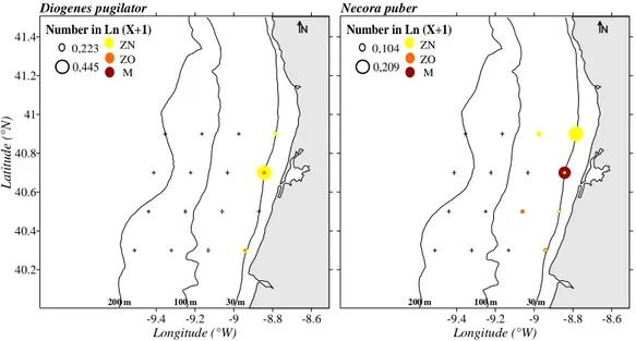
Figure 4- Horizontal distributions of Diogenes pugilator and Necora puber. The 30, 100 and 200 m
bathymetric lines are identified. The abundances are represented in Ln (X+1) with X in ind.10 m-3. ZN
corresponds to the early zoeal stages, ZO to later zoeal stages, and M to megalopa. 36
Figure 5- Vertical distributions of Diogenes pugilator and Necora puber. T1, T2, T3 and T4 represent the
four sampled transects (respectively 40.9º N, 40.7º N, 40.5º N and 40.3ºN). The vertical distributions of
xviii
The abundances are represented in Ln (X+1) with X in ind.10 m . ZN corresponds to the early zoeal stages,
ZO to later zoeal stages, and M to megalopa. 37
Figure 6- Horizontal distributions of Upogebia spp. and Anapagurus spp.. The 30, 100 and 200 m
bathymetric lines are identified. The abundances are represented in Ln (X+1) with X in ind.10 m-3. ZN
corresponds to the early zoeal stages, ZO to later zoeal stages, and M to megalopa. 38
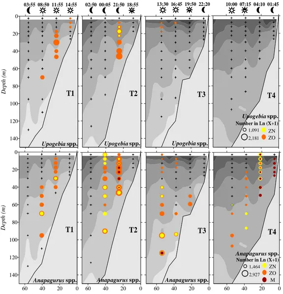
Figure 7- Vertical distributions of Upogebia spp. and Anapagurus spp.. T1, T2, T3 and T4 represent the
four sampled transects (respectively 40.9º N, 40.7º N, 40.5º N and 40.3ºN). The vertical distributions of
temperature are represented by the grey areas (scales in Fig. 3). The night and day periods are represented
by the moon and sun symbols, and the hours of the day are also represented above the night-day symbols.
The abundances are represented in Ln (X+1) with X in ind.10 m-3. ZN corresponds to the early zoeal stages,
ZO to later zoeal stages, and M to megalopa. 39
Figure 8- Horizontal distributions of Processa nouveli and Callianassa subterranea. The 30, 100 and 200
m bathymetric lines are identified. The abundances are represented in Ln (X+1) with X in ind.10 m-3. ZN
corresponds to the early zoeal stages, ZO to later zoeal stages, and M to megalopa. 40
Figure 9- Vertical distributions of Processa nouveli and Callianassa subterranea. T1, T2, T3 and T4
represent the four sampled transects (respectively 40.9º N, 40.7º N, 40.5º N and 40.3ºN). The vertical
distributions of temperature are represented by the grey areas (scales in Fig. 3). The night and day periods
are represented by the moon and sun symbols, and the hours of the day are also represented above the
night-day symbols. The abundances are represented in Ln (X+1) with X in ind.10 m-3. ZN corresponds to
the early zoeal stages, ZO to later zoeal stages, and M to megalopa. 41
Figure 10- Horizontal distributions of Eualus occultus, Pandalina brevirostris, Philocheras bispinosus,
Atelecyclus rotundatus, Liocarcinus spp. and Polybius henslowii. The 30, 100 and 200 m bathymetric lines are identified. The abundances are represented in Ln (X+1) with X in ind.10 m-3. ZN corresponds to the
xix
Figure 11- Vertical distributions of Eualus occultus and Pandalina brevirostris. T1, T2, T3 and T4
represent the four sampled transects (respectively 40.9º N, 40.7º N, 40.5º N and 40.3ºN). The vertical
distributions of temperature are represented by the grey areas (scales in Fig. 3). The night and day periods
are represented by the moon and sun symbols, and the hours of the day are also represented above the
night-day symbols. The abundances are represented in Ln (X+1) with X in ind.10 m-3. ZN corresponds to
the early zoeal stages, ZO to later zoeal stages, and M to megalopa. 44
Figure 12- Vertical distributions of Philocheras bispinosus and Atelecyclus rotundatus. T1, T2, T3 and T4
represent the four sampled transects (respectively 40.9º N, 40.7º N, 40.5º N and 40.3ºN). The vertical
distributions of temperature are represented by the grey areas (scales in Fig. 3). The night and day periods
are represented by the moon and sun symbols, and the hours of the day are also represented above the
night-day symbols. The abundances are represented in Ln (X+1) with X in ind.10 m-3. ZN corresponds to
the early zoeal stages, ZO to later zoeal stages, and M to megalopa. 45
Figure 13- Vertical distributions of Liocarcinus spp. and Polybius henslowii. T1, T2, T3 and T4 represent
the four sampled transects (respectively 40.9º N, 40.7º N, 40.5º N and 40.3ºN). The vertical distributions of
temperature are represented by the grey areas (scales in Fig. 3). The night and day periods are represented
by the moon and sun symbols, and the hours of the day are also represented above the night-day symbols.
The abundances are represented in Ln (X+1) with X in ind.10 m-3. ZN corresponds to the early zoeal stages,
ZO to later zoeal stages, and M to megalopa. 46
Figure 14- Horizontal distributions of Solenocera membranacea, Parthenope spp. and Goneplax
rhomboides. The 30, 100 and 200 m bathymetric lines are identified. The abundances are represented in Ln (X+1) with X in ind.10 m-3. ZN corresponds to the early zoeal stages, ZO to later zoeal stages, and M to
megalopa. 47
Figure 15- Vertical distributions of S. membranacea, Parthenope spp. and G. rhomboides. T1, T2, T3 and
T4 represent the four sampled transects (respectively 40.9º N, 40.7º N, 40.5º N and 40.3ºN). The vertical
xx
night-day symbols. The abundances are represented in Ln (X+1) with X in ind.10 m . ZN corresponds to
the early zoeal stages, ZO to later zoeal stages, and M to megalopa. 48
Chapter 3
Figure 1- Map of the northwest coast of Portugal: sampling position of the fixed station collected aboard
the RV Noruega, from 15-17 May 2002, during ProRecruit project. The 30, 100 and 200 m bathymetric
lines are also identified. 67
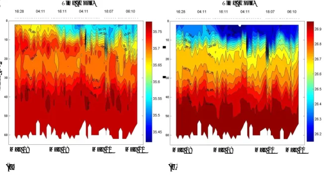
Figure 2- Sequence of CTD measurements of: (a) salinity and (b) density (σt, kg.m-3), during sampling at
the 69 h fixed station, 18-21 May 2002. X axis represents the sampling date; Y axis the depth (m). 71
Figure 3- Temperature (º C) measurement during sampling at the 69 h fixed station, 18-21 May 2002. X
axis represents the sampling date; Y axis the depth (m). 72
Figure 4- Average depth of distribution of Processa nouveli (ind. m-3) zoeal stages: ZI (first zoea) to ZIX
(ninth and last zoea), and M (megalopa). The grey areas represent the night period. 77
Figure 5- Average depth of distribution of Pandalina brevirostris (ind. m-3) zoeal stages: ZI (first zoea) to
ZVII (seventh and last zoea), and M (megalopa). The grey areas represent the night period. 78
Figure 6- Average depth of distribution of Philocheras bispinosus (ind. m-3) zoeal stages: ZI (first zoea) to
ZV (fifth and last zoea), and M (megalopa). The grey areas represent the night period. 79
Figure 7- Average depth of distribution of Anapagurus spp. (ind. m-3) zoeal stages: ZI (first zoea) to ZIV
(fourth and last zoea), and M (megalopa). The grey areas represent the night period. 81
Figure 8- Average depth of distribution of Pagurus bernhardus (ind. m-3) zoeal stages: ZI (first zoea) to
xxi
(second and last zoea), and M (megalopa). The grey areas represent the night period. 83
Figure 10- Average depth of distribution of Atelecyclus rotundatus (ind. m-3) zoeal stages: ZI (first zoea) to
ZV (fifth and last zoea), and M (megalopa). The grey areas represent the night period. 83
Figure 11- Average depth of distribution of Liocarcinus spp. (ind. m-3) zoeal stages: ZI (first zoea) to ZV
(fifth and last zoea), and M (megalopa). The grey areas represent the night period. 84
Figure 12- Average depth of distribution of Necora puber (ind. m-3) zoeal stages: ZI (first zoea) to ZV
(fifth and last zoea), and M (megalopa). The grey areas represent the night period. 85
Chapter 4
Fig. 1- Lysmata galapagensis. First zoea: A, total animal, lateral view; B, antennule; C, antenna; D,
mandibles; E, maxillule; F, maxilla; G, first maxilliped; H, second maxilliped; I, third maxilliped; J, first
pereiopod; K, fifth pereiopod; L, 5th pleomere and telson; L’, detail of the 5th pleomere. Scale bars: 0.5
mm (A); 0.1 mm (B-L’). 104
Fig. 2- Lysmata galapagensis. Second zoea: A, total animal, lateral view; A’, detail of 3rd pleomere, lateral
view; A’’, detail of 5th pleomere, dorsal view; B, carapace, dorsal view; C, antennule; D, antenna; E, first
pereiopod; E’, detail of first pereiopod dactylus setae; E’’, detail of basis of pereiopods; F, fifth pereiopod;
F’, detail of fifth pereiopod propodus lateral spine; F’’, detail of fifth pereiopod end of propodus and
dactylus; G, 5th pleomere and telson. Third zoea: H, detail of antennule flagella; I, detail of scaphocerite
distal segments; J, second pereiopod; K, fifth pereiopod; K’, detail of fifth pereiopod propodus and
dactylus; L, detail of 3rd pleomere, lateral view; M, telson and uropods; M’, endopod of uropods; M’’,
exopod of uropods. Scale bars: 0.5 mm (A, B, G, K); 0.1 mm (A’-A’’, C-F’’, H-J, K’-M’’). 108
Fig. 3- Lysmata galapagensis. Fourth zoea: A, total animal, dorsal view; A’, third pleomere, lateral view of
procurved spine; A’’, fifth pleomere, lateral view of dorso-lateral spines; A’’’, sixth pleomere, lateral view
xxii
pereiopod end of propodus and dactylus; I, telson and uropods. Scale bars: 0.5 mm (A-A’’’, D-F, H, I); 0.1
mm (B-C, G, H’). 111
Fig. 4- Lysmata galapagensis. Fifth zoea: A, carapace, dorsal view; B, maxillule; C, maxilla; D, third
maxilliped; D’, third maxilliped, detail of propodus and dactylus; E, third pereiopod; F, fourth pereiopod;
G, telson and uropods. Sixth zoea: H, total animal, lateral view; I, mandibles; J, first maxilliped; K, second
maxilliped; L, first pereiopod; M, second pereiopod; N, third pereiopod; O, fourth pereiopod. Scale bars: 1
mm (H); 0.5 mm (A, D, E-G, L-O); 0.1 mm (B-C, D’, I-K). 115
Fig. 5- Lysmata galapagensis. Seventh zoea: A, total animal, lateral view; B, carapace, dorsal view; C,
maxillule; D, maxilla; E, first maxilliped; F, second maxilliped; G, third maxilliped; H, first pereiopod; I,
third pereiopod; J, fourth pereiopod; K, fifth pereiopod, propodus and dactylus; L, first pleopod; M,
second pleopod; N, third pleopod; O, fourth pleopod; P, fifth pleopod; Q, telson and uropods. Scale bars: 1
mm (A); 0.5 mm (B, J); 0.1 mm (C-I, K-Q). 119
Fig. 6- Lysmata moorei. First zoea: A, total animal, lateral view; B, antennule; B’, antennule, detail of short
aesthetasc; C, antenna; D, mandibles; E, maxillule; F, maxilla; G, first maxilliped; H, second maxilliped; I,
third maxilliped; J, first pereiopod; K, fifth pereiopod; L, 5th pleomere and telson; L’, detail of the 5th
pleomere. Scale bars: 0.5 mm (A, L); 0.1 mm (B-K, L’). 123
Fig. 7- Lysmata moorei. Second zoea: A, total animal, lateral view; B, antennule; C, antenna; D,
mandibles; E, first pereiopod; E’, detail of basis of pereiopods; F, second pereiopod; G, fifth pereiopod.
Third zoea: H, carapace, dorsal view; I, detail of antennule flagella; J, detail of scaphocerite distal
segments; K, second pereiopod; L, fifth pereiopod; L’, detail of fifth pereiopod propodus and dactylus; M,
telson and uropods; M’, detail of endopod and exopod of uropods. Scale bars: 0.5 mm (A, H); 0.1 mm
(B-G, I-M’). 127
Fig. 8- Lysmata moorei. Fourth zoea: A, total animal, lateral view; B, carapace, dorsal view; C, antennule; D, antenna; E, mandibles; F, first pereiopod; G, second pereiopod; H, third pereiopod; I, fifth pereiopod,
xxiii
and uropods. Scale bars: 0.5 mm (A-B, I); 0.1 mm (C-H, I’-J). 130
Fig. 9- Lysmata moorei. Last zoea: A, total animal, lateral view; B, carapace, dorsal view; C, mandibles; D,
maxillule; E, maxilla; F, first maxilliped; G, second maxilliped; H, third maxilliped, propodus and
dactylus; I, first pereiopod, propodus and dactylus; J, second pereiopod; K, third pereiopod, propodus and
dactylus; L, fourth pereiopod; M, fifth pereiopod, propodus and dactylus; N, first pleopod; N’, first
pleopods, detail of spines; O, second pleopod; O’, second pleopod, detail of appendix interna; P, third
pleopod; Q, fourth pleopod; R, fifth pleopod; S, telson and uropods. Scale bars: 1 mm (A); 0.5 mm (B, H,
J, L, S); 0.1 mm (C-G, I, K, M-R). 133
Chapter 5
Fig. 1- Clibanarius aequabilis. First zoea: a, total animal, dorsal view; a’, detail of rostrum, lateral view; b,
antennule; c, antenna; d, mandible; e, maxillule; f, maxilla; g, first maxilliped; h, second maxilliped; i, third
maxilliped; j, telson; j’, detail of posterior margin of telson. Scale bars: 0.1 mm. 155
Fig. 2- Clibanarius aequabilis. Second zoea: a, detail of rostrum, lateral view; b, antennule; c, mandible; d,
first maxilliped; e, third maxilliped; f, first to third pereiopod. Third zoea: g, total animal, dorsal view; h,
antennule; i, antenna; j, maxilla; k, second maxilliped; l, third pereiopod; m, fourth pereiopod; n, fifth
pereiopod; o, telson and uropods; o’, detail of endopod of uropods. Scale bars: 1.0 mm (g); 0.1 mm (b-f,
h-o’). 158
Fig. 3- Clibanarius aequabilis. Fourth zoea: a, total animal, lateral view; b, antennule; c, antenna; d,
mandible; e, maxillule; f, maxilla; g, first maxilliped; h, third maxilliped; i, first pereiopod; j, second
pereiopod; k, third pereiopod; l, fourth pereiopod; m, fifth pereiopod; n- q, pleopods of abdominal somites
2-5; r, telson and uropods. Scale bars: 1.0 mm (a); 0.1 mm (b-r). 161
Fig. 4- Clibanarius aequabilis. Megalopa: a, total animal, dorsal view; a’, detail of rostrum, lateral view; b,
antennule; c, antenna; d, mandibles; e, maxillule; f, maxilla; g, first maxilliped; h, second maxilliped; i,
xxiv
Fig. 5- Clibanarius aequabilis. Megalopa: a, first pereiopod; a’, detail of chela of first pereiopod; b, second
pereiopod; b’, detail of dactylus of second pereiopod; c, third pereiopod; d, fourth pereiopod; d’, detail of
dactylus of fourth pereiopod; e, fifth pereiopod; e’, detail of propodus and dactylus of fifth pereiopod; f- i,
pleopods of abdominal somites 2-5; j, telson and uropods. Scale bars: 0.1 mm (a-e, f-j); 0.05 mm (e’). 167
Fig. 6- Clibanarius erythropus. First zoea: a, total animal, lateral view; a’, detail of rostrum, dorsal view; b,
antennule; c, antenna; d, mandible; e, maxillule; f, maxilla; g, first maxilliped; h, second maxilliped; i, third
maxilliped; j, telson; j’, detail of posterior margin of telson. Scale bars: 1.0 mm (a’); 0.1 mm (a-j’). 169
Chapter 6
Fig. 1- Ilia nucleus. First zoea: A, general aspect, frontal view; A’, detail of setae on carapace; B, antennule
and antenna; C, mandibles; D, maxillule; E, maxilla; F, first maxilliped; G, second maxilliped; G’, detail of
second maxilliped endopod; H, dorsal view of abdomen and telson; H’, detail of furcal spine; H’’, detail of
furcal setae. Scale bars: 0.1 mm. 196
Fig. 2- Ilia nucleus. Second zoea: A, general aspect, lateral view; B, antennule and antenna; C, mandibles; D, maxillule; E, maxilla; F, first maxilliped; G, second maxilliped; H, third maxilliped; I, pereiopods; J,
dorsal view of abdomen and telson. Scale bars: 0.5 mm (A); 0.1 mm (B-J). 198
Fig. 3- Ilia nucleus. Third zoea: A, general aspect, frontal view; B, antennule and antenna; B’, detail of
terminal aesthetascs and seta; C, mandibles; D, maxillule; E, maxilla; F, first maxilliped; G, second
maxilliped; H, third maxilliped; I, pereiopods; J, dorsal view of abdomen and telson; J’, pleopods. Scale
bars: 0.1 mm. 200
Fig. 4- Ilia nucleus. Fourth zoea: A, general aspect, lateral view; B, antennule and antenna; C, mandibles; D, maxillule; E, maxilla; F, first maxilliped; G, second maxilliped; H, third maxilliped; I, pereiopods; J,
xxv
A’’, general aspect, lateral view; B, antennule; C, antenna; D, mandibles; E, maxillule; F, maxilla; G, first
maxilliped; H, second maxilliped; I, third maxilliped. Scale bars: 1.0 mm (A’’); 0.1 mm (A-I). 205
Fig. 6- Ilia nucleus. Megalopa: A, first pereiopod; B, second pereiopod; B’, detail of dactylus of second
pereiopod; C, third pereiopod; D, fourth pereiopod; E, fifth pereiopod; F, sternum; G, dorsal view of
abdomen and telson; H, telson and uropods; I, pleopods. Scale bars: 0.5 mm (A-E, G); 0.1 mm (B’, F,
xxvi
Page Chapter 2
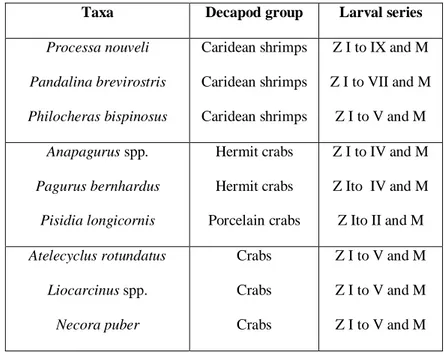
Table 1- List of the selected taxa, respective decapod crustacean group, distribution range when adult, and
the grouping of the larval stages. 26
Table 2- Coefficients of significant explanatory variables of the two steps model (presence or absence
predicted using a Logistic Model in the first step, values of abundance modelled with a Generalized
Addictive Model in the second step) describing the distribution of the decapod larvae selected taxa
collected with the LHPR net in the horizontal grid of stations. Levels of significance are represented as
***p<0.001, **p<0.01, *p<0.05; n.s. not significant. 33-34
Chapter 3
Table 1- List of the selected taxa, respective decapod crustacean group, and larval series. 69
Table 2- Coefficients of significant explanatory variables of the two steps model (presence or absence
predicted using a Logistic Model in the first step, values of abundance modelled with a Generalized
Addictive Model in the second step) describing the larval vertical distribution of the Caridea selected
species collected with the Neuston and LHPR nets in the fixed station. Levels of significance: ***p<0.001,
**p<0.01,*p<0.05; n.s. not significant. 73
Table 3- Coefficients of significant explanatory variables of the two steps model (presence or absence
predicted using a Logistic Model in the first step, values of abundance modelled with a Generalized
Addictive Model in the second step) describing the larval vertical distribution of the Anomura selected
species collected with the Neuston and LHPR nets in the fixed station. Levels of significance: ***p<0.001,
xxvii
predicted using a Logistic Model in the first step, values of abundance modelled with a Generalized
Addictive Model in the second step) describing the larval vertical distribution of the Brachyura selected
species collected with the Neuston and LHPR nets in the fixed station. Levels of significance: ***p<0.001,
**p<0.01,*p<0.05; n.s. not significant. 75
Chapter 4
Table 1 Comparison of relevant morphological characters of L. seticaudata, L. galapagensis and L. moorei
zoeal stages. 137
Table 2 Staging of the “Cosmopolitan Clade”/ “Lysmata Clade” larval morphological characters based on
the three studied species. 139
Chapter 5
xxviii Bir.- biramous
Bottom- depth of the station CL- carapace length
CTD- conductivity-temperature-depth recorders CW- carapace width
CWls- carapace width with lateral spines D- dactylus
Decr. post.- decreasing in size posteriorly
DEPTH_CAT – upper limit of the depth stratum DIST_COAST - distance to the coast
DL- dactylus length
DVM- Diel vertical migration GAM- Generalized Addictive Model Hour1- night and day
Hour2- night, dawn, day, dusk HOUR_WAVE - hour of the day
ISWM – Intermediate Salinity Water Mass IPC – Iberian Poleward Current
LHPR- Longhurst Hardy plankton recorder LM- Logistic Model
M- megalopa or decapodid
MAX_TOW_DEPTH – Maximum depth towed in the station MID_DEPTH – Midpoint of depth stratum
xxix n.s.- not significant P- propodus p.-page Part.- partially pc- pseudochaetae PL- propodus length RDL- rostro-dorsal length REAL_TIME – Hour of the day RL (or R)- rostrum length SAL – salinity
Seg.- segmented
Sub-ac.pr- sub-acute processes T- transect
TEMP – temperature TL- total length Unir.- uniramous Unseg.- unsegmented
WIBP - Western Iberia Buoyant Plume WMD- Weighted Mean Depth
Z- zoea
ZN- early zoeal stages ZO- old zoeal stages
CHAPTER 1
__________________________________________________
General Introduction
- 2 -
1.1- The importance of decapod crustaceans
Tropical and temperate coastal zones are inhabited by 110000 species of benthic invertebrates, of which approximately 80% have complex life cycles with a pelagic larval phase, a vital period for gene-flow, recruitment and consequent renovation of populations (Thorson 1964, McConaugha 1992). The Phylum Crustacea is one of the largest taxa in the animal kingdom only exceeded by insects and gastropods, and the Decapoda Order is the largest one with around 50000 species (Tudge 2000), a number that stills increasing (e.g. dos Santos et al. 2008a). Most of the decapods are found in the sea or adjacent brackish waters, some species invaded the freshwater, and a small number of species occupied the terrestrial habitats (e.g. Anger 2001). The diversity of decapod species reflects the diversity in their life styles, even knowing that most decapods are benthic they can live in the floors of the oceans from the high latitudes to the tropical environments, in mangroves, in coral reefs, polar and deep seas or hydrothermal vents. They are also a relevant part of the food web in the marine ecosystems.
A high number of species of decapods are commercially exploited, and in Portugal they constitute important fisheries resources (e.g. dos Santos 1999). An adequate fisheries management strategy for a species with economical value requires the knowledge of the species life cycle, so in the case of decapods the plankton studies give information about the distribution and abundance during their pelagic larval phase (e.g. dos Santos 1998). The exploited stock depends mainly on the larval stages survival (e.g. Roughgarden et al. 1988, Queiroga et al. 1994, dos Santos 1999), so the larval phase is a vital period in the life cycle of most decapod species and it is crucial for genetic flux, recruitment and consequent populations and stocks preservation (Paula 1993).
- 3 -
1.2- Decapod crustaceans life cycle: ecology
The decapod crustacean larvae are active swimmers, that can actively regulate their vertical position in the water column, controlling the extent range and direction of their horizontal dispersal (Queiroga & Blanton 2005), maintaining a favourable position to the adequate and necessary transport (e.g. Forward et al. 1997, Christy & Morgan 1998). The transport processes are the decisive components of the supply mechanism of larvae to habitats where settlement and juvenile development will occur (e.g. Botsford 1986), separating the ontogeny in time and in space (different environments), exposing the larvae to different mortality factors (Queiroga & Blanton 2005). The understanding of temporal and spatial larval movement patterns is fundamental to study decapods ecology leading to the design of effective conservation and resource management strategies (e.g. Pittman & McAlpine 2001, Mace & Morgan 2006). The larval vertical migrations are the strategy adopted by decapod larvae for the adequate and necessary transport at a life cycle phase (e.g. Oishi & Saigusa 1997, Christy & Morgan 1998, Paula 1998, Queiroga 1998).
The Diel Vertical Migration (DVM) behaviour seems to be the rule for decapod larvae, but it is rarely described in detail (see Queiroga & Blanton 2005 for examples), being most of the studies obtained by the neuston and/or discrete depth levels sampling (e.g. Shanks 1985, Jamieson & Phillips 1988, Abelló & Guerao 1999). Dos Santos et al. (2007, 2008b) showed that decapod zoeae and megalopae, as well as Chthamalus stellatus cyprids, displayed the DVM in the upwelling ecosystem adjacent to the Ria de Aveiro lagoon system. Pineda et al. (2007) concluded that the vertical swimming behaviour, changes in buoyancy, and ontogenetic changes in vertical position in the water column influenced the horizontal larval movements, and recently, dos Santos et al. (2008b) verified the diel vertical migration in
- 4 -
decapod crustacean larvae over the Portuguese upwelling ecosystem, corroborating the hypothesis presented by the models developed for the study area (Marta-Almeida et al. 2006, Peliz et al. 2007).
The ontogenetic migrations defined as the change in the average depth of distribution during the larval life period, is obligatory for benthic crustacean that hatch close to the bottom, feed in the surface, and must return to the adult habitat (Queiroga & Blanton 2005). Most of the times, the studies describing the ontogenetic migrations (see Queiroga & Blanton 2005 for
details and references), considered brachyuran species, and in the study area, Queiroga (1996)
described the ontogenetic variations in the vertical distribution of Carcinus maenas.
For the complete understanding of the larval transport and settlement locations is essential to describe the local hidrography and the three-dimensional current fields, as well as the growth, survival and behavioural responses of larvae to their environment (e.g. Botsford et al. 1994). The northwest coast of Portugal is characterised by complex mesoscale variability and a strong seasonality (Peliz et al. 2002, Relvas et al. 2007). Dos Santos et al. (2008b) studied the decapod larvae and discussed their results with the modelling studies for the area (Marta-Almeida et al. 2006, Peliz et al. 2007), confirming that the larval dispersal in the inner and middle shelf is made alongshore and the observed retention greatly depends on the diel vertical migration behaviour. Moreover, the authors supposed that the horizontal distribution patterns of several species retained over the shelf were associated with their presumable settlement areas. The apparent retention of decapod larvae close to parental populations was recently discussed by Morgan et al. (2009) and seems to be more frequent than considered until present date.
- 5 -
1.3- Decapod crustaceans life cycle: morphology
As referred before, the majority of decapods have complex life cycles, with an indirect development, and after the embryonic development within the egg the larvae are release, floating in the water column (e.g. Anger 2001). These pelagic larvae usually differ entirely in their morphology and habits from juvenile and adults, showing their own evolutionary adaptations. The larval development of crustaceans is described as the sequence of morphologically distinct stages.
To avoid misunderstandings and due to the particular importance of the concepts, according to Anger (2001):
- phase: is defined as the sequence of morphologically equal developmental stages, e.g. all naupliar, zoeal or megalopal stages combined as the “larval phase”;
- instar: is defined as a numerical stage, e.g. the appearance of a new instar may not be associated with morphological changes;
- stage: is defined as a distinguishable morphologically different instar, e.g. the roman numbers to identify successive stages in a given phase zoea I, zoea II, etc.;
- metamorphosis: is defined as the sudden and dramatic changes in the morphology of two subsequent stages, e.g. the transition from nauplius to zoea, from zoea to the megalopa, from the megalopa to juvenile.
During the larval development of decapod crustacean there are three possible phases separated by one metamorphosis: the nauplius, the zoea and the decapodid or megalopa (Williamson1969), depending on the appendages used by the larva for swimming (cephalic, thoracic and abdominal, respectively).
- 6 -
The nauplius is considered the most ancestral type of larva in the Crustacea, and in decapods, it is only present in the Suborder Dendrobranchiata. All the other decapods have this phase in the egg, during the embryonic development. The nauplius swims with the three pairs of cephalic appendages: antennules, antennae and mandibules. In a subsequent stage of the naupliar phase, the metanauplius, can develop other appendages however those are nonfunctional; the nauplius has a rudimentary small median eye.
The Suborder Pleocyemata has an embryonic development longer than the Dendrobranchiata because the nauplius develops within the egg, that when hatches releases a zoea. This phase is the most common in the plankton collections, and as a consequence it is the most studied (e.g. Paula 1993). The zoea presents as locomotion appendages the biramous thoracic appendages. It has compound eyes, in general sessile in the first zoea and mobile in subsequent stages. The number of stages in the zoeal phase is greatly variable, varying between groups (from two zoeal stages in Majidae crabs, to 15 or more in the Palinura lobsters) but also within a species (e.g. Anger 2001).
The megalopa or decapodid is characterized by the presence of functional pleopods as swimming appendages. Generally this phase has only one stage, which is the last larval stage that will metamorphose to a juvenile. It is a stage more similar to the adult (e.g. Paula 1993, dos Santos 1999), and presents a more or less uniform shape among the various taxonomical groups.
The decapod crustacean larvae may have different forms, but usually present stable developmental patterns (Paula 1993). The analysis of bio-ecological processes involved in the larval phase of the life cycle, the hydrological factors, the duration of the larval development,
- 7 -
and the number of larval stages in each larval series, as well as their morphological characters should be assigned as adaptive strategies to the coastal ecosystems. It can be supposed that the larval hydrodynamic form may correspond to different dynamic patterns, so having in mind the decapod crustacean larvae we grouped them in four different types:
Figure 1. A: Zoea I of Callianassa tyrrhena; B: Zoea II of Palinurus elephas; C: Zoea I of Nephrops
norvegicus; D: Zoea I of Carcinus maenas. In: dos Santos (1999).
Type A (Figure 1A): carapace and pleon laterally flattened; triangular telson. Suborder Dendrobranchiata, Infraorders Caridea, Stenopodidea and Thalassinidea, all shrimps sensu lato.
Type B (Figure 1B): the body strongly dorso-ventrally flattened; with very long appendices, laterally projected. Infraorder Palinura, the lobsters.
- 8 -
Type C (Figure 1C): with a laterally flattened but slightly rounded carapace; a cylindrical pleon (or abdomen); a bifurcated or triangular telson. Infraorders Astacidea and Anomura, the Norway lobster and the hermit crabs.
Type D (Figure 1D): a carapace almost spherical; usually with dorsal and lateral spines, and a rostrum ventrally projected; cylindrical telson or as a furca. Infraorder Brachyura, the crabs.
1.4- Objectives
The aim of present thesis is to describe the horizontal and vertical distribution patterns of decapod crustacean larvae in the Portuguese upwelling ecosystem, as well as to present the morphological larval descriptions from laboratory cultured material of the three selected larval forms of decapod larvae: the caridean shrimps, the hermit crabs, and the crabs. In the working plan of present thesis five specific objectives related with the larval ecology and morphology of decapod crustacean larvae were delineated: (1) to verify if the larval vertical distribution in the water column is related with the day cycle variation and (2) to describe the vertical migration rhythms of the considered larval forms (larval form and stage), (3) to establish an average depth of distribution through the larval development and (4) to verify the existence of ontogenetic vertical migration in the water column. The last objective is to make the morphological description of the larval development of three selected taxa of the three selected larval forms (a caridean shrimp, a hermit crab and a crab). To fulfil the ecological and morphological aims of present dissertation, five different studies were conducted:
- dos Santos et al. (2008b) studied the decapod larvae and hypothesised that the horizontal distribution patterns of several decapod species retained over the shelf were
- 9 -
related to their presumable settlement areas. So, in Chapter 2 our objective was to test the hypothesis that the decapod crustacean larval distribution, in the studied coastal upwelling ecosystem, clearly shows a retention strategy being coincident with the adults’ distributional ranges. As a result, we also pretend to demonstrate that the larvae concentrated very close to the shore will be the inner shelf species larvae; those concentrated somewhere in the middle shelf, will include the shelf species larval stages; and those that will appear over the continental shelf break, will concentrate the shelf slope species. To verify our hypothesis we analyse the relationships between the larval distributions of 15 selected taxa with the physical environment in the Western Iberia upwelling ecosystem.
- dos Santos et al. (2008b) described the diel vertical migration of decapod larvae. Knowing this, in Chapter 3 we pretend to analyse in detail the vertical distribution patterns of the larval stages of 9 species, in order to determine their ontogenetic vertical migration and also to describe the behavioural responses of each stage to the environment.
- present work in Chapter 4 pretends to describe the available zoeal characters of two closely related trans-isthmian caridean shrimp species, Lysmata moorei (Rathbun, 1901) and L. galapagensis Schmitt 1924, using laboratory cultured material. Both descriptions are compared with the larval features previously described for the only species in the “Cosmopolitan Clade” with their larvae known, L. seticaudata (Calado et al. 2004), and with the Eretmocaris larval genus (Bate 1888) given the evident resemblances between L. galapagensis larvae and E. corniger from the Tropical Eastern Pacific.
- 10 -
- the hermit crabs Clibanarius aequabilis and C. erythropus are both present in the eastern Atlantic, and present work pretends to describe and compare the complete larval series of Clibanarius aequabilis with the previously described larval stages of other species of the genus, in particularly with those of C. erythropus. These two species do not co-occur in the same area, but a careful comparison was required, so Chapter 5 also presents a redescription of C. erythropus larval morphology according to modern standards.
- finally, the Chapter 6 describes in detail the four zoeal stages and the megalopa of the crab I. nucleus from laboratory reared material.
1.5- Structure of this Dissertation
As the objectives list indicates, this dissertation is organized in seven chapters where the ecological and morphological aspects of decapod crustacean larvae are studied. Chapters two and three present the ecological results, while chapters four to six present the morphological results. Present chapter, the “Introduction”, includes a general introduction to the study of decapod crustacean larvae, with the state of the art, the presentation of the objectives of this thesis, and a brief description of the study area were the research cruise was carried.
The second chapter, the “Circulation patterns and decapod larvae distribution in a coastal upwelling ecosystem”, analyses the spatial distribution of decapod larvae off the northwest Portuguese shelf. This chapter is in preparation for publication.
- 11 -
The third chapter, the “Ontogenetic vertical migration behaviour of decapod larvae in the Portuguese upwelling ecosystem”, describes the ontogenetic vertical migration behaviour of decapod larvae on the western Iberia upwelling ecosystem. This chapter is also in preparation for publication.
The fourth chapter, “Shedding light over the larval genus Eretmocaris – morphological larval features of two closely related trans-isthmian Lysmata species using laboratory cultured material”, the morphological larval description of the zoeal stages of the caridean shrimps of the genus Lysmata was made. This chapter was submitted to “Systematics and Biodiversity” for publication.
The fifth chapter, “Complete larval development of the hermit crabs Clibanarius aequabilis and Clibanarius erythropus (Decapoda: Anomura: Diogenidae), under laboratory conditions, with a revision of the larval features of genus Clibanarius”, presents the complete morphological larval description of the hermit crabs of the genus Clibanarius. This chapter was published in Helgoland Marine Research.
The sixth chapter, “Complete larval development of the crab Ilia nucleus (Linnaeus, 1758) (Decapoda, Brachyura, Leucosiidae) reared under laboratory conditions”, describes the complete morphological larval development of the brachyuran crab of the species Ilia nucleus. This chapter was published in Scientia Marina.
- 12 -
1.6- Study area
In the Portuguese coast, until the beginning of the 21st century, Queiroga (1996) and dos Santos (1999) were the only authors studying the decapods larval distribution over the continental shelf, so most of the studies considering the spatio-temporal distribution of planktonic larval stages of decapod crustaceans were carried in estuaries and nearshore zones (e.g. Flores et al. 2002, Almeida & Queiroga 2003) and generally had as objective the brachyuran larval stages being the exceptions Paula’s works with Mira estuarine species (Paula 1987, 1989, 1993, 1998), dos Santos description of decapods occurring in the Portuguese coastal area (dos Santos 1999) and Pereira et al. (2000) description of the decapods larval fluxes at Aveiro coastal lagoon.
Queiroga (1996) studied the distribution and drift of the crab larvae of Carcinus maenas over the continental shelf off northern Portugal, demonstrating that the first zoea was clearly associated with the estuarine inlets, the older stages were dispersed progressively offshore, and the megalopal stage experienced an onshore transport. Sustained on these results a survey was conducted in May 2002 in the adjacent area to the Aveiro coastal lagoon, and several studies addressing the planktonic larval stages distribution resulted from the collected data (the most recent: dos Santos et al. 2008b, Moreno et al. 2009, Garrido et al. 2009).
The area adjacent to Aveiro coastal lagoon, the northwest coast of Portugal, is characterised by complex mesoscale variability and a strong seasonality (Peliz et al. 2002, Relvas et al. 2007). The Western Iberia Shelf is a seasonal upwelling system where the northerlies (upwelling favourable) are observed in summer (June to September), developing the typical upwelling phenomena like coastal jets and long filaments (e.g. Peliz et al., 2002). the presence