Vol.58, n.4: pp. 547-552, July-August 2015 http://dx.doi.org/10.1590/S1516-8913201500462
ISSN 1516-8913 Printed in Brazil
BRAZILIAN ARCHIVES OF BIOLOGY AND TECHNOLOGY
A N I N T E R N A T I O N A L J O U R N A L
Heat Shock Protein Hspa5 Interacts with and Protects
Tyrosinase Activity
Hongmin Tang
*and Peifu Zhou
Institute of Ethnic-minority Medicine; College of Chemistry & Environmental Science; Guizhou Minzu University, Guiyang - China
ABSTRACT
In this study, heat shock protein, Hspa5 was cloned, expressed and purified subsequently confirmed that it interacted with the tyrosinase (TYR) in vitro. Then, using the crystal structure of the homologous protein from the bacteria as a template, a homology model of human TYR was constructed. This model was further applied to investigate the molecular docking with Hspa5. The model showed that the interaction between the TYR and Hspa5 was mainly maintained by some hydrogen bonds in a quite low energy state. The results indicated that TYR was protected in different denaturation conditions by Hspa5. It was concluded that Hspa5 served as a molecular chaperone of TYR, which could help to better understand the molecule regulation mechanism of TRY in many kinds of diseases.
Key words: Molecular chaperone, Hspa5, tyrosinase, melanin, melanosome
*Author for correspondence: [email protected]
INTRODUCTION
Melanin is a common pigment that is found in bacteria, fungi, plants and animals. In mammals, including human, melanin is mainly located in horn cells of epidermis and hairs for coloring them. This coloring protects these cells from the ultraviolet radiation and internal overheating. Melanin is a derivative of tyrosine and synthesized by a series of enzymes in melanosome, a post-Golgi organelle. Tyrosinase (TYR) catalyzes the first two steps of the melanin synthesis pathway, hydroxylation of L-tyrosine to L-3, 4-dihydroxyphenylalanine (L-DOPA, monophe-nolase or cresolase activity, EC1.14.18.1) and the subsequent oxidation of L-DOPA to dopaquinone (diphenol oxidase or catecholase activity, EC1.10.3.1) (Lerner et al. 1949; Sanchez-Ferrer et al. 1995).
The glycan-specific oxidoreductase ERp57 is cross-linked to TYR when calnexin and calreticulin are associated. This timing coincide with the formation of disulfide bonds within TYR and the cleavage of its signal sequence (Wang et al. 2005). Finally, the mature TYR is translocated to melanosome to catalyze melanin biosynthesis (Sarangarajan et al. 2001).
TYR plays critical role in melanin synthesis and its dysfunction is linked to many diseases. Firstly, mutations in the TYR encoding gene causes oculocutaneous albinism Type 1 (OCA1), an autosomal recessive disorder characterized by the reduced melanin pigment in the hair, skin and eyes (Dolinska et al. 2014). Till now, more than 100 types of mutations have been found in OCA1 (Oetting et al. 2003), most of which are located in lumenal domain and some in transmembrane domain of TYR (Popescu et al. 2005). These mutants could not be transported to melanosome (Halaban et al. 2000; Toyofuku et al. 2001). Another infrequent temperature sensitive albinism was caused by missense mutation like R422Q, R402Q (Simeonov et al. 2013). At 37°C, these mutated TYRs retained in the endoplasmic reticulum and were possibly degraded by proteasomes with no pigment production. In contrast, in pigmented tissues at lower temperatures (32°C), the enzyme was translocated into the endosomes where it produced pigment (Halaban et al. 2000). Secondly, TYR is also related to malignant melanoma, which accounts for 75% of all deaths associated with skin cancer (Jerant et al. 2000). The increased melanin in melanoma and many melanoma cell lines are mainly attributed to the over-expression of TYR (Brichard et al. 1993; Bertolotto et al. 1996). While in amelanotic melanoma cell lines, TYR is normally expressed but fails to reach the melanosome, the organelle for melanin synthesis, because it is retained in the endoplasmic reticulum (ER) and then degrades (Halaban et al. 1997). Fundamentally, the main threat to melanoma cell and melanocyte is from the imbalance of antioxidant system. TYR can use O2- that go into the melanocyte to protect the cell from cytotoxic effect of O2- usually. However, this balance is broken by either deviant expression or translocation of TYR, which ultimately lead to the exposure of the cells to the attack of O2- (Mastore et al. 2005). Studies have shown that the incidence of Parkinson disease is positively correlated to melanoma. This might be due to that the
dopamine, a molecule, which plays a critical role in pathological mechanism of Parkinson disease, shares some enzymes and substrates with melanin during their synthesis. Therefore, the dysfunction of TYR affects dopamine synthesis directly or indirectly (Tianhong et al. 2011).
Therefore, the precise mechanism transporting TYR is critical for understanding the TYR-related diseases. In view of this, the aim of this study was to verify that if Hspa5 serve as a molecular chaperone of TYR and the interaction mechanism
between them.
MATERIAL AND METHODS
Materials, strains and growth conditions ExTaq DNA polymerase, pFL-B31cl vector, Hspa5 cDNA, T4 DNA ligase, restriction endonuclease
NdeI and XhoI were purchased from YAHX Biotechnology Limited Company. TYR and L-DOPA were purchased from Sigma-Aldrich. E. coli DH5a and E. coli BL21 (DE3) were purchased from Transgen Biotech and grown in Luria-Bertani (LB) broth or on LB agar. When necessary, ampicillin (YAHX Biotechnology Limited Company, Beijing, China) was added at a final concentration of 100 µg/mL.
Cloning, expression, and purification of Hspa5
Gene (NM_005347) encoding human Hspa5 protein was amplified from cDNA. The PCR product was purified, digested and ultimately ligated to the pFL-B31cl vector that was digested with same restriction endonucleases to produce plasmid pFL-B31cl-hspa5. Once the identity was confirmed by sequencing, it was transformed into
E. coli BL-21 (DE3) cells for protein expression.
The overnight culture was 1:100 diluted, and the cells were induced with 0.6 mM IPTG (isopropyl β-D-thiogalactoside) after reaching an OD600 of 0.6-0.8 at 23°C. Hspa5 protein was purified by Ni-NTA (Ni2+-nitrilotriacetate) resin (Qiagen) affinity chromatography according to the manufacturer’s manual.
In vitro protein-protein interaction
TYR was denatured in 1.5 M guanidine hydrochloride solution.
1.2, 4.8 and 4.8 M, respectively based on the preliminary experiment. Samples were separated by native-PAGE with 4% stacking gel and 10% separation gel, respectively.
Homology modeling and molecular docking
The crystal structure of TYR (4j6u.1.A) from
Bacillus megaterium was used as a model and the
homology modeling was accomplished by SWISS-MODEL on-line (Marco et al. 2014). HEX 8.0.0 was used for molecular docking and the models were further analysed by PyMOL.
TYR activity assay
TYR activity assay was carried out using L-DOPA as the substrate according to published protocols (Xiao-Hong Huang 2006). In brief, the absorbance at 405 nm was monitored over time at 25°C and the enzyme activity was calculated according to the following formula: α = k / (10-6 ε × V × 2.5 × 0.1). In which, α, k, ε and V represent the TYR activity, rake ratio of absorbance, molar absorption coefficient of L-DOPA and volume of reaction, respectively.
RESULT
Cloning, expression and purification of Hspa5
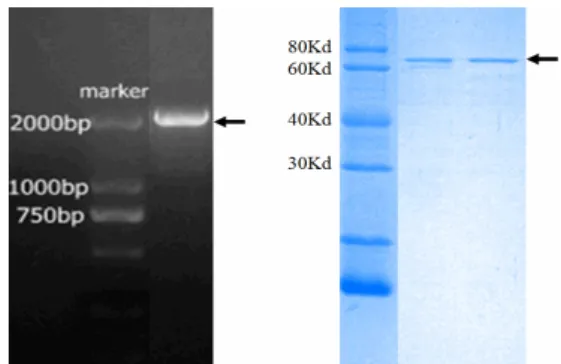
The cDNA fragment (1895 bp) of Hspa5 was amplified and the product was shown in Fig.S1A. It was cloned into the plasmid pFL-B31cl and the recombinant plasmid was transformed into E. coli
BL-21(DE3) strain for protein expression. The identity of the insert and the fidelity of the amplification were confirmed by DNA sequencing. As shown in Figure S1B, the 70 Kd recombinant Hspa5 (C-terminal His-tagged) protein was purified by Ni-NTA resin affinity chromatography according to the manufacturer’s manual.
Hspa5 interacts with TYR in vitro
Protein interaction is the prerequisite for a molecular chaperone. Here, native polyacrylamide gel electrophoresis was used to test the interaction between Hspa5 and TYR. As shown in Figure 1, incubation of TYR with Hspa5 formed a new protein band (right line) that corresponded to tyrosinase-Hspa5 complex according to the molecular weight; the negative control BSA could not band with TYR (Left line). This clearly indicated that Hspa5 interacted with TYR in vitro.
Figure S1 - Cloning, expression and purification of Hspa5. A. PCR product of ORF Hspa5. B. SDS-PAGE of purified recombinant protein Hspa5. The black arrows indicate the products.
Figure 1 - Hspa5 interacts with TYR in vitro. Left line represents the control that contains TYR and BSA. Right line represents the sample contains TYR and Hspa5.
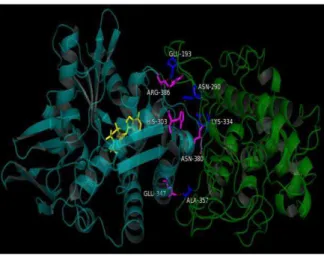
TYR docks with Hspa5 through intermolecular hydrogen bonds
hydrogen bonds, which were contributed by Glu193, Asn290, Lys334 and Ala357 of TYR, and His303, Asn380, Arg386 and Glu347 of Hspa5, respectively.
Figure 2 - Computer-generated model of TYR complexed with Hspa5. This model is based on HEX 8.0.0 docking analysis. The green molecular is TYR and the cyanic one represents Hspa5 with an ATP (yellow stick).
Figure 3 - Computer-generated interaction interface between TYR with Hspa5. The green molecular is TYR, and the cyanic one represents Hspa5 with an ATP (yellow stick). Red dashed lines represent the intermolecular hydrogen bonds. The amino acids involved in intermolecular hydrogen bond were highlighted as blue or purple in TYR or Hspa5 respectively.
Hspa5 protects TYR in different denaturation conditions
To further confirm that Hspa5 served as a molecular chaperone for TYR, the effect of Hspa5 was tested on TYR activity under different denaturation conditions. As show in Figure 4, TYR activity declined as time extended in buffer
containing protein denaturation guanidine hydrochloride. Hspa5, but not BSA, alleviated TYR activity loss in a dose dependent manner. Similarly, Hspa5 could protect TYR activity in dose-dependent manner under a 55°C thermal denaturation condition (Fig. 5).
Figure 4 - Hspa5 protects TYR under protein denaturation solution. Simply,different concentrations of Hspa5 (from 1.6 to 9.6 M) were mixed with 1.6 M of TYR in Tris-HCl buffer (pH7.5). BAS was used as a negative control. Afterwards, guanidine hydrochloride was added to a final concentration of 1.5 M and incubated at 25ºC. TYR activity were measured at different time points and compared with TYR activity at 0 min without Hspa5 or BAS. Each reaction is triplicate. Symbols: TYR only (●), with 1.6 M of Hspa5 (△), 3.2 M of Hspa5 (◆),
6.4 M of Hspa5 (□), 9.6 M of Hspa5 (▼) and 2.6 M of
BSA (×).
Figure 5 - Hspa5 protects TYR under 55°C thermal denaturation condition. In general, different concentrations of Hspa5 (from 1.6 to 9.6 M) were mixed with 1.6 M of TYR in Tris-HCl buffer (pH7.5). BAS was used as a negative control. All reactions were incubated at 55°C. TYR activity were measured at different time points and compared with TYR activity at 0 minute without Hspa5 or BAS. Each reaction is triplicate. Symbols: TYR only (●), with 1.6 M of Hspa5 (△), 3.2 M of Hspa5 (◆), 6.4 M
of Hspa5 (□), 9.6 M of Hspa5 (▼) and 2.6 M of BSA
DISCUSSION
TYR is a key enzyme in melanin synthesis, whose dysfunction was linked to many diseases such as oculocutaneous albinism Type 1 (OCA1), melanoma. The main cause of TYR dysfunction is that variegated TYR mutants are trapped in the endoplasmic reticulum instead of transporting into the melanosome, where the melanin is synthetized. Hence, in these conditions, some important proteins must be missing to participate in the TYR transport. These proteins could be important for revealing the mechanism of TYR involved diseases.
Based on the results of this study, it seemed that TYR interacted with heat shock protein Hspa5, which was in accordance with the results from co-immunoprecipitation experiment previously done (Wang et al. 2005). In addition, a three-dimensional structure model of TYR was constructed by using B. megaterium TYR (4j6u.1.A), a protein sharing 29% sequence identity with human TYR, as a homology modeling template. The produced model covered 67.3% (from R116 to Q453) of human TYR. The truncated human TYR model may not absolutely mimic the native protein. However, a bunch of low energy interaction models strongly indicate a potential interaction between Hspa5 and TYR. Lastly, the protection of TYR activity from Hspa5 in different denaturation conditions strengthens the conclusion that Hspa5 could serve as a heat shock protein of TYR. All these results would help to further understand the TYR transportation in the relevant diseases.
CONCLUSION
In this work, heat shock protein Hspa5 was successfully cloned, expressed and purified. The native polyacrylamide gel electrophoresis analysis suggested that it interacted with TYR in vitro. Further, a homology model of human TYR was made based on the crystal structure of the homologous protein from B. megaterium, and protein docking analysis showed that TYR interacted with Hspa5 in a quite low energy state in a virtual model. More importantly, results showed that Hspa5 protected TYR in different denaturation conditions. These results all together indicated that Hspa5 served as a molecular chaperone of TYR, which further showed the
relevance of Hspa5 to The Cotranslational Maturation.
ACKNOWLEDGEMENTS
We deeply appreciate the help of Dr. Sen Li (Beijing Normal University, P.R. China) throughout this study. The present investigation was supported by Abroad Joint Research Fund of Guizhou Province (Grant No.[2011]7034).
REFERENCES
Bertolotto C, Bille K, Ortonne JP, Ballotti R. Regulation of tyrosinase gene expression by cAMP in B16 melanoma cells involves two CATGTG motifs surrounding the TATA box: implication of the microphthalmia gene product.J Cell Biol. 1996; 134: 747-755.
Brichard V, Van Pel A, Wolfel T, Wolfel C, De Plaen E, Lethe B, et al. The tyrosinase gene codes for an antigen recognized by autologous cytolytic T lymphocytes on HLA-A2 melanomas. J Exp Med. 1993; 178: 489-495.
Costin IP, Crina P, Raymond AD, Stefana MP. Soluble Tyrosinase is an Endoplasmic Reticulum (ER)-associated Degradation Substrate Retained in the ER by Calreticulin and BiP/GRP78 and Not Calnexin. J Biol Chem. 2005; 280: 13833-13840.
Dolinska MB, Kovaleva E, Backlund P, Wingfield PT, Brooks BP, Sergeev YV. Albinism-causing mutations in recombinant human tyrosinase alter intrinsic enzymatic activity.PLoS One. 2014; 9: e84494. Halaban R, Cheng E, Zhang Y, Moellmann G, Hanlon
D, Michalak M, et al. Aberrant retention of tyrosinase in the endoplasmic reticulum mediates accelerated degradation of the enzyme and contributes to the dedifferentiated phenotype of amelanotic melanoma cells.Proc Natl Acad Sci. 1997; 94: 6210-6215. Halaban R, Svedine S, Cheng E, Smicun Y, Aron R,
Hebert DN. Endoplasmic reticulum retention is a common defect associated with tyrosinase-negative albinism.Proc Natl Acad Sci. 2000; 97: 5889-5894. Jerant AF, Johnson JT, Sheridan CD, Caffrey TJ. Early
detection and treatment of skin cancer. Am Fam Physician. 2000; 62: 357-368.
Lerner AB, Fitzpatrick TB, Calkins E, Summerson WH. Mammalian tyrosinase: preparation and properties. J Biol Chem. 1949; 178: 185-195.
Mastore M, Kohler L, Nappi AJ. Production and utilization of hydrogen peroxide associated with melanogenesis and tyrosinase-mediated oxidations of DOPA and dopamine. FEBS J. 2005; 272: 2407-2415.
Oetting WS, Fryer JP, Shriram S, King RA. Oculocutaneous albinism type 1: the last 100 years. Pigment Cell Res. 2003; 16: 307-311.
Popescu CI, Paduraru C, Dwek RA, Petrescu SM. Soluble tyrosinase is an endoplasmic reticulum (ER)-associated degradation substrate retained in the ER by calreticulin and BiP/GRP78 and not calnexin.J Biol Chem. 2005; 280:13833-13840.
Sanchez FA, Rodriguez-Lopez JN, Garcia CF, Garcia CF. Tyrosinase: a comprehensive review of its mechanism.Biochim Biophys Acta. 1995; 1247:1-11. Simeonov DR, Wang X, Wang C, Sergeev Y, Dolinska
M, Bower M, et al. DNA variations in oculocutaneous albinism: an updated mutation list and current outstanding issues in molecular diagnostics.Hum Mutat. 2013; 34:827-835.
Tianhong P, Xingqun L, Joseph J. The association
between Parkinson’s disease and melanoma.
International Journal of Cancer. 2011; 128:2251-2260.
Toyofuku K, Wada I, Valencia JC, Kushimoto T, Ferrans VJ, Hearing VJ. Oculocutaneous albinism types 1 and 3 are ER retention diseases: mutation of tyrosinase or Tyrp1 can affect the processing of both mutant and wild-type proteins. FASEB J. 2001; 15:2149-2161.
Wang N, Daniels R, Hebert DN. The cotranslational maturation of the type I membrane glycoprotein tyrosinase: the heat shock protein 70 system hands off to the lectin-based chaperone system.Mol Biol Cell. 2005; 16:3740-3752.
XiaoHong H, QingxiC, Qin W, KangKang S, Jun W, Li S, et al. Inhibition of the activity of mushroom tyrosinase by alkylbenzoic acids. Food Chemistry. 2006; 94: 1-6.