Protein 90 and Ribosomal Gene Sequences
Mona Hoppenrath1,2, Brian S. Leander1*
1Department of Zoology, University of British Columbia, Canadian Institute for Advanced Research, Program in Integrated Microbial Biodiversity, Vancouver, Canada, 2Forschungsinstitut Senckenberg, Deutsches Zentrum fu¨r Marine Biodiversita¨tsforschung, Wilhelmshaven, Germany
Abstract
Background:Interrelationships among dinoflagellates in molecular phylogenies are largely unresolved, especially in the deepest branches. Ribosomal DNA (rDNA) sequences provide phylogenetic signals only at the tips of the dinoflagellate tree. Two reasons for the poor resolution of deep dinoflagellate relationships using rDNA sequences are (1) most sites are relatively conserved and (2) there are different evolutionary rates among sites in different lineages. Therefore, alternative molecular markers are required to address the deeper phylogenetic relationships among dinoflagellates. Preliminary evidence indicates that the heat shock protein 90 gene (Hsp90) will provide an informative marker, mainly because this gene is relatively long and appears to have relatively uniform rates of evolution in different lineages.
Methodology/Principal Findings:We more than doubled the previous dataset of Hsp90 sequences from dinoflagellates by generating additional sequences from 17 different species, representing seven different orders. In order to concatenate the Hsp90 data with rDNA sequences, we supplemented the Hsp90 sequences with three new SSU rDNA sequences and five new LSU rDNA sequences. The new Hsp90 sequences were generated, in part, from four additional heterotrophic dinoflagellates and the type species for six different genera. Molecular phylogenetic analyses resulted in a paraphyletic assemblage near the base of the dinoflagellate tree consisting of only athecate species. However,Noctilucawas never part of this assemblage and branched in a position that was nested within other lineages of dinokaryotes. The phylogenetic trees inferred from Hsp90 sequences were consistent with trees inferred from rDNA sequences in that the backbone of the dinoflagellate clade was largely unresolved.
Conclusions/Significance:The sequence conservation in both Hsp90 and rDNA sequences and the poor resolution of the deepest nodes suggests that dinoflagellates reflect an explosive radiation in morphological diversity in their recent evolutionary past. Nonetheless, the more comprehensive analysis of Hsp90 sequences enabled us to infer phylogenetic interrelationships of dinoflagellates more rigorously. For instance, the phylogenetic position ofNoctiluca, which possesses several unusual features, was incongruent with previous phylogenetic studies. Therefore, the generation of additional dinoflagellate Hsp90 sequences is expected to refine the stem group of athecate species observed here and contribute to future multi-gene analyses of dinoflagellate interrelationships.
Citation:Hoppenrath M, Leander BS (2010) Dinoflagellate Phylogeny as Inferred from Heat Shock Protein 90 and Ribosomal Gene Sequences. PLoS ONE 5(10): e13220. doi:10.1371/journal.pone.0013220
Editor:Richard Cordaux, University of Poitiers, France
ReceivedJune 7, 2010;AcceptedSeptember 6, 2010;PublishedOctober 8, 2010
Copyright:ß2010 Hoppenrath, Leander. This is an open-access article distributed under the terms of the Creative Commons Attribution License, which permits unrestricted use, distribution, and reproduction in any medium, provided the original author and source are credited.
Funding:This work was supported by a postdoctoral scholarship from the Deutsche Forschungsgemeinschaft (Grant Ho3267/1-1), by operating grants from the National Science and Engineering Research Council of Canada (NSERC 283091-09), the National Science Foundation’s Assembling the Tree of Life (NSF# EF-0629624), and by the Canadian Institute for Advanced Research, Program in Integrated Microbial Biodiversity. The funders had no role in study design, data collection and analysis, decision to publish, or preparation of the manuscript.
Competing Interests:The authors have declared that no competing interests exist.
* E-mail: [email protected]
Introduction
Dinoflagellates comprise an extraordinary lineage of protists (unicellular eukaryotes) in regard to overall diversity in cell morphology and nutritional modes (e.g., phagotrophy, ‘klepto-phototrophy’, photoautotrophy, mixotrophy, and parasitism) [1–3]. Both heterotrophic and photoautotrophic members of the group are abundant and ecologically important components of marine planktonic communities. Dinoflagellates are morphologi-cally distinct from other eukaryotes in the structure of their (dinokont) flagellar apparatus and (dinokaryotic) nucleus (i.e., permanently condensed chromosomes without typical eukaryotic histones and with an extranuclear spindle that passes through cytoplasmic channels) [1,4,5].
characters when making inferences about dinoflagellate evolution-ary history [12,13,16]. As such, inferences based on morphology have yet to be adequately tested with molecular markers that provide sufficient signal at the deepest levels in the dinoflagellate phylogenetic tree.
Ribosomal DNA (rDNA) sequences are most useful for resolving (‘‘genus’’ level) relationships near the tips of the dinoflagellate tree [12,17–21]. Deeper branches receive either no or poor statistical support in trees inferred from rDNA for several reasons: (1) a large number of highly conserved regions; (2) strong evolutionary rate heterogeneity among sites in variable regions; (3) high levels of compositional heterogeneity among some of the sequences; (4) high levels of homoplasy within variable regions; and (5) non-independently evolving sites in paired helix regions [12–14,22]. Moreover, taxon sample biases and taxon identification are reoccurring problems – fewer than 150 species of about 2,500 known species have so far been sequenced, with a strong bias towards photosynthetic taxa [14]. Although some effort has been made to increase the representation of heterotrophic and uncultivated taxa in the datasets over the past five years [18,19,21,23], the taxon bias remains.
Understanding the phylogeny of athecate (unarmored) dinofla-gellates is particularly problematic because (1) their patterns of amphiesmal vesicles are more difficult to discern than in thecate (armored) dinoflagellates, (2) many of them are heterotrophic and uncultivated, (3) they are widely polyphyletic in molecular phylogenetic analyses, and (4) many of them have been misclassified [13,14,21,24,25]. Nonetheless, detailed re-evaluations of morphology combined with molecular phylogenetic studies of several athecate taxa over the past ten years has resulted in descriptions of new genera and improved re-descriptions of existing genera [12,17,20,21,23,26–30].
The phylogenetic position of the (athecate) Noctilucales is especially controversial. These free-living dinoflagellates possess a dinokaryon only during part of their lifecycle and sometimes possess a highly distinctive trophont stage consisting of an inflated balloon-like cell with a feeding tentacle. Molecular phylogenetic analyses of rDNA sequences and heat shock protein gene (Hsp90) sequences plus the absence of a dinokaryon in the trophont stage suggested that Noctiluca was an early diverging lineage of dinoflagellates that retained several ancestral states for the group as a whole (e.g., a pre-dinokaryotic nucleus) [31–34]. However, the molecular phylogenetic position of Noctiluca is inconsistent in different analyses, and these cells possess several very novel morphological features, so some authors have questioned the interpretation that this lineage is basal among dinoflagellates [13,16].
The major subgroups of dinoflagellates are largely recognized from patterns of either amphiesmal vesicles or thecal plates, called ‘‘tabulation patterns’’. This morphology-based criterion has been used to identify several monophyletic groups of dinoflagellates, some of which have been corroborated with molecular phyloge-netic data, such as the Suessiales, the Gonyaulacales, the Dinophysiales, the Prorocentrales, and the Gymnodiniales sensu stricto [12–14,20,21,24,29,35,36]. Several lineages previously classified within the ‘‘Gymnodiniales’’ have been removed from this subgroup upon closer examination with electron microscopy and molecular phylogenetic analyses [37]. The tabulation pattern found in the Suessiales (represented byPolarellaandSymbiodinium) forms an intermediate between the tabulation patterns found in some athecate taxa (previously lumped within the Gymnodiniales) and several thecate subgroups, like the Peridiniales and the Gonyaulacales. Although taxon sampling is far from complete, molecular phylogenetic analyses indicate that the Peridiniales is
paraphyletic and might form a stem group from which the Gonyaulacales, Dinophysiales and Prorocentrales evolved [13]. Moreover, the highly distinctive morphology of the Prorocentrales indicates that the group is monophyletic, but molecular phyloge-netic data did not corroborate this inference [13,14,24,38–40] until analyses of mitochondrial genes were performed [25,41].
Along these lines, molecular phylogenetic analyses of mito-chondrial gene sequences (cob + cox1) concatenated with SSU rDNA recover basal positions for Amphidinium (athecate) and
Heterocapsa(thecate) [25]; some paleontological data also support this hypothesis [13]. Although the general morphology of
AmphidiniumandHeterocapsadoes not immediately indicate a close relationship between them, both genera contain species that possess body scales [42–44]. Scales are unusual in dinoflagellates and are known only in these two genera plusLepidodinium[45,46]. Perhaps significantly, Oxyrrhis, which is a sister lineage to dinokaryotes (syn. ‘‘core’’ dinoflagellates), also possesses scales on the cell body and the flagella [11,47–49]. The putative phylogenetic distribution of this character suggests that the most recent ancestor ofOxyrrhisand dinokaryotes also possessed body scales.
However, inferences about morphological character evolution in dinoflagellates depend on a robust molecular phylogenetic framework, especially at the deepest levels that relate the major subgroups (i.e., ‘‘orders’’). Accomplishing this requires exploration of different molecular markers, which is the primary aim of this study. We have chosen to significantly expand the current heat-shock protein 90 (Hsp90) dataset for dinoflagellates by more than doubling the taxon sample in a manner that enhances broader representation of the major subgroups. Hsp90 is a highly conserved molecule that functions as a chaperone for protein folding and plays a key role in cellular signal transduction networks in all eukaryotes [50]. Stechmann and Cavalier-Smith [51] predicted that Hsp90 could become an important ‘‘universal’’ phylogenetic marker for eukaryotes because it is relatively long (1,800 bp) and evolves relatively uniformly in very different lineages. These authors advocated that Hsp90 should be sequenced from a broad selection of eukaryotic taxa and included within multi-gene phylogenetic analyses. The relatively homoge-nous branch lengths in trees inferred from Hsp90 sequences also helps reduce methodological artifacts associated with long-branch attraction.
Hsp90 datasets have been used previously for inferring dinoflagellate relationships [10,33,52,53]. The first dinoflagellate Hsp90 sequences were used to examine the relationships between the three major alveolate subgroups, which resulted in a strongly supported framework [52]; a few subsequent studies have used Hsp90 sequences to address the internal phylogeny of dinoflagel-lates [10,33,53]. One of these studies used Hsp90 sequences to explore the evolution of plastid diversity within dinoflagellates, which reinforced that there were several independent plastid replacements as suggested earlier using comparative morphology and analyses of rDNA sequences [53,54]. A concatenated analysis of SSU rDNA sequences with Hsp90 sequences demonstrated considerably higher statistical support values for almost all of the deep nodes when compared to trees inferred from SSU rDNA alone [53]. Most recently, Hsp90 gene sequences were used to evaluate the controversial phylogenetic position of N. scintillans, and the authors of this study concluded thatN. scintillans diverges very early within dinoflagellates [33]. However, all of these studies were limited by the very few Hsp90 sequences available at the time.
sequences from 17 different species of dinoflagellates, covering as many orders as possible; consequently, the Hsp90 gene data set for dinoflagellates was more than doubled. Moreover, in order to be able to concatenate the Hsp90 data with LSU and SSU rDNA sequences, we supplemented the new Hsp90 sequences with three new SSU rDNA sequences and five new LSU rDNA sequences. All of these data enabled us to address the broad phylogenetic interrelationships of dinoflagellates and will contribute to future analyses using larger multi-gene datasets.
Results
New Hsp90 sequences were generated from dinoflagellates representing seven different orders, including the first sequences from the Phytodiniales and the Suessiales and from the genera
Akashiwo, Diplopsalis, Peridinium, Polarella, Protoperidinium, Scrippsiella,
Spiniferodinium, Thecadinium and Togula. Only three of the 12 previously known Hsp90 sequences from dinoflagellates were from heterotrophic species, namely Crypthecodinium,Lessardia, and Nocti-luca; in this study, we generated four additional sequences from heterotrophic dinoflagellates, namelyDiplopsalis lenticula, Protoperidi-niumsp.,P. steidingeraeandP. crassipes. An Hsp90 sequence from the phototrophicPyrocystis lunulais available in GenBank, but the length of this sequence was too short to include it in our phylogenetic analyses. A sequence representing the type species of each genus is particularly important in dinoflagellates in order to maintain taxonomic stability in the phylogenetic trees. Accordingly, we generated new Hsp90 sequences from six type species: Akashiwo sanguinea, Gymnodinium fuscum, Polarella glacialis, Spiniferodinium galei-formis, Thecadinium kofoidii andTogula britannica. All 17 of the new Hsp90 sequences contained the diagnostic indel for dinoflagellates [10]. The new rDNA sequences generated in order to complete the combined phylogenetic analyses represent the first SSU rDNA sequences fromAmphidinium mootonorumandSpiniferodinium galeiformis
and the first LSU rDNA sequence fromThecadinium kofoidii. Six different alignments were constructed and analyzed: (1) SSU rDNA (35 taxa); (2) LSU rDNA (30 taxa); (3) Hsp90 DNA, 3rd codon positions excluded (40 taxa); (4) amino acid sequences inferred from the Hsp90 DNA sequences (40 taxa); (5) Hsp90 DNA, 3rd codon positions excluded, concatenated with SSU rDNA (34 taxa); and (6) Hsp90 DNA, 3rd codon positions excluded, concatenated with SSU rDNA and LSU rDNA (27 taxa). The resulting trees from datasets 3 to 6 are presented as Figures 1, 2, 3, 4, respectively. The statistical support values from the analyses of the SSU rDNA alone (dataset 1, Figure S1) were added to the corresponding nodes in Figure 3 (dataset 5). Analyses of the LSU rDNA sequences alone (dataset 2) resulted in a poorly resolved phylogeny (Figure S2).
The monophyly of dinoflagellates and dinozoans (i.e., the most recent ancestor of dinoflagellates and perkinsids and all of its descendants) received high support in all of the analyses (Figures 1– 4). The statistical support values for basal nodes within dinokaryotes were low in all of the analyses, except for a few basal nodes in the tree inferred from dataset 3 (Hsp90 DNA, 3rd codon positions excluded) (Figure 1). The Gonyaulacales and the Prorocentrales received modest to strong support in all of the analyses, especially in trees inferred from datasets including rDNA sequences (Figures 3, 4). TheProtoperidinium/Diplopsalisclade and theKareniaclade were strongly supported in trees inferred from Hsp90 sequences (Figures 1, 2).Togula britannicaandSpiniferodinium galeiformisformed a strongly supported clade in trees inferred from Hsp90 sequences alone and in trees inferred from datasets including both Hsp90 and SSU rDNA (Figures 1, 2, 3). Unexpectedly,Polarella glacialisand
Gymnodinium simplexdid not cluster together in the trees inferred from
datasets 3–5 (Figures 1, 2, 3) but did cluster strongly together in the tree inferred from dataset 6 (Figure 4).
Genera of athecate species branched as a paraphyletic assemblage near the base of the dinoflagellate tree in all of the analyses (Figures 1, 2, 3, 4). Moreover, in all of the trees inferred from DNA sequences, the Karenia/Karlodinium clade formed the earliest diverging lineage among the dinoflagellates included in the analyses (Figures 1, 3, 4); the tree inferred from amino acid sequences had an anomalous topology, whereby the Togula/ Spiniferodiniumclade formed the earliest diverging lineage (Figure 2).
Amphidinium carterae, which is a representative of the Amphidinium
sensu stricto, also branched near the base of the dinoflagellate tree in all of the analyses, albeit with weak statistical support (Figures 1– 4). Nonetheless, neitherNoctilucanorHeterocapsaever branched in a basal position relative to the other core dinoflagellates in the analyses (Figures 1, 2, 3, 4). Instead, Noctiluca branched in a position that was deeply nested within other lineages of dinokaryotes, especially within the trees inferred from Hsp90 DNA sequences (Figure 1). In order to gain additional insight into how well the Hsp90 data supported the phylogenetic position of
Noctiluca relative to dinokaryotes, we performed AU tests for comparing the likelihoods of two alternative topologies differing in the relative position of this species: (1)Noctilucapositioned as shown in Figure 1, and (2)Noctilucapositioned as the nearest sister lineage to all dinokaryotes in the analysis (e.g., afterOxyrrhisand before the
Karlodinium/Karenia clade, Figure 1). Topology 2 was strongly rejected by the AU test in the datasets that incorporated Hsp90 DNA sequences (Pvalue for the AU test = 461026) and topology 1 was supported (Pvalue for the AU test = 1.00).
Discussion
General phylogenetic patterns among athecate dinokaryotes
All trees inferred from the data generated in this study have nearly the same taxon composition in order to make the most direct comparison possible between the different phylogenetic markers employed. As outlined in the Results section, several topological differences were detected in trees inferred from Hsp90 sequences (including concatenations with rDNA, Figures 1, 2, 3, 4) and trees inferred from rDNA sequences alone (additional Figure 1 and published trees from previous studies). Some of these differences were also recognized in previous studies that explored Hsp90 as a phylogenetic marker for dinoflagellates [10,53]. Shalchian-Tabrizi et al. [53] also noticed that although the branching order in trees inferred from SSU rDNA and Hsp90 sequences was generally congruent, the statistical support values for most of the deep nodes were considerably higher in the Hsp90 analyses. However, analyses of Hsp90 amino acid sequences produce topologies that are different from those derived from analyses using Hsp90 DNA sequences (excluding the third codon positions), which can be attributed to a more conserved and thus weaker level of phylogenetic signal in the amino acid dataset [55]. The authors of previous molecular phylogenetic studies of rDNA sequences concluded that the Gymnodiniales are polyphy-letic and that loss of a theca occurred multiple times independently [13,14,16,54]. Not surprisingly, this scenario is also reflected in our phylogenetic analyses of rDNA sequences and our analyses of Hsp90 DNA sequences concatenated with rDNA sequences (Figures 3, 4). Zhang et al. [25] suggested that either the
formed the earliest diverging branch in trees inferred from LSU rDNA and (3)Karlodiniumformed the earliest diverging branch in trees inferred from a combination of SSU and LSU rDNA. This last topology is consistent with our studies of Hsp90 sequences, whereby theKarenia/Karlodiniumclade formed the earliest diverging branch among dinokaryotes in all of the analyses of DNA sequences (Figures 1, 3, 4). Amphidinium and Akashiwo never branched as the earliest diverging lineage, and Heterocapsa and
Noctiluca were consistently nested more deeply within the tree of dinokaryotes (Figures 1, 2, 3, 4).
Our phylogenetic analyses of the Hsp90 amino acids (dataset 4, Figure 2) resulted in athecate genera (i.e., the Gymnodiniales) branching as a paraphyletic assemblage that encompassed the most recent ancestor of all dinokaryotes. Because our study contained 12 species from nine different genera of athecate dinoflagellates, this paraphyletic distribution of athecate dinoflagellates is particularly compelling; this phylogenetic pattern is also consistent with a previous study of Hsp90 sequences that contained representatives of four athecate genera [53]. Therefore, our new sequences and molecular phylogenetic analyses provide additional support for the hypothesis Figure 1. Bayesian tree inferred from 40 Hsp90 DNA sequences (3rd codon positions excluded; dataset 3), 984 unambiguously aligned sites and a GTR+I+G+8 model of nucleotide substitutions.Numbers above the branches denote ML bootstrap percentages, and numbers below the branches denote Bayesian posterior probabilities. Black circles denote bootstrap percentages and posterior probabilities of 100% and 1.00, respectively.
that the initial evolutionary radiation of dinoflagellates involved athecate dinoflagellates that subsequently gave rise to several thecate lineages, perhaps independently (e.g., the Prorocentrales, Gonyaula-cales and Peridiniales). This hypothesis is also consistent with the phylogenetic results derived from dataset 3 (Figure 1), dataset 5 (Figure 3) and dataset 6 (Figure 4); these trees show Karenia,
Karlodinium,Gymnodinium, andAmphidinium mootonorumbranching as a paraphyletic assemblage that incorporates the most recent ancestor of
all dinokaryotes. In some of the analyses,SpiniferodiniumandAkashiwo
were also part of this athecate assemblage (Figures 3, 4). However, the statistical support values for the nodes near the backbone of the trees inferred from all of the datasets were generally modest at best.
LSU rDNA, dataset 6) (Figure 4). Other phylogenies suggest that
G. simplex belongs into the Suessiales [13]; overall, a more confident placement ofG. simplexrequires, in part, a more detailed morphological investigation of this species.
The molecular phylogenetic position ofNoctiluca There is significant debate about the phylogenetic position of
Noctiluca scintillans among dinoflagellates, mainly because this lineage possesses an unusual collection of morphological features.
The molecular phylogenetic analyses published so far (e.g., SSU rDNA, LSU rDNA, b-tubulin, and Hsp90) suggest that N. scintillans diverges very early within dinoflagellates, and most studies show this species branching as the nearest sister lineage to dinokaryotes. Some of the morphological features in this lineage (e.g., the absence of a nucleus with permanently condensed chromosomes in the trophont stage) have, accordingly, been interpreted as concordant evidence for a sister relationship between Noctiluca and dinokaryotes [31–33]. Moreover, the Figure 3. Bayesian tree inferred from 34 Hsp90 DNA sequences (3rd codon positions excluded) concatenated with SSU rDNA sequences (dataset 5), 2365 unambiguously aligned sites and a GTR+I+G+8 model of nucleotide substitutions.Numbers above the branches denote ML bootstrap percentages, and numbers below the branches denote Bayesian posterior probabilities. Black circles denote bootstrap percentages and posterior probabilities of 100% and 1.00, respectively. Numbers within the ovals compare the statistical support values from the analyses of dataset 5 (bold and to the right) and the analyses of the SSU rDNA sequences alone (dataset 1; to the left).
published Hsp90 sequence from N. scintillans was previously analyzed within the context of other dinoflagellate sequences and shown to be the first branch to diverge from the other taxa in the analyses [33]. However, these analyses were limited by the very small taxon sample available at the time. We were able to re-evaluate these analyses with a much larger sample of Hsp90 sequences from dinoflagellates and show that N. scintillans never
occupied a basal position and was instead more deeply nested within dinokaryotes (Figures 1, 2, 3). AU tests provided additional support for this inference.
Although the previous molecular phylogenetic analyses suggest-ing a basal position forN. scintillanshave been questioned by some authors [13,16], several other authors have used this framework to (mis)interpret different aspects of the biology ofN. scintillans. For Figure 4. Bayesian tree inferred from 27 Hsp90 DNA sequences (3rd codon positions excluded) concatenated with SSU rDNA sequences and LSU rDNA sequences (dataset 6), 2847 unambiguously aligned sites and a GTR+I+G+8 model of nucleotide substitutions. Numbers above the branches denote ML bootstrap percentages, and numbers below the branches denote Bayesian posterior probabilities. Black circles denote bootstrap percentages and posterior probabilities of 100% and 1.00, respectively.
instance, Fukuda and Endoh [33] stated that Liu and Hastings [56] discovered the most ancestral type of luciferase gene in N. scintillans. However, this was only one of two alternative interpretations posed by Liu and Hastings [56] and was based on the assumption thatN. scintillanshad already been demonstrat-ed to be among the earliest diverging dinokaryotes. The alternative interpretation posed was that the condition in N. scintillanswas a derived state in this lineage: ‘‘The ancestral system may have had two genes, which fused inNoctiluca…’’ [56].
Moreover, Fukuda and Endoh [32,33] attempted to reconstruct the early evolution of dinokaryotes based on the properties of the gametes inN. scintillans; we think this approach is problematic for several reasons. First, a comparison of trees inferred from ribosomal DNA sequences to trees inferred from Hsp90 sequences with a sufficient taxon sample (i.e., this study) demonstrate that the phylogenetic position of this lineage within dinoflagellates has not been confidently established. Thus, at this time, the characters in
N. scintillanscannot be interpreted to be ancestral for dinokaryotes as a whole. Second, these authors characterized their observations ofN. scintillansas representing the complete life cycle of this species without accounting for previously reported discrepancies [32]. For instance, the authors describe the gametes as being isogamous and having two flagella that are visible with light microscopy [32]. However, TEM was required to demonstrate that the swarmer cells of N. scintillans had a distinctly heteromorphic flagellation, with one long flagellum and one very short flagellum oriented to the left side of the cell [57]. The short flagellum is not visible with light microscopy, which is why Zingmark [58] previously described the gametes as being uniflagellated.
Contradictory observations in the literature also led Schnepf and Drebes [59] to re-investigate sexual reproduction in N. scintillansand conclude that although a few microgametes with two flagella were present, generally the microgametes possess only a single longitudinal flagellum and do not undergo fusion. Schnepf and Drebes agreed with Uhlig [60], who reasoned that the appearance of gamete fusion and the presence of two long flagella is a consequence of incomplete cytokinesis. The possibility of an anisogamous (or nearly oogamous) sexual cycle was also suggested, but the author’s explicitly stated that definitive evidence is unavailable [59]. Until the fusion of gametes and karyogamy is convincingly demonstrated, the mode of sexual reproduction inN. scintillanswill remain speculative. The ‘‘isogamy hypothesis’’ and the transformation of the zygote into a mature trophont characterized by Fukuda and Endoh [32,33] also need to be more convincingly described. Perhaps the best way to establish a more confident phylogenetic position and life cycle for the Noctilucales is to move beyond N. scintillans and characterize more species within the ‘‘order’’ at both the ultrastructural and molecular phylogenetic levels [61].
Concluding Remarks
The resolution of interrelationships between the major lineages of dinoflagellates was modest at best when inferred from Hsp90 sequences alone or in concatenation with rDNA sequences. The high degree of sequence conservation and the consistently poor to modest resolution of the deepest nodes in trees inferred from Hsp90 and rDNA sequences supports the hypothesis that dinoflagellates underwent an explosive radiation in morphological diversity relatively recently in their evolutionary history. However, the lack of sufficient phylogenetic signal in the markers analyzed so far for dinoflagellates could be explained in other ways as well (e.g., mutational saturation over a large period of time). Nonetheless, the more comprehensive analysis of Hsp90 sequences presented here enabled us to re-address several phylogenetic
interrelationships of dinoflagellates, such as the phylogenetic position ofN. scintillans. Currently, there are no Hsp90 sequences available for the Dinophysiales, the Blastodiniales, and the Syndiniales, and the taxon sampling within the other ‘‘orders’’ is far from being an adequate representation for the overall biodiversity within these groups. In our opinion, the Hsp90 dataset for dinoflagellates should be expanded with the inclusion of
Dinophysis species, Pfiesteria-like species, woloszynskioid species, additional noctilucoid species (e.g.,SpatulodiniumandKofoidinium), and additionalProrocentrumspecies that represent the two separate clades inferred from rDNA phylogenies. Moreover, the incorpo-ration of Hsp90 sequences from additional athecate taxa, like
Gyrodinium,Polykrikos,Takayama, andApicoporus, will help verify the main phylogenetic pattern we observed in this study, namely that athecate dinoflagellates form a paraphyletic assemblage that includes the most recent ancestor of all dinokaryotes. The generation of additional Hsp90 sequences will also contribute significantly to future multi-gene analyses of dinoflagellate interrelationships, and the present study is an essential step in that direction.
Materials and Methods
Strain collection and culture conditions
The strains used in this study were either (1) isolated from natural samples (e.g., the plankton or intertidal sand) and brought into culture or (2) acquired from culture collections and colleagues (see Table 1 and acknowledgments). The strains we isolated were collected from Helgoland, German Bight, North Sea, Germany [62,63]; Boundary Bay, Vancouver, Canada; and Pachena Beach, Vancouver Island, Canada. Cultures were maintained at 17uC under low light conditions in f/2-medium [64].
The cultures of heterotrophic dinoflagellates were grown at room temperature and normal daylight conditions on a plankton wheel at 1–2 rpm and fed with either the diatomDitylum brightwellii
(Diplopsalis lenticulaandProtoperidinium steidingerae) or the dinoflagel-lateLingulodinium polyedrum(Protoperidinium crassipes). Cultures were transferred every 5 to 7 days by pouring approximately one half of the culture into a new flask containing medium and prey cells. The food cultures were grown at 17uC under low light conditions in f/ 2-medium [64]. See Gribble and Anderson [18,22] for details of the protocol used for strain isolation and culture establishment.
Cells from cultures received from culture collections were harvested immediately for DNA extraction.
DNA extraction, PCR amplification, cloning, and sequencing
as reported previously [21] and in Table 2. PCR products of the expected size were gel isolated and cloned into pCR2.1 vector using a TOPO TA cloning kit (Invitrogen Corporation, CA, USA). One clone was completely sequenced with ABI big-dye reaction mix using both vector primers, internal primers in both directions (for SSU) and some times specific internal primers designed for the taxon (for Hsp90).
GenBank accession codes of the used new and already published sequences are shown in Table 3.
Molecular phylogenetic analyses
Six different alignments were constructed for phylogenetic analysis (Table 3): (1) SSU rDNA (35 taxa and 1,381
unambiguously aligned characters); (2) LSU rDNA (30 taxa and 482 unambiguously aligned characters); (3) Hsp90 DNA, first two codon positions (40 taxa and 984 unambiguously aligned characters); (4) amino acid sequences inferred from the Hsp90 DNA sequences (40 taxa and 511 unambiguously aligned characters); (5) Hsp90 DNA, first two codon positions, concatenated with SSU rDNA (34 taxa and 2,365 unambigu-ously aligned characters); and (6) Hsp90 DNA, first two codon positions, concatenated with SSU rDNA and LSU rDNA (27 taxa and 2847 unambiguously aligned characters). Unambigu-ously aligned sequences were confirmed by eye, and all gaps were excluded from the alignments prior to phylogenetic analyses.
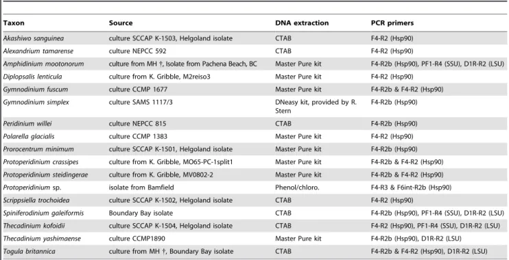
Table 1.Information about the dinoflagellate species from which sequences were generated in this study.
Taxon Source DNA extraction PCR primers
Akashiwo sanguinea culture SCCAP K-1503, Helgoland isolate CTAB F4-R2 (Hsp90)
Alexandrium tamarense culture NEPCC 592 CTAB F4-R2 (Hsp90)
Amphidinium mootonorum culture from MH{, Isolate from Pachena Beach, BC Master Pure kit F4-R2b (Hsp90), PF1-R4 (SSU), D1R-R2 (LSU)
Diplopsalis lenticula culture from K. Gribble, M2reiso3 Master Pure kit F4-R2 (Hsp90)
Gymnodinium fuscum culture CCMP 1677 Master Pure kit F4-R2b & F4-R2 (Hsp90)
Gymnodinium simplex culture SAMS 1117/3 DNeasy kit, provided by R.
Stern
F4-R2b (Hsp90)
Peridinium willei culture NEPCC 815 CTAB F4-R2b (Hsp90)
Polarella glacialis culture CCMP 1383 Master Pure kit F4-R2 (Hsp90)
Prorocentrum minimum culture SCCAP K-1501, Helgoland isolate Master Pure kit F4-R2b (Hsp90)
Protoperidinium crassipes culture from K. Gribble, MO65-PC-1split1 Master Pure kit F4-R2b & F4-R2 (Hsp90)
Protoperidinium steidingerae culture from K. Gribble, MV0802-2 Master Pure kit F4-R2b & F4-R2 (Hsp90)
Protoperidiniumsp. isolate from Bamfield Phenol/chloro. F4-R3 & F6int-R2b (Hsp90)
Scrippsiella trochoidea culture SCCAP K-1502, Helgoland isolate CTAB F4-R2 (Hsp90)
Spiniferodinium galeiformis Boundary Bay isolate CTAB F4-R2b (Hsp90), PF1-R4 (SSU), D1R-R2 (LSU)
Thecadinium kofoidii culture SCCAP K-1504, Helgoland isolate CTAB F4-R2 (Hsp90), PF1-R4 (SSU), D1R-R2 (LSU)
Thecadinium yashimaense culture CCMP1890 Master Pure kit F4-R2b (Hsp90), D1R-R2 (LSU)
Togula britannica culture from MH{, Boundary Bay isolate CTAB F4-R2b & F4-R2 (Hsp90), D1R-R2 (LSU)
CCMP = Provasoli-Guillard National Centre for Culture of Marine Phytoplankton, Hsp90 = heat shock protein 90 sequence, lsu = large subunit ribosomal DNA sequence, MH = Mona Hoppenrath, NEPCC = North East Pacific Culture Collection (now CCCM = Canadian Center for the Culture of Microorganisms), SAMS = Scottish Association for Marine Science (CCAP = Culture Collection of Algae and Protozoa), SCCAP = Scandinavian Culture Collection of Algae & Protozoa, ssu = small subunit ribosomal DNA sequence,
{
= dead/lost.
doi:10.1371/journal.pone.0013220.t001
Table 2.PCR primers used in this study.
Gene Primer name Primer sequence 59-39 Citation
Hsp90 F4 GGAGCCTGATHATHAAYACNTTYTA this study
F6int AAYAARMMNAARCCNHTNTGGATG this study
R2 CGCCTTCATMATNCSYTCCATRTTNGC [10]
R2b GCCTTCATDATNCKYTCCATRTT this study
R3 GATGACYTTNARDATYTTRTTYTGYTG [10]
SSU PF1 GCGCTACCTGGTTGATCCTGCC [70] (modified)
R4 GATCCTTCTGCAGGTTCACCTAC [70] (modified)
LSU D1R ACCCGCTGAATTTAAGCATA [71]
R2 ATTCGGCAGGTGAGTTGTTAC [19]
Phylogenetic relationships were inferred from all six alignments using maximum likelihood (ML) and Bayesian inference (BI) methods with the programs RAxML v7.04 [65] and MrBayes
v3.12 [66,67], respectively. ML and BI analyses of the nucleotide alignments (i.e., alignments 1–3 and 5–6) were built under a GTR+I+G+8 model as suggested by the criteria implemented in
Table 3.Taxa and their accession numbers used for the different alignments and phylogenetic analyses.
Taxon SSU rDNA LSU rDNA Hsp90 (Hsp+SSU) Combined (Hsp+SSU) Combined (Hsp+SSU+LSU)
Ciliates, apicomplexans &Perkinsus (outgroups)
Blepharisma** M97909 x AY390395 x x
Cryptosporidium parvum AF093489 AE040725 AY423866 included included
Eimeria tenella EF210325 x AAB97088 included x
Halteria grandinella AY00744 x AY391253 included x
Paramecium tetraurelia EF502045 x AAG00569 included x
Perkinsus marinus AF126013 AY876319 AY391259 included included
Tetrahymena bergeri AF364039 x AY391257 included x
Tetrahymena pyriformis EF070254 x AAG00567 included x
Theileria parva AF013418 AF218825 AAA30132 included included
Toxoplasma gondii M97703 L25635.1 AAQ24837 included included
Dinoflagellates &Oxyrrhis(ingroup)
Oxyrrhis marina x x AAR27544 x x
Akashiwo sanguinea AF276818 AF260396 GU295192 included included
Alexandrium tamarense AB088333 AY438021 AM184118 included included
Alex. tamarenseUBC x x GU295210 x x
Amphidinium carterae AF274251 AY455669 EU876701 included included
Amphidinium mootonorum GU295202 GU295205 GU295199 included included
Crypthecodinium cohnii M64245 FJ939575 AAM02974 included included
Diplopsalis lenticula x EF152794 GU295193 x x
Gymnodinium chlorophorum AM184122 AF200669 AM184119 included included
Gymnodinium fuscum AF022194 AF200676 GU295194 included included
Gymnodinium simplex DQ388466 AF060901 GU295211 included included
Heterocapsa triquetra AF022198 AF260401 AAR27541 included included
Karenia brevis AF172714 AF200677 AM184117 included included
Karenia mikimotoi AF022195 AF200682 AM184120 included included
Karlodinium micrum AF172712 AF200675 AM184121 included included
Kryptoperidinium foliaceum AF274268 EF052684 AAV32830 included included
Lessardia elongata AF521100 x AY391256 included x
Noctiluca scintillans AF022200 x AB297471 included x
Peridinium willei AF274272 AF260384 GU295195 included included
Polarella glacialis AF099183 AY571373 GU295196 included included
Prorocentrum micans M14649 AF260377 AAR27546 included included
Prorocentrum minimum AY421791 AF260379 GU295201 included included
Protoperidinium crassipes AB261515 EF152846 GU295197 included included
Protoperidinium steidingerae x DQ444231 GU295198 x x
Protoperidiniumsp. x x GU295212 x x
Scrippsiella trochoidea AF274277 AF260393 GU295213 included included
Spiniferodinium galeiformis GU295203 GU295206 GU295214 included included
Thecadinium kofoidii GU295204 GU295207 GU295215 included included
Thecadinium yashimaense AY238477 GU295209 GU295200 included included
Togula britannicaUBC x GU295208 GU295216 included included
Togula britannica AY443010 AY455679 X included included
**B. intermediumfor Hsp90;B. americanumfor SSU rDNA.
ModelTest v0.1.1 [68]. Two alternative topologies differing in the relative position ofNoctiluca, in the analyses of the Hsp90 DNA, were generated with TreeView. Approximately unbiased (AU) tests were performed with CONSEL [69] using the likelihoods calculated with RAxML v7.04 with the same models and parameters indicated above. ML and BI analyses of the amino acid alignment (i.e., alignment 4) was analyzed under a WAG model of substitution considering corrections for site-to-site rate variation (gamma) with eight categories of rate variation and proportion of invariable sites. In order to assess topological support, 500 bootstrap replicates were performed with RAxML on each alignment with the parameters described above.
Bayesian analyses consisted of two independent Markov Chain Monte Carlo (MCMC) runs of 2,000,000 generations were calculated with trees sampled every 50 generations and with a prior burn-in of 100,000 generations (i.e. the first 2,000 sampled trees were discarded). The convergence diagnostic for all six alignments was within 1.0 (60.005). A majority rule consensus tree was constructed from 38,001 post-burn-in trees. Posterior probabilities correspond to the frequency at which a given node was found in the post-burn-in trees.
Introns in the dinoflagellate Hsp90 sequences
Introns were present in only 3 of 17 hsp90 genes sequenced from genomic DNA. The hsp90 gene ofPeridinium willeicontained one canonical intron near the 59 end of the template sequence between residues 467 and 563 (97 bases). The hsp90 gene of
Polarella glacialis contained one non-canonical intron near the 59
end of the template sequence between residues 112 and 245 (134 bases). The hsp90 gene of Thecadiniium yashimaensecontained one canonical intron near the 59end of the template sequence between residues 355 and 643 (289 bases).
Supporting Information
Figure S1 Maximum likelihood (ML) tree inferred from 35 SSU rDNA sequences (dataset 1), 1,381 unambiguously aligned sites and a GTR+I+G+8 model of nucleotide substitutions. Numbers above the branches denote ML bootstrap percentages, and numbers below the branches denote Bayesian posterior probabil-ities. Black circles denote bootstrap percentages and posterior probabilities of 100% and 1.00, respectively.
Found at: doi:10.1371/journal.pone.0013220.s001 (.16 MB EPS)
Figure S2 Maximum likelihood (ML) tree inferred from 30 LSU rDNA sequences (dataset 2), 482 unambiguously aligned sites and a GTR+I+G+8 model of nucleotide substitutions. Numbers above the branches denote ML bootstrap percentages, and numbers below the branches denote Bayesian posterior probabilities. Black circles denote bootstrap percentages and posterior probabilities of 100% and 1.00, respectively.
Found at: doi:10.1371/journal.pone.0013220.s002 (1.38 MB EPS)
Acknowledgments
We would like to thank Rowena Stern (University of British Columbia, Vancouver, Canada) for providing some DNA extracts from dinoflagellates and Kristin Gribble (Marine Biological Laboratory, Woods Hole, USA) for providing theProtoperidiniumandDiplopsaliscultures.
Author Contributions
Conceived and designed the experiments: MH BL. Performed the experiments: MH BL. Analyzed the data: MH BL. Contributed reagents/materials/analysis tools: BL. Wrote the paper: MH BL.
References
1. Taylor FJR (1987) General group characteristics, special features, short history of dinoflagellate study. In: Taylor FJR (Ed.) The biology of Dinoflagellates Botanical Monographs, Vol. 21, Blackwell Scientific Publications, Oxford. 2. Hackett JD, Anderson DM, Erdner DL, Bhattacharya D (2004) Dinoflagellates:
a remarkable evolutionary experiment. Am J Bot 91(10): 1523–1534. 3. Taylor FJR, Hoppenrath M, Saldarriaga JF (2008) Dinoflagellate diversity and
distribution. Biodivers Conserv 17: 407–418.
4. Fensome RA, Taylor FJR, Norris G, Sarjeant WAS, Wharton DI, et al. (1993) A classification of living and fossil dinoflagellates. Am Mus Nat Hist, Micropale-ontology special publication number 7: 1–351.
5. Hoppenrath and Saldarriaga (2008) Available: http://tolweb.org/Dinoflagel lates/2445.
6. Maroteaux L, Herzog M, Soyer-Gobillard MO (1985) Molecular organization of dinoflagellate ribosomal DNA: molecular implications of the deduced 5.8S rRNA secondary structure. Biosystems 18: 307–319.
7. Cavalier-Smith T (1993) Kingdom Protozoa and its 18 phyla. Microbiol Rev 57: 953–994.
8. Patterson D (1999) The diversity of eukaryotes. Am Nat 154: 96–124. 9. Leander BS, Keeling PJ (2003) Morphostasis in alveolate evolution. Trends Ecol
Evol 18: 395–402.
10. Leander BS, Keeling PJ (2004) Early evolutionary history of dinoflagellates and apicomplexans (Alveolata) as inferred from hsp90 and actin phylogenies. J Phycol 40: 341–350.
11. Saldarriaga JF, McEwan ML, Fast NM, Taylor FJR, Keeling PJ (2003) Multiple protein phylogenies show thatOxyrrhis marinaandPerkinsus marinusare early branches of the dinoflagellate lineage. Int J Syst Evol Microbiol 53: 355– 365.
12. Daugbjerg N, Hansen G, Larsen J, Moestrup Ø (2000) Phylogeny of some of the major genera of dinoflagellates based on ultrastructure and partial LSU rDNA sequence data, including the erection of three new genera of unarmoured dinoflagellates. Phycologia 39: 302–317.
13. Saldarriaga JF, Taylor FJR, Cavalier-Smith T, Menden-Deuer S, Keeling PJ (2004) Molecular data and the evolutionary history of dinoflagellates. Europ J Protistol 40: 85–111.
14. Murray S, Flø Jørgensen M, Ho SYW, Patterson DJ, Jermiin LS (2005) Improving the analysis of dinoflagellate phylogeny based on rDNA. Protist 156: 269–286.
15. Taylor FJR (1980) On dinoflagellate evolution. BioSystems 13: 65–108.
16. Taylor FJR (2004) Illumination or confusion? Dinoflagellate molecular phylogenetic data viewed from a primarily morphological standpoint. Phycol Res 52: 308–324.
17. Flø Jørgensen M, Murray S, Daugbjerg N (2004) Amphidinium revisited. I. Redefinition ofAmphidinium(Dinophyceae) based on cladistic and molecular phylogenetic analyses. J Phycol 40: 351–365.
18. Gribble KE, Anderson DM (2006) Molecular phylogeny of the heterotrophic dinoflagellates, Protoperidinium, Diplopsalis and Preperidinium (Dinophyceae), inferred from large subunit rDNA. J Phycol 42: 1081–1095.
19. Yamaguchi A, Kawamura H, Horiguchi T (2006) A further phylogenetic study of the heterotrophic dinoflagellate genus,Protoperidinium(Dinophyceae) based on small and large subunit ribosomal RNA gene sequences. Phycol Res 54: 317–329.
20. Hoppenrath M, Leander BS (2007) Morphology and phylogeny of the pseudocolonial dinoflagellates Polykrikos lebouraeandPolykrikos herdmanae n. sp. Protist 158: 209–227.
21. Hoppenrath M, Bachvaroff TR, Handy SM, Delwiche CF, Leander BS (2009) Molecular phylogeny of ocelloid-bearing dinoflagellates (Warnowiaceae) as inferred from SSU and LSU rDNA sequences. BMC Evol Biol 9: 116. 22. Gribble KE, Anderson DM (2007) High intraindividual, intraspecific, and
interspecific variability in large-subunit ribosomal DNA in the heterotrophic dinoflagellatesProtoperidinium,DiplopsalisandPreperidinium(Dinophyceae). Phyco-logia 46: 315–324.
23. Sparmann SF, Leander BS, Hoppenrath M (2008) Comparative morphology and molecular phylogeny of taxa of the new marine benthic dinoflagellate genus
Apicoporus, classified formerly withinAmphidiniumsensu lato. Protist 159: 383– 399.
24. Edvardsen B, Shalchian-Tabrizi K, Jakobsen KS, Medlin LK, Dahl E, et al. (2003) Genetic variability and molecular phylogeny of Dinophysis species (Dinophyceae) from Norwegian waters inferred from single cell analyses of rDNA. J Phycol 39: 395–408.
25. Zhang H, Bhattacharya D, Lin S (2007) A three-gene dinoflagellate phylogeny suggests monophyly of Prorocentrales and a basal position forAmphidiniumand
Heterocapsa. J Mol Evol 65: 463–474.
26. De Salas MF, Bolch CJS, Botes L, Nash G, Wright SW, et al. (2003)Takayama
27. Flø Jørgensen M, Murray S, Daugbjerg N (2004) A new genus of athecate interstitial dinoflagellates, Togula gen. nov., previously encompassed within
Amphidinium sensu lato: Inferred from light and electron microscopy and phylogenetic analyses of partial large subunit ribosomal DNA sequences. Phycol Res 52: 284–299.
28. Murray S, Flø Jørgensen M, Daugbjerg N, Rhodes L (2004) Amphidinium
revisited. II. Resolving species boundaries in theAmphidinium operculatumspecies complex (Dinophyceae), including the descriptions ofAmphidinium trullasp. nov. andAmphidinium gibbosumcomb. nov. J Phycol 40: 366–382.
29. Hoppenrath M, Leander BS (2007) Character evolution in polykrikoid dinoflagellates. J Phycol 43: 366–377.
30. Hoppenrath M, Yubuki N, Bachvaroff S, Leander BS (2010) Reclassification of
Pheopolykrikos hartmannii as Polykrikos (Dinophyceae) based partly on the ultrastructure of complex extrusomes. Europ J Protistol 46: 29–37.
31. Litaker RW, Tester P, Colorni A, Levy MG, Noga EJ (1999) The phylogenetic relationship ofPfiesteria piscicida, cryptoperidiniopsoid sp.Amyloodinium ocellatum
and aPfiesteria-like dinoflagellate to other dinoflagellates and apicomplexans. J Phycol 35: 1379–1389.
32. Fukuda Y, Endoh H (2006) New details from the complete life cycle of the red-tide dinoflagellateNoctiluca scintillans(Ehrenberg) McCartney. Eur J Protistol 42: 209–219.
33. Fukuda Y, Endoh H (2008) Phylogenetic analyses of the dinoflagellateNoctiluca scintillansbased onb-tubulin and Hsp90 genes. Eur J Protistol 44: 27–33. 34. Ki J-S (2010) Nuclear 28S rDNA phylogeny supports the basal placement of
Noctiluca scintillans(Dinophyceae; Noctilucales) in dinoflagellates. Europ J Protistol 46: 111–120.
35. Handy SM, Bachvaroff TR, Timme RE, Coats DW, Kim S, et al. (2009) Phylogeny of four dinophysiacean genera (Dinophyceae, Dinophysiales) based on rDNA sequences from single cells and environmental samples. J Phycol 45: 1163–1174.
36. Hastrup Jensen M, Daugbjerg N (2009) Molecular phylogeny of selected species of the order Dinophysiales (Dinophyceae) – testing the hypothesis of a dinophysioid radiation. J Phycol 45: 1136–1152.
37. Lindberg K, Moestrup Ø, Daugbjerg N (2005) Studies on woloszynskioid dinoflagellates I: Woloszynskia coronata re-examined using light and electron microscopy and partial LSU rDNA sequences, with description ofTovelliagen. nov. andJadwigiagen. nov. (Tovelliaceae fam. nov.). Phycologia 44: 416–440. 38. Grzebyk D, Sako Y, Berland B (1998) Phylogenetic analysis of nine species of
Prorocentrum (Dinophyceae) inferred from 18S ribosomal DNA sequences, morphological comparisons, and description ofProrocentrum panamensissp. nov. J Phycol 34: 1055–1068.
39. Mohammad-Noor N, Moestrup Ø, Daugbjerg N (2007) Light, electron microscopy and DNA sequences of the dinoflagellateProrocentrum concavum(syn.
P. arabianum) with special emphasis on the periflagellar area. Phycologia 46: 549–564.
40. Hoppenrath M, Leander BS (2008) Morphology and molecular phylogeny of a new marine sand-dwelling Prorocentrum species, P. tsawwassenensis sp. nov. (Dinophyceae, Prorocentrales), from British Columbia, Canada. J Phycol 44: 451–466.
41. Murray S, Ip CL-C, Moore R, Nagahama Y, Fukuyo Y (2009) Are prorocentroid dinoflagellates monophyletic? A study of 25 species based on nuclear and mitochondrial genes. Protist 160: 245–264.
42. Iwataki M, Hansen G, Sawaguchi T, Hiroishi S, Fukuyo Y (2004) Investigation of body scales in twelveHeterocapsaspecies (Peridiniales, Dinophyceae), including a new speciesH. pseudotriquetrasp. nov. Phycologia 43: 394–403.
43. Sekida S, Okuda K, Katsumata K, Horiguchi T (2003) A novel type of body scale found in two strains ofAmphidiniumspecies (Dinophyceae). Phycologia 42: 661–666.
44. Tamura M, Takano Y, Horiguchi T (2009) Discovery of a novel type of body scale in the marine dinoflagellate,Amphidinium cupulatisquamasp. nov. (Dinophy-ceae). Phycol Res 57: 304–312.
45. Watanabe MM, Suda S, Inouye I, Sawaguchi T, Chihara M (1990)Lepidodinium viridegen. et sp. nov. (Gymnodiniales, Dinophyta), a green dinoflagellate with a chlorophyllA- andB-containing endosymbiont. J Phycol 26: 741–751. 46. Hansen G, Botes L, de Salas M (2007) Ultrastructure and large subunit rDNA
sequences of Lepidodinium viride reveal a close relationship to Lepidodinium chlorophorumcomb. nov. ( =Gymnodinium chlorophorum). Phycol Res 55: 25–41.
47. Clarke KJ, Pennick NC (1972) Flagellar scales inOxyrrhis marinaDujardin. Br phycol J 7: 357–360.
48. Clarke KJ, Pennick NC (1976) The occurrence of body scales inOxyrrhis marina
Dujardin. Br phycol J 11: 345–348.
49. Slamovits CH, Saldarriaga JF, Larocque A, Keeling PJ (2007) The highly reduced and fragmented mitochondrial genome of the early-branching dinoflagellate Oxyrrhis marina shares characteristics with both apicomplexan and dinoflagellate mitochondrial genomes. J Mol Biol 372: 356–368. 50. Young JC, Mosarefi I, Hard EU (2001) Hsp90: a specialized but essential
protein-folding tool. J Cell Biol 154: 267–273.
51. Stechmann A, Cavalier-Smith T (2003) Phylogenetic analysis of eukaryotes using heat-shock protein Hsp90. J Mol Evol 57: 408–419.
52. Fast NM, Xue L, Bingham S, Keeling PJ (2002) Re-examining alveolate evolution using multiple protein molecular phylogenies. J Eukaryot Microbiol 49: 30–37.
53. Shalchian-Tabrizi K, Minge MA, Cavalier-Smith T, Nedreklepp JM, Klaveness D, et al. (2006) Combined heat shock protein 90 and ribosomal RNA sequence phylogeny supports multiple replacements of dinoflagellate plastids. J Eukaryot Microbiol 53: 217–224.
54. Saldarriaga JF, Taylor FJR, Keeling PJ, Cavalier-Smith T (2001) Dinoflagellate nuclear SSU rRNA phylogeny suggests multiple plastid losses and replacements. J Mol Evol 53: 204–213.
55. Breglia SA, Slamovits CH, Leander BS (2007) Phylogeny of phagotrophic euglenids (Euglenozoa) as inferred from Hsp90 gene sequences. J Eukaryot Microbiol 54: 86–92.
56. Liu L, Hastings JW (2007) Two different domains of the luciferase gene in the heterotrophic dinoflagellateNoctiluca scintillansoccur as two separate genes in photosynthetic species. Proc Natl Acad Sci 104: 696–701.
57. Ho¨hfeld I, Melkonian M (1995) Ultrastructure of the flagellar apparatus of
Noctiluca miliarisSuriray swarmers (Dinophyceae). Phycologia 34: 508–513. 58. Zingmark RG (1970) Sexual reproduction in the dinoflagellateNoctiluca miliaris
Suriray. J Phycol 6: 122–126.
59. Schnepf E, Drebes G (1993) Anisogamy in the dinoflagellate Noctiluca? Helgola¨nder Meeresunters 47: 265–273.
60. Uhlig G (1972) Entwicklung vonNoctiluca miliaris. Scientific movie C 897/1965, accompanying publication Inst Wiss Film Go¨ttingen, 15 p.
61. Go´mez F, Moreira D, Lo´pez-Garcı´a (2010) Molecular phylogeny of noctilucoid dinoflagellates (Noctilucales, Dinophyceae). Protist 161: 466–478.
62. Hoppenrath M (2004) A revised check-list of planktonic diatoms and dinoflagellates from Helgoland (North Sea, German Bight). Helgol Mar Res 58: 243–251.
63. Hoppenrath M, Elbra¨chter M, Drebes G (2009) Marine Phytoplankton. Selected microphytoplankton species from the North Sea around Helgoland and Sylt. Kleine Senckenberg-Reihe, Band 49, 264 p.
64. Guillard RRL, Ryther JH (1962) Studies of marine planktonic diatoms. I.
Cyclotella nanaHustedt andDetonula confervaceaCleve. Can J Microbiol 8: 229–239. 65. Stamatakis A (2006) RAxML-VI-HPC: maximum likelihood-based phylogenetic analyses with thousands of taxa and mixed models. Bioinformatics 22: 2688–2690.
66. Ronquist F, Huelsenbeck JP (2003) MRBAYES 3: Bayesian phylogenetic inference under mixed models. Bioinformatics 19: 1572–1574.
67. Altekar G, Dwarkadas S, Huelsenbeck JP, Ronquist F (2004) Parallel metropolis coupled Markov chain Monte Carlo for Bayesian phylogenetic inference. Bioinformatics 20: 407–415.
68. Posada D (2008) jModelTest: phylogenetic model averaging. Mol Biol Evol 25: 1253–1256.
69. Shimodaira H, Hasegawa M (2001) CONSEL: for assessing the confidence of phylogenetic tree selection. Bioinformatics 17: 12461247.
70. Leander BS, Clopton RE, Keeling PJ (2003) Phylogeny of gregarines (Apicomplexa) as inferred from small subunit rDNA and beta-tubulin. Int J Syst Evol Microbiol 53: 345–35471.