Arq. Bras. Med. Vet. Zootec., v.69, n.1, p.95-100, 2017
Bone marrow bi-hypoplasia in a dog with a sertoli cell tumor
Hipoplasia dupla de medula óssea em um cão com tumor de células de sertoli]
P.C.L.G. Valente, R.M. Couto, C.O. Gamba, A.V. Vasconcelos, F.O.P. Leme, R. Ecco, P.R.O. Paes
Universidade Federal de Minas Gerais ˗ Belo Horizonte, MG
RESUMO
Um cão Poodle, macho, de 20 anos, não castrado, apresentou prostração, apatia, andar cambaleante, falta de apetite e infestação por carrapatos. Nesse animal, foi diagnosticado tumor de células de Sertoli em um testículo não descendente, utilizando-se citologia, histopatologia e imuno-histoquímica. Pancitopenia com anemia moderada não regenerativa, leucopenia e trombocitopenia intensas foram detectadas no hemograma. Na avaliação citológica e histopatológica da medula óssea, havia celularidade de 30%, constituída pelas linhagens eritroide (59%) e linfoide (40%) e por mastócitos (1%), com ausência de células das linhagens granulocítica, monocítica e megacariocítica. Em exames post mortem, mudanças relacionadas à hemostasia foram encontradas. A ausência de micro-organismos nos testes moleculares e a concentração sérica de estrogênio acima dos valores de referência confirmaram hiperestrogenismo como a possível causa da pancitopenia. A literatura descreve hiperestrogenismo em tumores de células de Sertoli induzindo pancitopenia associada com hipoplasia da medula óssea de todas as linhagens hematopoiéticas. Em contraste, no presente caso, as células precursoras eritróides estavam preservadas na medula óssea, embora não houvesse reticulócitos no sangue. Assim, o relato apresentado deve ser considerado em futuras investigações de pancitopenia induzida por hiperestrogenismo em tumor de células de Sertoli.
Palavras-chave: cão, hiperestrogenismo, pancitopenia
ABSTRACT
A 20-year-old unneutered male poodle presented prostration, apathy, staggering gait, lack of appetite and tick infestation. The dog was diagnosed with a Sertoli cell tumor in an undescended testicle by cytological, histopathological and immunohistochemical tests. Pancytopenia with moderate nonregenerative anemia, leukopenia and severe thrombocytopenia were detected in the complete blood count. Cytological and histopathological evaluation of the bone marrow revealed a cellularity of 30%, with erythroid (59%), lymphoid (40%) and mast cells (1%), and an absence of granulocytic, monocytic and megakaryocytic lineage cells. In post-mortem examinations, changes related to hemostatic disorders were found. The absence of microorganisms in molecular tests and an estrogen serum concentration over reference values confirmed hyperestrogenism as a possible cause of pancytopenia. The literature describes a Sertoli cell tumor hyperestrogenism that induced pancytopenia, along with bone marrow hypoplasia of all hematopoietic lineages. In contrast, in the present case, the erythroid precursor cells were preserved in the bone marrow, although there were no reticulocytes circulating in the blood. This case, therefore, should be considered in future investigations of pancytopenia induced by Sertoli cell tumor hyperestrogenism.
Keywords: dog, hyperestrogenism, pancytopenia
INTRODUCTION
The testicles are the second most frequent site of neoplasia in unneutered male dogs and the three main testicular tumors are Sertoli cell tumors, seminomas and interstitial cell tumors, which affect mainly older dogs and dogs with undescended testicles (Ciaputa et al., 2012). Animals with a Sertoli cell tumor may present hyperestrogenism with a bilateral symmetrical alopecia, hyperpigmentation, gynecomastia, galactorrhea, pendulous prepuce, and atrophy of the penis and the contralateral testicle (Peters et al., 2001). Additionally, high serum levels of estrogen are considered myelotoxic in dogs because they cause bone marrow hypoplasia with pancytopenia (Sherding et al., 1981; Suess et al., 1992; Sanpera et al., 2002; Sontas et al., 2009). The prognosis for a bone marrow hypoplasia secondary to hyperestrogenism varies from reserved to unfavorable because dogs tend to not respond well to treatment and may also present with generalized secondary infections and hemorrhages (Sherding et al., 1981; Sanpera et al., 2002). The present report describes the changes caused by the myelotoxic action of the hyperestrogenism secondary to a Sertoli cell tumor in a cryptorchid dog.
CASE REPORT
A 20-year-old, unneutered male poodle with a history of prostration, apathy, tottering gait and lack of appetite was presented to the Veterinary Hospital of the Universidade Federal de Minas Gerais (UFMG). At the general clinical examination, the dog was dehydrated, had many ticks and exhibited a capillary refill time (CRT) of 3 seconds, tachypnea and pale mucous membranes. There were also hemorrhages in the periocular area, alopecia, cryptorchidism and a firm tumor in the right inguinal area. Clinical suspicion was testicular neoplasia. Blood samples were collected for a complete blood count (CBC) and a biochemical profile. Fine needle aspiration of the inguinal mass was performed for cytological examination. A CBC, cytological tests and histopathological tests of the bone marrow were performed.
The CBC, carried out with an automated analyzer, revealed pancytopenia; moderate normocytic, normochromic anemia (28%
mean corpuscular volume: 70.4fL, RI: 60-77fL; mean corpuscular hemoglobin concentration: 33.2g/dL, RI: 31–36g/dL); decreased red blood cell width (RDW) (11.7%, RI: 12-15%); and absence of reticulocytes. Also, there were morphological changes such as anisocytosis, polychromasia and Howell-Jolly bodies; features that indicate a nonregenerative anemia. The leukocyte count revealed an intense leukopenia (total leukocyte count: 490/mm³, RI: 6,000-17,000/mm³) represented by lymphocytes (40%), eosinophils (30%), monocytes (20%) and segmented neutrophils (10%). The platelet examination indicated marked thrombocytopenia (platelet count: 5,000/mm³ RI: 175,000-500,000/mm³).
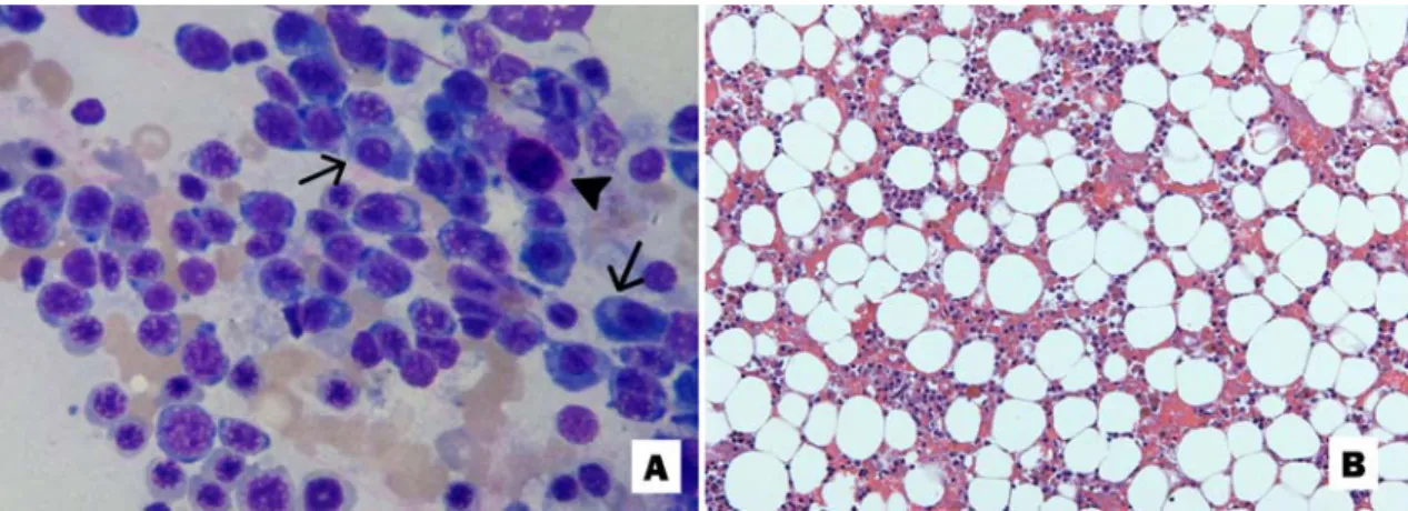
The myelogram revealed a small concentration of particles, absence of monocytic and megakaryocytic granulocyte lineage cells, and increased iron stores in macrophages (Fig. 1A). The differential examination revealed erythroid (59%), lymphoid (40%) and mast cells (1%) with the following distribution: rubriblasts (2%), prorubricytes (3%), rubricytes (38%), metarubricytes (16%), lymphocytes (18%), plasma cells (22%) and mast cells (1%). Histopathological examination of bone marrow cells confirmed the absence of megakaryocytic and granulocytic cells and revealed a 30% cellularity (Fig. 1B). Based on the clinical and laboratory findings, a bone marrow bi-hypoplasia of granulocytic and megakaryocytic lineages was diagnosed.
The evaluation of the serum biochemical profile revealed increased alkaline phosphatase activity (735U/L, RI: 20-156U/L), hypoalbuminemia (1.6g/dL, RI: 2.3 to 3.1g/dL) and hyperglobulinemia (5.0g/dL, RI: 2.7 to 4.4g/dL).
After a day of hospitalization, the animal died, and a complete necropsy was performed. An external examination revealed extensive alopecia on the back extending laterally to the right with varying amounts of suffusion of the skin and the absence of one testicle in the scrotum (cryptorchidism). The undescended testicle, which was located in the right inguinal region, was markedly enlarged and firm and was enclosed in a tense tunica albuginea. On the cut surface, the parenchyma was totally replaced by a lobulated and whitish tumor interspersed with dark red areas (Fig. 2B). In the subcutaneous tissue, there were many ecchymoses and petechiae. Additionally, an increase in size of the iliac and axillary lymph nodes was found. The parietal and visceral pleural surfaces, the serous surface of the stomach, intestines, and kidney capsule had many petechiae. The cortices of the kidneys were diffusely yellowish-white.
Samples of multiple organs were routinely processed for histopathology. In the lymph nodes, there was a moderate to marked draining hemorrhage within the lymphatic sinus and moderate hemosiderosis. There was an intense chronic congestion associated with hyperplasia of the bile ducts and moderate hemosiderosis in the liver. The kidneys presented membranous glomerulonephritis with a mild multifocal glomerulosclerosis, mild multifocal interstitial fibrosis and plasma cell infiltration. Diffuse and marked atrophy was present in the descended testicle. The undescended testicle had a well-demarcated, encapsulated and expansive neoplastic cell proliferation. The cells were
arranged perpendicularly to the basement membrane, providing a tubular pattern interspersed with abundant collagenous stroma. Diffuse arrangements of neoplastic cells could be seen in multifocal areas. These cells resembled normal Sertoli cells and were characterized by mild cellular pleomorphism and anisokaryosis. The nuclei contained loose chromatin and prominent nucleoli, and the cytoplasm was abundant, eosinophilic and elongated, with some containing fine vacuoles. Multifocal areas of coagulative necrosis were also found (Fig. 2C).
A biotin-peroxidase system with identification of the secondary antibody by Advance HRP enzyme polymer was used for the immunohistochemical procedure (Ramos-Vara e Miller, 2014). The cytoplasm of neoplastic cells stained diffusely and intensely for vimentin and revealed negativity for c-kit. These results supported the diagnosis of Sertoli cell tumor (Peters et al., 2001, Yu et al., 2009).
To investigate the possible causes of bone marrow hypoplasia and hematological disorders found in the case, the serum concentration of total estrogen was determined, which resulted in 178.0pg/mL (RI for males: < 50pg/mL), indicating hyperestrogenism. Additionally, blood and bone marrow PCR tests were performed searching for Ehrlichia spp., Ehrlichia canis, Babesia spp., Anaplasma platys and Leishmania spp., according to protocols described previously (Lachaud et al., 2002; Birkenheuer et al., 2003; Bulla et al., 2003). All reactions were negative for these infectious agents.
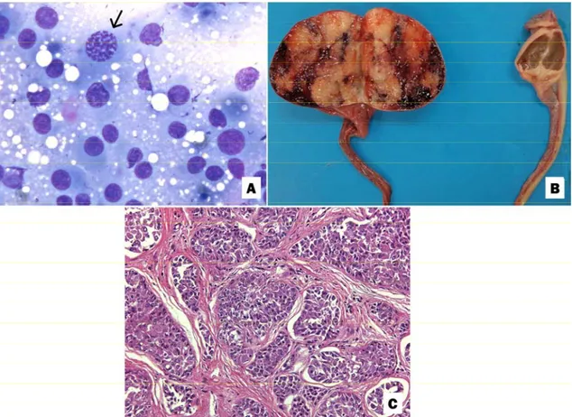
Figure 2. Testicle of a dog with a Sertoli cell tumor. A, Testicular aspirates. Intense population of round cells with moderate anisocytosis and anisokaryosis as well as poorly distinguished and vacuolated cytoplasm. A mitotic figure is visualized (arrow). Giemsa stain, 1000x. B, On the left, the right testicle is enlarged, and the parenchyma is replaced by solid and whitish neoproliferation interspersed with dark red areas (Sertoli cell tumor). On the right, the size of the left testicle is markedly reduced and is brown at the cut surface (atrophy). C, The parenchyma is replaced by neoplastic cell proliferation separated by abundant fibrous tissue. These cells are arranged perpendicularly to the basement membrane, characterizing the intratubular type. Hematoxylin and eosin stain, 200x.
DISCUSSION
The gross and microscopic alterations in the kidneys and liver were not accompanied by changes in serum urea, creatinine, total protein, or enzymatic activities of alanine aminotransferase (ALT) and aspartate aminotransferase (AST). The serum activity of alkaline phosphatase over the reference values (735U/L, RI: 20-156U/L) was not accompanied by elevated gamma-glutamyltransferase (GGT) activity (11.7U/L, RI: 0-25U/L). In cholestasis, an increase in GGT concentration occurs later than an increase in alkaline phosphatase (Stockham and Scott, 2011). The same behavior of liver enzymes has been observed in another case of hyperestrogenism associated with a
in protein fractions are most likely associated with the responses of negative acute-phase proteins (albumin) and positive acute-phase proteins (globulins) or with the protein loss caused by the glomerular lesions (Stockham and Scott, 2011).
The increased concentration of iron stores in the hematopoietic organs may be correlated with blood loss and erythrophagocytosis. In particular, little of the iron that had been stored in the destroyed red blood cells was utilized in the synthesis of new red blood cells, as the anemia was classified as nonregenerative. This iron may have been unavailable, which suggests association with anemia of the inflammatory disease, (anemia of the chronic disease), also caused by the neoplasia (Stockham and Scott, 2011).
Testicular tumors have different embryonic origins: Sertoli cells are of epithelial origin, Leydig cells are of mesenchymal origin and seminomas are derived from spermatogenic cells of the seminiferous epithelium (Ciaputa et al., 2012). Histopathological techniques may not differentiate these tumors; therefore, incubations with anti-vimentin and CD117 (c-kit) could help with diagnostic characterization and discrimination (Peters et al., 2001, Yu et al., 2009). In this study, the neoplastic cells showed positive immunostaining for vimentin and negative for CD 117, confirming a Sertoli cell origin.
Dogs with a Sertoli cell tumor may show clinical signs of feminization attributable to paraneoplastic hyperestrogenism. In this case, changes present are: alopecia and atrophy of the contralateral testis. The myelotoxicity of high levels of estrogen, endogenous or exogenous, is widely described in dogs and other animals such as ferrets. However, the action mechanism of this hormone in bone marrow is not well known (Sanpera et al., 2002). Several hypotheses have been considered: a reduction in the number of hematopoietic stem cells, an inhibition of the differentiation of these cells, changes in the utilization of iron by erythroid precursors, and a reduction of the bone marrow response to erythropoietin (Brockus, 1998).
In all reports of hematological changes induced by high levels of estrogen, bone marrow hypoplasia was found in all hematopoietic lineages (Sherding et al., 1981; Suess et al., 1992; Sanpera et al., 2002; Sontas et al., 2009); some cases were initially bi-hypoplastic, with granulocytic or megakaryocytic cells, but became widely hypoplastic after several weeks (Sherding et al., 1981, Suess et al., 1992). In
contrast, in the present case, erythroid precursor cells in the bone marrow were preserved, although there were no reticulocytes in the circulating blood. Thus, the reported case presents a previously unseen hematologic response, which should be further considered in the investigation of the mechanisms of a Sertoli cell tumor hyperestrogenism with induced pancytopenia. Furthermore, the present report contributes to the issue since few publications have been made in recent years.
REFERENCES
BIRKENHEUER, A.J.; LEVY, M.G.;
BREITSCHWERDT, E.B. Development and evaluation of a seminested PCR for detection and differentiation of Babesia gibsoni (Asian genotype) and B.canis DNA in canine blood samples. J. Clin. Microbiol., v.40, p.4172-4177, 2003.
BROCKUS, C.W. Endogenous estrogen myelotoxicity associated with functional cystic ovaries in a dog. Vet. Clin. Pathol., v.27, p.55-56, 1998.
BULLA, C.; TAKAHIRA, R.K.; ARAUJO JUNIOR, J.P. et al. The relationship between the degree of thrombocytopenia and infection with Ehrlichia canis in an endemic area. Vet. Res., v.34, p.1-6, 2003.
CIAPUTA, R.; NOWAK, M.; KIELBOWICZ, M. et al. Seminoma, sertolioma, and leydigoma in dogs: clinical and morphological correlations. Bull. Vet. Inst. Pulawy, v.56, p.361-367, 2012. LACHAUD, L.; MARCHERGUI-HAMMAMI, S.; CHABBERT, E. et al. Comparison of six PCR methods using peripheral blood for detection of canine visceral leishmaniasis. J. Clin. Microbiol., v.40, p.210-215, 2002.
PETERS, M.A.J.; TEERDS, K.J.; VAN DER GAAG, I. et al. Use of antibodies against LH receptor, 3β-hydroxysteroid dehydrogenase and vimentin to characterize different types of testicular tumour in dogs. J. Reprod. Fertil., v.121, p.287-296, 2001.
SANPERA, N.; MASOT, N.; JANER, M. et al. Oestrogen-induced bone marrow aplasia in a dog with a Sertoli cell tumour. J. Small Anim. Pract., v.43, p.365-369, 2002.
SHERDING, R.G.; WILSON, G.P.; KOCIBA, G.J. Bone marrow hypoplasia in eight dogs with Sertoli cell tumor. J. Am. Vet. Med. Assoc., v.178, p.497-500, 1981.
SONTAS, H.B.; DOKUZEYLU, B.; TURNA, O.; EKICI, H. Estrogen-induced myelocicity in dogs: a review. Can. Vet. J., v.50, p.1054-1058, 2009.
STOCKHAM, S.L.; SCOTT, M.A. Fundamentos de patologia clínica veterinária. 2.ed. Rio de Janeiro: Guanabara Koogan, 2011. 729p.
SUESS, R.P.J.R.; BARR, S.C.; SACRE, B.J.; FRENCH, T.W. Bone marrow hypoplasia in a feminized dog with an interstitial cell tumor. J. Am. Vet. Med. Assoc., v.200, p.1346-1348, 1992.
YU, C.H.; HWANG, D.N.; YHEE,