A reusable Eu
3+complex for naked-eye
discrimination of methanol from ethanol with a
ratiometric fluorimetric equilibrium in
methanol/ethanol mixtures
João P. Leala, b, Filipe A. Almeida Pazc, Ricardo F. Mendesc, Tiago Moreirad, Mani Outisd,
César A. T. Laia*,d, Bernardo Monteiro*a, b, Cláudia C. L. Pereira*d
[a] Dr, J.P. Leal; Dr. B., Monteiro*
Departamento de Engenharia e Ciências Nucleares Centro de Ciências e Tecnologias Nucleares (C2TN), Instituto Superior Técnico.
Estrada Nacional 10, 2695-066 Bobadela, Portugal. E-mail: bernardo.monteiro@ctn.tecnico.ulisboa.pt
[b] Dr, J.P. Leal; Dr. B., Monteiro*
Departamento de Engenharia e Ciências Nucleares
Centro de Química Estrutural (CQE), Instituto Superior Técnico. Estrada Nacional 10, 2695-066 Bobadela, Portugal.
E-mail: bernardo.monteiro@ctn.tecnico.ulisboa.pt
Homepage: https://cqe.tecnico.ulisboa.pt/members/13317 [c] Dr. F.A.A. Paz; Dr. R.F. Mendes
Departamento de Química
CICECO - Instituto de Materiais de Aveiro Universidade de Aveiro, 3810-193 Aveiro, Portugal. [d] Dr, T. Moreira; Dr. C.A.T. Laia*; Dr. C.C.L. Pereira*
Departamento de Química
Associated Laboratory for Sustainable Chemistry-Clean Processes and Technologies - LAQV-REQUIMTE
Universidade Nova de Lisboa, 2829-516 Caparica, Portugal. E-mail: ccl.pereira@fct.unl.pt
E-mail: catl @fct.unl.pt Homepage:
Supporting information for this article is given via a link at the end of the document.
Abstract
Immediate naked eye distinction of methanol from ethanol can be performed by simple dissolution, in each of these solvents, of either [Na][EuFOD4] (1) complex or in mixture of products from the reaction between [P6,6,6,14][Eu(FOD)4] and NaOPhMe3, referred as 2, ([P6,6,6,14]+ = trihexyltetradecylphosphonium cation, FOD- = 1,1,1,2,2,3,3-heptafluoro-7,7-dimethyloctane-4,6-dionate). Additionally, an easy, low-cost, efficient and fast spectrofluorimetric method for the detection and quantification of methanol in mixtures with ethanol is described. This method is based on the changes in the Eu3+ luminescence in the visible region due to the interaction of the [P6,6,6,14][Eu(FOD)4] complex with these alcohols. A limit of detection as low as 15% (w/w) of methanol in mixtures of ethanol/methanol is discussed considering a linear calibration curve.
Introduction
Certain compounds present a variation of the absorption and emission spectra depending on the solvent where they are dissolved. This effect is called solvatochromism and was previously presented by Binnemans and co-workers as a new tool to distinguish structurally similar compounds. Strong solvent effects were observed for lanthanide complexes containing the hemicyanine chromophore. This effect was explained based on dipolar interactions between the solvent and the complexes when a series of n-alcohols are used as solvent.1
Recently, Cui and co-workers reported that the color of two isostructural Tb- and Eu-MOFs (with the ligand 5,5′,5″-(1,3,5-triazine2,4,6-triyl)tris(azanediyl)triisophthalate) gradually changed from colorless to golden, to dark orange and to dark red as the immersion time increased in ethanol, acetonitrile, and diethyl ether. Moreover, these two MOFs presented solvatochromism in
the presence of diethyl ether vapors. This allows these porous structures to be used as ethanol, acetonitrile, and diethyl ether sensors by the naked eye.2
We have reported, among other studies, the solvatochromic properties within the [P6,6,6,14] [Ln(NTA)4] family (NTA = naphthoyltrifluoroacetonate and [P6,6,6,14]+ = trihexyltetradecylphosphonium).3,4 In the case of the Eu3+ ionic liquid, the ligand absorption spectra did not present a significant solvatochromic effect. However, comparing the pure Eu+ compound (liquid state, at 65 ºC) and when dissolved in cyclohexane, methanol, and toluene a redshift and band broadening was found for the pure compound. This result indicates aggregation of β-deketonate ligands (NTA) in the ionic liquid form and is responsible for the yellowish color of the pure liquid compound while colorless in solution. The Gd3+ analogue presented the same behavior and the Tb3+ had an irrelevant solvatochromic effect (Δλmax ≈ 5 nm). However, in the case of the Dy analogue a clear solvatochromic effect is observed (Δλmax ≈ 71 nm) with a blueshift in non-alcohol solvents (cyclohexene and toluene) when compared to methanol and 1-buthanol. Unlike what was observed for the Eu3+ complex, in the case of the Dy3+ the solvent interacts preferentially with the ligands through hydrogen bonds, which weakens the ligand– metal bond leading to the observed solvatochromic effect.
Two-dimensional layered structures based on Cd (II) and Zn (II) exhibit multichromism such as solvatochromism, thermochromism and piezochromism when subjected to various external stimuli such as solvent, heat, and mechanical grinding or pressure. Interestingly, these framework materials proved to be useful for visual detection of DMF, DMA, DMSO, CH3CN, acetone and Et3N, but not in case of exchange with water, ethanol or methanol.5
Complexes of Zn (II) octa(carbazolyl)phthalocyanines revealed a pronounced solvent effect presenting green color in DMF, THF, pyridine, acetone, acetonitrile and DCM, a blue color in
hexane, cyclohexane, EtOH, isopropanol, and 1- pentanol, and a purple color in CHCl3, CCl4, benzene, toluene and p-xylene. This color change was explained by the transparency shift region of phtalocyanines in the 400−600 nm region, which is responsible for their common green color.6 Solvatochromic effects has been difficult to find between ethanol and methanol no doubt due to their similarities in terms of polarity, density and coordinating character.
Methanol is a highly toxic substance, but it is unfortunately very difficult to differentiate from other alcohols (ex. ethanol) without performing chemical analyses. Methanol quantification is typically based on the use of advanced techniques such as head space gas chromatography (GC)7-8910, GC-flame ionization11 detection, high-performance liquid chromatography (HPLC), Raman12,13 and infra-red spectroscopies.14 A bioenzymatic analytical system was developed consisting of two biosensors, one based on alcohol dehydrogenase (ADH) that responds only to the ethanol and the second one based on alcohol oxidase (AOX) that responds to both methanol and ethanol.15
This report summarizes our efforts to differentiate ethanol from methanol by simple naked eye observation or under a UV-light lamp. We developed an approach for a simple spectrofluorometric method to quantify methanol in the presence of a large excess of ethanol with limit of detection of 15%. The method is based on the solvent displacement from an Eu3+ complex dissolved in ethanol which in the presence of methanol produces an intense purple color, easily quantified and even visible at the naked eye. Besides this and with an equipment as simple as a UV-light lamp we propose a method to distinguish ethanol from methanol using a stable luminescent Eu3+ complex allowing its reutilization in several analysis.
Selectivity of this method for methanol detection in mixtures with other n-alcohols is also discussed.
Results and Discussion
[Na][Eu(FOD)4]. Strong solvatochromic effect was primarily observed for the complex [Na]
[Eu(FOD)4] (1) as shown in the luminescence spectra in Figure 1. The change in the coordination sphere, attributed to a decrease of symmetry caused by solvent change, is observable by an increment in band intensity that corresponds to the 5D07F0 strictly forbidden transition at 579 nm. The 5D07F0 band intensity of the [Eu(FOD)4]- moiety (FOD- = 1,1,1,2,2,3,3-heptafluoro-7,7-dimethyloctane-4,6-dionate) responds differently to ethanol and methanol. We associate this effect with the higher coordination character of methanol, which together with a labile Eu-O bond from one of the β-diketonate ligands, leads to a more asymmetric Eu3+ structure. The splitting of the 5D07F2 transition band in methanol can be attributed to the ‘crystal field effect’ of the Eu3+ ion in a C1 symmetry and is constituted by five intense and well-defined bands at 611, 612, 619, 623 and 633 nm. The Stark splitting of the ground state level of 7F2 into ‘2J+1=5’ sublevels with J=2 demonstrates that the Eu3+ ions occupy the possible lowest local site symmetry. In opposition, a lower number of J-splitting in ethanol (see ESI for details) represents a higher site symmetry of the Eu3+ ions.16
Figure 1. Luminescence spectrum of [Na][Eu(FOD)4] with a concentration of 0.1 mM in methanol (black line) and ethanol (red dotted line) upon excitation with λexcitation = 350 nm. Inset: Picture taken under 366 nm UV light that evidences a brighter orange emission in ethanol solution.
After several attempts, we were able to obtain poor quality crystals of [Na][Eu(FOD)4](H2O) with the structure being unveiled by single-crystal X-ray diffraction studies as depicted in Figure 2. The complex crystallizes in the centrosymmetric P21/n space group, with the asymmetric unit being composed of four independent FOD- anionic linkers connected to a Eu3+ cation, plus one Na+ metal center interacting with three FOD- and one disordered water molecule. The Eu3+ ion is octacoordinated, {EuO8}, to all four FOD- ligands with the overall coordination geometry resembling a distorted square antiprism. Remarkably, three FOD- ligands are orientated in the same direction, allowing the first -CF2- groups to interact with the Na+ ion which lies in a “pocket” of the complex close to the Eu3+ ion. Experimental and simulated (from single-crystal
data) powder X-ray diffraction patterns of compound [Na][Eu(FOD)4](H2O) agree well with the proposed structure (See ESI for details).
Figure 2. Ball and stick representation of the [Na][Eu(FOD)4](H2O) complex (1).
[P6,6,6,14][Eu(FOD)4] + NaOPhMe3. For the previously reported [P6,6,6,14][Eu(NTA)4], when
dissolved in protic solvents (e.g., 2-propanol or ethanol) a pronounced interaction between NTA -and the solvent was evaluated.3 The emission spectra agrees with a high symmetry point group for Eu3+ which breaks down if the compound is dissolved in polar solvents (e.g., n-alcohols).3,17 Also, we recently reported that the [P6,6,6,14][Eu(FOD)4] ionic liquid undergoes a disturbance in the coordination sphere of the Eu3+ ion, with a disruption of the local symmetry when heated up to ca. 80 °C.18 This can be explained by the fact that the inclusion of fluorine substituents in the organic ligand, with a high electron-withdrawing effect, reduces the charge density on the oxygen atoms inducing partial dissociation of one of the ligands.19 The interaction of the acidic
[P6,6,6,14]+ counter-ion with the labile oxygen from one FOD- ligand forms an electron delocalized six-membered ring with Eu3+ with a red/purple color. Since this red colored complex is only seven coordinated, we made several attempts to block the reversibility of this process. The method used consisted on coordinating an additional ligand to the red compound in order to fill the coordination sphere, preventing the labile oxygen to coordinate again to the Eu3+ center, and thus obtaining a red eight-coordinated neutral Eu3+ complex. This could not be achieved by neutral donor ligands like alcohols. Color irreversibility for long periods could only be achieved after the addition of anionic ligands with a highly electron donor nature such as 2,4,6-trimethylphenolate (Scheme 1). Sodium 2,4,6-2,4,6-trimethylphenolate was added stoichiometrically and without solvent to slightly heated [P6,6,6,14][Eu(FOD)4]. The viscous light-yellow material turned immediately to dark purple, showing color stability for several months after cooling to ambient temperature. The mixture of the reaction between [P6,6,6,14][Eu(FOD)4] and NaOPhMe3 (hereafter coined as 2) was characterized by ESI-MS using methanol and ethanol as solvents (Table S1 - ESI). Remarkably, other molecules such as azide (N3-) or methoxide (CH3O-) only stabilized the colored form for short periods, turning into a light-yellow viscous liquid within just a few hours.
It is worth to mention that when using newly purchased (P6,6,6,14])Cl reagent, it usually has a basic character and the [P6,6,6,14][Eu(FOD)4] compound does not change color when heated. It is necessary to either wash with water the phosphonium reagent, or in alternative the final complex, until they reach a neutral pH in order to guarantee that no excess of basic FOD- is present. Additionally, we have prepared the [P6,6,6,14][Eu(FOD)3(DBM)] (DBM = dibenzoylmethanate)20 and another side comment that worth mention is that if even only one FOD- ligand is substituted the thermochromism is lost.
We have previously observed that the reaction product between NaFOD and (P6,6,6,14])Cl yielded, when heated, in an irreversible way the purple P6,6,6,14FOD compound.18 According to the preformed solubility tests, this organic purple compound was more stable (maintained the pinkish color) in MeOH than in EtOH (although after ethanol evaporation the color was regained again).18 This behavior lead us to test the reaction product 2 in these two solvents and the results obtained are depicted in the equilibrium proposed in Scheme 1.
Scheme 1. Solvent dependent equilibrium of 2. The colors used in the scheme (except for NaOPhMe3, which is a white powder) are an approximation of the colors of the compounds ([P6,6,6,14][Eu(FOD)4] 2 is light yellow, Na[P6,6,6,14][Eu(FOD)3OPhMe3] is most certainly light yellow and P6,6,6,14FOD is purple.
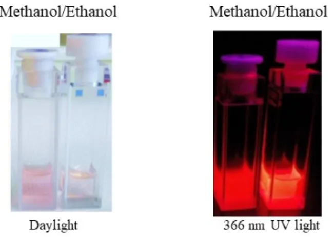
Like was previously observed for the purple P6,6,6,14FOD compound, in ethanol mixture 2 dissociates giving a yellowish solution while in methanol it kept the purple/pinkish (Figure 3, left). This means that the reaction with NaOPhMe3 is more extent in methanol than in ethanol forming [Eu(FOD)3OPhMe3]- and P6,6,6,14FOD (supported by ESI-MS). This can be explained by
the ability of the ethanol to interfere with the P-O bond of the P6,6,6,14FOD compound, yielding free FOD- that is able to replace [OPhMe3]- from de asymmetric [Eu(FOD)3OPhMe3]- complex, ultimately increasing the concentration of the emissive [P6,6,6,14][Eu(FOD)4] in 2. The mechanism behind this proposal is reinforced by the fact that methanol, with a higher coordination ability to Eu3+ than ethanol, prevents any existing FOD- in the mixture to coordinate to [Eu(FOD)3OPhMe3]-, thus stabilizing the purple color (Figure 3, left).21
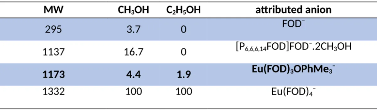
According to ESI-MS data, higher concentrations of [Eu(FOD)3OPhMe3]-, and consequently lower concentrations of [P6,6,6,14][Eu(FOD)4] (4.4 % in methanol vs 1.9 % in ethanol) result in a less effective sensitization mechanism of the Eu3+ complex and, consequently, a poorer energy transfer with the concomitant establishment of a P-O interaction between the free FOD- moiety and the [P6,6,6,14]+ cation (Figure 3, right). This explains the ESI-MS result showing the detection of neutral P6,6,6,14FOD which is exclusively found in methanol (16.7 % in methanol vs 0 % found in ethanol -Table 1).
Figure 3. Picture under 366 nm UV light of 2 in methanol and ethanol with the same concentration (1 mM), with corresponding absorption spectra in figure 4.
In summary, Figure 3 evidences the more intense purple/pink color of mixture 2 in methanol as the P6,6,6,14FOD content is higher. On the other hand, in ethanol the solution has a more yellow/pink color, as the purple/pinkish P6,6,6,14FOD content is lower. Under UV light, the solutions also have different emission intensities, colors and brightness has the less emissive [Eu(FOD)3OPhMe3]- moiety is more abundant in methanol and the more emissive [P6,6,6,14] [Eu(FOD)4] is more abundant in ethanol.
Table 1 ESI–MS analysis results in the negative mode. Molecular weight (MW), percentage of peak area in methanol ethanol and the attributed anionic specie.
MW CH3OH C2H5OH attributed anion
295 3.7 0 FOD–
1137 16.7 0 [P6,6,6,14FOD]FOD–.2CH3OH
1173 4.4 1.9 Eu(FOD)3OPhMe3–
1332 100 100 Eu(FOD)4–
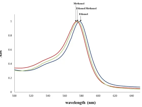
Absorption Spectra. In the UV-vis spectra of 2, the band with max. between 574–578 nm corresponds to the phosphorane like compound P6,6,6,14FOD formed during the 2,4,6-trimethylphenolate addition to the complex [P6,6,6,14][Eu(FOD)4] (Scheme 1).18 ESI–MS confirms the presence of this neutral organic compound while Eu3+ is hepta–coordinated with three FOD -units plus one 2,4,6–trimethylphenolate unit. When 2 is dissolved in methanol the maximum absorption spectra in the visible region is centered at 574 nm to which corresponds a higher amount of the purple P6,6,6,14FOD compound. The final color of the solution results from a balance of different amounts of Na[Eu(FOD)3OPhMe3] (light yellow), [P6,6,6,14][Eu(FOD)4], (light
yellow), NaOPHMe3 (white) and P6,6,6,14FOD (purple) in each of the alcohol solutions. In ethanol, due to a lower amount of P6,6,6,14FOD (purple), and a relative higher amount of light yellow Eu3+ complexes, the solution has a slightly higher absorption wavelength (578 nm). In 1:1 mixture of ethanol and methanol the solution has a max. of 576 nm, exactly between the middle of 574 and 578 nm observed for pure methanol and ethanol, respectively.
Figure 4. Normalized absorption spectra of 2 in methanol (red line, max. = 574 nm) in 1:1 mixture of ethanol:methanol (green line, max. = 576 nm ) and ethanol (blue line, max. = 578 nm). Photoluminescence. Figure 5 shows the room temperature excitation spectra of 2, monitored within the intra-4f6, 5D07F2 transition observed at 612 nm. The spectrum displays a large broad
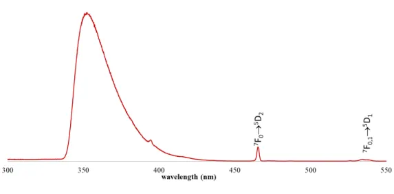
band ascribed to the excited states of the ligands (335-425 nm), with three components peaking around 355, 395, 420 nm and the 7F0,1 5D1,2 transitions of the Eu3+ ion.
Figure 5. Excitation spectra of 2 at room temperature in methanol monitored at 612 nm.
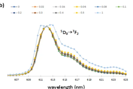
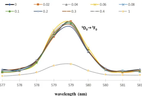
The luminescence spectra of 2 was recorded by fixing the excitation wavelength at 350 nm (Figure 6). The 5D07F0, and 5D07F2 transitions (forbidden and hypersensitive electric and dipole transitions, respectively) are the ones most affected by local site symmetry. The spectra have significant differences for the ethanol and methanol solutions. This solvatochromic effect prompted us to further investigations with special attention to the 5D07F0 transition that systematically increases its intensity upon addition of methanol to an ethanolic solution, ranging from 0.02 to 0.50 molar fractions (Figure 1 and Figure 3 and Electronic Supplementary Information for additional details). The emission is distributed in the 577-625 nm spectral range, with lines associated with 4f-4f transitions of the 5D0 excited state to 7F0―2, with the strongest peak at 612 nm attributed to the 5D07F2 transition.22-2324 Addition of methanol favors the
formation of [Eu(FOD)3OPhMe3]- in a higher extent as reflected in the 5D07F0 and 5D0F2 band intensities and shapes. A gradual decrease in the intensity of 5D07F2 is observed during the addition of methanol (see the ESI) because of the gradual FOD- ligand replacement by phenolate. The line splitting observed for higher concentrations of methanol indicates a high level of asymmetry around the Eu3+ centre.23 A single oxygen donor atom of the 2,4,6-trimethylphenolate is less polarizable than the coordinating carbonyl groups of chelating β-diketonates, resulting in a decrease in the intensity of the hypersensitive 5D07F2 transition.23 The 5D07F0 band intensity peaks reaches its maximum when the complex is solubilized in methanol showing, for both solutions, a labile coordination between Eu3+ and the solvents, which grows stronger with methanol. This process modifies the geometry of the complexes to a more asymmetric structure around the Eu3+ center (Figures 6a and 6b). A similar behavior was found for methanol/1-butanol and methanol/1-propanol mixtures allowing this method to be used in other mixtures of methanol/n-alcohol.
Figure 6. Normalized luminescence spectrum (towards the most intense 5D07F2 band) of scheme 1 reaction products; in mixtures methanol in ethanol with the methanol molar fractions ranging from 0 to 1, with λirradiation = 350 nm. a) range 577-600 nm b) range 605-625 nm.
Luminescence of 2 shows that ethanol interferes (i.e., changes the Eu3+ symmetry) less than methanol, thus constituting an accurate and precise method for quantification of the latter (Figure 7). The 5D07F1 transition reflects directly the crystal-field splitting of the 7F1 level (see ESI for details). Asymmetry arises as a balance between the concentrations of [Eu(FOD)4]- and [Eu(FOD)3OPhMe3]- complexes. It is further directly dependent to the ratio of methanol and ethanol in the solvent mixture.
Figure 7 Sensing assay as the normalized luminescence response in the 5D07F0 transition range towards addition of different amounts (molar fraction χ) of ethanol to Eu3+ of 2 in methanol upon irradiation at 350 nm.
Chemical Characterization. The positive mass spectra, of the ESI-MS analysis, are identical and show only one peak with m/z of 484 Da that corresponds to the [P6,6,6,14]+ cation. In the negative mode, the most abundant specie is the [Eu(FOD4)]- anion with m/z of 1332 Da, meaning only that this anion is the most easily ionizable. In methanol a peak appears with a 1137 Da mass that could be attributed to [P6,6,6,14FOD2]- anion, [(P6,6,6,14FOD)–FOD]-, which was detected with two solvent molecules. The MS2 spectra of this isolated peak show the loss of 32-unit mass that corresponds to methanol loss. It was not possible to see the loss of the second methanol unit since the resulting peak was too small to perform MS3. Both negative spectra, with methanol and ethanol, show a peak mass at 1173 that can be associated to the anionic moiety [Eu(FOD)3OPhMe3]-. Considering in each case the peak at 1332 as 100 %, the peak at 1173 has an intensity of 4.4 % in methanol and 1.9 % in ethanol (Table 1). This difference is related with a
higher concentration of this anion in methanol, which is in accordance with the photoluminescence data.
2, when heated, present an incredibly high thermal stability with decomposition temperature starting close to 300 °C, almost 100 °C higher than the starting compound [P6,6,6,14][Eu(FOD)4], Figure 8. This can be explained by the previously proved presence of [P6,6,6,14][FOD] in the products mixture with temperature decomposition slightly lower than 300 ºC (blue line, Figure 7). The low weight loss up to 200 ºC is expected for highly fluorinated complexes that usually presents a minimum of adsorbed solvent retained in the structure. The remaining mass residues are due to EuOF phases and sodium oxide phases that are stable at 600 ºC.
Figure 8. Thermogravimetric analysis of [P6,6,6,14][Eu(FOD)4], products of reaction of [P6,6,6,14] [Eu(FOD)4] and NaOPhMe3, [P6,6,6,14][FOD] and P6,6,6,14OPhMe3.
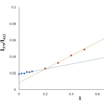
Sensing Assays. A ratiometric method was used to calculate the ratio of the luminescence intensities in order to determine the methanol concentration in methanol/ethanol mixtures.
Calibration assays were performed by adding different amounts of methanol to a solution of 2 in ethanol and correlate the methanol concentration with the normalized intensity of the 5D07F0 transition (λemision = 579 nm, Figure 6).
For these assays, 2 was solubilized in a quartz cuvette in 2 ml of ethanol. The calculated amounts of methanol were added to obtain a final molar fraction (χ) of methanol in the solvent mixture of: 0.02, 0.04, 0.08, 0.08, 0.1, 0.2, 0.3, 0.4 and 0.5 as represented in Figure 9.
Figure 9. Calibration curves with linear behavior for methanol estimation in ethanol/methanol mixtures. χ molar fraction of methanol in ethanol.
There are numerous methods for the determination of limit of detection, many of which are described in a review by Belter et al.25 A linear trend for the calibration curve of the molar fraction, χ, of methanol in ethanol was then observed in the 0.2-1 range (Graphic 1). The
calibration curve with χ from 0.2 exhibited a linear trend with, at least, R2=0.9993 with a limit of detection (LOD) of 0.207 (LOD=3*/m, is standard deviation of noise and m is calibration curve slope, ESI for details) and sensitivity of 0.0819.26,27
Accuracy assays were highly reproducible (in triplicate–ESI for details) and were carried out by measuring the I(5D07F0)/I(5D07F2) ratio after the addition of increasing amounts of methanol to a solution of 2 in ethanol. 2 was fully recovered after the removal of the solvents, thus showing a good stability of the sensitizing molecule which allowed three consecutive calibration assays (ESI for details).
For molar ratios below 0.2 we were able to observe an irregular increment on the 5D07F0 transition which did not allow an accurate determination of the sensitivity and limit of detection. From the calibration curve we can only speculate that this method has, still, some sensitivity for concentrations of methanol lower than 0.2 (15% w/w).
The methanol-sensing study of 2 demonstrates that the characteristic luminescence intensity of Eu3+ (5D07F0, 5D07F1 and 5D07F2) is modified immediately as the methanol can efficiently stabilize the asymmetric β-diketonate complex promoting a more pronounced purple color due to higher amounts of [P6,6,6,14][FOD]. In contrast, ethanol breaks the P-O interaction of [P6,6,6,14] [FOD], allowing coordination of the 4th FOD- anionic ligand to Eu3+ and increasing local metal symmetry. Methanol is a more strongly coordinating solvent to Eu3+, and with the simultaneous presence of a highly electron donor ligands such as [OPhenMe3]-, FOD- is removed leading to a 5D07F0 band increase.
Selectivity assays were performed by testing other alcohols like 1-propanol and 1-butanol as substituents of ethanol (Fig. 10). For 2, the highest Eu3+ complex asymmetry was found for methanol solutions, while addition of methanol, even in very low concentrations to all the other
solutions modified immediately the luminescence profile (Figure S7, see ESI for details). In opposition, addition of ethanol, 1-propanol and 1-butanol to a methanol solution of 2 didn´t produce significant changes in the luminescence spectrum of the emissive specie.
Figure 10 Normalized luminescence (upon excitation at 350 nm) of 2 in methanol (dark line), in ethanol (dash blue line), in 1-propanol (red line) and in 1-butanol (square dot red line).
Conclusions
Here, we observe unique photoluminescent solvatochromism between methanol and ethanol, visible at naked eye.
This highly reproducible ratiometric methodology is based in the changes of the intensities of the hypersensitive electric dipole transition bands 5D07F0, and 5D07F2, which are highly sensitive to the coordination geometry around Eu3+ and can be rationalized by the I(5D07F0)/I(5D07F2) ratio as
a function of the methanol molar concentration in ethanol. The changes observed in these bands could be explained by the presence of [P6,6,6,14][Eu(FOD)4] and NaOPhMe3 in ethanol that, in the presence of increasing amounts of methanol, change to a mixture of increasing amounts of [Eu(FOD)3OPhMe3]- and [P6,6,6,14][FOD]. These modifications lead to a decrease in the Eu3+ coordination symmetry, quantified by the I(5D07F0)/I(5D07F2) ratio. This is further detectable by the naked eye due to a color change in the solution because of the increasing concentrations of [P6,6,6,14][FOD].
The methanol-sensing studies reported in this manuscript show that the mixture of the reaction between [P6,6,6,14][Eu(FOD)4] and NaOPhMe3 can be used as an approach to a fast and low-cost sensitivity method to determine the methanol content in methanol/ethanol mixtures from as low as χ of 0.2 that corresponds to 15 % (w/w). In terms of limit of detection this method is still not competitive when compared with other analytical methods that are currently used like Raman spectroscopy or even available in bienzymatic disposable kits.
In summary, we have successfully designed a new optical method based on solvatochromic effects of a Eu3+ complex, that allows quantification of methanol quantification in mixtures of ethanol/methanol.
Experimental Section
MaterialsReagent grade chemicals were obtained from Aldrich and used without further purification. Microanalyses for C and H were carried using a Thermo Finnigan–CE Instruments Flash EA 1112 CHNS series. FT–IR spectra (range 4000 – 400 cm−1) were collected using a drop of sample between KBr round cell windows on a Thermo Scientific Nicolet iS50 FT–IR
spectrometer, by averaging 32 scans at a maximum resolution of 4 cm−1. TGA curves were obtained using a Thermal Analysis Ta Q500–2207, with a scanning rate of 5 °C min–1, with samples weighing around 6 mg in Aluminum crucibles. The calibration of the TGA equipment was made following the recommendation described in the manufacturer’s manual. Electrospray Ionization Mass Spectrometry (ESI–MS). ESI–MS was performed using a Bruker HCT quadrupole ion trap mass spectrometer. Sample solutions approximately 10−5 M in acetonitrile were introduced to the ESI source via a syringe pump at a flow rate of 150 mL min−1. The heated capillary temperature was set to 250 °C and the cover gas (N2) to a flow rate of 2 L min−1. Both positive and negative modes were detected to see the existing cations and anions. Spectroscopic Measurements. UV/vis absorbance spectra were performed using a UV−vis−NIR Varian Cary 5000 spectrophotometer within the spectral range 200 – 800 nm. NMR studies were performed on a Bruker Avance III 400 using deuterated methanol and dichloromethane as solvents.
Sodium 2,4,6–trimethylphenoxide, NaOPhMe3, was carried out using standard Schlenk line and dry box techniques in an atmosphere of N2 to avoid hydrolysis. Small portions of freshly cut metallic sodium was added to a THF solution of 2,4,6–trimethylphenol (1g) and the resulting mixture was left at room temperature under stirring. When the evolution of H2 ended, the supernatant was decanted, and the solvent evaporated under reduced pressure yielding NaPhO Me3 as a white powder.
[P6,6,6,14][Eu(FOD)4] was prepared according to a procedure already reported by us.
NaFOD(0,0767 g, 0,241mmol) was added stoichiometrically to a solution of Eu(FOD)3 (0,250 g, 0,241 mmol) in methanol. After 2 hours of reaction at room temperature, 1 equivalent of
P6,6,6,14Cl (0,117 g, 0,241 mmol) previously dissolved in a minimum of CH2Cl2 was added dropwise to the solution and left under magnetic stirring for one hour. The solvent was then removed under reduced pressure and the resultant oily solid was dissolved in CH2Cl2. NaCl was removed by centrifugation and the [P6,6,6,14][Eu(FOD)4] was recovered as a neat light yellow oil after solvent evaporation under reduced pressure with an yield of 80 %. Anal. Calcd. for [PC32H68][Eu(C10H10O2F7)4]: C, 47.61; H, 5.99%. Experimental; C, 47.69; H, 6,31.
[(P6,6,6,14)(FOD)] + Na[Eu(FOD)3(OPhMe3)] reaction mixture, (2), was prepared by mixing in
a flask under N2 stoichiometric amounts of [P6,6,6,14][Eu(FOD)4] (200 mg; 1.1 mmol) and NaOPhMe3 (17 mg; 1.1 mmol). Within a few minutes, at room temperature, the pale-yellow oil starts to turn to orange with red spots where the powder of the NaOPhMe3 sticks to the walls. In order to get a uniform oil this mixture was heated to 50 °C, under stirring, for 30 min forming fluid purplish red oil. 1H–NMR (ppm): 6.14 (s, PhHOMe3–), 4.89 (s, Hα–FOD), 3.04 [s, – PhO(CH3)3], 2.21 (t, +P6,6,6,14, Hα), 1.52–0.84 (m, +P6,6,6,14), 0.97 (s, -CH3 FOD-). 13C-NMR (ppm): 199 (O=C-C(CH3)3, FOD-). 31P-NMR (ppm): 33.37 (+P6,6,6,14).
Supporting Information. Details of general procedures of the experiments, crystallographic, structure and tables refinement data of complex Na[Eu(FOD)4], photoluminescence spectrum of complexes, 31P-NMR and 1H-NMR spectrum of 1, electrospray-mass spectra of anions of 2 UV−vis spectra of 2 in ethanol and methanol and 1:1 mixtures, and calibration curve of sensor.
AUTHORS INFORMATION Corresponding Authors
*Cláudia C. L. Pereira. LAQV-REQUIMTE, Departamento de Química, Universidade Nova de Lisboa, 2829-516 Caparica, Portugal. E-mail: ccl.pereira@fct.unl.pt.
*Bernardo Monteiro. Centro de Ciências e Tecnologias Nucleares (C2TN), DECN, Instituto Superior Técnico, Estrada Nacional 10, 2695-066 Bobadela, Portugal; E-mail: bernardo.monteiro@ctn.tecnico.ulisboa.pt.
*César A. T. Laia. LAQV-REQUIMTE, Departamento de Química, Universidade Nova de Lisboa, 2829-516 Caparica, Portugal. E-mail: catl@fct.unl.pt.
ORCID
João P. Leal: 0000-0003-1235-0107
Filipe A. Almeida Paz: 0000-0003-2051-5645 Ricardo F. Mendes: 0000-0001-8242-324X Tiago Moreira: 0000-0002-5880-1754 Mani Outis: 0000-0003-1940-8703 César A. L. Laia: 0000-0001-6410-6072 Bernardo Monteiro: 0000-0001-6434-365X Cláudia C. L. Pereira: 0000-0003-3421-8676 Conflict of interests
The authors declare no competing financial interests.
Development Fund (ERDF), through COMPETE 2020 - Operational Programme for Competitiveness and Internationalization (OPCI), and by national funds, through FCT -Fundação para a Ciência e a Tecnologia I.P. This work was supported by the Associated Laboratory for Sustainable Chemistry-Clean Processes and Technologies- LAQV which is financed by national funds from FCT/MEC (UID/QUI/50006/2013) and co-financed by the ERDF under the PT2020 Partnership Agreement (POCI-01-0145-FEDER – 007265) The NMR spectrometers are part of The National NMR Facility, supported by Fundação para a Ciência e a Tecnologia (RECI/BBB-BQB/0230/2012). Financial support from Fundação para a Ciência e a Tecnologia and Portugal 2020 to the Portuguese Mass Spectrometry Network (LISBOA-01-0145-FEDER-402-022125) is acknowledged. This work has been supported by Fundação para a Ciência e a Tecnologia through the contract nº IST-ID/077/2018 (Bernardo Monteiro), SFRH/BD/120985/2016 (Mani Otis) and PTDC/QEQ-QIN/3007/2014 (Tiago Moreira). C2TN/IST authors gratefully acknowledge the FCT support through the UID/Multi/04349/2013 project). Cláudia C. L. Pereira thanks to Fundação para a Ciência e a Tecnologia, MCTES, for the Norma transitória DL 57/2016 Program Contract.
Table of Contents
FULL PAPER
The reaction product between [P6,6,6,14][Eu(FOD)4] and NaOPhMe3forms an equilib-rium in solution that allows for an easy, low-cost, efficient and fast spectrofluorimet-ric method for the detection and quantification of methanol in mixtures with ethanol based on the changes in the Eu3+ luminescence in the visible region.
Luminescence
João P. Leal, Filipe A. Almeida Paz, Ricardo F. Mendes, Tiago Moreira, Mani Outis, César A. T. Laia*, Bernardo Monteiro*, Cláudia C. L. Pereira*
Page No. – Page No.
A reusable Eu3+ complex for
naked-eye discrimination of methanol from ethanol with a ratiometric fluorimetric equilibrium in methanol/ethanol mix-tures
1[] K. Binnemans, C. Bex, A. Venard, H. De Leebeeck, C. Gorller-Walrand, J. Mol. Liq. 1999, 83, 283-294. 2[] Z. Cui, X. Zhang, S. Liu, L.Zhou, W. Li, J.Zhang, Inorg. Chem. 2018, 57, 11463−11473.
3[] C. C. L. Pereira, S. Dias, I. Coutinho, J. P. Leal, L. C. Branco, C. A. T. Laia, Inorg. Chem. 2013, 52,
3755−3764.
4[] C. C. L. Pereira, J. T. Coutinho, L. C. J. Pereira, J. P. Leal, C. A. T. Laia, B. Monteiro, Polyhedron 2015,
91, 42–46.
5[] A. Dey, A. Garai, V. Gude K. Biradha, Crystal Growth & Design 2018, 18, 6070-6077.
6[] S. A. Majeed, B. Ghazal, D. E. Nevonen, P. C. Goff, D. A. Blank, V. N. Nemykin, S. Makhseed, Inorg.
Chem. 2017, 56, 11640−11653.
7[] M. L. Wang, J. T. Wang and Y. M. Choongy, J. Food Compos. Anal. 2004, 17, 187–196. 8[] R. Caruso, G. L. Gambino and M. Scordino, Nat. Prod. Commun., 2011, 6, 1939–1943. 9[] G. Ai, T. Sun and X. Dong, Rapid Commun. Mass Spectrom. 2014, 28, 1674–1682.
10[] Y. C. Zhang, N. B. Lin, X. S. Chai, Z. Li and G. D. Barnes, Food Chem. 2015, 183, 169–172. 11[] M.-L. Wanga, J.-T. Wang and Y.-M. Choong, J. Food Compos. Anal., 2004, 17, 187-196.
12[] I. H. Boyaci, H. E. Genis, B. Guven, U. Tamer, N. Alper, J. Raman Spectrosc. 2012, 43, 1171–1176. 13[] D. I. Ellis, H. Muhamadali, Y. Xu, R. Eccles, I. Goodallc, R. Goodacre, Analyst, 2019, 144, 324–330. 14[] P. Ramasami, S. Jhaumeer-Laulloo, F. Cadet, P. Rondeau, Y. Soophul, Int J Food Sci Nutr. 2005, 56,
177-183.
15[] B. Bucur, G. L. Radu, C. N. Toader, Eur. Food Res. Technol. 2008, 226, 1335-1342.
16[] K. K. Kar (ed.), Composite Materials: Processing, Applications, Characterizations. Springer Verlag,
Berlin–Heidelberg, 2017.
17[] A. V. Mudring, S. Tang, ‐ Eur. J. Inorg. Chem. 2010, 2569–2581.
18[] B. Monteiro, M. Outis, H. Cruz, J. P. Leal, C. A. T. Laia, C. C. L. Pereira, Chem. Commun. 2017, 53,
850-853.
19[] E. Wolcan, L. Villata, A. L. Capparelli, Mario R. Féliz, Photochem. Photobiol. Sci. 2004, 3, 322-327. 20[] J. P. Leal, M. Outis, M. H. Casimiro, L. M. Ferreira, F. Fernandes, B. Monteiro, C. A. T. Laia, C. C. L.
Pereira, Eur. J. Inorg. Chem. 2017, 3429–3434.
22[] K. Binnemans, in: Handbook on the Physics and Chemistry of Rare Earths (Eds.: K.A. Gschneidner, Jr.,
J.-C.G. Bünzli and V.K. Pecharsky), Elsevier, Amsterdam, 2005, Chapter 225.
23[] K. Binnemans, Coord. Chem. Rev. 2015, 295, 1-45.
24[] D. Prodius, A.-V. Mudring, Coord. Chem. Rev. 2018, 363, 1–16.
25[] M. Belter, A. Sajnog, D. BaraIkiewicz, Talanta 2014, 129, 606−616.
26[] Analytical Methods Committee, Recommendations for the definition, estimation and use of the
detection limit, Analyst 1987, 199-203.
![Figure 1. Luminescence spectrum of [Na][Eu(FOD) 4 ] with a concentration of 0.1 mM in methanol (black line) and ethanol (red dotted line) upon excitation with λ excitation = 350 nm](https://thumb-eu.123doks.com/thumbv2/123dok_br/18141076.871076/6.918.205.716.107.472/figure-luminescence-spectrum-concentration-methanol-ethanol-excitation-excitation.webp)
![Figure 2. Ball and stick representation of the [Na][Eu(FOD) 4 ](H 2 O) complex (1).](https://thumb-eu.123doks.com/thumbv2/123dok_br/18141076.871076/7.918.321.602.248.571/figure-ball-stick-representation-na-eu-fod-complex.webp)
![Figure 8. Thermogravimetric analysis of [P 6,6,6,14 ][Eu(FOD) 4 ], products of reaction of [P 6,6,6,14 ] [Eu(FOD) 4 ] and NaOPhMe 3 , [P 6,6,6,14 ][FOD] and P 6,6,6,14 OPhMe 3 .](https://thumb-eu.123doks.com/thumbv2/123dok_br/18141076.871076/17.918.219.697.513.868/figure-thermogravimetric-analysis-fod-products-reaction-naophme-ophme.webp)