UNIVERSIDADE DA BEIRA INTERIOR
Ciências da Saúde
Developing a NGS panel for diagnosis of Lyme
disease and its co-infections
Eduardo Augusto Coelho
Dissertação para obtenção do Grau de Mestre em
Ciências Biomédicas
(2º ciclo de estudos)
Orientador: Prof.ª Doutora Isabel Theriaga Gonçalves Co-orientador: Doutor Gonçalo Doria
iii
Dedicatória
Este trabalho é dedicado a todos aqueles que, de alguma forma, fizeram parte do meu caminho e que me ajudaram a ser pessoa que sou hoje.
v
Agradecimentos
Em primeiro lugar, obviamente, gostaria de agradecer à minha família por me dar todas as ferramentas possíveis para atingir esta etapa. Para o meu Pai e para a minha Mãe, faltam-me as palavras para expressar o quão grato estou por todo o apoio e conforto que sempre faltam-me deram. Vocês são um verdadeiro exemplo para mim e acho que a melhor maneira de vos agradecer é provar, todos os dias, que o vosso esforço deu frutos. Aos meus irmãos, companheiros de brincadeira e de sofrimento, quero apenas relembrar que são indispensáveis na minha vida e que farei sempre o possível para vos ajudar, como sei que farão por mim. Aos meus avós, aos que me dão todo o apoio e aos que infelizmente não podem assistir a mais uma conquista na minha vida, agradeço por tudo o que fizeram por mim e por me fazerem sentir especial todos os dias em que estamos juntos. Gosto muito de todos vocês e obrigado por me acompanharem de perto o meu crescimento.
À minha namorada, Marta, quero agradecer pelo apoio incondicional que me dás sempre que preciso. Ajudaste-me muito a evoluir como pessoa e a melhorar a perceção do que me rodeia. Que venham muitos mais anos de felicidade, sempre de mãos dadas!
À minha orientadora, Professora Doutora Isabel Theriaga Gonçalves, gostaria de agradecer sinceramente por toda a disponibilidade e prontidão em ajudar-me sempre que eu necessitei.
Ao Professor Doutor Ilídio Correia, diretor do Mestrado em Ciências Biomédicas na UBI, gostaria de agradecer por me ter ajudado a abrir os horizontes, numa altura em que a motivação parecia escassa.
Queria agradecer a toda a equipa da STAB VIDA, principalmente ao CEO Orfeu Flores pela oportunidade que me deu em desenvolver este projeto em âmbito empresarial. Ao Gonçalo Doria, pilar no desenvolvimento deste projeto, o meu obrigado por todos os esclarecimentos e críticas construtivas que tanto me ajudaram a evoluir como investigador.
Por último, não por serem menos importantes, mas porque sei que leem esta página até ao fim, queria agradecer a todos os meus amigos que me têm acompanhado nos últimos anos. Ao Gustavo, fiel companheiro de casa, ao Rui e ao Miguel pela companhia e ajuda nesta reta final, ao Tiago por todos os conselhos, obrigado e um grande abraço. Aos meus irmãos da Desertuna, companheiros de trabalho e de diversão, muito obrigado por me darem tanta bagagem, tanto a nível pessoal como a nível profissional, para o meu futuro.
vii
Resumo
A doença de Lyme, também conhecida como borreliose de Lyme, é a doença transmitida por carraças mais comum no hemisfério norte, com 300.000 novos casos estimados anualmente só nos Estados Unidos da América. Para além da transmissão de Borrelia burgdorferi sensu lato (s.l.), bactéria responsável pela doença de Lyme, as carraças do complexo Ixodes ricinus são vetores de outras infeções, sendo as mais comuns a babesiose e a anaplasmose granulocítica humana. A presença de coinfecções pode causar manifestações clínicas mais severas e o seu incorreto diagnóstico pode levar a um tratamento inapropriado. O único método aceite para o diagnóstico da borreliose de Lyme, atualmente, é um teste sorológico baseado numa abordagem de dois níveis no qual é realizado um imunoensaio enzimático complementado por um western blot. Este método indireto para a deteção de Borrelia burgdorferi s.l. carece de sensitividade na fase inicial da infeção, devido ao tempo necessário para os anticorpos serem produzidos.
Ao longo das últimas décadas, vários estudos usando métodos diretos, tais como cultura e
Polimerase Chain Reaction (PCR), foram realizados com o objetivo de desenvolver um
método de diagnóstico alternativo para esta doença infeciosa. Contudo, estes testes demonstraram uma taxa elevada de falsos negativos.
Neste estudo, foi criado um painel de Next Generation Sequencing (NGS), para a plataforma MiSeq da Illumina, que permite o diagnóstico simultâneo da doença de Lyme e das suas coinfecções mais frequentes. Este painel inclui sete pares de primers específicos para um fragmento de um gene de cada uma das espécies patogénicas incluídas, em regiões que permitem a distinção entre genoespécies. O painel foi testado na preparação das bibliotecas para sequenciação com amostras de sangue total e com amostras de ADN extraído do sangue total. A par do teste com o painel desenvolvido, com o intuito de avaliar a sensibilidade do mesmo, os dois tipos de amostras foram também testados com primers específicos para as regiões V3 e V4 do gene 16S do ARN ribossomal, amplamente usados na análise de microbiomas.
Devido à dificuldade em obter amostras de pacientes com doença de Lyme e com as outras infeções abrangidas pelo painel, neste trabalho, foram testadas cinco amostras de sangue de pacientes diagnosticados com babesiose, juntamente com os controlos positivo e negativo. A condição que demonstrou melhores resultados foi aquela em que foi usado ADN extraído de sangue em combinação com o painel, com a qual foi possível identificar ADN de Babesia
microti nas cinco amostras de pacientes infetados.
Apesar da necessidade de testar o método em amostras de pacientes com doença de Lyme e com as restantes infeções incluídas no painel desenvolvido, os resultados obtidos neste trabalho demonstram-se promissores para a futura utilização deste método como alternativa aos exames atuais, especialmente na fase inicial da infeção.
viii
Palavras-chave
ix
Resumo Alargado
A doença de Lyme, também conhecida como borreliose de Lyme, é a doença transmitida por carraças mais comum no hemisfério norte, com 300.000 novos casos estimados anualmente só nos Estados Unidos da América. Para além da transmissão de Borrelia burgdorferi sensu lato (s.l.), bactéria responsável pela doença de Lyme, as carraças do complexo Ixodes ricinus são vetores de outras infeções, sendo as mais comuns a babesiose e a anaplasmose granulocítica humana. A presença de coinfecções pode causar manifestações clínicas mais severas e o seu incorreto diagnóstico pode levar a um tratamento inapropriado. O único método aceite pela
Food and Drug Administration (FDA) para o diagnóstico da borreliose de Lyme, atualmente, é
um teste sorológico baseado numa abordagem de dois níveis no qual é usado um imunoensaio enzimático complementado por um Western blot. Basicamente, neste método, se o imunoensaio enzimático der um resultado negativo, exclui-se a hipótese de doença. Caso o resultado seja positivo, a amostra de soro é submetida a um Western blot para deteção de anticorpos IgM ou IgG, consoante o tempo passado desde o início da infeção. Este método indireto para a deteção de Borrelia burgdorferi s.l. carece de sensitividade na fase inicial da infeção, devido ao tempo necessário para os anticorpos serem produzidos, uma vez que os anticorpos IgM e IgG podem demorar entre 2 a 4 e 4 a 6 semanas, respetivamente, a atingirem uma concentração mínima para ser detetada por este teste.
Ao longo das últimas décadas, vários estudos usando métodos diretos, tais como cultura e PCR, foram desenvolvidos com o objetivo de se alcançar um método de diagnóstico alternativo para esta doença infeciosa. Contudo, apesar dos vários tipos de amostras testadas, tais como, líquido cefalorraquidiano, líquido sinovial, sangue e urina, estes testes demonstraram uma taxa elevada de falsos negativos.
Neste estudo, foi criado um painel de Next Generation Sequencing (NGS), para a plataforma MiSeq da Illumina, que permite o diagnóstico simultâneo da doença de Lyme e das suas coinfecções mais frequentes. As espécies abrangidas por este painel são: Borrelia burgdorferi
s.l., responsável pela doença de Lyme, Anaplasma phagocytophilum, responsável por causar
anaplasmose granulocítica humana, Babesia microti, responsável por causar babesiose,
Bartonella henselae, responsável por causar bartonelose, Coxiella burnetii, causadora da
febre Q, Ehrlichia canis, responsável por causar ehrlichiose e Rickettsia rickettsii, conhecida por causar a febre da carraça. Este painel inclui sete pares de primers específicos para um fragmento de um gene de cada uma das espécies patogénicas incluídas, em regiões que permitem a distinção entre genoespécies, informação que pode ser importante na compreensão das manifestações clínicas.
O painel foi testado na preparação das bibliotecas para sequenciação com amostras de sangue total e com amostras de ADN extraído do sangue total. A par do teste com o painel desenvolvido, com o intuito de avaliar a sensibilidade do mesmo, os dois tipos de amostras
x
foram também testados com primers específicos para as regiões V3 e V4 do gene 16S do ARN ribossomal, amplamente usados na análise de microbiomas.
Devido à dificuldade em obter amostras de pacientes com doença de Lyme e de pacientes com as outras infeções abrangidas pelo painel, neste trabalho, foram testadas cinco amostras de sangue de pacientes diagnosticados com babesiose,. Para além destas amostras, fornecidas pelo Centers for Disease Control and Prevention (CDC), dois controlos, um positivo e um negativo foram também testados. Como controlo negativo, foi usada uma amostra de sangue, escolhida de forma aleatória, de um grupo de indivíduos que não vivem em zonas endémicas para estas doenças e que não se recordam de terem sido mordidos por carraças. Para o controlo positivo, os fragmentos dos genes avaliados no painel foram amplificados individualmente a partir de ADN genómico de cada uma das espécies através de um PCR, usando só o respetivo par de primers específicos. Os fragmentos obtidos foram clonados e usados na transformação em células de E.coli. Posteriormente, estas células foram inseridas numa alíquota juntamente com sangue do controlo negativo, tentando mimetizar uma infeção.
No PCR da preparação das bibliotecas para a sequenciação, cada uma das amostras foi testada com quatro condições diferentes, relativamente ao tipo de amostra e aos primers usados. A condição que demonstrou melhores resultados foi aquela em que foi usado ADN extraído do sangue em combinação com o painel, na qual foi possível identificar ADN de
Babesia microti nas cinco amostras de pacientes infetados. Na outra condição em que o
painel foi usado, diretamente no sangue total, apenas foi possível detetar a presença do agente patogénico em três dos cinco pacientes e verificou-se a amplificação de produtos de PCR não específicos. Nas duas condições em que os primers específicos para o gene 16S do ARN ribossomal foram usados, só se observaram resultados positivos em duas das cinco amostras.
Apesar da necessidade de otimizar e testar o método em amostras de pacientes com doença de Lyme e com as restantes infeções incluídas no painel desenvolvido, os resultados obtidos neste trabalho demonstram-se promissores para a futura utilização deste método como alternativa aos exames atuais, especialmente na fase inicial da infeção.
xi
Abstract
Lyme disease, also known as Lyme borreliosis, is the most common-tick borne disease in the northern hemisphere, with 300,000 cases estimated each year only in the United States. In addition to the transmission of Borrelia burgdorferi sensu lato (s.l.), the bacteria responsible for Lyme disease, ticks of the Ixodes ricinus complex are vectors for other infections, with the most common being babesiosis and human granulocytic anaplasmosis. The presence of co-infections may cause more severe clinical manifestations and their misdiagnosis may lead to an inappropriate treatment. The only accepted method to diagnose Lyme borreliosis, currently, is a serologic test based in a two-tier approach using an enzyme immunoassay (EIA) complemented with a Western immunoblot. This indirect method for Borrelia burgdorferi s.l. detection suffers from lack of sensitivity in the early stage of the disease, due to the time window needed for antibodies to be produced.
Over the past few decades, many studies have been carried using direct methods, such as culture and PCR, in order to develop an alternative diagnostic method for this infectious disease, however this tests also suffer from a high rate of false negatives.
In this study, a NGS panel was developed, for the Illumina's Miseq platform, which allows the simultaneous diagnose of Lyme disease and its most common co-infections. This panel includes seven specific primer pairs that target a gene fragment of each of the included pathogenic species, in regions that allow the genospecies distinction.
The panel was tested in the library preparation for sequencing using samples of whole blood and samples of extracted DNA from the whole blood. Along with the developed panel, in order to evaluate its sensibility, both types of samples were also tested with specific primers that target the V3 and V4 regions of the 16S ribosomal RNA gene, widely used in microbiome analysis.
Due to the difficulty to obtain samples from patients with Lyme disease and with the other infections covered by the panel, in this study, five samples of whole blood from patients diagnosed with babesiosis were tested, along with a positive and a negative controls.
The condition that has shown better results was the one using extracted DNA from the blood combined with the panel, which detected Babesia microti DNA in all five samples of the infected patients.
Despite the need to test this method in samples from patients with Lyme disease and the remaining infections included in the developed panel, the results obtained in this study are promising for the future use of this method as an alternative to the current tests, especially in the initial phase of infection.
xii
Keywords
xiii
Table of contents
I. INTRODUCTION ... 1
1.Lyme disease ... 3
1.1. Identification of a new pathology... 3
1.2. Pathogen ... 3
1.3. Vector ... 4
1.3.1. The life cycle of Ixodes ricinus complex ... 5
1.4. Co-infections ... 5
1.5. Epidemiology ... 6
1.6. Clinical Manifestations ... 6
1.6.1. Erythema migrans ... 6
1.6.2. Stages of Lyme disease ... 7
1.7. Diagnosis... 9
1.7.1. Direct methods ... 9
1.7.2. Indirect methods ... 11
1.8.Treatment ... 12
2.Next Generation Sequencing ... 12
2.1.Sequencing overview ... 12
2.2.Illumina MiSeq workflow ... 13
2.2.1.Sample preparation ... 13
2.2.2. Cluster generation ... 13
2.2.3. Sequencing ... 14
2.2.4. Data analysis ... 15
II. AIM ... 17
III. MATERIAL AND METHODS... 21
1. Biological material ... 23
1.1. Genomic DNA ... 23
1.2. Blood samples ... 23
1.2.1. Positive control ... 23
2. Cell preparation for positive control and PCR ... 24
2.1.Target Genes... 24
2.2.Primers ... 25
2.3.Cloning ... 27
2.4.Transformation ... 27
2.4.1.Confirmation of correct insertion ... 27
3. Agarose Gel Electrophoresis ... 27
4. DNA Extraction ... 28
5. Library Preparation for NGS sequencing ... 28
5.1.Amplicon PCR ... 28
xiv
5.1.2.Extracted DNA + Multiplex conditions (DNA+Mx) ... 29
5.1.3.Whole blood + 16S primers conditions (B+16S) ... 29
5.1.4.Extracted DNA + 16S primers conditions (DNA+16S) ... 30
5.1.5.Amplification verification and purification ... 30
5.2.Index PCR... 30 6. Data Analysis ... 32 6.1.Software ... 32 6.1.1.Qiime2 2018.6 ... 32 6.1.2.BLAST2GO... 33 6.1.3.Python ... 33 IV. RESULTS ... 35
1. Library preparation results ... 37
1.1.Amplicon PCR and purification ... 37
1.2.Index PCR and purification ... 40
2. NGS results ... 41
2.1. Unspecific amplification ... 41
2.2. Cut-off definition ... 41
2.3. Bar graphs ... 42
V. DISCUSSION AND CONCLUSION ... 49
1. Comparison of the four different conditions ... 51
1.1.General overview ... 51
1.2.Whole blood used in multiplex PCR (B+Mx) ... 51
1.3.Extracted DNA used in multiplex PCR (DNA+Mx) ... 51
1.4.Whole blood used in 16S PCR (B+16S) ... 52
1.5.Extracted DNA used in 16S PCR (DNA+16S) ... 52
2. Unspecific amplification in multiplex PCR ... 52
3. Conclusion ... 53
VI. FUTURE PERSPECTIVES ... 55
xv
List of Figures
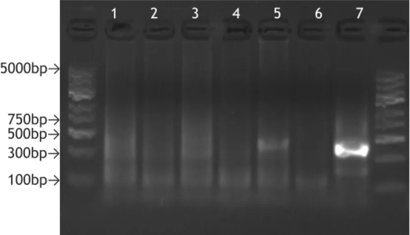
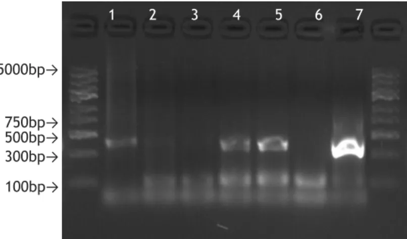
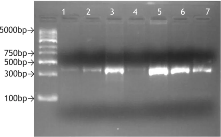
Figure 1 - Geographical distribution of the four ticks with greater importance in transmission of Lyme disease worldwide (Adapted from(17)). ... 5 Figure 2- Examples of Erythema migrans. . ... 7 Figure 3- Illumina's cluster generation process. ... 14 Figure 4- Agarose gel (1.5%) electrophoresis, stained with GelRed, of the PCR products obtained with B+Mx condition. ... 37 Figure 5- Agarose gel (1.5%) electrophoresis, stained with GelRed, of the PCR products obtained with B+Mx condition, after purification ... 37 Figure 6- Agarose gel (1.5%) electrophoresis, stained with GelRed, of the PCR products obtained with DNA+Mx condition ... 38 Figure 7- Agarose gel (1.5%) electrophoresis, stained with GelRed, of the PCR products obtained with DNA+Mx condition, after purification. ... 38 Figure 8- Agarose gel (1.5%) electrophoresis, stained with GelRed, of the PCR products obtained with B+16S condition ... 38 Figure 9- Agarose gel (1.5%) electrophoresis, stained with GelRed, of the PCR products obtained with B+16S condition, after purification. ... 39 Figure 10- Agarose gel (1.5%) electrophoresis, stained with GelRed, of the PCR products obtained with DNA+16S condition ... 39 Figure 11- Agarose gel (1.5%) electrophoresis, stained with GelRed, of the PCR products obtained with DNA+16S condition, after purification. ... 39 Figure 12- Agarose gel (1.5%) electrophoresis, stained with GelRed, of the PCR products obtained in the second PCR of library preparation. B+Mx condition. ... 40 Figure 13- Agarose gel (1.5%) electrophoresis, stained with GelRed, of the PCR products obtained in the second PCR of library preparation, after purification ... 40
xvii
List of Tables
Table 1- Identification of the species gathered for this study, the target gene for each species and the size of the amplicon obtained in PCR. ... 24 Table 2- Designation, sequences and melting temperatures of the primers designed and 16S primers used in 16S metagenomic sequencing library preparation.. ... 26
xix
List of Abbreviations
U.S.A. United States of America
bp base pairs
EM Erythema migrans
s.l. Sensu lato
CDC Centers for Disease Control and Prevention
PCR Polymerase chain reaction
ACA Acrodermatitis chronica atrophicans
spp. Species
s.s. Sensu stricto
FDA United States Food and Drug Administration EIA Enzyme immunoassay
IFA Immunofluorescent assay
WB Western blot
IgM Immunoglobulin M
IgG Immunoglobulin G
HGP Human Genome Project NGS Next generation sequencing AGE Agarose gel eletrophoresis TAE Tris-Acetate-EDTA
3
1. Lyme disease
Lyme disease is a zoonosis, which is a disease from animals that can be transmitted to humans. This disorder has become a major concern in the last four decades among the medical community in the United States of America(U.S.A.) and in Europe. Although Lyme Disease was initially considered an inflammatory joint disorder, soon became identified as a multisystemic disorder affecting as well the skin, nervous system and heart (1).
1.1. Identification of a new pathology
Lyme arthritis, the initial designation of the disorder that nowadays is preferentially referred to as Lyme disease or Lyme borreliosis, was first suggested as an unrecognized pathology in 1975. The first suspicions started when two mothers, from Old Lyme, Connecticut, within a month, informed the State Health Department and Yale Rheumatology Clinic about the strange prevalence of arthritis in a small community. The first mother only reported children cases, who have been diagnosed with juvenile rheumatoid arthritis, and both mothers highlighted the fact that most of people suffering from this symptoms lived in the same neighborhood or close together (2). By geographic clustering of reported cases and, in some patients, their association with the characteristic skin lesion, Steere et al. refer that Lyme arthritis has been affecting people in eastern Connecticut since 1972, with the majority of the new cases taking place in the summer and early fall (2-4).
1.2. Pathogen
Borrelia burgdorferi is a gram negative bacteria member of eubacterial phylum Spirochaetes.
This phylum's name is due to the morphology of its organisms which show a spiral body. This irregularly coiled spirochetes range from 10 to 30 μm in length and from 0.18 to 0.25 μm in diameter (5).
This spirochete was first isolated in 1982 by Burgdorfer et al. by dissection of adult Ixodes
dammini, now known as Ixodes scapularis, collected in Shelter Island, New York, a known
endemic region of Lyme disease. More than a half of the ticks (61%) contained spirochetes, which were principally distributed in the midgut. No spirochetes were found in the salivary glands (5).
The theory suggested by Burgdorfer et al., that this spirochete was the etiological agent of Lyme disease, was solidly supported by subsequent isolation of identical spirochetes from blood, skin, and cerebrospinal fluid of patients with signs and symptoms suggestive of Lyme disease (6, 7).
The complete genome of Borrelia burgdorferi (strain B31), was first sequenced by Fraser et
al., in 1997. It consists in a linear chromosome with 910,725 base pairs (bp) and 12 linear and
4
proteins with biosynthetic activity, which makes the bacteria dependent on the host to acquire its nutritional needs. Besides, no recognizable toxins are encoded by Borrelia
burgdorferi genome, its pathogenic effect is caused by adhesion to host cells, migration
through tissues and evasion of immune clearance (8-10).
1.3. Vector
The identification of erythema migrans (EM) as a symptom of this disorder, a skin lesion that has previously been associated with tick bites in Europe (11), though any arthritis episodes have been related with it, suggested that the vector responsible for the disease transmission was this arthropod. This theory was supported by the data obtained by geographical clustering of epidemiologic studies. The patients were from rural regions with heavily wooded areas, the peak onset of new cases being reported was between summer and early fall and the occurrence of the disorder onset in elements of the same family usually didn't occur in the same year. Besides, some patients remembered a tick bite in the region where the skin rash appeared and one of them even took the tick to identification (Ixodes scapularis) (12). Nowadays, there are four different species of ticks that are known to be competent vectors for Lyme Disease, with all of them belonging to Ixodes ricinus complex. This complex is a paraphyletic group with a geographical distribution throughout almost the entire globe (13). This ticks' saliva has the ability to inhibit the alternative pathway of the complement of the host, which will lead to an absence of efficient rejection by the host, increasing the chance of a successful blood meal (14).
In the U.S.A., the blacklegged tick, Ixodes scapularis, is the responsible for the cases in eastern and upper midwestern regions while Ixodes pacificus is the vector in Pacific Coast. Across the Atlantic, Ixodes ricinus and Ixodes persulcatus are the principal vectors in Europe and Asia, respectively, but unlike U.S.A., there are regions where both are present (15-17) (Figure 1).
5
1.3.1. Figure 1 - Geographical distribution of the four ticks with greater
importance in transmission of Lyme disease worldwide (Adapted
from(17)).The life cycle of Ixodes ricinus complex
The life cycle of the four ticks early referred, has a duration of two to three years, and present different seasonality. From the eggs, ticks pass through three developmental stages: larvae, nymph and adult taking one blood meal at each one of them. Since there is no evidence for transovarial transmission of B. burgdorferi sensu lato (s.l.), ticks depend on infected hosts to be infected. After acquiring the pathogen, ticks are able to transmit it to the next host (16, 18, 19).
Nymphs, which are the principal responsible for transmitting the pathogen to humans, due to its small size that may go unnoticed, are active from early spring to mid-summer for I.
ricinus, I. persulcatus and I. pacificus with the possibility of a second peak in the autumn for I. ricinus, while I. scapularis has its peak from early summer to early fall. The fact that the
peaks of nymphal and larvae stages activity differ by about three months, leads to the wide transmission of B. burgdorferi s.l. in the hosts of this two stages which are mainly small mammals, such as mice and shrews, and birds. Adult ticks are known to feed in larger mammals, being the most commonly described the white-tailed deer which shows great importance in supporting tick populations (16, 18, 20).
1.4. Co-infections
Ixodes ricinus is the most widespread and abundant ixodid tick in western Europe and is
frequently associated with bites in humans (21). This small hard tick is known to be a vector for a large variety of pathogenic species concerning both physicians and veterinaries. The most frequently reported co-infections of Borrelia burgdorferi s.l. in humans are caused by
Babesia microti, known to cause babesiosis, and Anaplasma phagocytophilum, the responsible
agent for human granulocytic anaplasmosis (22, 23). The presence of co-infections can cause greater disease severity and the misdiagnosis may lead to inappropriate treatment (23). There are other species that have already been reported as co-infections, although with less
6
frequency that the ones described before. These include Rickettsia monacensis and Rickettsia
helvetica, that cause spotted fever ricketsiosis, Bartonella henselae, responsible for
cat-scratch disease and Coxiella burnetii, the agent of Q fever (24-28).
1.5. Epidemiology
Lyme disease is the most common tick-borne disease in the northern hemisphere. This disorder can affect both genders and all ages, but in a surveillance report performed in the U.S.A., children and older adults are the most affected, with males showing a slightly higher incidence (15). This disorder is estimated to present 300,000 new cases every year just in the United States, but despite the high incidence rates, few cases of death have been reported(16).
In the United States of America Lyme Disease has become a notifiable disorder in 1991. Since then, surveillance reports for Lyme Disease have been performed by the Centers for Disease Control and Prevention (CDC). The latest report describes a total of 275,589 cases of Lyme between 2008 and 2015, with approximately 76% of the cases confirmed and the other 24% marked as probably. The highest peak of disease reported cases, was observed in the beginning of July in all the years covered by the study. Although states with high risk of infection tend to stabilize or even decrease the number of reported cases, neighbor states' events have been increasing(15).
Unlike the information in U.S.A., epidemiology of Lyme Disease is still very unclear due to not being included, until June of the present year, in the list of diseases with epidemiologic surveillance in Europe. However, through the analysis of published studies of Lyme borreliosis in different countries in Europe, Skyes and Makiello, calculated the weighted mean incidence rate obtaining the value of 22.05/100,000 person-years. The incidence rates for this disease show a wide variation not only between countries, but also in regions of the same country. The country with more cases reported is Sweden, with 464/100,000 person-years while in Italy only 0.001/100,000 person-years cases were described(29). That being said, approximately 91,000 new cases are estimated to occur in Western Europe every year.
Hereafter, incidence of Lyme borreliosis in European Union will be better understood, since there will be an standardization in surveillance which will open the way to a better understanding of data acquired from the different countries.
1.6. Clinical Manifestations
1.6.1. Erythema migrans
The most characteristic symptom of Lyme Disease is the formation of a skin rash, called erithrema migrans. This lesion was first described in Europe by Lipschütz (30), but it was Afzelius the first to suggest that this symptom was related to a tick bite(11).
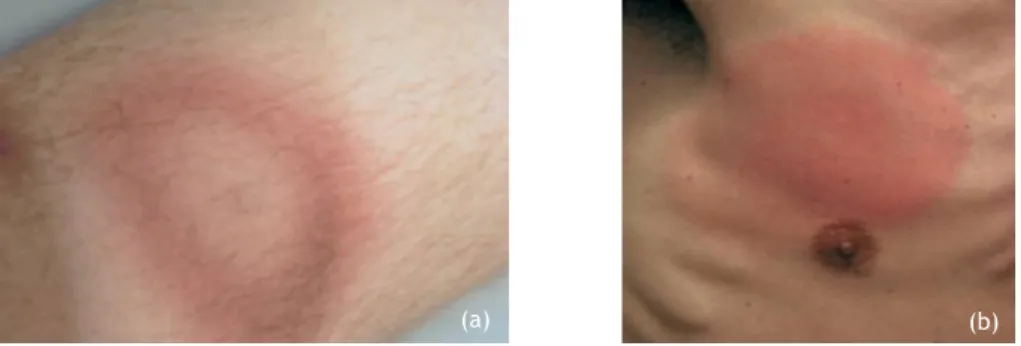
7 This skin lesion occurs in about 75% of the patients diagnosed with Lyme borreliosis and appears from 3 to 20 days after the tick bite (12, 15). EM, most of the times begins in the tick bite site as a red papule or macule and expands forming a red ring, with partial central clearing, resulting in a bull's eye shape, its characteristic form. However, sometimes central clearing is not verified, resulting in a large red spot which makes it more difficult to distinguish between similar lesions (Figure 2). Although it occurs frequently in the thigh, groin, and axilla, EM has been observed at any region of the body, with a diameter of at least 5 cm, being the most common size observed of about 16 cm (12, 31, 32). Generally, only one lesion appears, however, multiple skin rashes occurring simultaneously have been reported in approximately 20% and 10% of patients from U.S.A. and Europe respectively (31). This symptom, even in untreated patients, tends to fade within 3 to 4 weeks, but it may reappear (32, 33).
Although initially this symptom was described as exclusive for Lyme Disease patients, cases of tick bite, outside the Ixodes ricinus complex, shown an identical skin lesion, and so, besides being a good indicator of Lyme borreliosis, no conclusive diagnosis must be taken by its identification (34).
Figure 2- Examples of Erythema migrans. (a)- EM on the lower leg presenting central
clearing. (b)- EM on the right breast without central clearing. (Adapted from (35)).
1.6.2. Stages of Lyme disease
In 1989, for clinical purposes, Steere described three stages to characterize the development of the illness, that are still used nowadays, being that two of them represent early disease and the other late disease (33).
Early Localized Infection:
In this stage, B. burgdorferi spreads locally in the skin resulting in EM for the majority of the patients. In Europe, EM has been described to expand slower than the cases reported in the U.S. and usually, no other symptoms are reported along with the skin lesion (36, 37). The two principal species causing Lyme disease in Europe, B.afzelii and B.garinii, present different evolution in the skin lesion provoked. B.garinii EM tends to spread faster than the one caused by B.afzelii and it's usually itchy, while B.afzelii, sometimes cause a rare skin manifestation,
8
known as borrelial lymphocytoma, located often in the ear lobe in children and on the nipple in adults (38).
The symptoms that usually occur at this stage are: fever, fatigue, malaise, headache, myalgias, arthralgias and regional lymphadenopathy (17, 33). According to Berger et al., is at this stage, more than any other, that B.burgdorferi can be cultured from the skin lesions with higher success rate (39). In Early localized infection the mean response of peripheral blood mononuclear cells, to B.burgdorferi antigens, is low and specific antibodies to the spirochete are lacking (40, 41).
Early Disseminated Infection:
Days to weeks after its transmission to the patient, B.burgdorferi sensu stricto may spread through blood or lymph and affect multiple systems like musculoskeletal, neurologic, lymphatic and respiratory. It can also affect the eyes, heart, liver and kidneys (33). The appearance of multiple EM is a sign that the spirochete is disseminating(42). Despite of the variety of regions that can be affected by this spirochete, the most frequently described in this phase are skin, nervous and musculoskeletal systems (32).
At this stage, patients may start to have symptoms of acute Lyme neuroborreliosis, which include headaches and mild neck stiffness as manifestations of lymphocytic meningitis, radiculoneuritis and cranial neuropathy (43). This last one, can lead to Bell's palsy, a common manifestation described at this stage, affecting one or both sides of the face (42, 43). Several weeks after the infection onset, some untreated patients can develop cardiac abnormalities, with the most common being asymptomatic atrioventricular block from first to third degree, which are often reversible after antibiotic treatment (44, 45).
In Europe, B.garinii infection is more associated with dissemination to both central and peripheral nervous systems. One of its manifestations is Bannwarth syndrome, characterized by painful radiculoneuritis and lymphocytic pleocytosis in the cerebrospinal fluid, which is often followed with peripheral paresis (46, 47).
Neurological abnormalities can also occur in infections that have B.afzelii as its cause, but the clinical features are usually less specific and harder to diagnose (46). In the case
B.afzelii, which rarely disseminates through other organs, the skin, not only on the region of
the tick bite, is the most affected (17). Late infection:
In this stage the spectrum of disease manifestations varies more than at any other phase when typical Europe and U.S.A. cases are compared (17, 42). In U.S.A., the typical manifestations are intermittent swelling of large joints accompanied by pain, with knees being the most affected, that can occur for several years. However, in some cases, patients present persistent synovitis for 4 to 5 years (17). The negative results of polymerase chain reaction (PCR) in this patients propose that there is no active infection, suggesting that in genetically susceptible patients, B.burgdorferi may induce an immune response with
9 autoreactive features that continues to occur, for months to years, after the bacteria has been killed (33, 48). This post-infectious persistence of the symptoms is described as antibiotic refractory Lyme arthritis (49).
Arthritis caused by Lyme borreliosis in late infection in Europe may also occur, but this manifestation tends to appear more frequently in the early stages of the disease (42). Acrodermatitis chronica atrophicans (ACA) is probably the most common manifestation of late disease and is almost exclusively caused by B.afzelii infection (42). It can occur several years after the tick bite, which most of the times leads the patient to not associate it with the event. This clinical manifestation, that occurs principally in elderly women, starts with a inflammatory phase, resulting in a characteristically bluish-red discoloration of the skin, often in one extremity of the body, that may continue through years to decades (50). Although the culture of B.burgdorferi spirochetes is rare in patients at late stage of the disease, a previous study reported the successful isolation of spirochetes from a patient with ACA for more than 10 years, suggesting that spirochetes may survive in human body for extensive periods of time (51).
Patients with late infection, with B.garinii as the infectious agent, tend to present severe chronic encephalomyelitis, resulting in cranial nerve paralysis, cognitive difficulties or paraparesis. In the U.S.A. chronic neurological disease has also been reported, though with less-severe abnormalities. In both cases, diagnosis is supported by the presence of intrathecal antibodies (52, 53).
1.7. Diagnosis
With the exception of EM which is diagnosed clinically, the other manifestations of Lyme borreliosis are normally diagnosed accordingly the characteristic clinical symptoms of the disease along with serological testing (54).
The methods that are currently used in laboratorial diagnosis of Lyme disease are divided in two different approaches: the direct methods, to detect B. burgdorferi s.l., and the indirect ones that detect an immunological response against this pathogen (55).
1.7.1. Direct methods
The direct methods used in diagnosis include culture of B. burgdorferi and PCR. This approaches are still challenging in obtaining a correct diagnosis due to a low amount of this bacteria in most clinical samples, resulting in a low sensitivity. Although this tests can give important information of the infection, currently, none of them is used as a common practice for Lyme disease diagnosis (55).
10 Culture:
Culture of Borrelia species (spp.) has been essential for comprehension of Lyme borreliosis, and remains the gold standard for diagnosis confirmation. Despite its importance, culture is not a common practice to diagnose Lyme disease for several reasons. This include the long incubation time and low sensitivity of this method, due to the scarcity of bacterial burden in patients and difficult growth of the spirochete (55). Due to slowly replication of Borrelia, cultures are only considered negative after 8 to 12 weeks (56).
Positive cultures depends of the specimen, the stage of the disease and the Borrelia species involved in the infection. Culture of skin biopsies from EM presents a sensitivity of 40 to 60% (55). For infections caused by B.burgdorferi sensu stricto (s.s.), better results are obtained in skin biopsies from patients recently infected with small EM lesion, while in B.afzelii infection, successful culture occurs principally from skin biopsies from larger lesions, in patients infected up to 30 days (57, 58).
Polymerase chain reaction:
PCR has been a method with great importance for detection of microorganisms in various types of samples obtained from patients. PCR detection of B.burgdorferi s.l. has been of interest for Lyme disease study for almost three decades. This method presents high specificity in Borrelia spp. detection and likewise culture, presents the advantage of detecting infection sooner than serological tests (59). Despite being a good support for confirmation of diagnosis made from serological tests, this method is not a common practice in laboratorial diagnosis due to lack of sensitivity (18, 55). Thus, a negative PCR test does not exclude the possibility of having the disease and a positive result may not necessarily mean that the patient has an active infection (60).
The first PCR used in specific amplification of Borrelia burgdorferi s.s. from culture was first reported in 1989 (61), and since then, many studies have been carried to detect Borrelia species in patients' samples. This include skin biopsies, blood, cerebrospinal fluid, synovial fluid and urine. The sensitivity of PCR detection varies a lot accordingly to the specimens used and the time they were collected (55). Also different target genes have been described used in Borrelia spp. detection PCR, being the most frequently used the16S ribosomal RNA (rrs), the flagellin (flaB), recA and p66 genes encoded on the chromosome and the ospA gene encoded on a linear plasmid (62).
PCR testing in skin biopsy samples of patients presenting EM has good sensitivity results (≈69%, ranging from 36% to 88%), but in this cases, clinical diagnosis its usually enough to confirm the infection (18, 59, 62). However, this test may be a good option to obtain a clear diagnosis in patients with dubious shapes of EM and it can test if there is presence of co-infections in patients reporting unusual clinical manifestations (63).
11 Blood and cerebrospinal fluid PCR detection, described in MEDLINE-indexed studies from 1991 to 2003, have a low mean sensitivity (≈14%, ranging from 0% to 100% and 38% ranging from 12% to 100% respectively) (59). However, this lack of sensitivity may be associated with incorrect timing in sample collecting accordingly to the stage of the disease, since recent studies have shown much better results (63, 64).
PCR detection of Borrelia spp. in synovial fluid present a high sensitivity, (mean 78%, ranging from 42% to100%), and has been used to support diagnosis from serological testing in late stages of Lyme disease, when patients suffer from arthritis (18, 59). In case of urine samples, diagnosis by PCR showed poor results, thus, this specimen is not reliable for correct diagnosis (18, 55).
1.7.2. Indirect methods
The indirect methods are based on the detection of the host's immunological system response against to the microorganism causing the disease. Regarding Lyme disease, currently, the antibody-based assays are the only method approved by the United States Food and Drug Administration (FDA) for laboratorial diagnostic tests (55, 65). However, the practices used in different laboratories, and different interpretation of the test results may lead to low specificity of this method. In 1995, in order to improve specificity of serological testing in Lyme disease diagnosis, Centers for Disease Control and Prevention presented a two-tier approach consisting in an enzyme immunoassay (EIA) or, less frequently, an immunofluorescent assay (IFA), complemented with a Western immunoblot (WB). Basically, if a result is negative by a sensitive EIA or IFA no further test is needed, on the other hand, if this test is positive or equivocal, the sample must be submitted to a standardized Western immunoblot for detection of immunoglobulin M (IgM) or immunoglobulin G (IgG) antibodies to
B.burgdorferi in serum. In the first 4 weeks of infections both IgM and IgG testing are
recommended, however, after this period only IgG test should be performed. To be considered positive, the IgM WB must have at least 2 of the three signature bands, while in the IgG WB 5 of 10 signature bands are needed (66).
The main limitation of two-tier serological tests is that in early localized infection, many false negative results are obtained, clearly due to the time window of the specific antibodies to be produced. IgM antibodies can take 2-4 weeks to be produced in quantities that enable test detection, while IgG antibodies take 4-6 weeks. Consequently, in this stage of the disease (stage 1), serological testing presents a relatively low sensitivity in patients with early localized infection, approximately 46%, but in patients with stage 2 or stage 3 of the infection, early disseminated and late disease, the sensitivity of this method increases to approximately 90% and 99%, respectively (65). False positives results may be obtained in patients with disease that are known to produce antibodies that cross react in serological tests for B.burgdorferI (67).
12
1.8. Treatment
For treatment of patients with early localized (stage 1) or early disseminated disease (stage 2), presenting EM and associated symptoms, without specific neurologic symptoms or advance atrioventricular heart block, doxycycline, amoxicillin or cufuroxine axetil have shown remarkable effectiveness , thus, this antibiotics are the principal recommended. Doxycycline is usually the antimicrobial agent recommended in this stages of the disease, since it presents also the advantage of being effective for the treatment of some co-infections like Human granulocytic anaplasmosis. In the case of early Lyme disease presenting acute neurologic symptoms, intravenously ceftriaxone or cefotaxime, are often prescribed (45).
Macrolide antibiotics should not be used as first-line therapy since they present less effectiveness, however, for patients who are intolerant or should not take the antibiotics previously referred, macrolide antibiotics like azithromycin, clarithromycin and erythromycin can be used (45).
2. Next Generation Sequencing
2.1. Sequencing overview
In 1990, a very ambitious project, named The Human Genome Project (HGP), has been launched with the purpose of sequencing, with high accuracy, almost entirely the euchromatic part of the human genome. This project was carried out by the International Human Genome Sequencing Consortium, a collaboration between twenty centers distributed in six different countries. This project was able to assemble approximately 99% of the euchromatic sequence of the human genome, in a total of 2,85 billion nucleotides with an error associated of only 1 event per 100,000 bases (68). The HGP was carried out using Sanger sequencing and it took 13 years to its completion, with an estimated cost of 3 billion dollars (68, 69).
With successful results, this project was able to provide reference sequences, not only for the human genome but also for simpler organisms with smaller genomes (68). However, it consumed huge amount of time and resources. Therefore, the demand for faster, higher throughput, and cheaper technologies increased significantly among the scientific community (69, 70). The reference sequences obtained, led to the development of new approaches to re-sequencing in which smaller reads are mapped to the reference to indentify genetic variation (71). This new approaches for sequencing became known as Next Generation Sequencing (NGS).
Different NGS methods were thus developed after the completion of the HGP, providing many improves to Sanger sequencing, being the most important the ability of sequencing millions of DNA fragments simultaneously (massively parallel sequencing) with a high throughput (70). The differences of the launched NGS platforms are mainly based in the approach used in the
13 sequencing reaction. Second generation sequencing instruments can be categorized in 4 different types accordingly to the type of sequencing used. This include pyrosequencing, sequencing by synthesis, sequencing by ligation and ion semiconductor sequencing. The third generation of sequencing presents novel approaches with the ability to sequence at a single molecule level. These systems bring several advantages in some fields, highlighting the long read sequences obtained (up to hundred thousand bp), the portability and speed of this devices, and the possibility of collecting and analyzing sequencing data in real time. However, although this methods have been arousing curiosity among the scientific community, this systems are not still widely used due to some accuracy problems(70, 72). The creation of NGS platforms has made sequencing accessible to more labs, rapidly increasing the amount of research regarding nucleic acids. These instruments have shown a lot of applications in fields such as genetic diseases research, personalized medicine and clinical diagnostics. Nowadays, due to being cost effective, the NGS instruments that are most commonly used in clinical diagnostics are Illumina's MiSeq and the Ion Personal Genome Machine (PGM), which use sequencing by synthesis and ion semiconductor sequencing methods, respectively(73).
2.2. Illumina MiSeq workflow
The Illumina sequencing workflow is similar in the various instruments, suitable for different applications, developed by the company. This workflow is divided in four steps: sample preparation, cluster generation, sequencing and data analysis (71).
2.2.1. Sample preparation
In the sample preparation step, adaptors are added to the extremities of the DNA fragments. This adaptors provide a complementary region to the insertion of the sequencing binding site, the indexes that will allow the correct read attribution to the respective sample and the region complementary to the fixed oligos present in the flow cell. This insertion is performed by an eight cycle PCR (71).
2.2.2. Cluster generation
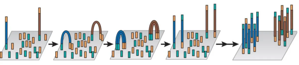
The cluster generation step is where the DNA fragment is amplified. Illumina MiSeq uses a flow cell with one lane, which is coated with two different oligos. The adaptor region of one of the strands of the DNA fragment hybridizes with one of this oligos. Then, a DNA polymerase generates the complement of the hybridized strand creating a double stranded molecule. This molecule is after denatured, and only the complementary sequence stays fixed to the cell while the original template is washed. After this process the bridge amplification starts to occur. Basically, the strand that is fixed to the cell bends over and the adaptor region present in the opposite extremity of the fixed region hybridizes with the second type of fixed oligo.
14
The DNA polymerase will then generate a double stranded bridge that after being denatured results in two single stranded copies which at this point are both fixed to the flow cell (Figure 3). This process is repeated sequentially for millions of clusters simultaneously resulting in a massive amplification of the fragments. Finally, to begin the sequencing step, the reverse reads are washed (71).
Figure 3- Illumina's cluster generation process: the complementary strand of the original
template that hybridized with the first type of oligo is fixed to the flow cell. This strand bends over and hybridization of the second type of oligo with the complementary extremity of the strand occurs. The DNA polymerase generates a double stranded bridge which, after being denatured, results in two strands fixed to the cell. This process is repeated sequentially for millions of clusters simultaneously resulting in massive amplification of all the fragments (Adapted from (72)).
2.2.3. Sequencing
In the Illumina's MiSeq sequencing by synthesis approach all four nucleotides are added simultaneously. This nucleotides are reversibly fluorescent labeled and have the 3'-OH group chemically blocked, allowing only one base incorporation at a time. The forward read sequencing starts with the extension of the first sequencing primer. After nucleotide incorporation, the remaining nucleotides are washed away and the signal from laser-induced excitation of the fluorophores is read from each cluster, with an associated quality value for each base call. The fluorescent molecule and the terminator group are then cleaved and washed away and a new cycle commences. The length of the read is determined by the number of cycles. After finishing the forward read, the attached strand folds over, bridge amplification occurs and after being denatured, the forward read is washed. Reverse reads are then obtained through the same process as forward reads. In the end of the sequencing run, a base calling algorithm assigns the sequences and a quality value (phred score) to each read. The error rate in this system can increase as the reaction proceeds due to incomplete removal of the fluorescent molecule which will cause background noise in the acquired signal. Thus, it is important to bear in mind the chosen size of the fragment, in order to obtain an overlap of at least 50 bp between forward and reverse reads to overcome the lack of quality of the reads ends (71).
15
2.2.4. Data analysis
At the end of the sequence process, millions of reads were generated. MiSeq sequencer automatically attributes the reads to the correct sample in the library pool, based on the combination of indexes used. The first step in the analysis must be a quality control of the reads, in order to understand what parameters to use in filtering to obtain accurate results. After filtering, forward and reverse paired reads are merged to obtain a contiguous sequence. Further analysis should be performed conveniently for the intended application (71).
19 Several studies have been showing that the two-tier serological testing for Lyme disease, the only type of test approved for its diagnosis by FDA, show inaccuracy in detecting Lyme disease in the early localized stage of the infection. Since the antibodies against the pathogen can take weeks to be produced, efforts have been made, throughout the last decades, in order to present direct methods as an alternative to the diagnosis at this stage of the disease. This methods include culture and PCR. Despite different approaches that have been described, concerning the specimen and the methods used, the results obtained with this tests also suffer from a high rate of false negatives.
The present study intends to develop a method suitable for the diagnosis of Lyme borreliosis, specially in an early phase of the infection, using Illumina's MiSeq Next Generation Sequencing platform. The reason why this technology was chosen was based in its ability to sequence DNA present in low concentrations and due to being cost-effective making it applicable to diagnostic practices.
Since many reports have described cases of patients infected with more than one pathogen transmitted by Ixodes ricinus complex, this study aims to create a panel capable of detecting not only the presence of Borrelia spp. but also the presence of the most common co-infections. This panel will target fragments of genes that enable the determination of the genospecies responsible for the infection, which may give important information for the understanding of the clinical manifestations and to appropriate treatment.
23
1. Biological material
1.1. Genomic DNA
Genomic DNA from Borrelia spp. and from the species responsible for the co-infections of Lyme disease were obtained by contacting other researchers/institutes. Borrelia burgdorferi,
Borrelia afzelii and Ehrlichia canis genomic DNAs were provided by Instituto de Higiene e
Medicina Tropical (IHMT-UNL). Genomic DNA of Ehrlichia chaffeensis and Anaplasma
phagocytophilum HGE1 was provided by Prof. Dr. Ulrike Munderloh from Department of
Entomology, University of Minnesota. Genomic DNA of Bartonella henselae Houston-1 and
Bartonella henselae Marseille were provided by Prof. Dr. Volkhard Kempf, from Institute for
Medical Microbiology and Infection Control, Frankfurt. Genomic DNA of Coxiella burnetti was provided by Prof. Dr. Federico Capuano from Department of Food Inspection from Istituto Zooprofilattico Sperimentale del Mezzogiorno. Genomic DNA of Rickettsia rickettsii was provided by Tina Clark, Microbiologist at Laboratory of Intracellular Parasites, NIAID, NIH. Genomic DNA of Babesia microtii Gray was purchased from the ATCC-LGC Standards Partnership (Spain).
1.2. Blood samples
Blood samples from six anonymized individuals were used in this work. Samples from five patients with Babesiosiswere provided by CDC (Atlanta, U.S.A.). This patients' blood has been tested positive, in CDC, for the presence of Babesia species by qPCR. The sixth sample, used as the negative control, is the blood of an individual belonging to a group who never reported a tick bite and from a non-endemic area, which means it is most likely not to have Lyme disease nor co-infections. All blood samples were collected in EDTA vacutainer tubes.
1.2.1. Positive control
For the positive control used in this work, once the purpose was to evaluate the seven primer pairs from the panel, an aliquot of the negative control was spiked with approximately 100,000 E.coli cells from each of the ten different transformed cells containing the amplicon of the different genes selected from each species. The E.coli cells transformation procedure, better explained later in this chapter, was successful for all the fragments of the target genes, with exception of the fragment of the 18S gene from Babesia microti, since the results from Sanger sequencing after cell transformation showed the loss of the majority of the region where the primer should hybridize (data not shown). The estimated number of cells/mL was calculated using the OD value at 600nm of each sample, obtained with Nanodrop equipment, multiplied by 8 x 10⁸, as suggested Agilent Genomics BioCalculator.
24
2. Cell preparation for positive control and PCR
2.1. Target Genes
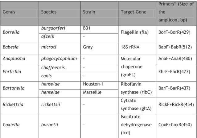
Table 1- Identification of the species gathered for this study, the target gene for each species
and the size of the amplicon obtained in PCR. (* Primer sequences are in the Table 2, Section 3.2.)
Genus Species Strain Target Gene
Primers* (Size of the
amplicon, bp)
Borrelia burgdorferi B31 Flagellin (fla) BorF+BorR(429)
afzelii -
Babesia microti Gray 18S rRNA BabF+BabR(512)
Anaplasma phagocytophilum - Molecular
chaperone (groEL)
AnaF+AnaR(480)
Ehrlichia chaffeensis - EhrF+EhrR(477)
canis -
Bartonella henselae Houston-1 Riboflavin
synthase (ribC) BarF+BarR(437)
henselae Marseille
Rickettsia rickettsii - Cytrate
synthase (gltA) RickF+RickR(454)
Coxiella burnetii -
Isocitrate dehydrogenase (icd)
CoxF+CoxR(450)
The interest genes chosen to be sequenced from each of the ten species were amplified, from the respective genomic DNAs, through a standard PCR using the species specific designed primers, in the following conditions:
[] Reagents Volume 10 x PCR Reaction Buffer 2.5 μL 25 mM MgCl2 1.5 μL 5 mM dNTP 1μL 10 mM Fwd Primer 1 μL 10 mM Rev Primer 1 μL
10 U/μL Surf Hot Taq Polymerase 0.2 μL 1-100 ng/ μL Extracted DNA 2 μL
Water (mQ) 15.8 μL
25
To confirm if amplification occurred, the PCR products were visualized through an agarose gel electrophoresis (AGE) (conditions described in section 3).
All the samples containing a band, in the gel, for the expected size were purified using Magnetic Beads (MCLAB, San Francisco, U.S.A.) to remove, mostly, the primer-dimers formed in the reaction. The purification was performed accordingly to the manufacturer's protocol, using a 1:1.8 DNA/Magnetic Beads ratio. To ascertain if successful purification occurred, another AGE was performed.
The PCR products were then sequenced by Sanger method at STAB VIDA Lda., in order to verify the correct amplification of the desired sequences. All the sequences generated by Sanger sequencing throughout this work were assembled and analyzed with Sequencher 4.10.1 and FinchTV Version1.4.0 Software, respectively.
2.2. Primers
Species specific primers were designed and analyzed using Oligo Explorer 1.1.1 and Oligo Analyzer 1.0.2 software respectively, and finally a primer BLAST was performed in NCBI platform. The primers were acquired from STAB VIDA, Lda. (Portugal). Since the downstream application intended was NGS sequencing using Illumina MiSeq System, a common overhang adaptor was inserted in all the primers as described in 16S Metagenomic Sequencing Library Preparation (74). Forward adaptor sequence: TCGTCGGCAGCGTCAGATGTGTATAAGAGACAG and reverse adaptor sequence: GTCTCGTGGGCTCGGAGATGTGTATAAGAGACAG. The MiSeq Reagent v3 was used with the 600 cycle kit to obtain read lengths of 2x300bp. Primers were designed to obtain amplicons with a size around 450 bp, with the purpose of having an overlap of at least 50 bp between forward and reverse reads.
PCR program
Time Temp.
1. Initial Denaturation 15 min 95ºC 2. Denaturation 30 sec 95ºC 3. Annealing 30 sec 55ºC 4. Elongation 1 min 72ºC 5. Final Elongation 5 min 72ºC
6. Hold ∞ 4ºC
26
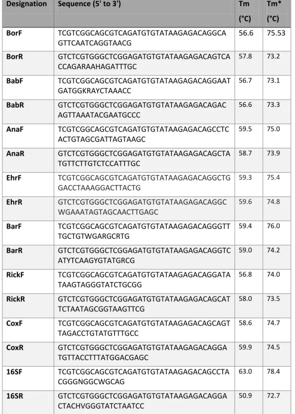
Table 2- Designation, sequences and melting temperatures of the primers designed and 16S
primers used in 16S metagenomic sequencing library preparation(74). All sequences include the common forward and reverse adaptors needed for hybridization to the flow cell in NGS. Tm refers exclusively to the melting temperature of the primer sequence designed to hybridize with the target, while Tm* refers to the melting temperature of the whole primer, include the adaptor sequence.
Designation
Sequence (5' to 3')
Tm
(°C)
Tm*
(°C)
BorF
TCGTCGGCAGCGTCAGATGTGTATAAGAGACAGGCA
GTTCAATCAGGTAACG
56.6
75.53
BorR
GTCTCGTGGGCTCGGAGATGTGTATAAGAGACAGTCA
CCAGARAAHAGATTTGC
57.8 73.2BabF
TCGTCGGCAGCGTCAGATGTGTATAAGAGACAGGAAT
GATGGKRAYCTAAACC
56.7 73.1BabR
GTCTCGTGGGCTCGGAGATGTGTATAAGAGACAGAC
AGTTAAATACGAATGCCC
56.6 73.3AnaF
TCGTCGGCAGCGTCAGATGTGTATAAGAGACAGCCTC
ACTGTAGCGATTAGTAAGC
59.5 75.0AnaR
GTCTCGTGGGCTCGGAGATGTGTATAAGAGACAGCTA
TGTTCTTGTCTCCATTTGC
58.7 73.9EhrF
TCGTCGGCAGCGTCAGATGTGTATAAGAGACAGGCTG
GACCTAAAGGACTTACTG
59.3 75.4EhrR
GTCTCGTGGGCTCGGAGATGTGTATAAGAGACAGGC
WGAAATAGTAGCAACTTGAGC
59.6 74.8BarF
TCGTCGGCAGCGTCAGATGTGTATAAGAGACAGGGTT
TGCTGTWGARGCRTG
59.4 76.0BarR
GTCTCGTGGGCTCGGAGATGTGTATAAGAGACAGGTC
ATYTCAAGYGTATGRCG
59.0 74.2RickF
TCGTCGGCAGCGTCAGATGTGTATAAGAGACAGGATA
TAAGTAGGGTATCTGCGG
56.8 74.0RickR
GTCTCGTGGGCTCGGAGATGTGTATAAGAGACAGCAT
TCTAATAGCGGTAAGTTCG
58.0 73.5CoxF
TCGTCGGCAGCGTCAGATGTGTATAAGAGACAGCAGT
TAGACCTGTATGTTTGCC
58.6 74.7CoxR
GTCTCGTGGGCTCGGAGATGTGTATAAGAGACAGGA
TGTTACCTTTATGGACGAGC
59.9 74.516SF
TCGTCGGCAGCGTCAGATGTGTATAAGAGACAGCCTA
CGGGNGGCWGCAG
63.0 78.416SR
GTCTCGTGGGCTCGGAGATGTGTATAAGAGACAGGA
CTACHVGGGTATCTAATCC
50.9 72.727
2.3. Cloning
The cloning process was performed using the CloneJET PCR Cloning Kit, purchased from Thermo Scientific (Waltham, MA, USA). Briefly, pJET1.2/blunt is a linearized cloning vector capable to accept inserts from 6 bp to 10 kb. The Sticky-End Cloning Protocol, which consists in a blunting reaction of the amplicons and a ligation reaction to the cloning vector, was followed as described by the manufacturer, with exception of the added volume of nuclease free water, due to lack of starting concentration of DNA.
2.4. Transformation
The transformation step was performed using E.coli NZY5α competent cells, purchased from NZYTech (Lisboa, Portugal). This cells show similar properties to DH5α, which are suitable for high efficiency transformation. The transformation protocol was performed accordingly to the manufacturer's. Briefly, 10μL of the ligation reaction were added to a tube containing 100μL of competent cells. After the incubation and heat-shock, SOC medium was added and 250μL of the transformed cells were inoculated on LB agar (Miller) plates. A competent cells control plasmid solution provided with the NZY5α was used as a positive control and milli-Q H2O was used as the negative control. The cell culture was incubated in CO2 incubator overnight at
37ºC with 5% CO2.
2.4.1. Confirmation of correct insertion
From each of the ten LB agar plates, two different colonies were picked and inoculated in 2 mL of LB Broth medium containing 100 μg/mL ampicillin and incubated at 37ºC o/n with agitation. Then, 2μL of each tube were used directly on a standard PCR using the pJET1.2F and pJET1.2R pair of primers provided in the CloneJET PCR Cloning Kit. After seeing the PCR products in the AGE, one of the two colonies of each species was selected to sequence by Sanger. All sequences were confirmed to be completely inserted in E.coli cells with exception of both Babesia microti colonies, which lacked the final region of the fragment, needed for primer to hybridize.
To verify the presence of the primers in the sequences, pDRAW32 1.0 Revision 1.1.133 ACACLONE Software was used.
3. Agarose Gel Electrophoresis
All the AGE performed throughout this work used 1,5% agarose gel prepared in Tris-Acetate-EDTA (TAE). The nucleic acids were stained with GelRed (1:50000) which is a stable fluorophore that intercalates the nucleic acids without impair their migration in the gel. The complex dsDNA-GelRed when excited with UV light emits fluorescence that is captured by the camera present in the trasilluminator equipment. The conditions of the AGE were 120 Volt (V) for 20 minutes in TAE 1x.
28
4. DNA Extraction
Extraction of genomic DNA from the blood samples was performed with the GE illustra blood genomicPrep Mini Spin Kit purchased from GE Healthcare (Little Chalfont, United Kingdom). The DNA extraction was performed as described in the manufacturer's protocol for samples between 50-300µl. Briefly, in this procedure, a chaotropic agent is used to extract de DNA from nucleated blood cells and promote the selective binding of DNA to the silica membrane column. The protease used in the kit is proteinase K, which is active even in the presence of detergents and chelating agents, like EDTA. The kit also provides a low ionic strength elution buffer which allows the DNA to be stored. From each sample of whole blood, 200µl were used in the DNA extraction procedure, since this is the indicated volume to obtain the optimal performance.
5. Library Preparation for NGS sequencing
The seven samples, five from Babesiosis patients and the positive and negative controls, were submitted to four different conditions: whole blood used in multiplex PCR with the primers designed (B+MX), extracted DNA also used in multiplex PCR (DNA+M) and extracted DNA with the multiplex PCR and the PCR using 16S primers.
5.1. Amplicon PCR
The four distinct PCR had different reagents and PCR programs. The reactions using whole blood were performed using the enzyme Hemo KlenTaq (New England BioLabs, Massachusetts, U.S.A.), while in the reactions using extracted DNA the enzymes Surf HotTaq DNA Polymerase (STAB VIDA, Portugal) and KAPA Hifi HotStart ReadyMix (KAPA Biosystems, were used for multiplex PCR and 16S PCR respectively.
5.1.1. Whole blood + Multiplex conditions (B+Mx)
[] Reagents Volume
5 x Hemo KlenTaq Reaction Buffer 10 μL
10 mM dNTP 1μL
10 μM Fwd Multiplex Primers 1.5 μL 10 μM Rev Multiplex Primers 1.5 μL
n.a. Hemo KlenTaq 4 μL
Whole Blood 10 μL
Water (mQ) 22 μL