$'
REV. HOSP. CLÍN. FAC. MED. S. PAULO 56(6):169-172, 2001 NOVEMBER-DECEMBER
From the Department of Dermatology, Hospital das Clínicas, Faculty of Medicine, University of São Paulo.
CONIZATION, FROZEN SECTION EXAMINATION, AND
PLANNED HYSTERECTOMY IN THE TREATMENT OF
HIGH-GRADE CERVICAL INTRAEPITHELIAL
NEOPLASIA
Jesus Paula Carvalho, Filomena Marino Carvalho, Katia Maciel Pinceratoand Elsa A. Gay Pereyra
RHCFAP/3056
CARVALHO JP et al. - Conization, frozen section examination, and planned hysterectomy in the treatment of high-grade cervical intraepithelial neoplasia. Rev. Hosp. Clín. Fac. Med. S. Paulo 56(6):169-172, 2001.
Purpose: We tested the role of frozen section examination of the cone specimen in the evaluation of the resection margin status
and to rule out invasion in patients with high-grade cervical intraepithelial neoplasia.
Methods: Twenty-five patients with cervical intraepithelial neoplasia underwent conization followed by frozen section
examination and planned hysterectomy. The results of the definitive paraffin exam were compared with frozen section examination.
Results: In the evaluation of the margins by frozen section examination, 16 patients (64%) had positive cone margins and 9
(36%) had negative margins. The definitive paraffin examination of margin status was concordant in all the cases. Intraoperative diagnosis of invasion was made in 5 cases, and 1 of these was microinvasive. Among the remaining 20 cases, we detected 2 additional microinvasive carcinomas after paraffin study, so the diagnosis of the frozen section examination was concordant with the paraffin sections in 23/25 cases (92%). Two cases of microinvasive carcinoma were diagnosed as cervical intraepithelial neoplasia by frozen section examination and had less than 2 mm stromal invasion.
Conclusions: In high-grade cervical intraepithelial neoplasia, frozen section examination can provide immediate and precise
evaluation of the cone margin status in high-grade cervical intraepithelial neoplasia. It can identify frank invasion and permit adequate treatment in a one-stage procedure. In early microinvasive disease, frozen section examination fails to detect the area of invasion but reliably detects clear resection margins.
DESCRIPTORS: Cervical neoplasia. Intraepithelial neoplasia. Frozen section examination. Cervical conization.
Hysterectomy.
High-grade cervical intraepithelial neoplasia (CIN 3) is the last step of neoplastic transformation in the cervi-cal epithelium before invasion occurs. An inadequate or insufficient treatment can result in poor prognosis and death. The treatment strategies for CIN have changed considerably in the last decades, moving from the classical sur-gical conization to office procedures such as large loop excision of the trans-formation zone (LLETZ)1 ,2. However,
incomplete excision of CIN continues to be the most vulnerable point of this kind of treatment3 -4.
Incomplete excision of CIN ranges from 15% to 50%5 -6 and presents a significant problem if follow-up is in-sufficient. Unfortunately, the disease progresses to invasive carcinoma in
many patients after an incomplete treatment for CIN.
In Brazil, women from very distant places are referred to Hospital das Clinicas for treatment of many dis-eases. We use this opportunity for screening them for cervical cancer and its precursor lesions.
fol-%
REV. HOSP. CLÍN. FAC. MED. S. PAULO 56(6):169-172, 2001 NOVEMBER-DECEMBER
low-up. Our policy is to choose the most efficient kind of treatment with least probability of recurrence.
For young patients with satisfactory colposcopy, the treatment of choice is large loop excision of the transforma-tion zone (LLETZ). On the other hand, for older patients with large lesions, endocervical involvement, or suspicion of invasion, the classic surgical coniza-tion is a better opconiza-tion. In this high-risk group of patients, we tested the role of frozen section examination (FSE) of the cone specimen in order to evaluate the margin status and to rule out inva-sion, thus providing adequate treatment in a one-stage procedure.
Little attention has been paid to the role of FSE in cervical intraepithelial neoplasia. Some authors have demon-strated high concordance with the de-finitive paraffin exam in grading the lesion, but there are few studies regard-ing the evaluation by FSE of the resec-tion margin status7 -8.
METHODS
We studied prospectively 25 con-secutive patients who underwent coni-zation and planned hysterectomy who had the histological diagnosis of cervi-cal intraepithelial squamous neoplasia grade 3 (CIN 3) and concomitant be-nign gynecologic disease, such as leio-myoma, abnormal uterine bleeding, or extensive CIN with suspicion of inva-sion. Patient ages ranged from 24 to 69 years, median 42 years. In these cases, particularly those with suspicion of in-vasion, the objective of the FSE was to evaluate the extent of invasion, thus permitting the adequate treatment of invasive carcinoma. Our second objec-tive was to verify the accuracy of the FSE for evaluating the resection mar-gin status.
We performed cold knife coniza-tion under general anesthesia, carefully excising the entire visible lesion. The
vertical size of the cone ranged from 1 cm to 2.5 cm.
The cone specimens were excised in one piece and sent immediately for FSE. The pathologist inked the cone with different colors in order to iden-tify the anterior and posterior borders. The endocervical margin was selected for frozen section (Fig. 1). The remain-ing specimen was sectioned sequentially into 3-mm pieces.
The pathologist selected for FSE the fragments corresponding to the grossly suspicious areas noted in the colposcopic image. The fragments not undergoing frozen section were processed in the tra-ditional paraffin-embedded method, with inclusion of the entire material for his-tological examination.
In the patients with the diagnosis of invasion from FSE, the treatment was radical hysterectomy with pelvic lym-phadenectomy (Wertheim-Meigs sur-gery). In those without signs of inva-sion, we performed a simple hysterec-tomy.
The results of the definitive paraf-fin exam were compared with those of the FSE. We evaluated the accuracy of the diagnosis of invasion and the resec-tion margin status. The complementary specimen from the hysterectomy was studied in order to determine the pres-ence of residual disease and its relation with the final diagnosis from the cone specimens.
RESULTS
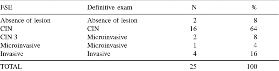
Intraoperative diagnosis of invasion was made in 5 cases; 1 of these was microinvasive, and all were confirmed in the definitive diagnosis. Among the remaining 20 cases, we detected 2 ad-ditional microinvasive carcinomas after paraffin study; therefore, the diagnosis of the FSE was concordant with the per-manent sections in 23/25 cases (92%) (Table 1). The 2 cases of microinvasive carcinoma diagnosed as CIN 3 by FSE had less than 2 mm of stromal invasion. In the 4 cases of invasive carcinoma, FSE allowed us to identify the invasion and perform a radical hysterectomy with lymphadenectomy (Wertheim-Meigs surgery) in the same surgical procedure. One of these cases was the youngest pa-tient (24 years old).
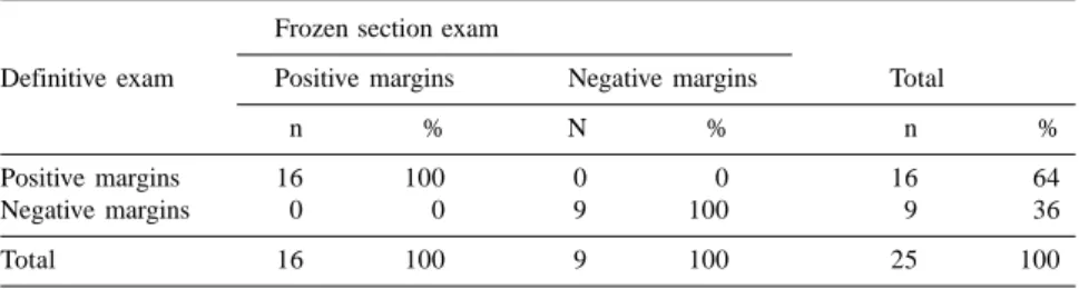
In the evaluation of cone margins by FSE, 16 patients (64%) had positive surgical margins (Fig. 2) and 9 (36%) had negative margins. There were 1 lat-eral and 15 endocervical margins in-volved. The definitive paraffin exami-nation of margin status was concord-ant in all cases (Table 2).
Table 1 - Comparison of intraoperative diagnosis by frozen section examination
(FSE) and definitive paraffin exam.
FSE Definitive exam N %
Absence of lesion Absence of lesion 2 8
CIN CIN 16 64
CIN 3 Microinvasive 2 8
Microinvasive Microinvasive 1 4
Invasive Invasive 4 16
TOTAL 25 100
%
REV. HOSP. CLÍN. FAC. MED. S. PAULO 56(6):169-172, 2001 NOVEMBER-DECEMBER
In the 16 cases with positive mar-gins, we found residual disease in the uterus in 11 (68.8%). In all cases of negative resection margins, the uterus did not have lesions (Table 3).
DISCUSSION
The two most undesirable occur-rences following conservative CIN treatment are positive margins and un-diagnosed invasive disease. In these two situations, the treatment is consid-ered incomplete and the risk of recur-rence is very high8.
The advent of conservative meth-ods of CIN treatment such as LLETZ has allowed reduction in cost and per-mitted the preservation of the best cer-vical function; however, its use is as-sociated with new problems, such as high recurrence rates, especially in older women or in those with extensive endocervical lesions7.
In patients with incomplete exci-sion of CIN, additional treatment ranges from simple observation to hys-terectomy. Expectant management is rational only for those patients with good conditions for adequate follow-up. However, this requirement is very difficult to guarantee in our country due to the socioeconomic conditions of the assisted population. In our institu-tion, we have endeavored to treat all cases in a one-stage procedure. Conse-quently, we frequently have decide be-tween performing the option of coni-zation—with the accompanying risk of leaving positive surgical margins—or to perform a total hysterectomy. Hys-terectomy, the treatment of choice in high-grade CIN, has the associated risk of inadequate treatment of invasive car-cinoma, which in our study was diag-nosed in 16% of the cases.
The adoption of FSE provides a so-lution for the two vulnerable points in the treatment of high-risk CIN. First, the lesion can be shown to have been entirely removed when negative cone margins are found; second, the diagno-sis of invasive carcinoma can be made with good accuracy and treated ad-equately in a one-stage procedure.
Our study showed a high level of tumor involvement at the cone margins in extensive CIN 3 (64%). Others have noted the frequent occurrence of posi-tive resection margins in cervical conizations, particularly in the en-docervical conizations9. Using FSE, we were able to accurately diagnose the resection margin status of all of our cases.
Eleven out of 16 patients with posi-tive resection margins had residual dis-ease in the uterus, and they probably would have been of high risk of recur-rence if management had been expect-ant.
Using FSE, we were able to iden-tify all 4 cases of invasive carcinoma, but only 1 of the 3 microinvasive cases. The 2 underdiagnosed cases had an in-vasion of less than 2 mm. However, these lesions have a low risk of metas-tasis and can be treated as CIN with good results if the resection margins are clear.
CONCLUSION
FSE can provide immediate and precise evaluation of the cone margin status in high-grade CIN. It can iden-tify frank invasion, permitting adequate treatment in a one-stage procedure. In early microinvasive disease, FSE fails to correctly grade the lesion, but FSE reliably detects clear resection margins. We believe FSE should be included in the treatment plan of women with high risk of tumor involvement in resection margins and who cannot participate an adequate follow-up.
Table 2 - Margin status by frozen section examination and definitive paraffin
exam.
Frozen section exam
Definitive exam Positive margins Negative margins Total
n % N % n %
Positive margins 16 100 0 0 16 64
Negative margins 0 0 9 100 9 36
Total 16 100 9 100 25 100
Table 3 - Relationship between cone margin status and residual disease in uterus.
Frozen section cone margins status
Residual disease Positive Negative Total
in uterus
n % N % n %
Absent 5 31.2 9 100 14 56
Present 11 68.8 0 0 11 44
Total 16 100 9 100 25 100
%
REV. HOSP. CLÍN. FAC. MED. S. PAULO 56(6):169-172, 2001 NOVEMBER-DECEMBER
RESUMO RHCFAP/3056
CARVALHO J P e col. – Conização, exame de congelação e histerec-tomia planejada no tratamento de neoplasia intra-epitelial de alto grau. Rev. Hosp. Clín. Fac. Med.
S. Paulo 56(6):169-172, 2001.
Objetivos: Foi avaliado o papel do
exame intra-operatório de congelação no diagnóstico de invasão e no estado das margens cirúrgicas em pacientes com neoplasia intra-epitelial de alto grau.
Casuística e Método: Vinte e
cin-co pacientes cin-com neoplasia intra-epitelial de alto grau foram submetidas a conização cervical seguida de histe-rectomia. O resultado do exame
defi-nitivo em parafina foi comparado com o exame de congelação.
Resultados: Dezesseis pacientes
(64 %) tiveram margens comprometi-das e 9 (36 %) livres. O diagnóstico de-finitivo na parafina foi concordante em todos os casos no diagnóstico do esta-do das margens. Em cinco casos foi diagnosticada invasão, sendo 1 caso de carcinoma microinvasivo. Entre os 20 casos restantes foram detectados 2 car-cinomas microinvasivos adicionais após o exame em parafina. Desta for-ma o exame intra-operatório de conge-lação foi concordante com o exame definitivo em 23/25 casos (92 %). Dois casos de carcinoma microinvasivo fo-ram diagnosticados como neoplasia
intra-epitelial de alto grau no exame de congelação e apresentavam invasão estromal menor que 2 cm.
Conclusões: O exame
intra-operató-rio de congelação é capaz de predizer o estado das margens cirúrgicas em to-dos os casos de neoplasia intra-epitelial de alto grau assim como o diagnóstico de invasão franca. Nos casos de carci-noma microinvasivo este exame falha na detecção de invasão, entretanto é capaz de garantir margens livres de ressecção.
DESCRITORES: Carcinoma de
células escamosas. Neoplasia intra-epitelial escamosa. Exame de conge-lação. Conização cervical. Histerectomia.
REFERENCES
1. JONES HW - Treatment of cervical intraepithelial neoplasia. Obstet Gynecol 1990;33:826-836.
2. WRIGHT TC JR, GAGNON S, RICHART RM et al. - Treatment of cervical intraepithelial neoplasia using the loop electrosurgical excision procedure. Obstet Gynecol 1992;79:173-178. 3. LUBICZ S, EZEKWECHE C, ALLEN A et al. - Significance of cone
biopsy margins in the management of patients with cervical neoplasia. J Reprod Med 1984;29:179-184.
4. TOWNSEND DE - Cervical cone margins as a predictor for residual dysplasia in post-cone hysterectomy specimens. Obstet Gynecol 1994;84:898.
5. BALDAUF JJ, RITTER J, CUENIN C et al. - Therapeutic results of conization with diathermy. Contracept Fertil Sex 1999;27:140-146.
6. CHAN KS, YU KM, LOK YH et al. - Conservative management of patients with histological incomplete excision of cervical intraepithelial neoplasia after large loop excision of transformation zone. Chin Med J 1997;110:617-619.
7. HANNIGAN EV, SIMPSON JS, DILLARD JR et al. - Frozen section evaluation of cervical conization specimens. J Reprod Med 1986;31:11-14.
8. TORRES JE, MOORMAN J, SHIU A et al. - Colposcopically directed conization for frozen-section examination in the management of cervical intraepithelial neoplasia. J Reprod Med 1983;28:123 125. 9. PATERSON-BROWN S, CHAPPATTE OA, CLARK SK et al. - The significance of cone biopsy resection margins. Gynecol Oncol 1992;46:182-185.