Male and female runners demonstrate different
sagittal plane mechanics as a function of static
hamstring flexibility
D. S. Blaise Williams III1,2, Lee M. Welch3
ABSTRACT | Background: Injuries to runners are common. However, there are many potential contributing factors to
injury. While lack of lexibility alone is commonly related to injury, there are clear differences in hamstring lexibility between males and females. Objective: To compare the effect of static hamstring length on sagittal plane mechanics
between male and female runners. Method: Forty subjects (30.0±6.4 years) participated and were placed in one
of 4 groups: lexible males (n=10), inlexible males (n=10), lexible females (n=10), and inlexible females (n=10). All subjects were free of injury at the time of data collection. Three-dimensional kinematics and kinetics were collected while subjects ran over ground across 2 force platforms. Sagittal plane joint angles and moments were calculated at the knee and hip and compared with a 2-way (sex X lexibility) ANOVA (α=0.05). Results: Males exhibited greater peak
knee extension moment than females (M=2.80±0.47, F=2.48±0.52 Nm/kg*m, p=0.05) and inlexible runners exhibited greater peak knee extension moment than lexible runners (In=2.83±0.56, Fl=2.44±0.51 Nm/kg*m, p=0.01). For hip lexion at initial contact, a signiicant interaction existed (p<0.05). Flexible females (36.7±7.4º) exhibited more hip lexion than inlexible females (27.9±4.6º, p<0.01) and lexible males (30.1±9.5º, p<0.05). No differences existed for knee angle
at initial contact, peak knee angle, peak hip angle, or peak hip moment. Conclusion: Hamstring lexibility results in
different mechanical proiles in males and females. Flexibility in the hamstrings may result in decreased moments via active or passive tension. These differences may have implications for performance and injury in lexible female runners.
Keywords: biomechanics; gender; hamstrings; running.
HOW TO CITE THIS ARTICLE
Williams DSB III, Welch LM. Male and female runners demonstrate different sagittal plane mechanics as a function of static hamstring lexibility. Braz J Phys Ther. 2015 Sept-Oct; 19(5):421-428. http://dx.doi.org/10.1590/bjpt-rbf.2014.0123
1 VCU RUN LAB, Department of Physical Therapy, Virginia Commonwealth University, Richmond, Virginia, USA 2 Department of Kinesiology and Health Sciences, Virginia Commonwealth University, Richmond, Virginia, USA 3 B. Young Physical Therapy, Fuquay-Varina, North Carolina, USA
Received: Mar. 18, 2015 Revised: May 25, 2015 Accepted: June 30, 2015
Introduction
Running is one of the most popular competitive, recreational, and itness activities worldwide. In fact, running is a component of, or training modus for, most Olympic and non-Olympic sports. In 2012, roughly 51.4 million Americans ran at least once with approximately 29.4 million of these running at least 50 days per year1. The health beneits of running include reducing the risks of (i) chronic disease, (ii) disability, (iii) pain, and (iv) health care costs2-4. However, with the continued popularity of running, there has been a corresponding maintenance in the rate of running-related injuries5. The majority of these injuries can be attributed to overuse3. As a result, these injuries force an estimated 46% to 65% of runners to stop running and seek medical treatment each year.
The highest risk factor for injuries in runners is weekly mileage. In particular, it is believed that the
422 Braz J Phys Ther. 2015 Sept-Oct; 19(5):421-428
the methodology between these two studies is not consistent, it is dificult to draw speciic conclusions regarding the hamstrings’ role in running injury, but it does raise questions regarding the speciic effects of hamstring lexibility on running mechanics and injuries. While muscle lexibility may play a role in injury, single anatomical factors are not likely to predict rates or incidences of injuries in runners.
Flexibility has been deined as the ability of muscular tissue to lengthen, given that the articulation travels through the entire movement’s span15. Lower extremity alignment, with respect to hamstring lexibility and its correlation to risk of injury, has been studied extensively12,14,16. In an open chain, the hamstrings are the primary lexors of the knee, while acting as secondary extensors of the hip. During running, the hamstrings act to slow down hip lexion in the last half of the swing phase (just prior to initial contact) and to extend the hip during the stance phase17. Additionally, the hamstrings decelerate tibial extension momentum just before initial contact18. Therefore, simultaneous hip lexion and knee extension during late swing result in substantial elongation and eccentric contraction of the biarticular hamstrings, causing extremely high loads during the elongated position of the hamstrings during late swing19. Due to energy transfer between phases and the important concentric and eccentric functions of the hamstrings, the lexibility of this group of muscles is not only an important factor inluencing running biomechanics, but also a potential factor related to injury during running18.
The relationship between hamstring lexibility and injury is poorly understood because the mechanism of tissue damage likely depends on multiple factors, such as joint biomechanics, tissue mechanics, intensity of exercise, fatigue, and tissue structure. It has been shown that simulated hamstring shortening inluences gait adversely when the popliteal angle is greater than 15 degrees from full knee extension20. These abnormal characteristics were demonstrated by increases in the parameters of walking effort, posterior pelvic tilt, and knee lexion during the stance phase of gait. These were also associated with decreases in walking speed, stride length, step length, hip lexion, pelvic obliquity and rotation, as well as premature ankle dorsilexion and plantarlexion in stance20. While normal hamstring inlexibility would not likely be as extreme, some of these biomechanical effects would result from existing hamstring inlexibility.
In addition to the above kinematic and spatiotemporal characteristics, knee joint moment is another
important biomechanical factor that must also be taken into consideration when considering running biomechanics as it relates to static hamstring length. As the hamstring muscles are elongated during late swing prior to initial contact, the moment around the knee is signiicantly increased. With the hip in 0° extension, maximum knee lexion moment (internal) occurs at full knee extension. With the hip at 90°, there is some variation in position of maximum knee torque with some individuals producing maximum knee torque with the knee near 30-45° and some with the knee at full extension21. Furthermore, those with decreased hamstring lexibility exhibit greater knee lexion moment at short muscle lengths and decreased moment at long muscle lengths when compared to individuals with increased hamstring lexibility22. Regardless, at initial contact during running, the knee is close to the maximum torque and the hamstring is substantially elongated, resulting is high loads on this muscle during late swing and early stance.
Differences between the sexes may also play a role in running biomechanics. It has been shown that female recreational runners, when compared to males, demonstrate signiicantly greater peak hip adduction, hip internal rotation, and knee abduction angles. Thus, female runners exhibit signiicantly different lower extremity mechanics at the hip and knee in the frontal and transverse planes23. Additionally, it has been demonstrated that women have less knee lexion angle and more knee valgus angle as well as greater quadriceps activation, and lower hamstring activation as compared to their male counterparts during the stance phase of running, side cutting, and cross cutting24. It is unknown whether changes in lexibility of the hamstrings result in different biomechanical proiles in men compared to women.
sagittal plane mechanics in male and female runners. We hypothesize that hamstring lexibility will result in similar changes in running mechanics when compared between males and females.
Method
Individuals in this study were recruited from the University, surrounding communities, and local running clubs, resulting in a sample of convenience of runners who were asymptomatic at the time of data collection. Each subject gave their written informed consent for participation in the study, which was approved by the University and Medical Center Institutional Review Board, Greenville, NC, USA (UMCIRB 10-0437). An a priori power analysis was conducted utilizing data consistent with the variables of interest in the current study (α=0.05, β=0.80). Each variable was used independently for the power analysis, and peak hip angle was found to require the largest number of subjects to obtain signiicance. Based on this analysis, a sample size of 8 subjects per group was established for comparisons with adequate statistical power. In order to account for attrition and protect from type II error, the study included a total of 40 male and female subjects ranging in age from 18-50 years. Participants were placed in groups based on hamstring length, measured as the number of degrees lacking from zero, where zero is full knee extension with the hip at 90 degrees (popliteal angle). All subjects in this study had hamstrings that were classiied as either lexible or inlexible. There were 4 groups consisting of 10 individuals in each group: lexible males, inlexible males, lexible females, and inlexible females sampled from a larger group of 99 runners collected in the current study. All subjects with tight hamstrings had a popliteal angle >29° away from zero. All subjects with lexible hamstrings had a popliteal angle <10° away from zero (Table 1). The values of 10 and 29° were chosen, as they were 1 standard deviation from the mean for the previously mentioned group of 99 runners ranging in age from 29 to 81 years. Participants ran
a minimum of 10 miles (16 kilometers) per week for at least 6 months prior to this study. Subjects were excluded from this study if they had any cardiovascular or neurological compromise, current lower extremity musculoskeletal injury, joint replacement, or joint fusion. Runners were not excluded from the study if they had previous lower extremity injuries related to running.
Static hamstring lexibility for both lower extremities was measured by two researchers using a standard goniometer with the subject supine on a mat table. One researcher maintained the knee and hip to be measured in a 90° lexed position and moved the knee into a terminal knee extension position to perform the range of motion measurement. Once terminal knee extension was obtained, the second researcher used a hand-held dynamometer to push the leg being measured with an average force of 10-12 pounds into the patient’s end range (Figure 1). The average of
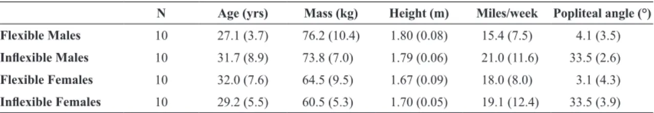
Table 1. Subject demographics.
N Age (yrs) Mass (kg) Height (m) Miles/week Popliteal angle (°)
Flexible Males 10 27.1 (3.7) 76.2 (10.4) 1.80 (0.08) 15.4 (7.5) 4.1 (3.5)
Inlexible Males 10 31.7 (8.9) 73.8 (7.0) 1.79 (0.06) 21.0 (11.6) 33.5 (2.6)
Flexible Females 10 32.0 (7.6) 64.5 (9.5) 1.67 (0.09) 18.0 (8.0) 3.1 (4.3)
Inlexible Females 10 29.2 (5.5) 60.5 (5.3) 1.70 (0.05) 19.1 (12.4) 33.5 (3.9)
Presented in mean (SD).
Figure 1. Measurement of hamstring lexibility. Measurements
424 Braz J Phys Ther. 2015 Sept-Oct; 19(5):421-428
3 measurements was taken for each lower extremity. The contralateral leg remained lat (extended) on mat table during each measurement. All subjects included in the study had symmetrical range of motion (±5°) between right and left limbs. Therefore, only the right limb was utilized in all subjects for comparison between groups.
A three-dimensional running analysis was completed on subjects eligible for participation. A standing calibration trial was collected during which static joint (greater trochanters, medial and lateral knees, medial and lateral maleoli, and medial and lateral forefoot) and segment tracking (calcaneus, shank, thigh, and pelvis) retrorelective markers were placed on bilateral lower extremities (Figure 2)25. The static joint markers were used to establish joint centers, segment geometry, and segment coordinate systems. Static markers were removed before the dynamic data collection. During the dynamic data collection, subjects were asked to run along a 16-meter runway at a speed of 3.35 m/s (±5%). Running speed was measured using photocells located 6 meters apart. A ixed running speed was used in order to decrease differences in lower extremity biomechanics and spatiotemporal parameters related to differences in forward velocity. Subjects were instructed to run with their normal
running gait. Kinematic data were collected at 240 Hz with a 9-camera motion analysis system (Qualisys Inc., Glastonbury, CT, USA). Qualisys software was used to reconstruct 3-dimensional coordinates for each marker. Two force plates (AMTI, Watertown, MA, USA) mounted on the loor of the runway recorded ground reaction forces (GRF) at a sample frequency of 1200 Hz. Kinematic data was time synchronized with GRF data at the time of collection. Subjects were required to run across the force plates for a minimum of 10 successful trials for the right lower extremity. A trial was considered successful if the subject ran with a natural gait over the force plates within the given velocity range while striking at least one of the force plates with their entire right foot.
Pelvis, thigh, shank, and foot segments were created using Visual 3D Software (C-motion Inc., Bethesda, MD, USA). Data were analyzed between initial contact and toe-off on the right limb and normalized to 100 data points, with each data point representing 1% of the stance phase of running. A second-order recursive Butterworth ilter was used to ilter marker data at 12 Hz and GRF data at 50 Hz. For this study, knee motion was deined as the tibia moving relative to the femur, and hip motion was deined as the femur moving relative to the pelvis. Visual 3-D software was used to calculate joint rotations via Cardan sequencing in which motion about the X-axis was deined as lexion/extension at the hip and knee. Joint moments were calculated at the hip and knee. Joint moments were normalized to subject mass and height. Mean joint angle and moment curves were created bilaterally at the hip and knee in the sagittal plane for each group. Peak lexion angles and extension moment values at the hip and knee were calculated. Sagittal plane hip and knee angle at initial contact were also calculated. Data plots were visually assessed for normality and variance homogeneity. Shapiro-Wilk test of normality was used to determine data normality on all variables. Based on the above test, all dependent variables were normally distributed.
Joint angles and joint moments were compared between the groups. These data were analyzed using a 2-factor (sex (df=1), lexibility (df=1), within-subjects (df=36)) analysis of variance (α=0.05) to determine differences between groups for peak knee lexion, peak hip lexion, peak knee extension moment, peak hip extension moment, knee lexion angle at initial contact, and hip lexion angle at initial contact. Post hoc t-tests (α=0.05) were utilized for individual comparisons.
Figure 2. Retrorelective marker placement. A total of 39 markers
Results
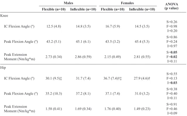
All results are presented in Table 2. Males demonstrated greater peak knee extension moment than females (M:2.80±0.47, F:2.48±0.61 Nm/kg*m). Inlexible runners demonstrated greater peak knee extension moment than lexible runners (In:2.83±0.56, Fl:2.44±0.51 Nm/kg*m).
A signiicant interaction existed for hip lexion at initial contact (p=0.03). Speciically, lexible females exhibited more hip lexion than inlexible females (p<0.01) and lexible males (p=0.05) (Figure 3). Interestingly, lexible females not only landed in more lexion but also remained in roughly the same degree of lexion during loading response (Δ=0.4°).
No differences existed for knee angle at initial contact or peak knee angle. Similar to hip motion, no differences existed for peak hip angle or peak hip moment.
Discussion
The purpose of this study was to compare the effect of static hamstring length on sagittal plane mechanics in male and female runners. Mechanical differences existed primarily in lexible females. This is the irst study to demonstrate that differences in lexibility result in different mechanical compensations between males and females. This understanding may help deine speciic interventions for female runners in an attempt to improve performance or reduce injuries.
At the knee, males exhibited greater peak knee extension moment when compared to females.
Figure 3. Sagittal plane hip angle during stance. Note that lexible
females demonstrate greater hip lexion at initial contact that does not exhibit the same lexion absorption as the other groups. FF=lexible females; FM=lexible males; IF=inlexible females; IM=inlexible males.
Table 2. Dependent variables.
Males Females ANOVA
(p value) Flexible (n=10) Inlexible (n=10) Flexible (n=10) Inlexible (n=10)
Knee
IC Flexion Angle (º) 12.5 (4.8) 14.8 (3.5) 16.7 (5.9) 14.5 (3.5) S=0.26F=0.98 I=0.20
Peak Flexion Angle (º) 43.2 (5.1) 45.1 (6.1) 43.5 (3.2) 45.4 (5.3) S=0.86F=0.24 I=0.97
Peak Extension
Moment (Nm/kg*m) 2.73 (0.34) 2.86 (0.59) 2.15 (0.49) 2.81 (0.55)
S=0.05
F=0.02
I=0.11
Hip
IC Flexion Angle (º) 30.1 (9.5)‡ 31.7 (7.4) 36.7 (7.4)†‡ 27.9 (4.6)† S=0.55F=0.13 I=0.03
Peak Flexion Angle (º) 35.2 (10.3) 37.2 (8.1) 37.1 (7.4) 31.0 (3.2)
S=0.38 F=0.40 I=0.11
Peak Extension
Moment (Nm/kg*m) 1.58 (0.41) 1.69 (0.34) 1.76 (0.40) 1.49 (0.23)
S=0.91 F=0.46 I=0.09
426 Braz J Phys Ther. 2015 Sept-Oct; 19(5):421-428
While differences in running mechanics have been demonstrated between sexes23, the majority of these differences were observed in joint movement (kinematic variables) and in the secondary planes of motion (frontal and transverse). Speciically, females demonstrate signiicantly greater peak hip adduction, hip internal rotation, and knee abduction angles23. Females also demonstrate less knee lexion angle, associated with greater quadriceps activation and lower hamstring activation when compared to males during running and cutting activities24. Females are often termed as “quad-dominant” and less able to activate their hamstrings25,26. As running is a series of single-leg landings (squats), the hamstrings are necessary to aid in extension moment at the knee by eccentrically controlling anterior motion of the tibia4,26,27. If females have reduced hamstring activity, this may partially explain the reduction in knee extension moment. This further requires that the knee extension moment be produced by the quadriceps and may place increased stress on the patellofemoral joint, a common injury among female runners28. If a runner does not use their hamstrings adequately (magnitude or timing), which may be the case in females, this may explain why females do not produce as much knee extension moment during stance. Further evaluation of hamstring activation in these individuals is necessary to explain this further.
Inlexible runners demonstrated higher peak knee moment than lexible runners. This is consistent with previous work showing that poor hamstring lexibility is associated with higher knee extension moments4. As the hamstrings are eccentrically active in controlling lexion of the knee, decreased length of these muscles may result in passive tension and similar control of knee lexion. Therefore, an individual with inlexible hamstrings could demonstrate increased knee extension moment due to the passive tension of this tight group of muscles. Additionally, as hamstring lexibility decreases, the knee extensors may need to counteract the tighter lexor muscles prior to initial contact, further increasing the extension moment at the knee throughout the stance phase.
Flexible females demonstrated the greatest amount of hip lexion at initial contact (Table 2). Interestingly, the females remained in increased hip lexion during loading response but only lexed an additional 0.4º over this time. This, in combination with a large hip extension moment (1.76 Nm/kg*m) results in increased joint stiffness at the hip joint. While not signiicant, this group demonstrated a similar pattern at the knee
where the lexible females lexed approximately 4 degrees less than the other groups. Speciically, lexible females demonstrated the least knee lexion excursion from initial contact to peak (Δ=26.8°). This creates a stiffer knee resulting in less shock attenuation and potential increases in impact forces. We suggest that this passive lexibility results in a need for the female runners to stabilize the hip joint. The question remains as to whether this is a positive compensation based on performance or injuries in this group. While many of the runners in both the lexible and inlexible groups had a history of running injuries, the number of subjects in the current study is not adequate to establish causation. A much larger cohort of runners followed prospectively is necessary to establish strong relationships between hamstring lexibility and lower extremity injuries in runners.
Previous research has shown that acute changes in hamstring lexibility result in minimal changes in mechanics during running29. Limited data exists on mechanical characteristics of runners based on hamstring lexibility, independent of intervention. It would be expected that increased lexibility in runners would result in more hip lexion or knee extension at initial contact. Because females are typically quadriceps dominant, increased quadriceps activity along with decreased hamstring activity should biomechanically result in more hip lexion26,27. It would also seem reasonable to assume that the increased lexibility in these females would result in increased knee extension at initial contact. This may result in changes in stride length or stride frequency. While no such changes were recognized in the current study, further studies may focus on the effect of stretching protocols on stride length and stride frequency or the effect of stride manipulation on lower extremity mechanics (i.e. knee extension moments) as they relate to hamstring lexibility. In the current study, we saw no differences in stride length or frequency, which suggested that the differences in knee moment were related to other factors.
the proximal joint. Further understanding of how males and females respond to stretching or strengthening interventions of the hamstrings is necessary to answer this question.
The current is study is limited by its retrospective nature and the collection of data on a sample of convenience. This study only provides a baseline upon which other randomized, controlled studies can be compared. Further, the subjects in the current study were fairly young and, therefore, not affected by changes in musculoskeletal structure related to aging. It is possible that physiological changes in collagen and neuromuscular control as individuals age may result in further disparity in the biomechanics of running. The risk of type 1 error due to multiple comparisons should be considered in the current study. However, the number of comparisons is relatively small compared to similar biomechanical studies. Further, while there are 6 total comparisons within this study, they are spread across 2 joints (knee and hip), include both kinematics and kinetics, and occur at different times during the stance phase of gait. The lack of control of stride frequency in the current study may also have an impact on the overall utility of the results. However, there were no differences in stride frequency between groups in the current study.
In conclusion, male and female runners respond to landing with different mechanics based on their level of hamstring lexibility. Flexible females demonstrate the lowest knee extension moment and greatest amount of hip lexion, particularly at initial contact. Understanding how these mechanics affect performance and injury patterns may aid in the development of treatment programs focused on strength, increasing passive control, or gait training.
References
1. Sports and Fitness Industry Association. 2013 SFIA Sports
and Fitness Participation Topline Report. Silver Spring: SFIA; 2013.
2. Marti B, VaderJP, MinderCE, Abelin T. On the epidemiology
of running injuries. The 1984 Bern Grand-Prix study. Am J Sports Med. 1988;16(3):285-94. http://dx.doi.
org/10.1177/036354658801600316. PMid:3381988. 3. Fredericson M, Misra AK. Epidemiology and aetiology of
marathon running injuries. Sports Med. 2007;37(4-5):
437-9. http://dx.doi.org/10.2165/00007256-200737040-00043.
PMid:17465629.
4. Messier SP, LegaultC, SchoenlankCR, NewmanJJ, Martin DF, DeVitaP. Risk factors and mechanisms of knee injury in runners. Med Sci Sports Exerc. 2008;40(11):1873-9. http://
dx.doi.org/10.1249/MSS.0b013e31817ed272. PMid:18845979.
5. Taunton JE, RyanMB, ClementDB, McKenzieDC,
Lloyd-SmithDR, Zumbo BD. A prospective study of running
injuries: the Vancouver Sun Run “In Training” clinics.Br J Sports Med. 2003;37(3):239-44. http://dx.doi.org/10.1136/
bjsm.37.3.239. PMid:12782549.
6. Decoster LC, ScanlonRL, Horn KD, ClelandJ. Standing
and supine hamstring stretching are equally effective.J
Athl Train. 2004;39(4):330-4. PMid:15592605.
7. Thacker SB, GilchristJ, Stroup DF, Kimsey CDJRJr. The impact of stretching on sports injury risk: a systematic review of the literature. Med Sci Sports Exerc. 2004;36(3):371-8.
http://dx.doi.org/10.1249/01.MSS.0000117134.83018.F7.
PMid:15076777.
8. Meroni R, CerriCG, LanzariniC, BarindelliG, Morte GD,
GessagaV, et al. Comparison of active stretching technique
and static stretching technique on hamstring flexibility. Clin J Sport Med. 2010;20(1):8-14. http://dx.doi.org/10.1097/
JSM.0b013e3181c96722. PMid:20051728.
9. Wang SS, Whitney SL, BurdettRG, JanoskyJE. Lower
extremity muscular flexibility in long distance runners.J Orthop Sports Phys Ther. 1993;17(2):102-7. http://dx.doi.
org/10.2519/jospt.1993.17.2.102. PMid:8467336.
10. Dalrymple KJ, Davis SE, Dwyer GB, Moir GL. Effect of
static and dynamic stretching on vertical jump performance
in collegiate women volleyball players. J Strength
Cond Res. 2010;24(1):149-55. http://dx.doi.org/10.1519/
JSC.0b013e3181b29614. PMid:20042927.
11. Hartig DE, HendersonJM. Increasing hamstring flexibility
decreases lower extremity overuse injuries in military basic
trainees. Am J Sports Med. 1999;27(2):173-6. PMid:10102097. 12. JönhagenS, NémethG, Eriksson E. Hamstring injuries in
sprinters. The role of concentric and eccentric hamstring muscle strength and flexibility.Am J Sports Med. 1994;22(2):262-6.
http://dx.doi.org/10.1177/036354659402200218. PMid:8198197. 13. Hreljac A, Marshall RN, Hume PA. Evaluation of lower
extremity overuse injury potential in runners.Med Sci Sports Exerc. 2000;32(9):1635-41.
http://dx.doi.org/10.1097/00005768-200009000-00018. PMid:10994917.
14. Hennessey L, Watson AW. Flexibility and posture assessment in relation to hamstring injury. Br J Sports Med.
1993;27(4):243-6. http://dx.doi.org/10.1136/bjsm.27.4.243.
PMid:8130961.
15. AquinoCF, GonçalvesGGP, Fonseca ST, Mancini MC. Analysis
of the relation between flexibility and passive stiffness of
the hamstrings. Rev Bras Med Esporte. 2006;12(4):175-9. 16. Wen DY, PufferJC, SchmalzriedTP. Lower extremity
alignment and risk of overuse injuries in runners.Med Sci Sports Exerc. 1997;29(10):1291-8. http://dx.doi.
org/10.1097/00005768-199710000-00003. PMid:9346158.
17. Mann RA, Hagy J. Biomechanics of walking, running, and sprinting. Am J Sports Med. 1980;8(5):345-50. http://dx.doi.
org/10.1177/036354658000800510. PMid:7416353.
18. Novacheck TF. The biomechanics of running. Gait
Posture. 1998;7(1):77-95.
http://dx.doi.org/10.1016/S0966-6362(97)00038-6. PMid:10200378.
19. Higashihara A, Ono T, Kubota J, Okuwaki T, Fukubayashi T. Functional differences in the activity of the hamstring
428 Braz J Phys Ther. 2015 Sept-Oct; 19(5):421-428
2010;28(10):1085-92. http://dx.doi.org/10.1080/02640414
.2010.494308. PMid:20672221.
20. WhiteheadCL, Hillman SJ, Richardson AM, Hazlewood ME,
RobbJE. The effect of simulated hamstring shortening on gait in normal subjects. Gait Posture. 2007;26(1):90-6. http://
dx.doi.org/10.1016/j.gaitpost.2006.07.011. PMid:16949826. 21. MohamedO, PerryJ, Hislop H. Relationship between wire
EMG activity, muscle length, and torque of the hamstrings. Clin Biomech. 2002;17(8):569-79. http://dx.doi.org/10.1016/
S0268-0033(02)00070-0. PMid:12243716.
22. Alonso J, McHugh MP, Mullaney MJ, Tyler TF. Effect of
hamstring flexibility on isometric knee flexion angle-torque
relationship. Scand J Med Sci Sports. 2009;19(2):252-6. http://
dx.doi.org/10.1111/j.1600-0838.2008.00792.x. PMid:18384490. 23. Ferber R, Davis IM, Williams DS3rd. Gender differences
in lower extremity mechanics during running.Clin Biomech. 2003;18(4):350-7. http://dx.doi.org/10.1016/
S0268-0033(03)00025-1. PMid:12689785.
24. MalinzakRA, ColbySM, Kirkendall DT, Yu B, Garrett WE. A comparison of knee joint motion patterns between
men and women in selected athletic tasks.Clin Biomech. 2001;16(5):438-45.
http://dx.doi.org/10.1016/S0268-0033(01)00019-5. PMid:11390052.
25. Williams DS, Isom W. Decreased frontal plane hip joint
moments in runners with excessive varus excursion at the
knee. J Appl Biomech. 2012;28(2):120-6. PMid:21975457. 26. YoudasJW, Hollman JH, Hitchcock JR, Hoyme GJ, Johnsen
JJ. Comparison of hamstring and quadriceps femoris
electromyographic activity between men and women during
a single-limb squat on both a stable and labile surface. J Strength Cond Res. 2007;21(1):105-11. http://dx.doi.
org/10.1519/00124278-200702000-00020. PMid:17313276.
27. Ebben WP. Hamstring activation during lower body
resistance training exercises.Int J Sports Physiol Perform.
2009;4(1):84-96. PMid:19417230.
28. Taunton JE, RyanMB, ClementDB, McKenzieDC,
Lloyd-SmithDR, Zumbo BD. A retrospective case-control analysis of 2002 running injuries. Br J Sports Med. 2002;36(2):95-101.
http://dx.doi.org/10.1136/bjsm.36.2.95. PMid:11916889.
29. Davis HammondsAL, LaudnerKG, McCawS, McLoda TA.
Acute lower extremity running kinematics after a hamstring
stretch. J Athl Train. 2012;47(1):5-14. PMid:22488225. 30. ShieldsRK, MadhavanS, Gregg E, LeitchJ, PetersenB,
SalataS, et al. Neuromuscular control of the knee during
a resisted single-limb squat exercise.Am J Sports Med.
2005;33(10):1520-6. http://dx.doi.org/10.1177/0363546504274150.
PMid:16009991.
Correspondence D. S. Blaise Williams III
Department of Physical Therapy West Hospital Building, Basement
Virginia Commonwealth University, Richmond, VA 23298, USA