Copyright
© ABE&M todos os dir
eitos r
eser
vados.
Total estradiol, rather than
testosterone levels, predicts
osteoporosis in aging men
Estradiol prediz melhor osteoporose que testosterona total em homens idosos
Ruth Clapauch1, Tatiana Martins Mattos1, Patrícia Silva1,
Lizanka Paola Marinheiro2, Salo Buksman3, Yolanda Schrank4
ABSTRACT
Objective: To study and establish sex hormone cutoff levels for osteoporosis risk in men over
50 years old. Methods: Case-control study of 216 men > 50 years, 110 with osteoporosis (O) and 106 with normal bone density (C). We measured estradiol (E2), sex hormone binding globulin (SHBG), total testosterone (TT) and albumin. Free testosterone (FT) and bioavailable testoster-one (BT) were calculated through Vermeulen’s formula. Results: There was no difference in TT between groups. Relative risks of osteoporosis were 1.89 for E2 < 37 pg/mL (p = 0.02); 1.91 for SHBG > 55 nmol/L (p = 0.019); 2.5 for FT < 7 ng/dL (p = 0.015); 2.7 for BT < 180 ng/dL (p = 0.0003).
Conclusions: In men over 50 years old, TT was not indicative of osteoporosis risk while E2 <
37 ng/mL was. SHBG > 55 nmol/L, FT < 7 ng/dL and BT < 180 ng/dL can represent additional indi-cations for osteoporosis screening in men over 50 years old. Arq Bras Endocrinol Metab. 2009;53(8):1020-5
Keywords
Male osteoporosis; estradiol; testosterone; free estradiol; free testosterone; SHBG
RESUMO
Objetivo: Estudar e estabelecer pontos de corte dos hormônios sexuais para risco de
osteopo-rose em homens após os 50 anos de idade. Métodos: Estudo caso-controle de 216 homens > 50 anos, 110 com osteoporose e 106 com densidade óssea normal. Foram dosados: estradiol (E2), globulina ligadora de hormônios sexuais (SHBG), testosterona total (TT) e albumina. Foram calculadas: testosterona livre (TLC) e testosterona biodisponível (TB) pela fórmula de Vermeu-len. Resultados: Não houve diferença na TT entre os grupos. Os riscos relativos de osteoporose foram de 1,89 para E2 < 37 pg/mL (p = 0,02); 1,91 para SHBG > 55 nmol/L (p = 0,019); 2,5 para TLC < 7 ng/dL (p = 0,015) e 2,7 para TB < 180 ng/dL (p = 0,0003). Conclusões: Em homens acima de 50 anos, TT não indicou risco de osteoporose, mas E2 < 37 pg/mL sim. SHBG > 55 nmol/L, TLC < 7 ng/dL e TB < 180 ng/dL podem representar indicações adicionais para pesquisa de osteoporose em homens acima de 50 anos. Arq Bras Endocrinol Metab. 2009;53(8):1020-5
Descritores
Osteoporose masculina; estradiol; testosterona; estradiol livre; testosterona livre; SHBG.
1 Divisão de Endocrinologia
Feminina e Andrologia, Setor de Endocrinologia, Hospital da Lagoa, Rio de Janeiro, RJ, Brasil
2 Instituto Fernandes Figueira
(IFF), Fundação Oswaldo Cruz (Fiocruz), Rio de Janeiro, RJ, Brasil
3 Instituto Nacional de Traumatologia
e Ortopedia (Into), Ministério da Saúde, Rio de Janeiro, RJ, Brasil
4 Diagnósticos da América,
Rio de Janeiro, RJ, Brasil
Correspondence to:
Tatiana Martins Mattos
Av. Joaquim Leite, 1/702 – Centro 27330-041 – Barra Mansa, RJ, Brasil
Received on June/6/2009 Accepted on Oct/16/2009
INTRODUCTION
M
ale osteoporosis has become recognized as an im-portant clinical and public health problem (1). It has been estimated that, in the United States, 1-2 million men have osteoporosis, and another 8-13 million have osteopenia (2), using World Health Or-ganization (WHO) current guidelines: bone mineralCopyright
© ABE&M todos os dir
eitos r
eser
vados.
being alcohol abuse, glucocorticoid excess and hypo-gonadism (2,4).
Hormonal changes are important factors for oste-oporosis development in aging men. Total testostero-ne (TT) levels drop while sex hormotestostero-ne binding glo-bulin (SHBG) rise, and as a consequence, free (FT) and bioavailable testosterone (BT, the sum of free and albumin-bound) levels drop even more. The lower the testosterone levels, the lower the aromatization to estradiol (E2). Recently, estrogen role in male bone homeostasis has been demonstrated through congeni-tal estrogen deficiency description: estrogen resistan-ce due to inactivating mutation in the estrogen alpha receptor gene (5,6) and aromatase (the enzyme that catalyzes androgens conversion into estrogens) defi-ciency (7-10). In both cases, lack of estrogen activity was associated with osteoporosis or severe osteopenia, demonstrated by low BMD at lumbar and femoral sites (3). Estrogen alpha receptor gene knockout studies in mice, as well as aromatase inhibitors use, proved that severe estrogen deficiency leads to bone mass reduc-tion (11).
However, the relative importance of testosterone
versus estrogen levels for bone health in men is not
clear (12). If estrogen is directly responsible for bone mass, even when testosterone levels fall with aging up to hypogonadal levels, men with genetically determi-ned high aromatase levels or estrogen receptor activity could be protected from osteoporosis. The opposite could also be true: men with normal testosterone le-vels but lower conversion to E2 or estrogen receptor activity could be at higher osteoporosis risk. The aim of this study was to compare E2, testosterone, SHBG levels and their calculated products (FT, BT, free E2 or E2/SHBG) for male osteoporosis prediction, through a case control study of osteoporotic men over 50 ye-ars compared to age and colour-matched control with normal BMD.
METHODS
All subjects took part in the Male Osteoporosis Detec-tion Program, conducted by the Instituto Nacional de Traumatologia e Ortopedia (Into) and sponsored by the Health Ministry. Through this program, men over 50 years from the city of Rio de Janeiro (RJ) were in-vited, by media announcements, to perform a free dual energy X-ray absorptiometry (DXA) scan with spine and hip BMD measurements. The criterion used for
os-teoporosis diagnosis was BMD -2.5 SD below average young adult peak in the spine or in the hip.
A subgroup of this population, 216 men, aged 50 to 84 years, was invited to the hormonal study: 110 were osteoporotic (group 1/Case) and 106 were age and colour-matched normal BMD control (group 2/ Control). Colour was auto-referred. This sample has been previously studied in relation to prevalence, clini-cal (13) and laboratorial hypogonadism diagnosis (14). Hormonal evaluation was performed at the Androlo-gy Sector of Hospital da Lagoa, Health Ministry, Rio de Janeiro. A written informed consent was obtained, following the project approved by the Ethics Commit-tee. Anamnesis and physical exam were performed at the first visit, and a blood collection for E2, TT, SHBG and albumin was scheduled to another day. FT and BT were calculated with Vermeulen formula, through the website http://www.issam.ch/freetesto.htm, using the dosages of TT, SHBG and albumin. If calculated FT (CFT) was < 6.5 ng/dL, a second blood collection was scheduled, with a minimum interval of one month from the first one. Late onset hypogonadism was diag-nosed when clinical symptoms were associated to two CFT dosages < 6.5 ng/dL.
TT was measured by chemiluminescence, using an automated Advia Centaur® kit (Bayer Diagnostics),
with an analytical sensitivity of 100 ng/dL (reference value in men ranges from 241 to 827 ng/dL); SHBG was also determined using a chemiluminescence kit, Immulite 1000® (Siemens), with an analytical
sensi-tivity of 0.2 nmol/L (reference value in men ranges from 13 to 71 nmol/L). E2 was measured by a chemi-luminescence kit (Biolab Merieux), with an analytical sensitivity of 9 pg/mL (reference value in men < 62 pg/mL). E2/SHBG was calculated as an index of free E2 levels. Albumin was determined using a colorimetric spectrophotometry kit (Targa BT Plus; Wiener Lab.), reference value ranges from 3.5 to 5. 5 g/dL.
Data were entered and validated using Excel for Windows XP (Microsoft® Office Professional 2007)
and analyzed in GraphPad Prism®, version 4.00 for
Copyright
© ABE&M todos os dir
eitos r
eser
vados.
RESULTS
Late onset hypogonadism was diagnosed in 28/110 osteoporotic men (25%) and in 13/106 men with normal BMD (12.2%, previously published results) (13). There was no difference in TT levels between osteoporotic (499 ± 15 ng/dL) and age and colour matched normal BMD men (538 ± 17 ng/dL, p = 0.0881).
E2 levels were lower in osteoporotic subjects (36.69 ± 1.59 pg/mL) compared to normal BMD men (42.26 ± 2.26 pg/mL; p = 0.04) (Figure 1). Osteoporosis risk was significant from E2 < 37 pg/mL on (OR = 1.89; 95%CI = 1.06-3.38; p = 0.02); for men with E2 < 36 pg/mL, the OR was 2.03 (95%CI = 1.14-3.63; p = 0.01) (Table 1).
Figure 1. Estradiol (E2) values in osteoporotic men (Case) compared to men with normal bone mineral density (Control).
Table 1. Cutoff points from which a higher risk of osteoporosis were observed
Values Cutoffs OR (95%CI) p-value
CFT (ng/dL) < 7 2.5 (1.36-4.71) 0.015
BT (ng/dL) < 180 2.7 (1.52-5.0) 0.0003
SHBG (nmol/L) > 55 1.91 (1.07-3.41) 0.019
E2 (pg/dL) < 37 1.89 (1.06-3.38) 0.02
< 36 2.03 (1.14-3.63) 0.01
CFT: calculated free testosterone; BT: bioavailable testosterone; SHBG: sex hormone binding globulin; E2: estradiol.
Figure 2. SHBG values in osteoporotic men (Case) compared to men with normal bone mineral density (Control).
36,69 ± 1,59 42,26 ± 2,26 * p = 0.04
Cases 300
200
100
0
E2
(pg/ml)
Controls
Mean SHBG levels were 64.52 ± 3.5 in
osteoporo-tic versus 52.72 ± 1.9 nmol/L (p = 0.0045) (Figure 2)
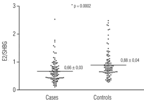
in control men. E2/SHBG relation was significantly different between groups: 0.66 ± 0.03 for
osteoporo-tic versus 0.88 ± 0.04 for normal BMD control (p =
0.0002) (Figure 3) even after excluding hypogonadal men (p = 0.0003).
64,52 ± 3,5 52,72 ± 1,9 * p = 0.0045
Cases
Cases
Controls
Controls 400
300
200
100
0
Mean SHBG
E2/SHBG
(nmol/L)
Figure 3. Estradiol/SHBG values in osteoporotic men (Case) compared to men with normal bone mineral density (Control).
* p = 0.0002
0,66 ± 0,03
0,88 ± 0,04
3
2
1
0
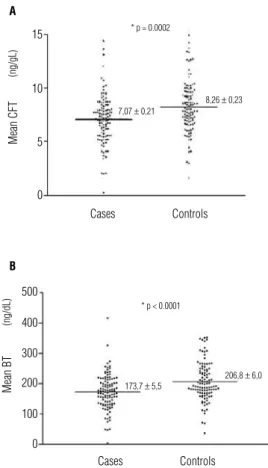
CFT, BT and SHBG were also significantly different between groups: mean CFT was 7.07 ± 0.21 in oste-oporotic versus 8.26 ± 0.23 ng/dL in normal BMD
men (p = 0.0002); mean BT values were 173.7 ± 5.5
versus 206.8 ± 6.0 ng/dL (p < 0.0001), respectively
(Figures 4A and B). Even after excluding hypogona-dal men from both groups, osteoporotic men remained with significantly lower CFT (p = 0.015) and BT (p = 0.007) values and higher SGHB levels (p = 0.02) com-pared with normal BMD men.
Subjects who had at the first dosage CFT values < 7 ng/dL or BT < 180 ng/dL or SHBG > 55 nmol/L had a relative risk for osteoporosis of 2.5 (1.36-4.71; p = 0.015); 2.7 (1.52-5.0; p = 0.0003) and 1.91 (1.07-3.41; p = 0.019), respectively (Table 1).
DISCUSSION
Copyright
© ABE&M todos os dir
eitos r
eser
vados.
However, TT may be a fracture risk marker. In the Dubbo Osteoporosis Epidemiology Study, 609 Austra-lian men > 60 years were followed for 5.8 years, after baseline serum TT, E2 and SHBG levels assessment. Fracture risk was significantly increased in men with re-duced TT and E2 levels. After further adjustment for major risk factors, lower testosterone was still associa-ted with increased fracture risk, particularly with hip and nonvertebral fractures (20). An explanation for this divergence can be found in a recent publication from MrOs Study, in which 1,245 Swedish community-dwelling men over 65 years had their frailty status as-sessed through criteria such as weakness, slowness, low activity, exhaustion, and shrinking/sarcopenia 4.1 years after enrollment and correlated to sex hormone levels. Low BT levels were independently associated with frail-ty, which can predispose to falls and consequent fractu-res (21). Testosterone action may predominate in mus-cle and E2 in bone, each one complementing the other for normal motor function. MrOS Study was designed to investigate if testosterone levels are associated with BMD and/or prevalent fractures in elderly men (22). A previous publication reported that low E2 levels could predict increased fractures risk independent of testosterone; on the contrary, men with low testostero-ne but normal E2 levels did not show an increased frac-tures risk. In multivariate analysis, free E2 was an inde-pendent fracture predictor in those elderly men (12).
Serum E2 was also shown to correlate better than testosterone in relation to BMD and its decrease in aging men (12). Different authors (12,23) reported that bone loss exceeded formation when E2 levels were 31 pg/mL or below in aging men. Cellular sex steroids action on bone tissue can be exerted through andro-gen and estroandro-gen receptors (ER) (24), which belong to nuclear receptor superfamily. Two estrogen receptors, ERα and ERβ have been cloned and are expressed in bone (25-27). Movérare and cols. (24), in a study with orchidectomized wild-type and ER-inactivated mice treated with 5α-dihydrotestosterone (a nonaromati-zable androgen), 17β E2, or vehicle, concluded that both ERα and androgen receptor (AR) but not ERβ
activation preserved trabecular bone amount. ERα ac-tivation resulted both in preserved thickness and tra-becular number. In contrast, AR activation exclusively preserved trabecular number, whereas thickness was unaffected. 17β E2 effects could not be mediated by AR. They concluded that ERα, but not AR or ERβ
activation resulted in preserved thickness, volumetric density and mechanical strength of cortical bone (24). Figure 4. Testosterone values in osteoporotic men (Case) and in men with normal
bone mineral density (Control): (A) CFT and (B) BT.
hip fractures (16). The bone loss mechanism in andro-gen-deficient men is similar to that induced by gonadal insufficiency in women: an imbalance in favor of bone resorption induces net bone loss, especially at cancellous bone sites with large remodeling surfaces (15).
We found no difference in TT levels between oste-oporotic and normal bone density men, which is con-sistent with other studies in which plasma TT levels were not related to BMD in different sites (17). In the MINOS study (18), 760 men, 40-85 years old, were analyzed to correlate testosterone and E2 levels with estimates of bone mechanical properties derived from areal BMD measured by DXA and TT was not asso-ciated with any bone variable. Amin and cols. (19), in a study with 405 elderly men from the Framingham cohort (mean age 75.7 years, range 68 to 96 years), found that 71 (17.5%) were hypogonadal and that BMD at any site did not differ in hypogonadal compa-red with eugonadal men. Hypogonadism diagnosis was based in TT levels which may be misleading, as men with “normal” TT may have lower FT and BT. Thus, TT is a poor marker for aging male gonadal function (14), as well as for osteoporosis risk.
7,07 ± 0,21
8,26 ± 0,23 * p = 0.0002
Cases Controls
15 A
B 10
5
0
Mean CFT
(ng/gL)
* p < 0.0001
173,7 ± 5,5
206,8 ± 6,0
Cases Controls
Mean BT
(ng/dL)
500
400
300
200
100
Copyright
© ABE&M todos os dir
eitos r
eser
vados.
Bioavailable E2, i.e., E2 not bound to SHBG, has been shown to decrease with age and not to be influen-ced by the body mass index (BMI), such as TT or total E2. To measure bioavailable E2, serum has to be equi-librated with tritiated hormones, and then SHBG and SHBG-bound steroids must be precipitated by adding an equal volume of a saturated aqueous solution of am-monium sulfate. Following the precipitation, the sam-ple has to be centrifuged and the tritiated hormone in the supernatant (not SHBG-bound) is quantified and related to the total amount of tritiated hormone. The percentage is applied to provide bioavailable E2 con-centrations (28). As tritiated E2 is not easily available, performing this measurement is not a usual practice. E2/SHBG relation could retrieve similar results, wi-thin normal laboratorial procedure. We found low E2/ SHBG relation to be highly associated to osteoporosis, as many other authors. Gennari and cols. (23), in a stu-dy of 200 elderly men, observed that those with total and bioavailable E2 below the median showed higher rates of bone loss at the lumbar spine and femoral neck. Another epidemiological study (29), found an associa-tion between higher levels of total and bioavailable E2 and lower fracture prevalence in older men. Estrogens in elderly men originate mainly from androgens peri-pheral conversion by aromatase (12), whose expression and activity can vary widely among subjects. This varia-tion can be genetically determined (30). Eriksson and cols. (31) demonstrated that CYP19 gene single nucle-otide polymorphism (SNP) rs2470152, which codes for aromatase, is strongly associated with serum E2 and estrone levels in men. Men with aromatase deficiency, who have normal androgen but undetectable estrogen levels, have low bone mass and areal density and res-pond to estrogen therapy with significant BMD increase (7,32,33).
In this study, CFT and BT were significantly lower and SHBG significantly higher in osteoporotic men compared to those with normal BMD. MrOs Study also showed that FT levels below the median were positive independent predictors of lower BMD and more prevalent fractures in elderly men (22). A study of 3,014 Swedish men also established a stronger frac-ture risk for free than for total androgen and estrogen levels (34): older men with high serum SHBG had an increased risk of fractures, with a hazard ratio of 1.32 per 1 SD increase. In a study of Lormeau and cols. (35) with 105 subjects, 65 of them suffering from idiopa-thic or secondary osteoporosis (mean age 53.9 years) and 40 age-matched control, SHBG were significantly
higher in osteoporotic patients than control, even after secondary osteoporosis (due to alcoholism or hypogo-nadism) exclusion. Also, SHBG was negatively correla-ted to femoral neck and lumbar spine BMD, to serum C-telopeptide of type I collagen (CTX), a bone remo-deling marker used to evaluate resorption (35). SHBG was significantly associated with fractures; the odds ra-tio of having a fracture was 2.04 (95%CI: 1.2-3.4; p < 0.01) for each increase of 1 SD in the patient’s SHBG level (35). SHBG levels are increased in a number of conditions like hyperthyroidism, severe malnutrition, hepatic cirrhosis or aging and may be, more importan-tly, genetically determined. Legrand and cols. (36) also refer a positive correlation between bone remodeling markers CTX, deoxypyridinoline (D-Pyr), and bone-specific alkaline phosphatase (bSAP) and serum SHBG (36). They suggest that the higher the SHBG, the lo-wer the BT and E2 levels, and the higher are bone re-modeling and resorption. Cummings and cols. (37), in a prospective study with postmenopausal women aged 65 years or older, observed that SHBG ≥ 34.7 nmol/L was associated to a relative risk of 2.0 for hip fractures and 2.3 for vertebral fractures. In this study, SHBG > 55 nmol/L was associated with a higher male osteopo-rosis risk.
In conclusion, TT levels were similar between oste-oporotic and normal BMD aging men. Total E2, E2/ SHBG, CFT, BT were significantly lower and SHBG were significantly higher in osteoporotic compared to normal BMD men. Cutoff values associated with hi-gher osteoporosis risk in men over 50 years old were: E2 < 37 ng/dL; SHBG > 55 nmol/L; CFT < 7 ng/dL; BT < 180 ng/dL.
In an aging man’s laboratorial exam, one or more of these values may represent an indication for osteoporo-sis screening. Limitations of the present study include the existence of possible confounders for plasma sex steroids values, such as obesity, that were not studied. Nevertheless, these confounders would not impair the predictive levels described.
Acknowledgements: Diagnósticos da América Laboratory kindly performed testosterone and SHBG measurements, while Insti-tuto Fernandes Figueira (IFF) performed E2 and albumin mea-surements. We thank Martha Suárez-Mutis, Epidemiology Doc-tor of Instituto Oswaldo Cruz (IOC), Fundação Oswald Cruz (Fiocruz), for the statistical analysis, and Sergio Koifman, chief professor of the Postgraduate Program of Public Health and En-vironment of the Escola Nacional de Saúde Pública of Fiocruz for the suggestions during the study.
Copyright
© ABE&M todos os dir
eitos r
eser
vados.
REFERENCES
1. Kelly JJ, Moses AM. Osteoporosis in men: the role of testoster-one and other sex-related factors. Curr Opin Endocrinol Diabetes. 2005;12(6):452-8.
2. Bilezikian JP. Osteoporosis in men. J Clin Endocrinol Metab. 1999;84(10):3431-4.
3. Rochira V, Balestrieri A, Madeo B, Zirilli L, Granata ARM, Ca-rani C. Osteoporosis and male age-related hypogonadism: role of sex steroids on bone (patho)physiology. Eur J Endocrinol. 2006;154(2):175-85.
4. Briot K, Cortet B, Trémollières F, Sutter B, Thomas T, Roux C, Audran M; Comité Scientifique du GRIO. Male osteoporosis: Diagnosis and fracture risk evaluation. Joint Bone Spine. 2009;76(2):129-33. 5. Smith EP, Boyd J, Frank GR, Takahashi H, Cohen RM, Specker B,
et al. Estrogen resistance caused by a mutation in the estrogen-receptor gene in a man. N Engl J Med. 1994;331(16):1056-61. 6. Rochira V, Balestrieri A, Madeo B, Spaggiari A, Carani C.
Congeni-tal estrogen deficiency in men: a new syndrome with different phenotypes; clinical and therapeutic implications in men. Mol Cell Endocrinol. 2002:193(1-2);19-28.
7. Carani C, Qin K, Simoni M, Faustini-Fustini M, Serpente S, Boyd J, et al. Effect of testosterone and estradiol in a man with aroma-tase deficiency. N Engl J Med. 1997:337(2);91-5.
8. Morishima A, Grumbach MM, Simpson ER, Fisher C, Qin K. Aro-matase deficiency in male and female siblings caused by a novel mutation and the physiological role of estrogens. J Clin Endocri-nol Metab. 1995:80(12);3689-98.
9. Maffei L, Murata Y, Rochira V, Tubert G, Aranda C, Vazquez M, et al. Dysmetabolic syndrome in a man with a novel mutation of the aromatase gene: effects of testosterone, alendronate, and estra-diol treatment. J Clin Endocrinol Metab. 2004;89(1):61-70. 10. Herrmann BL, Saller B, Janssen OE, Gocke P, Bockisch A, Sperling
H, et al. Impact of estrogen replacement therapy in a male with congenital aromatase deficiency caused by a novel mutation in the CYP19 gene. J Clin Endocrinol Metab. 2002;87(12):5476-84. 11. Couse JF, Korach KS. Estrogen receptor null mice: what have
we learned and where will they lead us? Endocrinology. 1999;20(3):358-417.
12. Ohlsson C, Vandenput L. The role of estrogens for male bone health. Eur J Endocrinol. 2009;160(6):883-89.
13. Clapauch R, Braga DJC, Marinheiro LP, Buksman S, Schrank Y. Risk of late-onset hypogonadism (andropause) in Brazilian men over 50 years of age with osteoporosis: usefulness of screening questionnaires. Arq Bras Endocrinol Metab. 2008;52(9):1439-47. 14. Clapauch R, Carmo AM, Marinheiro L, Buksman S, Pessoa I.
Labo-ratory diagnosis of late-onset male hypogonadism andropause. Arq Bras Endocrinol Metab. 2008;52(9):1430-8.
15. Vanderschueren D, Vandenput L, Boonen S, Lindberg MK, Bouillon R, Ohlsson C. Androgens and bone. Endocr Rev. 2004;25(3):389-425. 16. Tuck SP, Francis RM. Testosterone, bone and osteoporosis. Front
Horm Res. 2009; 37:123-32.
17. Ronde W, Pols HAP, Leeuwen JPTM, Jong FH. The importance of oestrogens in males. Clin Endocrinol. 2003;58(5):529-42. 18. Szulc P, Uusi-Rasi K, Claustrat B, Marchand F, Beck TJ, Delmas
PD. Role of sex steroids in the regulation of bone morphology in men. The MINOS study. Osteoporos Int. 2004;15(11):909-17. 19. Amin S, Zhang Y, Sawin CT, Evans SR, Hannan MT, Kiel DP, et
al. Association of hypogonadism and estradiol levels with bone mineral density in elderly men from the Framingham study. Ann Intern Med. 2000;133(12):951-63.
20. Meier C, Nguyen TV, Handelsman DJ, Schindler C, Kushnir MM, Rockwood AL, et al. Endogenous sex hormones and incident
fracture risk in older men: the Dubbo Osteoporosis Epidemiology Study. Arch Intern Med. 2008;168(1):47-54.
21. Cawthon PM, Ensrud KE, Laughlin GA, Cauley JA, Dam TT, Bar-rett-Connor E, et al. Sex hormones and frailty in older men: the osteoporotic fractures in men (MrOS) study. J Clin Endocrinol Metab. 2009 Oct;94(10):3806-15.
22. Mellström D, Johnell O, Ljunggren O, Eriksson AL, Lorentzon M, Mallmin H, et al. Free testosterone is an independent predictor of BMD and prevalent fractures in elderly men: MrOS Sweden. J Bone Miner Res. 2006; 21(4):529-35.
23. Gennari L, Merlotti D, Martini G, Gonnelli S, Franci B, Campagna S, et al. Longitudinal Association between Sex Hormone Levels, Bone Loss, and Bone Turnover in Elderly Men. J Clin Endocrinol Metab. 2003;88(11):5327-33.
24. Movérare S, Venken K, Eriksson AL, Andersson N, Skrtic S, Wergedal J, et al. Differential effects on bone of estrogen recep-tor alpha and androgen receprecep-tor activation in orchidectomized adult male mice. Proc Natl Acad Sci U S A. 2003;100(23):13573-8. 25. Vidal O, Kindblom LG, Ohlsson C. Expression and localization of
estrogen receptor-beta in murine and human bone. J Bone Miner Res. 1999;14(6):923-9.
26. Arts J, Kuiper GGJM, Janssen JMMF, Gustafsson JA, Löwik CWGM, Pols HAP, et al. Differential expression of estrogen recep-tors α and β mRNA during differentiation of human osteoblast SV-HFO cells. Endocrinology. 1997;138(11):5067-70.
27. Onoe Y, Miyaura C, Ohta H, Nozawa S, Suda T. Expression of estro-gen receptor β in rat bone. Endocrinology. 1997;138 (10):4509-12. 28. Dolomie-Fagour L, Gatta B, Nguyen TD, Corcuff JB. Bioavailable
estradiol in man: relationship with age and testosterone. Clin Chim Acta. 2008;398(1-2):145-47.
29. Barrett-Connor E, Mueller JE, Mühlen DG, Laughlin GA, Sch-neider DL, Sartoris DJ. Low levels of estradiol are associated with vertebral fractures in older men, but not women: The Rancho Ber-nardo Study. J Clin Endocrinol Metab. 2000;85(1):19-23. 30. Gennari L, Masi L, Merlotti D, Picariello L, Falchetti A, Tanini A, et
al. Polymorphic CYP19 TTTA repeat influences aromatase activity and estrogen levels in elderly men: effects on bone metabolism. J Clin Endocrinol Metab. 2004;89(6):2083-10.
31. Eriksson AL, Lorentzon M, Vandenput L, Labrie F, Lindersson M, Syvänen AC, et al. Genetic variations in sex steroid-related genes as predictors of serum estrogen levels in men. J Clin Endocrinol Metab. 2009;94(3):1033-41.
32. Vanderschueren D, Venken K, Ophoff J, Bouillon R, Boonen S. Clinical review: sex steroids and the periosteum – reconsidering the roles of androgens and estrogens in periosteal expansion. J Clin Endocrinol Metab. 2006;91(2):378-82.
33. Rochira V, Faustini-Fustini M, Balestrieri A, Carani C. Estrogen replacement therapy in a man with congenital aromatase defi-ciency: effects of different doses of transdermal estradiol on bone mineral density and hormonal parameters. J Clin Endocrinol Metab. 2000;85(5):1841-5.
34. Mellström D, Vandenput L, Mallmin H, Holmberg AH, Lorentzon M, Odén A, et al. Older men with low serum estradiol and high serum SHBG have an increased risk of fractures. J Bone Miner Res. 2008;23(10):1552-60.
35. Lormeau C, Soudan B, d’Herbomez M, Pigny P, Duquesnoy B, Cortet B. Sex hormone-binding globulin, estradiol, and bone turnover markers in male osteoporosis. Bone. 2004;34(6):933-9. 36. Legrand E, Hedde C, Gallois Y, Degasne I, Boux de Casson F,
Ma-thieu E, et al. Osteoporosis in men: a potential role for the sex hormone binding globulin. Bone. 2001;29(1):90-5.