In
fl
uence of expiratory positive airway pressure on
cardiac autonomic modulation at rest and in
submaximal exercise in COPD patients
C. da L. Goulart
1, E.A. San Martin
1, K.M.K. Mansour
1, P.B. Schneiders
1and A.L.G. da Silva
2,3 1Iniciac¸ão Científica, Curso de Fisioterapia, Universidade de Santa Cruz do Sul, Santa Cruz do Sul, RS, Brasil
2
Curso de Fisioterapia, Departamento de Educac¸ão Física e Saúde, Universidade de Santa Cruz do Sul, Santa Cruz do Sul, RS, Brasil
3
Programa de Reabilitac¸ão Pulmonar, Hospital Santa Cruz, Santa Cruz do Sul, RS, Brasil
Abstract
The aim of this study was to evaluate the effect of expiratory positive airway pressure (EPAP) on heart rate variability (HRV) indices at rest and during 6-min walk test (6MWT) in chronic obstructive pulmonary disease (COPD) patients. Fifteen moderate to severe COPD patients were randomized and evaluated with and without (Non-EPAP) a 5 cmH2O EPAP device. Respiratory rate (RR) was collected at rest (5 min), during the 6MWT (5 min), and at recovery (5 min). Indices of HRV were computed in the time domain, in the frequency domain, and nonlinear analysis. For EPAP and Non-EPAP during the 6MWT, we found an increased mean heart rate (HR) (P=0.001; P=0.001) while mean RR (P=0.001; P=0.015) and RR tri index decreased (P=0.006; P=0.028). Peripheral oxygen saturation (P=0.019) increased at rest only in the EPAP group. In EPAP, correlations were found between forced expiratory volume in 1 s (FEV1) and low frequency (LF) sympathetic tonus (P=0.05; r=–0.49), FEV1and high frequency (HF) parasympathetic tonus at rest (P=0.05; r=0.49), lactate at rest and LF during the 6MWT (P=0.02; r=–0.57), and lactate at rest and HF during 6MWT (P=0.02; r=0.56). Through a linear regression model, we found that lactate at rest explained 27% of the alterations of LF during 6MWT. The use of 5 cmH2O EPAP improved autonomic cardiac modulation and its complexity at rest in COPD patients. Although it did not influence the performance of the 6MWT, the EPAP device caused alterations in resting lactate concentration with an effect on sympatho-vagal control during the test.
Key words: Autonomic nervous system; HRV; COPD; Positive-pressure respiration; Exercise test
Introduction
The control of the autonomic nervous system (ANS) of patients with chronic obstructive pulmonary disease (COPD) is influenced by the sympathetic and parasympathetic systems, peripheral oxygen saturation (SpO2), pulmonary
stretch baroreceptors, and cardiac and pulmonary reflexes (1–3). In COPD patients the sympatho-vagal imbalance
is caused by alterations in the ANS (4), as well as by the bronchoconstriction mechanism, hypoxia, hypercapnia, and systemic inflammation (3,5,6), resulting in a higher risk of morbimortality (7).
Noninvasive ventilation (NIV) methods are frequently used in this population with the primary goal of increas-ing ventilation and SpO2, decreasing respiratory
complica-tions and dyspnea symptoms, reducing respiratory muscle load and the intrathoracic pressure during rest and phys-ical exercise, and incrementing their physphys-ical and func-tional performance. Scientific evidence suggests that the use of NIV can modulate autonomic control of heart rate (HR) (1,2,8).
Studies using bilevel positive pressure airway and continuous positive airway pressure (CPAP) have demon-strated that this method is effective for improving pulmo-nary ventilation, neural control of HR, and sympatho-vagal balance in COPD patients (2,9,10). Nicolini et al. (11) evaluated the effect of expiratory positive airway pressure (EPAP) on the 6-min walk test (6MWT) in COPD patients and found a beneficial effect of EPAP on their functional capacity. Müller et al. (12) highlighted that CPAP and EPAP reduce static hyperinflation in COPD patients, and that EPAP demonstrates equivalent effects to CPAP on static hyperinflation indirectly measured by inspiratory capacity and maximal inspiratory pressure (MIP). EPAP application relieves dynamic airway compression during expiration, reducing expiratory airflow limitation and thereby increasing inspiratory capacity in COPD patients (13,14).
No study has evaluated the effects of EPAP on the autonomic cardiac indices through the analyses of heart rate variability (HRV). It is known that linear and nonlinear
Correspondence: A.L.G. da Silva:<[email protected]>
Received October 26, 2017 | Accepted January 16, 2018
HRV indices are widely accepted as important tools to determine cardiac autonomic changes (2–9). Linear indices
of time domain such as mean respiratory rate (RR) interval (mean RR), RR interval standard deviation (STD RR), mean HR, and STD HR reflect HRfluctuations. Variables such as square root of the mean squared differences of successive RR intervals (RMSSD) and the HRV triangular index (RR tri index) reflect global HRV. In the frequency domain, low frequency (LF) and high frequency (HF) indexes indicate sympathetic and parasympathetic activity respectively, and LF/HF ratio indicates sympatho-vagal relation. The analysis of nonlinear components [approx-imate entropy (ApEn) and Shannon entropy] indicates ANS complexity (15). Therefore, our aim was to evaluate the effect of EPAP on the HRV indices at rest and during 6MWT in COPD patients. We hypothesize that a 5 cmH2O
EPAP application would provide a positive effect on auto-nomic modulation in COPD patients both at rest as during submaximal exercise.
Material and Methods
Study design
We performed a sectional and randomized cross-over study within the Programa de Reabilitac¸ão Pulmonar, Hospital Santa Cruz, Santa Cruz do Sul, RS, Brazil. All patients were formally invited to participate in the study by the responsible researcher and all volunteers signed an informed consent statement prior to participation. The study was approved by the Research Ethics Committee of the Universidade de Santa Cruz do Sul (protocol No. 1.514.705).
Subjects
Thirty patients with a clinical diagnosis of COPD by a pulmonary function test (forced expiratory volume in 1 s (FEV1)/forced vital capacity (FVC) ratio of 0.7; FEV160%
of predicted) with good cognitive function and without previous COPD exacerbation (15 days before the study) were included in the study. Patients with muscle-skeletal disorders or neurological conditions affecting the locomo-tor system in such a way that would preclude participation in the protocol, a clinical diagnosis of lung cancer, current alcoholism, complex cardiac arrhythmias, uncontrolled metabolic disease such as diabetes, thyroid changes, and systemic arterial hypertension (with or without the use of beta-blockers), or electrocardiogram alterations were excluded from the study (n=15; Figure 1).
Measurements
Patients were evaluated in a laboratory with 22°C and relative humidity between 50 and 60%. They were instructed to avoid stimulants and alcoholic drinks and not to perform exhausting physical exercise the day before the test; they were also instructed not to smoke or use bronchodilators for 6 h before the test. Patients underwent
a clinical evaluation to record gender, age, body mass index, smoking history, and COPD status by pulmonary function. The HRV signal was recorded at rest (5 min), during 6MWT (5 min) with and without EPAP, and during post-test recovery period (5 min). The order of the 6MWT with and without EPAP was determined by simple random-ization (draw of an envelope) with a 30-min resting period between the tests (Figure 1).
Pulmonary function
Pulmonary function was assessed using a digital spirom-eter (Microloops, MK8, Care Fusion, Germany), which provided measures of the FEV1. Spirometry was performed
according to the recommendations of the American Thoracic Society (16) and the results were analyzed according to the values predicted by Pereira et al. (17). The classification of severity of airflow limitation in COPD was performed according to the Global Initiative for Chronic Obstructive Lung Disease (GOLD) recommenda-tions and patients were classified as moderate (GOLD II), severe (GOLD III), or very severe (GOLD IV) (18).
Respiratory muscle strength (RMS)
RMS was assessed using a digital manometer (MDIs,
MVD300, Brazil), which provided measures of the MIP
and the maximum expiratory pressure (MEP). The assess-ment was performed according to the recommendations for the Brazilian population. The values were then compared with those described in the literature and reported as percentage of predicted values (19). The inspiratory muscle weakness was determined by an MIPo60 cmH2O (20).
Submaximal exercise capacity - 6MWT
The 6MWT was carried out according to the guidelines of the ATS (21), in order to measure the distance walked during the 6-min test on aflat 30-m corridor. The subjects were instructed to walk as far as possible, at a constant speed. The 6MWT was developed based on the study by Holland et al. (22). The percentage of the predicted distance walked was calculated considering gender, age, height, and weight of each patient based on the equation of Enright (23). SpO2, HR, blood pressure, perceived
exer-tion (using the Borg Scale), and distance walked (meters) were recorded at the beginning and end of every test as previously indicated.
EPAP device
The EPAP device consists of a Spring Loadedsvalve
(Kit EPAP) connected to a 20-mm inner diameter and 100-mm tube and a mouthpiece (11). The positive expira-tory pressure (PEP) set at 5 cmH2O, following previous
findings on application of 5 cmH2O PEP, was safe and
showed lower autonomic imbalance in COPD (2,11). Familiarization procedures were performed with the equip-ment and experiequip-mental protocols so patients could learn how to correctly perform the 6MWT with EPAP before the actual testing.
Lactate
Blood lactate was obtained at rest, during 6MWT (test peak), and during post-test recovery period (third minute after the end of the test) with and without EPAP. The measurement of blood lactate concentration was made by collecting a drop of capillary blood on a validated portable lactometer (Accutrend Plus) (24).
HRV
HR and RR intervals (iR-R) were recorded using a telemetric cardiac monitor (Polars S810i, Finland). An elastic band (Polar T31 transmitter) was placed around the patient’s thorax at the level of the lower third of the sternum, while the patient was in a sitting position; signals were continuously transmitted to the receiving unit by electromagnetic field. Recorded data were then trans-ferred to Kubios HRVs analysis software (version 2.2,
Finland) for subsequent analysis.
The signal processing was carried out as follows: at rest (5 min), during the 6MWT (we discarded the initial 60 s of data and selected the most stable signal, correspond-ing to the latter portion of the test), and post-exercise recovery (5 min) with and without EPAP. The HRV signal
collected in the time domain provided mean RR, STD RR, mean HR, STD HR, RMSSD, and RR tri index, in the fre-quency domain were LF, HF, and LF/HF ratio, and nonlinear analysis provided the ApEn and Shannon entropy indices.
Statistical analysis
Data were analyzed using the Sigmaplots statistical
package (version 11.0, Systat Software Inc., USA). Data were tested for normality through the Shapiro–Wilk test
and presented descriptively as mean and SD (parametric) or as median and minimum and maximum interval (non-parametric). Analysis of variance (ANOVA) for multiple comparisons with Tukey’spost hoc test was performed. Pearson correlation analysis was performed to investigate the correlations between variables. The analyses between the groups were performed by a Student’st-test. A linear regression model was used to determine the effect of the clinical variables on HRV parameters. Residuals were evaluated under the assumptions of normality, constant variance, and independence. Pp0.05 was considered significant.
Results
Clinical characteristics of the patients included in the study are shown in Table 1. We observed a greater prev-alence of patients with severe COPD in stage III and IV (60%), male gender, obesity, and only 33% with inspira-tory muscle weakness.
The mean HR increased significantly during the 6MWT in both groups (P=0.001), while the mean RR (P=0.001; P=0.015) and RR tri index decreased (P=0.006; P=0.028) demonstrating an increase of the HR and reduction of global HRV compared to rest (Table 2). In the EPAP group, we found a significant result in Shannon entropy at rest (P=0.016), showing a better ANS complexity com-pared to Non-EPAP at rest.
EPAP showed an improvement of SpO2at rest (P=
0.019; Figure 2A). However, no significant difference was found in the distance walked in meters in 6MWT between groups (P=0.481; Figure 2B).
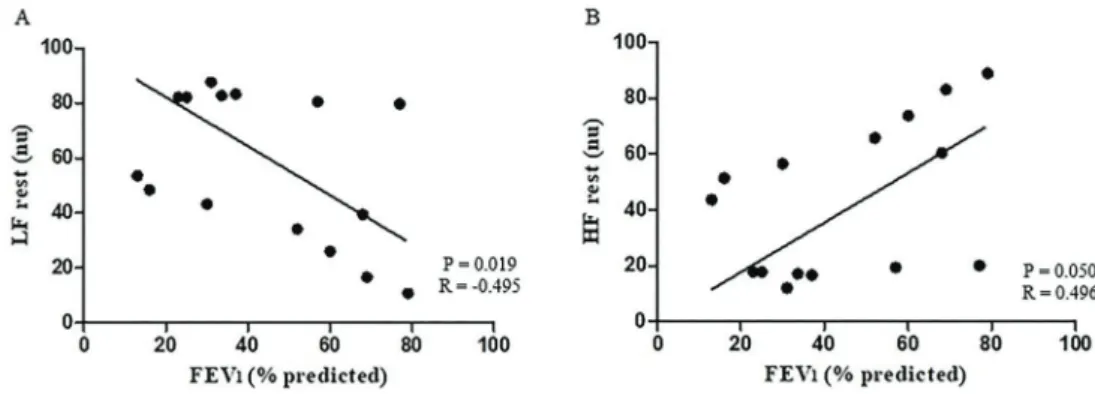
Moderate correlations were found between airway obstruction and the HRV parameters at rest with EPAP (Figure 3), demonstrating the influence and importance of airflow obstruction over the sympathetic and parasympa-thetic modulation.
Thesefindings were confirmed through a linear regres-sion model in which lactate at rest explained 27% of the alterations in LF during 6MWT (Table 3).
Discussion
Based on our study, we highlight the followingfindings: a) EPAP had a beneficial effect on SpO2 and in ANS
complexity, both during rest and compared to Non-EPAP; b) airway obstruction was associated with increased vagal modulation at rest with EPAP; c) resting blood lactate concentration was solely responsible for changes in sym-pathetic modulation during the 6MWT in COPD patients with EPAP. Thisfinding can be attributed to higher SpO2
and better response of muscle metabolism.
The effects of EPAP on HRV complexity during rest in COPD patients are unprecedented and cannot be com-pared to other scientific studies. One of the mainfindings of our study with 5 cmH2O EPAP was the increase of
SpO2, different from Müller et al. (12) study, in which they
applied 10 cmH2O EPAP and found no improvement
in SpO2. The main effect of the EPAP device consists in
increasing expiratory flow and decreasing pulmonary hyperinflation (11). EPAP demonstrated equivalent effects
to CPAP on static hyperinflation, improvements in inspira-tory muscle strength, and inspirainspira-tory capacity (12). The device has been used in COPD patients to assist elimina-tion of pulmonary secreelimina-tions and induce a reducelimina-tion in minute ventilation, respiratory rate, and physiological dead space (25,26). Reis et al. (2) highlights that treatment with a 5 cmH2O CPAP in COPD patients is safer and produces
a better response in ventilatory parameters and autonomic balance than when compared with a 10 cmH2O pressure.
An interestingfinding of our study was the association between VEF1 and parasympathetic and sympathetic
modulation, as observed by HF and LF indices during rest with EPAP. Our findings contradict previous studies on different autonomic tests like postural changing, iso-metric contraction, and during dynamic physical exercise (4,7,27,28). These findings are consistent with the increased parasympathetic cardiac modulation intimately associated with airway obstruction from the bronchocon-striction of these patients (29). It is important to note that none of the studies used the EPAP device. We speculate that the beneficial effects of EPAP on bronchoconstriction reduction altered the autonomic cardiac modulation with lower parasympathetic modulation in patients with higher airway obstruction.
Table 1.Clinical characteristics of chronic obstructive pulmonary disease (COPD) patients.
Variables COPD (n=15)
Age (years) 62.6±7.8
Gender
Male n (%) 12 (80.0)
BMI (kg/m2) 26.5±6.3
BMI classification, n (%)
Underweight 4 (26.7)
Eutrophic 3 (20.0)
Obese 8 (53.3)
Pulmonary volumes
FEV1(L/s) 1.1 (0.7–1.6)
FEV1(% predicted) 37.0 (25.0–68.0) Staging (GOLD), n (%)
Stage II, 6 (40.0)
Stage III 4 (26.7)
Stage IV 5 (33.3)
Respiratory muscle strength
MIP (cmH2O) 73.0±28.8
MIP (% predicted) 72.6±28.6
MEP (cmH2O) 115.0±40.2
MEP (% predicted) 105.5±33.6
Inspiratory muscle weakness (MIPo60 cmH2O), n (%)
Yes 5 (33.3)
No 10 (66.7)
In our study, during the 6MWT with EPAP, the COPD patients presented an increase in SBP, increased HR and a reduction on global HRV, and we observed that these indices went back to normal values during recovery. A single study demonstrated that a reduction on HR occurred in moderate to severe COPD patients after the 6MWT with EPAP (11). The authors did not evaluate the autonomic cardiac modulation through HRV indices analysis, even though these indices are important markers of cardiac alterations (30).
Our study is the first to evaluate the association between blood lactate concentration at rest and sympathetic
and vagal modulation during the 6MWT with EPAP, demonstrating that low levels of lactate at rest directly influence the autonomic sympathetic modulation during the 6MWT. This result was confirmed through linear regression. Lactate at rest and during low intensity exercise presents lower concentration, though it increases rapidly during exercise that requires an aerobic response (31), a result only found during 6MWT in the EPAP in our study. In COPD patients, the measurement of capillary blood lactate associated with the 6MWT can estimate muscular metabolic profile because its precocious increase before effort seems to be associated with muscular
Table 2.Comparisons within and between groups for all outcome measures.
Variables EPAP Non–EPAP Recovery Rest 6MWT Recovery
Rest 6MWT
SBP (mmHg) 116.6±11.1 137.3±21.2* 129.3±17.5 115.3±12.4 129.3±21.2 122.0±16.1 DBP (mmHg) 80.6±8.8 86.0±7.3 82.0±10.1 79.3±9.6 84.0±7.3 83.3±9.7 Lactate (mmol/L) 2.9±1.1 3.2±1.8 2.5±1.2 2.7±0.9 2.7±1.1 2.7±1.1 Borg–lower limb 7.3±2.0 9.5±3.1 8.1±2.3 7.3±1.7 9.0±3.0 7.4±2.6 Mean RR (ms) 729.3±123.8 556.1±109.1* 609.5±137.8 755.5±144.6 563.1±99.5* 708.4±130.0** STD RR (ms) 11.2 (9.1–24.0) 6.8 (3.5–11.1)* 10.9 (7.5–17.0) 1.7 (1.3–2.4) 1.5 (1.0–2.1) 1.9 (1.4–2.2) Mean HR (1/min) 84.4±13.4 111.9±22.3* 89.9±16.1** 82.1±15.3 109.8±20.0* 86.7±14.1** STD HR (1/min) 1.7 (1.3–2.6) 1.4 (1.1–1.9) 1.8 (1.3–2.2) 1.7 (1.3–2.4) 1.5 (1.0–2.1) 1.9 (1.4–2.2) RMSSD (ms) 16.6 (7.4–24.5) 6.3 (4.5–16.1) 12.5 (6.9–17.3) 18.2 (8.2–25.4) 8.6 (4.5–17.5) 10.3 (8.4–14.9) RR tri index 4.3 (2.8–6.5) 2.3 (1.6–3.2)* 3.5 (2.7–5.2) 4.0 (3.2–5.6) 2.5 (1.7–3.2)* 3.8 (2.7–4.9) LF (nu) 53.6 (35.5–82.1) 56.8 (33.8–70.1) 56.5 (48.9–75.7) 56.2 (31.8–76.2) 65.7 (40.9–75.3) 73.0 (51.2–83.8) HF (nu) 43.6 (17.8–64.4) 43.1 (29.4–65.5) 43.4 (23.6–50.9) 43.4 (23.5–68.1) 32.9 (24.3–57.9) 26.6 (16.1–48.4) LF/HF 1.2 (0.5–4.6) 1.3 (0.5–2.3) 1.3 (0.9–3.2) 1.2 (0.4–3.2) 2.0 (0.7–3.1) 2.7 (1.1–5.3)
ApEn 0.9±0.1 1.0±0 1.0±0 0.9±0 1.0±0.1 0.9±0.1
Shannon entropy 3.0±0.3 2.6±0.2 2.9±0.3 2.7±0.2# 2.8±0.3 2.8±0.2
SBP: systolic blood pressure; DBP: diastolic blood pressure; RR: respiratory rate interval; STD RR: RR interval standard deviation; HR: heart rate; STD HR: heart rate standard deviation; RMSS D: square root of the mean squared differences of successive RR intervals; RR tri index: HR V triangular index; LF: low frequency; HF: high frequency; nu: normalized units; LF/HF: ratio between LF power and HF power. * Po0.05, restvs6MWT. **Po0.05, 6MWTvsrecovery.#Po0.050 non-EPAP at restvsEPAP at rest. Comparisons within groups were done with ANOVA and Tukey’spost hocanalysis and between groups with Student’st-test.
Figure 3.Relationship between lung function and heart rate variability index at rest.A, Negative correlation between LF (nu) and FEV1 (% of predicted).B, Positive correlation between HF (nu) and FEV1(% of predicted). The Pearson correlation analysis was used for statistical analyses. FEV1: forced expiratory volume in 1 s; LF: low frequency; HF: high frequency; nu: normalized units.
metabolic alterations, as loss of the oxidative activity of the skeletal muscles is observed (32).
It has been described in the scientific literature that during discontinuous resistance exercise in an elderly population, the integrative responses between muscular metabolism and autonomic cardiac control are more evident at aerobic metabolism to anaerobic transition during exercise (33), and these factors, as aerobic capacity, may have an influence on cardiac autonomic control (34,35). In coronary artery disease and chronic heart failure patients, HRV is a feasible tool in clinical practice to determine anaerobic threshold and it can be safe and appropriate to determine
exercise intensity (36,37). Although having a different population and exercise protocol than our study, those findings may support the association between lactate at rest with autonomic modulation during 6MWT with EPAP in COPD patients.
The limitations that deserve to be listed are the rigid exclusion criteria and the difficulty in screening patients with 95% pure sinus beat. This study has a great signif-icance and importance for the clinical management of patients with COPD, as the new results on EPAP applica-tion may be used in research and in the clinicalfield, as well as to assess the effects on interventions frequently used in pulmonary rehabilitation.
Finally, the use of 5 cmH2O EPAP improved autonomic
cardiac modulation and ANS complexity during rest in COPD patients. Although it did not influence the perfor-mance of the 6MWT, EPAP caused alteration in resting lactate concentration with an effect on sympatho-vagal control during the test.
Acknowledgments
Grants were received from the University of Santa Cruz do Sul–UNISC and Santa Cruz Hospital.
References
1. Pinsky MR. Cardiovascular issues in respiratory care.Chest 2005; 128: 592S–597S, doi: 10.1378/chest.128.5_suppl_ 2.592S.
2. Reis MS, Sampaio LM, Lacerda D, De Oliveira LV, Pereira GB, Pantoni CB, et al. Acute effects of different levels of continuous positive airway pressure on cardiac autonomic modulation in chronic heart failure and chronic obstructive pulmonary disease.Arch Med Sci2010; 6: 719–727, doi: 10.5114/aoms.2010.17087.
3. Mazzuco A, Medeiros WM, Sperling MPR, de Souza AS, Alencar MC, Arbex FF, et al. Relationship between linear and nonlinear dynamics of heart rate and impairment of lung function in COPD patients.Int J Chron Obstruct Pulmon Dis 2015; 10: 1651–1661, doi: 10.2147/COPD.S81736. 4. Roque AL, Valenti VE, Massetti T, da Silva TD, Monteiro CB,
Oliveira FR, et al. Chronic obstructive pulmonary disease and heart rate variability: a literature update.Int Arch Med 2014; 7: 43, doi: 10.1186/1755-7682-7-43.
5. Borghi-Silva A, Arena R, Castello V, Simões RP, Martins LEB, Catai AM, et al. Aerobic exercise training improves autonomic nervous control in patients with COPD.Respir Med 2009; 103: 1503–1510, doi: 10.1016/j.rmed.2009. 04.015.
6. Chhabra SK, Gupta M, Ramaswamy S, Dash DJ, Bansal V, Deepak KK. Cardiac sympathetic dominance and systemic inflammation in COPD. COPD 2015; 12: 552–559, doi: 10.3109/15412555.2014.974743.
7. Nicolino J, Ramos D, Leite MR, Rodrigues FM, de Alencar Silva BS, Tacao GY, et al. Analysis of autonomic modulation after an acute session of resistance exercise at different intensities in chronic obstructive pulmonary
disease patients. Int J Chron Obstruct Pulmon Dis 2015; 10: 223–229.
8. Highcock MP, Shneerson JM, Smith IE. Increased ventila-tion with NiIPPV does not necessarily improve exercise capacity in COPD. Eur Respir J2003; 22: 100–105, doi: 10.1183/09031936.03.00292203.
9. Borghi-Silva A, Reis MS, Mendes RG, Pantoni CB, Simões RP, Martins LE, et al. Noninvasive ventilation acutely modi-fies heart rate variability in chronic obstructive pulmonary disease patients. Respir Med2008; 102: 1117–1123, doi: 10.1016/j.rmed.2008.03.016.
10. Skyba P, Joppa P, Oroľ̌i’n M, Tka’cova’R. Blood pressure and heart rate variability response to noninvasive ventilation in patients with exacerbations of chronic obstructive pulmo-nary disease.Physiol Res2007; 56: 527e33.
11. Nicolini A, Merliak F, Barlascini C. Use of positive expiratory pressure during six minute walk test: results in patients with moderate to severe chronic obstructive pulmonary disease.Multidiscip Respir Med2013; 8: 19, doi: 10.1186/ 2049-6958-8-19.
12. Müller PT, Christofoletti G, Koch R, Zardetti Nogueira JH, Pereira Patusco LA, Chiappa GR. CPAP and EPAP elicit similar lung deflation in a non-equivalent mode in GOLD 3-4 COPD patients.Clin Respir J2017.
13. Monteiro MB, Berton DC, Moreira MA, Menna-Barreto SS, Teixeira PJ. Effects of expiratory positive airway pressure on dynamic hyperinflation during exercise in patients with COPD. Respir Care2012; 57: 1405–1412, doi: 10.4187/ respcare.01481.
14. Visser FJ, Ramlal S, Dekhuijzen PN, Heijdra YF. Pursed-lips breathing improves inspiratory capacity in chronic obstructive Table 3. Results of linear regression with expiratory positive
airway pressure (EPAP) to determine the influence of lactate at rest on low frequency (LF; nu) during the 6-min walk test.
Variables bcoefficient P
Constant 86.43 o0.001
Lactate at rest (mmol/L) –12.06 0.027
pulmonary disease. Respiration 2011; 81: 372–378, doi: 10.1159/000319036.
15. Task Force of European Society of Cardiology. Heart rate variability. Standards of measurement, physiological inter-pretation, and clinical use. Task Force of the European Society of Cardiology and the North American Society of Pacing and Electrophysiology. Eur Heart J 1996; 17: 354–381, doi: 10.1093/oxfordjournals.eurheartj.a014868. 16. Gibson GJ, Whitelaw W, Siafakas N. Tests of overall
respiratory function. ATS/ERS statement on respiratory muscle testing. Am J Respir Crit Care Med 2002; 166: 518–624, doi: 10.1164/rccm.166.4.518.
17. Pereira CA, Sato T, Rodrigues SC. New reference values for forced spirometry in white adults in Brazil.J Bras Pneumol 2007; 33: 397–406, doi: 10.1590/S1806-371320070004 00008.
18. Global Initiative for Chronic Obstructive Lung Disease. Pocket Guide to COPD Diagnosis, Management and Prevention. Global Initiative for Chronic Obstructive Lung Disease; 2017. Available from: http://www.goldcopd.it/mate riale/2015/GOLD_Pocket_2017.pdf.
19. Neder JA, Andreoni S, Lerario MC, Nery LE. Reference values for lung function tests. II. Maximal respiratory pressures and voluntary ventilation. Braz J Med Biol Res 1999; 32: 719–727, doi: 10.1590/S0100-879X19990006 00007.
20. Beaumont M, Mialon P, Le Ber-Moy C, Lochon C, Péran L, Pichon R, et al. Inspiratory muscle training during pulmonary rehabilitation in chronic obstructive pulmonary disease: a randomized trial. Chron Respir Dis 2015; 12: 305–312, doi: 10.1177/1479972315594625.
21. American Thoracic Society/Statement.Guidelines for the 6-min Walk Test. Am J Respir Crit Care Med 2002; 166: 111–117, doi: 10.1164/ajrccm.166.1.at1102.
22. Holland AE, Spruit MA, Troosters T, Puhan MA, Pepin V, Saey D, et al. An official European Respiratory Society/ American Thoracic Society technical standard:field walking tests in chronic respiratory disease.Eur Respir J2014; 44: 1428–1446, doi: 10.1183/09031936.00150314.
23. Enright PL. The 6-min walk test. Respir Care 2003; 48: 783–785.
24. Santos DB, Viegas CAA. Correlation of levels of obstruction in COPD with lactate and 6-min walk test.Rev Port Pneumol 2009; 15: 11–25, doi: 10.1016/S0873-2159(15)30106-9. 25. Lien T, Wang J, Chang M, Kuo CD. Comparison of BiPAP
nasal ventilation and ventilation via iron lung in severe stable COPD. Chest 1993; 104: 460–466, doi: 10.1378/chest. 104.2.460.
26. Van der Schans C, de Jong W, de Vries G, Kaan WA, Postma DS, Koëter GH, et al. Effects of positive expiratory
pressure breathing during exercise in patients with COPD.
Chest1994; 105: 782–789, doi: 10.1378/chest.105.3.782.
27. Goulart CL, Simon JC, Schneiders PB, San Martin EA, Cabiddu R, Borghi-Silva A, et al. Respiratory muscle strength effect on linear and nonlinear heart rate variability parameters in COPD patients.Int J Chron Obstruct Pulmon Dis2016; 11: 1671–1677, doi: 10.2147/COPD.S108860. 28. Goulart CD, Cabiddu R, Schneiders PB, Antunes San Martin E,
Trimer R, Borghi-Silva A, et al. Is cardiac autonomic modulation during upper limb isometric contraction and Valsalva maneuver impaired in COPD patients?Int J Chron Obstruct Pulmon Dis 2017; 12: 849–857, doi: 10.2147/COPD.S130428.
29. Volterrani M, Scalvini S, Mazzuero G, Lanfranchi P, Colombo R, Clark AL, et al. Decreased heart rate variability in patients with chronic obstructive pulmonary disease.
Chest1994; 106: 1432–1437, doi: 10.1378/chest.106.5.1432.
30. Novak V, Saul JP, Eckberg DL. Task Force report on heart rate variability.Circulation1997; 96: 1056–1057.
31. Eastwood PR, Van Der Touw TJ, Sturdy GA, Jenkins SC, Hillman DR. Anaerobic metabolism of inspiratory muscles in COPD.Respirology 2006; 11: 32–40, doi: 10.1111/j.1440-1843.2006.00781.x.
32. Maltais F, Simard AA, Simard C, Jobin J, Desgagnes P, LeBlanc P. Oxidative capacity of the skeletal muscle and lactic acid kinetics during exercise in normal subjects and in patients with COPD.Am J Respir Crit Care Med1996; 153: 288–293, doi: 10.1164/ajrccm.153.1.8542131.
33. Simões RP, Mendes RG, Castello V, Machado HG, Almeida LB, Baldissera V, et al. Heart-rate variability and blood-lactate threshold interaction during progressive resistance exercise in healthy older men.J Strength Cond Res2010; 24: 1313–1320, doi: 10.1519/JSC.0b013e3181d2c0fe. 34. De Meersman RE, Reisman SS, Daum M, Zorowitz R,
Leifer M, Findley T. Influence of respiration on metabolic, hemodynamic, psychometric, and R-R interval power spectral parameters.Am J Physiol1995; 239: H1437–H14408. 35. Levy WC, Cerqueira MD, Harp GD, Johannessen KA,
Abrass IB, Schwartz RS, et al. Effect of endurance exercise training on heart rate variability at rest in healthy young and older men.Am J Cardiol1998; 82: 1236–1241, doi: 10.1016/ S0002-9149(98)00611-0.
36. Sperling MPR, Simões RP, Caruso FCR, Mendes RG, Arena R, Borghi-Silva A. Is heart rate variability a feasible method to determine anaerobic threshold in progressive resistance exercise in coronary artery disease?Braz J Phys Ther2016; 20: 289–297, doi: 10.1590/bjpt-rbf.2014.0165. 37. Leprêtre PM, Bulvestre M, Ghannem M, Ahmaidi S,