Printed in Brazil - ©2005 Sociedade Brasileira de Química 0103 - 5053 $6.00+0.00
S
ho
rt
R
e
p
o
rt
* e-mail: [email protected]
Diketopiperazines
Produced by an
Aspergillus fumigatus
Brazilian Strain
Niege A. J. C. Furtadoa, Mônica T. Pupoa, Ivone Carvalhoa, Vanessa L. Campoa, Marta Cristina T. Duarteb and Jairo K. Bastos*,a
a
Faculdade de Ciências Farmacêuticas de Ribeirão Preto, Universidade de São Paulo, Avenida do Café s/n, 14040-903 Ribeirão Preto- SP, Brazil
b
Centro Pluridisciplinar de Pesquisas Químicas, Biológicas e Agrícolas, Universidade de Campinas, Avenida Alexandre Casellato, 999, Vila Betel, 13081-970 Campinas- SP, Brazil
Sete dicetopiperazinas, correspondentes aos ciclos Phe, (L)-Pro-Gly, (L)-Pro-(L)-Pro, (L)-Pro-(L)-Val, (L)-4-OH-Pro-(L)-Leu, (L)-4-OH-Pro-(L)-Phe e (L)-Pro-(L)-Leu, foram isoladas do caldo da cultura do fungo Aspergillus fumigatus. As estereoquímicas relativas e absolutas foram determinadas, respectivamente, com base nos experimentos de NOESY e através de modificação do método de Marfey em CLAE.
Seven diketopiperazines, corresponding to the cyclos (L)-Phe, Gly, (L)-Pro-(L)-Pro, (L)-Pro-(L)-Val, (L)-4-OH-Pro-(L)-Leu, (L)-4-OH-Pro-(L)-Phe, and (L)-Pro-(L)-Leu, were isolated from the Aspergillus fumigatus fermentation broth. The relative and absolute stereochemistries were determined on the basis of NOESY experiments and by using a modified version of Marfey’s method using HPLC, respectively.
Keywords: diketopiperazines, Aspergillus fumigatus, absolute stereochemistry
Introduction
The search for secondary metabolites from filamentous
fungi has led to the discovery of many bioactive compounds.1
The structural novelty and diversity of fungal secondary metabolites make them a logical source of new leads for drug discovery and development. Among the 20 most commonly
prescribed medications, six are from fungal origin.2 The
versatility of microbial biosynthesis is conspicuous and the filamentous fungi, as well as the filamentous actinomycetes are the two most prolific groups capable of producing novel secondary metabolites.3 In the course of our screening program
for antibiotic producers, we selected a Brazilian strain of
Aspergillus fumigatus isolated from Pantanal (Mato Grosso
do Sul State) soil sample. The interest in A. fumigatus
metabolites is due to their broad range of biological activities.4
Moreover, Symoens et al.5 reported the high genetic
polymorphism of A. fumigatus isolates, and according
Debeaupuis et al.,6 the intraspecies variability at the genomic
level appears to be very high, reflecting the environmental diversity of this species. Varga and Tóth7 described a general
overview of the techniques used for the examination of the
genetic variability of A. fumigatus and suggested that
recombination played an important role in A. fumigatus
populations. Different strains of the same fungus species may
produce different compounds using the same medium.8 Also,
the production of secondary metabolites by fungi may vary with the biotope in which they grow and to which they are
adapted.9 Fumifungin and synerazol, new antifungal
antibiotics, were isolated from the culture broth of A.
fumigatus. Fumifungin was produced by a strain isolated from
a Himalayan soil sample10 and synerazol was produced by a
strain from Thailand soil sample.11 The study of a Brazilian
strain of A. fumigatus, aiming to isolate and identify
antimicrobial compounds, was undertaken based on the wide variability encountered for this species.
Experimental
Microorganisms
Aspergillus fumigatus
grade 40, desiccant activated) at 4 °C. The Staphylococcus
aureus ATCC 25923 and Micrococcus luteus ATCC 9341 strains, used for antimicrobial evaluation, were acquired from the American Type Culture Collection.
Metabolites production
A two-step culture was performed for secondary metabolites production, consisting of an initial inoculum of a suspension of 4 x 10⁶ conidia mL-1. The fungus was first
grown at 40 °C in five Erlenmeyer flasks containing 200 mL
of seed medium each.12 After 24 hours of the incubation on
rotary shaker at 120 rpm, the resulting mycelia were harvested, rinsed and transferred to a 30 liter Micros I fermenter (New Brunswick Scientific, New Brunswick, NJ, USA), containing 20 liters of fermentation medium (glycerol 1.5%, sucrose
1.5%, peptone 0.6%, yeast extract 0.15%, NaCl 3.0%, KH2PO4
0.06%, MgSO4. 7H2O 0.5%, CuSO4. 5H2O 0.0001%, FeSO4.
7H2O 0.0003% and dimethicone 0.01%). The initial pH value
of the medium was set at 6.0, and the fermentation was carried out at 40 °C for 144 hours at 200 rpm in the first 24 hours. After that, the stirring speed was increased to 250, 280 and 300 rpm at 24, 48 and 72h, respectively. Then, the stirring speed was kept at 300 rpm for additional 72 hours, and aeration rate was maintained at 9 L min-1.
Extraction and isolation of compounds
The culture broth was separated from mycelium by filtration and submitted to three times partition with chloroform. The resulting chloroform fractions were combined and concentrated under vacuum to eliminate the organic solvent. Then, the crude chloroform extract (960
mg) was submitted to flash chromatography13 over 11 g of
Si gel 60 (230-400 mesh), which was eluted with an isocratic mobile phase of methylene chloride-methanol 49:1. Thin-layer chromatography analysis of the 30 collected fractions allowed to assemble them into 19 fractions. Two active fractions (Fr-18 and Fr-19) were selected from guided bioassay, and further fractionated using an analytical HPLC system. Instrumentations consisted of a Shimadzu (SCL-10Avp, Japan) multisolvent delivery system, Shimadzu SPD-M10Avp Photodiode Array Detector, and an Intel Celeron computer for analytical system control, data collection and processing. Analytical chromatography of the two active fractions, as well as of the unfermented medium extract was carried out using isocratic gradient (methanol-water 35:65). A CLC-ODS (M) – 4.6 x 250 mm Shimadzu column was
used at a flow rate of 1.0 mL min-1. The spectral data from
the detector were collected over the 200-400 nm range of
the absorption spectrum. The Fr-18 (67.3 mg) yielded 1 (2.9
mg) and the Fr-19 (41 mg) afforded 2 (3.1 mg), 3 (1.4 mg), 4
(1.7 mg), 5 (3.2 mg), 6 (2.9 mg) and 7 (2.5 mg) after being purified by HPLC.
Structure determination of compounds
The structures of the compounds were determined by spectroscopic techniques, especially by detailed analyses
of their 1H and 13C NMR spectra, including 2D NMR
techniques (COSY, HMQC and HMBC), as well as mass spectrometry. The relative stereochemistries of the diketopiperazines were determined on the basis of the results of Nuclear Overhauser Effect Spectroscopy (NOESY)
experiments. A modified version of Marfey’s method14 was
used to determine the absolute stereochemistries of the individual diketopiperazines amino acids, which were obtained by acid hydrolysis. A small amount (100 mg) of
each diketopiperazine was hydrolysed in 6 mol L-1
hydrochloric acid at 105 °C, under reflux, for 24 hours. The acid was removed under vacuum, and the acid-free mixture of amino acids was derivatized with the addition of 1-fluoro-2,4-dinitrophenyl-5-L-alanine amide (FDAA, Sigma). For that, 100 mg of the mixture of free amino acids were reacted with a solution of 1% FDAA in acetone (80 μL) followed by
the addition of 0.5 M NaHCO3 solution (16 μL). Then, the
resulting mixtures were kept at 40 °C for one hour. The molar ratio of FDAA to diketopiperazine was set at 2.8:1.0. HPLC was carried out using a reversed-phase column
Nucleosil® 100-5 (4.6 mm x 250 mm ID) packed with 5 mm
spherical silica (Macherey-Nagel). A linear gradient starting with the mixture of triethylamine phosphate buffer (50 mmol
L-1, pH 3.5)-MeCN (9:1) and finishing with triethylamine
phosphate buffer (50 mmol L-1, pH 3.5)-MeCN (6:4), over
45 minutes (flow rate 1.0 mL min-1), was used to separate
the FDAA derivatives, which were detected by UV at 340 nm. The L-amino acids (Aldrich Co.), used as chromato-graphic standards, were derivatized as described above, and the diketopiperazine individual amino acids were identified by co-injection with the standards ones.
Bioassay
The antimicrobial activity of the crude extract was investigated by using both agar diffusion, using Petri dish template system, and bioautography methods. For the first procedure, an aliquot of the extract, free of solvent, was
dissolved (5.0 mg mL-1) in 50% dimethyl sulphoxide (DMSO,
v/v) aqueous solution and applied into the template holes on medium surface. Negative controls were run in parallel with DMSO aqueous solution and chloroform. Penicillin G (0.6075
(1.215 U, 50 μL, Sigma) for Staphylococcus aureus were used as positive controls. The inoculum was prepared by culturing each organism in antibiotic N° 1 agar medium (Merck) for 24 hours at 37 °C. The microorganisms were transferred to a 0.9% NaCl solution until they reached the turbidity equivalent to 0.5 McFarland standard. Each microorganism suspension (0.5%) was poured into antibiotic N° 1 agar medium, and distributed over the plates.
For bioautography, the crude extract was dissolved (30
mg mL-1) in ethyl acetate, from which 8 μL were applied on
silica gel G60 F254 thin layer chromatography plates. The
chromatograms were developed in chloroform-methanol (93:7, v/v). After elution, the plates were carefully dried for complete removal of the solvents. Suspensions of micro-organisms were prepared in saline solution by comparison
with the 0.5 McFarland standard. Afterwards, the suspensions were diluted (2%) in antibiotic N° 1 agar medium and distributed over the plates. Gentamicin discs were used as positive control. The thin layer chromatography plates were incubated overnight at 37 °C in a humid chamber. Subsequently, the plates were overlaid with a solution of 2,3,5-triphenyltetrazolium chloride (0.5%, m/v), and the growth inhibition was observed as clear zones on the
chromatograms.15 The obtained fractions were evaluated by
using bioautography technique, as described above, and the isolated compounds were evaluated in triplicate by
microdilution broth method16, for which the compounds
were solubilized in DMSO and diluted in tryptone soya
broth in the range of 15 nmol L-1 to 2.9 mmol L-1. The
inoculum was adjusted to each organism to achieve a cell
Aspergillus fumigatus
concentration of 103 colony forming units (CFU mL-1).
Penicillin G (Sigma) was used as positive control and the negative control was run for a solution of DMSO and tryptone soya broth. Also, it was included both inoculated wells, which controls the adequacy of the broth to support the growth of the organisms, and uninoculated wells, which was free of antimicrobial agent to verify the sterility of the medium. The microplates (96-well) were incubated at 37 °C for 24 hours. After that, 40 μL of 2,3,5- triphenyltetrazolium chloride (0.7%) in aqueous solution were added to indicate
the viability of microorganisms.17
Results and Discussion
The crude chloroform extract from A. fumigatus culture
broth exhibited activity against Micrococcus luteus and
Staphylococcus aureus, providing inhibition zones of 21 mm ± 0.577 and 25 mm ± 0.577, respectively, by using the agar diffusion method. The results of bioautography analysis displayed two large overlapping inhibition zones, at Rf values of 0.34 and 0.52.
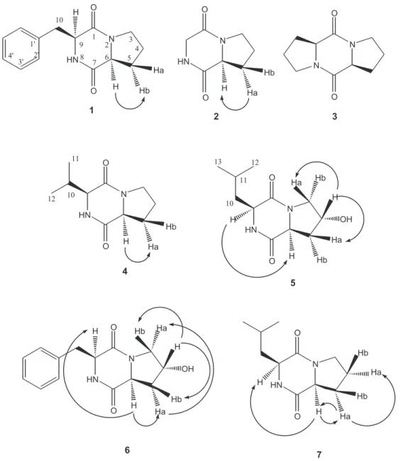
The chromatographic purification of chloroform extract, by using a combination of Si gel flash chroma-tography and high-performance liquid chromachroma-tography, furnished 7 diketopiperazines (Figure 1).
The identification of diketopiperazines 1-7 was
straightforward based on the presence of both the
characteristic 13C NMR chemical shifts for the amide
carbonyl groups (δ 167-172) and the 1H NMR signals of
the amino acids. The presence of proline moiety as one
of the components of 1-4 and 7 was deduced by the
presence of methylene multiplets in the spectra (δ 1.15 – 3.59, 6H) and by H-C correlations observed in the
HMBC experiments. Analysis of HMBC experiment showed the correlation of C-1 and C-6 with H-3 for compounds 1, 3, 6 and 7. Compounds 5 and 6 bear a
hydroxyproline moiety [δH 4.45 (H-4), δC 69.0 (C-4) and
δH 4.27 (H-4), δC 68.9 (C-4), respectively] instead of a
proline one. Analysis of the NMR spectra indicated that phenylalanine, glycine, valine and leucine were the
other amino acid moieties present in compounds 1-7
(Table 1, 2 and 3).
Diketopiperazine 1 bears the molecular formula
C14H16N2O2, which was established by mass spectrometry, using electrospray ionization (ESI). The molecular weight
of compound 1 was deduced as 244 by the quasimolecular
ions at m/z 245 [M + H]+, 267 [M + Na]+ and 283 [M + K]+.
The molecular formula of diketopiperazine 6 was assigned
as C14H16N2O3, which contains one additional oxygen in
relation to compound 1. These data were well consistent
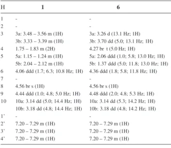
Table 1.1H NMR (400 MHz, CD
3OD, δ values) data of 1 and 6
H 1 6
1 -
-2 -
-3 3a: 3.48 – 3.56 m (1H) 3a: 3.26 d (13.1 Hz; 1H) 3b: 3.33 – 3.39 m (1H) 3b: 3.70 dd (5.0; 13.1 Hz; 1H) 4 1.75 – 1.83 m (2H) 4.27 br t (5.0 Hz; 1H)
5 5a: 1.15 – 1.24 m (1H) 5a: 2.06 ddd(1.0; 5.8; 13.0 Hz; 1H) 5b: 2.04 – 2.12 m (1H) 5b: 1.37 ddd (5.0; 11.8; 13.0 Hz; 1H) 6 4.06 ddd (1.7; 6.3; 10.8 Hz; 1H) 4.36 ddd (1.8; 5.8; 11.8 Hz; 1H)
7 -
-8 4.56 br s(1H) 4.56 br s(1H)
9 4.44 ddd (1.0; 4.8; 5.0 Hz; 1H) 4.48 ddd (2.0; 4.8; 5.3 Hz; 1H) 10 10a: 3.14 dd(5.0; 14.4 Hz; 1H) 10a: 3.14 dd(5.3; 14.2 Hz; 1H) 10b: 3.18 dd(4.8; 14.4 Hz; 1H) 10b: 3.18 dd(4.8; 14.2 Hz; 1H)
1’ -
-2’ 7.20 – 7.29 m(1H) 7.20 – 7.29 m (1H) 3’ 7.20 – 7.29 m (1H) 7.20 – 7.29 m(1H) 4’ 7.20 – 7.29 m(1H) 7.20 – 7.29 m(1H)
Table 2.1H NMR (400 MHz, CD
3OD, δ values) data of 2, 3, 4, 5 and 7
H 2 3 4 5 7
1 - - - -
-2 - - - -
-3 3.44 – 3.56 m (2H) 3.44 – 3.54 m (4H) 3.45 – 3.59 m (2H) 3a: 3.65 dd (4.2; 12.6 Hz; 1H) 3.47 – 3.51 m (2H) 3b: 3.43 dd (0.5; 12.6 Hz; 1H)
4 1.84 – 2.01 m (2H) 1.92 – 2.09 m(4H) 1.84 – 2.01 m(2H) 4.45 br t (4.2 Hz; 1H) 4a: 1.96 – 2.05 m (1H) 4b: 1.81 – 1.94 m (1H) 5 5a: 2.23 – 2.31 m (1H) 5a: 2.23 – 2.33 m(2H) 5a: 2.28 – 2.34 m (1H) 5a: 2.27 ddd (1.0; 6.5; 13.1 Hz; 1H) 5a: 2.24 – 2.34 m (1H) 5b: 1.84 – 2.01 m (1H) 5b: 1.92 – 2.09 m (2H) 5b: 1.84 – 2.01 m (1H) 5b: 2.07 ddd (4.2; 11.1; 13.1 Hz; 1H) 5b: 1.96 – 2.05 m (1H) 6 4.21 ddd (1.7; 6.8; 11.1 Hz; 1H) 4.31 – 4.36 m (1H) 4.21 ddd (2.2; 6.5; 12.1 Hz; 1H) 4.51 ddd (1.2; 6.5; 11.1 Hz; 1H) 4.25 ddd (1.7; 6.3; 11.1 Hz; 1H)
7 - - - -
-8 4.56 br s(1H) - 4.56 br s(1H) 4.56 br s(1H) 4.56 br s(1H)
9 9a: 4.06 dd (2.0; 16.9 Hz; 1H) 4.31 – 4.36 m (1H) 4.01 – 4.03 m (1H) 4.13 – 4.18 m (1H) 4.09 – 4.15 m (1H) 9b: 3.69 d (16.9 Hz; 1H)
10 - - 2.48 dsept(2.5; 6.9 Hz; 1H) 10a: 1.83 – 1.94 m(1H) 10a: 1.81 – 1.94 m (1H)
10b: 1.46 – 1.53 m (1H) 10b: 1.47 – 1.56 m (1H)
11 - - 0.93 d (6.9 Hz; 3H) 1.83 – 1.94 m (1H) 1.81 – 1.94 m (1H)
12 - - 1.07 d (6.9 Hz; 3H) 0.95 d (6.5 Hz; 3H) 0.94 d (6.5 Hz; 3H)
with theirs 1H and 13C NMR data. The 1H NMR spectra of 1
and 6 displayed signals of methylene benzylic protons
groups (H-10) attached to monosubstituted benzene rings
(δ 3.14- 3.18). These data suggested a phenylalanine
moiety for these compounds.
The 1H spectrum of the compound 2 showed the signals
at δ 4.06 (J 2.0 and 16.9 Hz, dd, 1H, H-9a) and δ 3.69 (J
16.9 Hz, d, 1H, H-9b), both protons are attached to a carbon at δ 45.8, as observed in HMQC experiment, indicating a glycine moiety for this compound.
The 1H spectrum of the compound 4 displayed two
doublets at δ 0.93 (J 6.9 Hz, 3H, H-11) and δ 1.07 (J 6.9 Hz, 3H, H-12) indicating the presence of two methyl groups.
Moreover, the signals at δ 2.48 (J 2.5 and 6.9 Hz, dsept,
1H, H-10) and δ 4.01-4.03 (m, 1H, H-9) allowed to propose
a valine moiety for this compound.
The molecular formula of diketopiperazine 5 contains
one additional oxygen in relation to compound 7. Their
1H and 13C NMR signals showed close correspondence,
except for the signals of both hydroxyproline and proline
residues, respectively. Their 1H spectra showed two
doublets at δ 0.95 (J 6.5 Hz, 3H, H-12) and δ 0.94 (J 6.5
Hz, 3H, H-12) for 5 and 7, respectively, as well as other two
doublets at δ 0.96 (J 6.5 Hz, 3H, H-13) and δ 0.95 (J 6.5
Hz, 3H, H-13), indicating the presence of two methyl groups attached to the molecules. In addition, the signals at δ 4.13- 4.18 (m, 1H, H-9), δ 1.83- 1.94 (m, 1H, H-10a), δ 1.46- 1.53 (m, 1H, H-10b) and δ 1.83- 1.94 (m, 1H, H-11)
of compound 5 and the signals at δ 4.09- 4.15 (m, 1H,
H-9), δ 1.81- 1.94 (m, 1H, H-10a), δ 1.47- 1.56 (m, 1H, H-10b)
and δ 1.81- 1.94 (m, 1H, H-11) for compound 7 allowed to
propose a leucine moiety for these compounds.
The spectra data for all compounds showed signals
due to one N-H proton (δ 4.56, br s, 1H), with exception of
diketopiperazine 3. The EIMS data showed peaks at m/z
195 [M + H]+, 217 [M + Na]+ and 233 [M + K]+, which
allowed to propose the structure of cyclo Pro-Pro for
compound 3.
1H and 13C NMR previously reported data for the
compounds 1, 4, 5, 6 and 7 are in agreement with obtained
data for isolated diketopiperazines.18-20
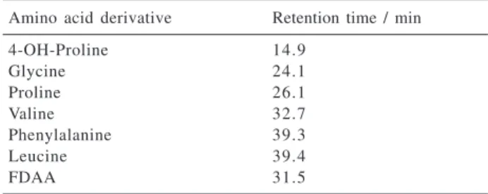
Regarding the absolute stereochemistries of the diketopiperazines, it was possible to determine that all diketopiperazines were biosynthesized from L-amino acids (Table 4). All the derivatized obtained from the hydrolysis of the diketopiperazines displayed the same retention times of the derivatized standard L-amino acids. The modified Marfey’s method was successfully applied to the determi-nation of the absolute configuration of amino acids. It was done by using a linear gradient starting with the mixture
of triethylamine phosphate buffer (50 mmol L-1, pH 3.5)/
MeCN (9:1) and finishing with triethylamine phosphate
buffer (50 mmol L-1, pH 3.5)/MeCN (6:4), over 45 minutes,
(flow rate 1.0 mL min-1). The original method describes a
linear gradient starting with the mixture of
triethyl-ammonium phosphate buffer (50 mmol L-1, pH 3.0)/MeCN
(9:1) and finishing with triethylammonium phosphate
buffer (50 mmol L-1, pH 3.0)/MeCN (1:1), over 60 minutes
(flow rate 2.0 mL min-1).14
Diketopiperazines corresponding to cyclic dipeptides have been isolated from microorganisms, sponges and from a variety of tissues and body fluids.18,21,22 These heterocyclic
compounds display several biological properties, such as: antimicrobial, antitumor, antiviral and plant growth regulation.19,23 Fdhila et al.20 reported the isolation of
DD-diketopiperazines cyclo (D)-Pro-(D)-Phe, (D)-Pro-(D)-Leu, (D)-Pro-(D)-Val and (D)-4-OH-Pro-(D)-Phe from marine
bacteria associated with cultures of Pecten maximus. These
DD-diketopiperazines presented a potent inhibitory
activity against the pathogen Vibrio anguillarum, while
the LL-enantiomers were completely inactive.20 Therefore,
the D-stereochemistry seems to be important for the biological activity.
Table 3.13C NMR (100 MHz, CD
3OD, δ values) data of compounds
1-7.
C 1 2 3 4 5 6 7
1 167.3 165.3 168.6 167.8 167.8 167.4 168.5
2 - - -
-3 45.9 45.1 45.8 46.1 56.0 55.6 46.0
4 23.1 22.1 23.2 23.2 69.0 68.9 23.2
5 29.7 28.2 28.8 29.4 37.0 39.2 28.8
6 60.4 58.6 61.7 60.1 58.0 58.0 60.1
7 171.3 170.8 168.6 172.6 171.9 171.6 172.6
8 - - -
-9 58.0 45.8 61.7 61.3 54.0 57.0 54.0
1 0 38.6 - - 30.1 38.0 38.4 38.2
1 1 - - - 16.3 25.0 - 25.0
1 2 - - - 18.3 21.0 - 21.4
1 3 - - - - 22.1 - 22.8
1 ’ 137.6 - - - - 137.8
-2 ’ 131.4 - - - - 131.3
-3 ’ 130.1 - - - - 130.0
-4’ 128.5 - - - - 128.9
-Table 4. HPLC reversed-phase retention times of the L-amino acid standard derivatives
Amino acid derivative Retention time / min
4-OH-Proline 14.9
Glycine 24.1
Proline 26.1
Valine 32.7
Phenylalanine 39.3
Leucine 39.4
Aspergillus fumigatus
All the isolated compounds inhibited the growth of
Staphylococcus aureus and Micrococcus luteus only at
the concentration of 2.9 mmol L-1. This result is
insignificant in comparison with penicillin, for which the
MIC values were established at 0.25 ηmol L-1 and 0.12
ηmol L-1, respectively. It is important to point out that the
literature reports24 the antimicrobial activity of the cycle
(L)-Pro-(L)-Phe against a clinical isolated strain of
Staphylococcus aureus at the concentration of 15 ηmol L-1 but, in the protocol used in this study, no activity was
detected by using the mentioned concentration.
The origin of diketopiperazines has been questioned, once several cyclic dipeptides have been found in fermentation broths and cultures of yeast, as well as in
lichens and fungi.25 It is known that diketopiperazines can
be generated via non-enzymatic cyclization of linear
dipeptides at extremes of temperature.26 It was checked
whether diketopiperazines would have been generated by heat sterilization and incubation of the medium culture during the fermentation process. However, these cyclic dipeptides were not detected in the HPLC profiles of the obtained extracts from the culture medium without the fungus.
To our knowledge, these cyclic dipeptides have not
been previously reported for A. fumigatus, with exception
of the cyclo (L)-Pro-(L)-Leu. 27
Acknowledgments
We are grateful to “Fundação de Amparo à Pesquisa do Estado de São Paulo (FAPESP)” Brazil for financial support (proc. 99/09850-8), to Dr. Suraia Said for providing the
strain of A. fumigatus, to Dr. Norberto P. Lopes for the
obtainment of MS data, as well as to Dr. Roberto G. S. Berlinck for providing the FDAA reagent and the authentic L-amino acids standards.
References
1. Baker, D. D.; Alvi, K. A.; Curr. Opin. Biotechnol.2004, 15, 576.
2. Czarnick, A. W.; Acc. Chem. Res.1996, 29, 112.
3. Donadio, S.; Monciardini, P.; Alduina, R.; Mazza, P.; Chiocchini, C.; Cavaleti, L.; Sosio, M.; Puglia, A.M.; J.
Biotechnol.2002, 99, 187.
4. Son, K.H.; Kim, Y. K.; Lee, H. W.; Lee, W. S.; Kim, S. U.; Jeong, T. S.; Kwon, B. M.; Bok, S. H.; Biotechnol. Lett.1996,
18, 1297.
5. Symoens, F.; Bertout, S.; Piens, M. A.; Burnod, J.; Renaud,F.; Nolard, N.; Chapuis, F.; Grillot, R.; EBGA Network.; J. Heart
Lung Transplant.2000, 20, 970.
6. Debeaupuis, J. P.; Sarfati, J.; Chazalet, V.; Latgé, J. P.; Infect.
Immun.1997, 65, 3080.
7. Varga, J.; Tóth, B.; Infect. Genet. Evol.2003, 3, 3.
8. Knight, V.; Sanglier, J. J.; DiTullio, D.; Braccili, S.; Bonner, P.; Waters, J.; Hughes, D.; Zhang, L.; Appl. Microbiol. Biotechnol.
2003, 62, 446.
9. Schulz, B.; Boyle, C.; Draeger, S.; Rommert, A. K.; Krohn, K.;
Mycol. Res.2002, 106, 996.
10. Mukhopadhyay, T.; Roy, K.; Coutinho, L.; Rupp, R. H.; Ganguli, B. N.; J. Antibiot.1987,40, 1050.
11. Ando, O.; Satake, H.; Nakajima, M.; Sato, A.; Nakamura, T.; Kinoshita, T.; Furuya, K.; Haneishi, T.; J. Antibiot.1991, 44, 382.
12. Jackson, M.; Karwoswski, J. P.; Humphrey, P. E.; Kohl, W. L.; Barlow, G. J.; Tanaka, S. K.; J. Antibiot.1993, 46, 34. 13. Still, C. S.; Kahn, M.; Mitra, A.; J. Org. Chem.1978, 43, 2923. 14. Marfey, P.; Ottesen, M.; Carlsberg Res. Commun.1984, 49,
585.
15. Hamburger, M. O.; Cordell, G. A.; J. Nat. Prod.1987, 50, 19. 16. Andrews, J. M.; J. Antimicrob. Chemother. 2001,48, 5. 17. Leverone, M.R.; Owen, T.C.; Tieder, F.S.; Stewart, G.J.; Lim,
D.U.; J. Microbiol. Methods1996, 25, 49.
18. Ström, K.; Sjögren, J.; Broberg, A.; Schnürer, J.; Appl. Environ.
Microbiol. 2002, 68, 4322.
19. Ienaga, K.; Nakamura, K.; Goto, T.; Tetrahedron Lett.1987,
28, 1285.
20. Fdhila, F.; Vázquez, V.; Sánchez, J. L.; Riguera, R.; J. Nat.
Prod.2003, 66, 1299.
21. De Rosa, S.; Mitova, M.; Tommonaro, G.; Biomol. Eng.2003,
20, 311.
22. Rudi, A.; Kashman, Y.; J. Nat. Prod. 1994, 57, 829. 23. Rhee, K. H.; Int. J. Antimicrob. Agents2004, 24, 423. 24. Graz, M.; Hunt, A.; Jamie, H.; Grant, G.; Milne, P.; Pharmazie
1999, 54, 772.
25. Prasad, C.; Peptides1995, 16, 151.
26. Holden, M. T. G.; Chhabra, S. R.; de Nys, R.; Stead, P.; Bainton, N. J.; Hill, P. J.; Manefield, M.; Kumar, N.; Labatte, M.; England, D.; Rice, S.; Givskov, M.; Salmond, G. P. C.; Stewart, G. S. A. B.; Bycroft, B. W.; Kjelleberg, S. A.; Williams, P.; Mol.
Microbiol. 1999,33, 1254.
27. Hanson, F. R.; Eble, T. E.; J. Bacteriol.1949, 58, 527.
Received: April 20, 2005 Published on the web: October 11, 2005