Isolation of
Escherichia coli
and
Salmonella
spp. from free-ranging wild animals
Renata de Oliveira Iovine
1, Catia Dejuste
2,3, Flávia Miranda
2, Claudia Filoni
1,3,4,
Marina Galvão Bueno
3,5, Vania Maria de Carvalho
11Laboratório de Biologia Molecular e Celular, Universidade Paulista, São Paulo, SP, Brazil.
2Projeto Tamanduá, São José dos Pinhais, PR, Brazil.
3Instituto Brasileiro para Medicina da Conservação, Recife, PE, Brazil.
4Laboratório de Diagnóstico Molecular, Universidade Estadual Paulista “Julio de Mesquista Filho”,
Botucatu, SP, Brazil.
5Laboratório de Patologia Comparada de Animais Selvagens, Universidade de São Paulo, São Paulo,
SP, Brazil.
Submitted: October 1, 2014; Approved: April 16, 2015.
Abstract
Increasing interactions between humans, domestic animals and wildlife may result in inter-species transmission of infectious agents. To evaluate the presence of pathogenicE. coliandSalmonellaspp. and to test the antimicrobial susceptibility of isolates, rectal swabs from 36 different free-ranging wild mammals were taken from two distinct natural sites in Brazil: Cantareira State Park (CSP, state of São Paulo) and Santa Isabel do Rio Negro Region (SIRNR, state of Amazonas). The swabs were randomly collected and processed for bacterial isolation, identification, characterization and anti-microbial resistance. EighteenE. colistrains from CSP and 20 from SIRNR were recovered from 14 and 22 individuals, respectively. Strains from animals captured in CSP, the site with the greatest anthropization, exhibited a higher range and percentage of virulence genes, including aneae+/bfpA+
strain. Antimicrobial resistance was verified in strains originating from both sites; however, in strains from SIRNR, aminopenicillins were almost the exclusive antimicrobial class to which strains exhib-ited resistance, whereas in CSP there were strains resistant to cephalosporins, sulfonamide, amino-glycoside, tetracycline and fluoroquinolone, in addition to strains exhibiting multidrug resistance. Two strains ofSalmonella entericathat are known to be associated with reptiles, serotypes Belem and 60:r:e,n,z15, were recovered only from Amazonian animals and showed susceptibility to all classes of antimicrobials that were tested. Although the potential impact of these pathogens on wild-life remains unknown, bacteria isolated from free-ranging wild animals may provide relevant infor-mation about environmental health and should therefore be more deeply studied.
Key words:E. coli,Salmonella, wild mammals, One Health, Brazil.
Introduction
Wildlife plays an important role within the context of the One World, One Health concept. Wild animals can serve as sentinels for infectious diseases and illnesses re-sulting from environmental contamination with toxic sub-stances; however, wildlife can also transmit pathogens to other animal species (directly and indirectly through envi-ronmental dispersal), including humans (Rabinowitzet al., 2009). Escherichia coli is one of the primary intestinal
commensal organisms found in endothermic animals and therefore it is widely disseminated in the environment. In-vestigating potential pathogenicE. colistrains and/or anti-biotic resistance may provide information regarding human activity in select ecological niches. Studies have indicated that living in proximity to humans may alter theE. coli
pop-ulations of wildlife intestinal microbiota (Rwego et al., 2008; Gordon and Cowling, 2003), which can in turn serve as a reservoir ofE. colistrains that are pathogenic to hu-mans (Bélangeret al., 2011).
Brazilian Journal of Microbiology 46, 4, 1257-1263 (2015) Copyright © 2015, Sociedade Brasileira de Microbiologia
ISSN 1678-4405 www.sbmicrobiologia.org.br
DOI: http://dx.doi.org/10.1590/S1517-838246420140843
Send correspondence to: V.M. Carvalho. Av. José Maria Whitaker 290, 04057-000 São Paulo, SP, Brazil. E-mail: vaniamc@uol.com.br.
Bacteria belonging to theSalmonellagenus are rec-ognized as important primary zoonotic pathogens. Unlike
E. coli,Salmonellaspp. are capable of persisting in aquatic environments for long periods of time, even in locations with high levels of eutrophication. The ubiquitous nature of
Salmonellapermits a cyclic exchange between host and en-vironment (Winfield and Groisman, 2003). Thus, wildlife can serve as reservoirs of diverse serotypes, especially with respect to reptilian species (Winfield and Groisman, 2003; Hoelzeret al., 2011).
Antimicrobial resistance in bacteria isolated from in-dividuals with no history of previous exposure to anti-microbial agents is indicative of the environmental dissemination of resistant strains and residual antimicrobial products through effluents from urban areas and animal production units (Costaet al., 2013). The present study was conducted to evaluate the presence of pathogenicE. coli
andSalmonellaspp. and to test antimicrobial susceptibility in isolates taken from fecal samples of free-ranging wild mammals in two distinct natural sites in Brazil.
Materials and Methods
Animals and biomes
A total of 14 different wild mammals (Table 1) from Cantareira State Park (CSP) (10 coati, 3 opossum, and 1 grey short-tailed opossum) and 22 (Table 2) from the Santa Isabel do Rio Negro Region (SIRNR) (18 bats, 3 rodents and 1 marsupial), all of them apparently healthy, were ran-domly captured using traps and were chemically restrained in strict accordance with protocols approved by the Re-search Ethics Committee (Permit Number: 140/12 CEP/ICS/UNIP). All activities were performed in full com-pliance with federal permits issued by the Brazilian
Minis-try of Environment (Permit Numbers:
CSP-260108-000.327/0 2008; SIRNR- SISBIO 27923-4). CSP (23°26’58.84" S; 46°38’7.94" O) is located in one of the world’s largest native areas of Atlantic forest within a met-ropolitan area (the city of São Paulo, which is the largest metropolis in South America), allowing for the possibility of close interactions between local fauna and visitors and domestic animals inhabiting the surrounding areas. In con-trast, SIRNR (0°24’51.61" S; 65° 1’10.23" O) is located in Amazonia and is accessible only via fluvial transportation. Human population density is low in this region.
Bacterial isolation and identification
Two rectal swabs were taken per animal. The swabs were delicately introduced into the rectum through rota-tional movements after cleaning the perianal area with gauze and sterile saline. The swabs were then immediately placed in Stuart transport medium (COPAN Venturi Transystem®) and kept under refrigeration. The first swab that was collected was seeded onto MacConkey Agar (Difco) to isolateE. coli, and the resulting colonies were
identified using Enterokit B (Probac do Brasil). To isolate
Salmonella spp., the second fecal swab sample was pro-cessed according to a previously published protocol (Mi-chaelet al., 2003). Colonies suggestive ofSalmonellaspp. were confirmed using an API 20E identification system (BioMerieux), and they were serotyped in the Reference Center forSalmonellaSerotyping (Instituto Adolfo Lutz, São Paulo, SP, Brazil).
E. colivirulence genotype and phylogenetic analysis
Strains identified asE. colithat were phenotypically different from each other were frozen in Brain Heart Infu-sion Medium (Difco) with 80% glycerol [Invitrogen; 1:1(v/v)] at -80 °C for further analysis. A PCR assay was used to test the isolates for the presence of virulence genes (VGs) known to be associated with diarrheagenicE. coli
(DEC) strains (eae, bfpA, stx1 and stx2) (Pollard et al.,
1990; Olsviket al., 1991; Gannonet al., 1993; Gunzburget al., 1995) and with extraintestinal pathogenic E. coli
(ExPEC) (papC,papEF,sfa,fyuA,iucD,hly,cnf1,cvaC,
traTandmalX) (Le Bouguenecet al., 1992; Johnson and Stell, 2000). Phylogenetic groups were determined using an established PCR-based method for assessingchuAand
yjaAgenes and the TSPE4.C2 DNA fragment (Clermontet al., 2000).
An EDL 933 strain was used as a positiveE. coli con-trol foreae, stx1, andstx2 genes, and an E2348/69 strain was used as the control for the bfpA gene. For ExPEC
genes, C7, JJ079 and BUTI 1-7-6 strains were used as posi-tive controls in all PCR reactions, and the JJ055 (derived from K12) strain was used as a negativeE. colicontrol (all were provided courtesy of Prof. Dr. James R. Johnson, Uni-versity of Minnesota). The controls used for phylogenetic determination were JJ055 (Group A), EC 15 (Group B1), JJ079 (Group B2), and EDL 933 (Group D).
Antimicrobial susceptibility test
Antimicrobial susceptibility was assessed using a disk diffusion method according to CLSI protocols (CLSI, 2013a; CLSI, 2013b). The following antimicrobials were tested: the amoxicillin (10 mg) and ampicillin (10 mg)
aminopenicillins; the cephalexin (30mg), cefoxitin (30mg)
and ceftiofur (30 mg) cephalosporins; the ciprofloxacin
(5 mg) and enrofloxacin (5 mg) fluoroquinolones; the
amphenicol chloramphenicol (30mg); the combined
sulfo-namide sulfametaxazol + trimethoprim (25mg); the
strepto-mycin (10mg) and gentamicin (10mg) aminoglycosides;
and tetracycline (30mg). Strains resistant to three or more
classes of antimicrobials were considered to be multidrug resistant (MDR) (Schwarzet al., 2010).
Enterobacteria
and
wildlife
1259
Table 1- Virulence genes, phylogenetic grouping and antimicrobial resistance inE. colistrains isolated from free-ranging wild mammals from Cantareira State Park.
Animal species Strain Virulence genes Phylogenetic group Antimicrobial resistance1
eae bfp stx1 stx2 papC papEF sfa fyuA iucD hly cnf1 cvaC traT malX A B1 B2 D
coati (Nasua nasua) 1/1 x x x x x Susceptible
1/2 x x x Amp
coati (Nasua nasua) 2/1 x x Amp
2/2 x x Amp, Amo
coati (Nasua nasua) 3 x x x x x Amp, Amo, Str, Tet, Sxt,
Cip2
coati (Nasua nasua) 4 x x x x Amp
coati (Nasua nasua) 5 x x x Susceptible
coati (Nasua nasua) 6/1 x x x x x x x x x Amp
6/2 x x x x Amp, Amo, Cfx, Tet, Sxt2
coati (Nasua nasua) 7/1 x x Amp
7/2 x x x Amp
coati (Nasua nasua) 8 x x x Amp, Cfx
coati (Nasua nasua) 9/1 x x Amp, Amo, Cfe, Str, Sxt2
9/2 x x x x Susceptible
opossum (Didelphis marsupialis)
10 x x x x Amp, Sxt
opossum (Didelphis marsupialis)
11 x x x Amp, Tet, Sxt
opossum (Didelphis marsupialis)
12 x x x Amp, Amo
grey short-tailed opossum (Monodelphis domestica)
13 x x Amp, Amo
1260
Iovine
et
al.
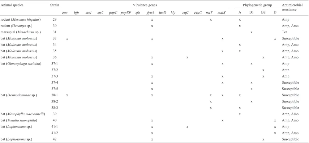
Table 2- Virulence genes, phylogenetic grouping and antimicrobial resistance inE. colistrains isolated from free-ranging wild mammals from Santa Isabel do Rio Negro Region.
Animal species Strain Virulence genes Phylogenetic group Antimicrobial
resistance1 eae bfp stx1 stx2 papC papEF sfa fyuA iucD hly cnf1 cvaC traT malX A B1 B2 D
rodent (Mesomys hispidus) 29 x x x Amp
rodent(Oecomyssp.) 30 x x Amp, Amo
marsupial (Metachirussp.) 31 x Tet
bat (Molossus molossus) 33 x x x x Susceptible
bat (Molossus molossus) 34 x Amp, Amo
bat (Molossus molossus) 35 x x Amp, Amo
bat (Molossus molossus) 36 x x x Amp, Amo
bat (Glossophaga soricina) 37/1 x x x Amp
37/2 x Amp
37/3 x x x Amp
37/4 x x x Susceptible
37/5 x x Susceptible
bat (Desmodontinaesp.) 38/1 x x x x x Susceptible
38/2 x x Susceptible
38/3 x x Susceptible
bat (Mesophylla macconnelli) 39 x Amp, Amo
bat (Tonatia saurophila) 40 x x x Amp, Amo
bat (Lophostomasp.) 41/1 x x x Amp
41/2 x x Amp, Amo
bat (Lophostomasp.) 42 x x Susceptible
Results and Discussion
Isolation and prevalence of VG and phylogenetic groups inE. coli
E. colistrains were isolated in 13 out of 14 (92%) and 13 out of 22 (59%) of the animals from CSP and SIRNR, re-spectively. The occurrence ofE. coliin the mammalian gut can vary enormously, but typicallyE. colimakes up more than 90% of gut microbiota in humans and only 56% of gut microbiota in wild mammals (Gordon and Cowling, 2003; Tenaillon et al., 2010). The recovery rate of E. coliwas higher in animals from CSP, which may reflect a more in-tense exposure to human contamination of the environ-ment. According to Gordon and Cowling (2003), the probability that a given host is carryingE. colidepends on the frequency of exposure, the likelihood that an exposure would result in the establishment of a population and, fi-nally, the average period of time that anE. colipopulation could remain in a host. These issues are relevant when eval-uating the importance of wildlife interactions with humans. Therefore, a total of 18 and 20E. colistrains were success-fully isolated from mammals in CSP and SIRNR, respec-tively. Characterizations of these strains are summarized in Tables 1 and 2.
This study verified that in the majority ofE. coli iso-lates from CSP, both the number and range of VGs were higher than in those isolated from SIRNR; the same was true for antimicrobial resistance patterns. Whereas in SIRNR only two out of 20 isolated strains (10%) contained three or more VGs, in CSP seven out of 18 (39%) showed this profile. Considering thatE. coliVGs are rare in the microbiota of wild animals but are not rare in humans (Tenaillonet al., 2010) and that the domestication status (which can be defined as living in proximity with humans beings) of an animal host is the principal environmental force that shapes the genetic structures of commensalE. colipopulations (Escobar-Páramoet al., 2006), strains re-covered from wild animals in CSP would reflect a broader anthropogenic action.
This hypothesis may be reinforced by the fact that the
eaegene was only present in 10% (2/20) of strains in ani-mals taken from SIRNR and in 28% (5/18) of those taken from CSP. However, none of the isolates were positive for either thestx1orstx2genes. Theeae+/stx-markers are as-sociated with the DEC, attaching and effacing ofE. coli,
and although strains with these markers are recognized as pathogens for humans and domestic animals, few studies have described their presence in wildlife (Bardiauet al., 2010; Obwegeseret al., 2012). Although a complete char-acterization of theeae+strains that were found in this sur-vey has not yet been performed, a search forbfpAgenes revealed one strain from CSP with the profileeae+/bfpA+.
CSP is a natural area with a high anthropogenic impact, and isolates with this profile are linked to typical EPEC, which are strains that have been epidemiologically associated
with human beings (Trabulsiet al., 2002). It is important to acknowledge that the strain was isolated from a coati, spe-cie that actively interacts with park visitors and their waste.
Furthermore, approximately 33% (6/18) of the strains isolated from CSP samples had some combination of the following genes:papEF, cvaC, traTandmalX.All of these genes have been correlated with the pathogenic potential of ExPEC (Johnsonet al., 2001; Maynardet al., 2004); how-ever, only a single strain (5.5%) from SIRN exhibited simi-lar findings (malX and traT). Previous studies have indicated that the presence of different combinations of VGs may provide a competitive advantage for the estab-lishment of bacteria in intestinal microbiota as well as in the development of extra-intestinal infections (Diard et al., 2010). P fimbria genes, such aspapCandpapEF,are im-portant indicators of pathogenicity and have been associ-ated with isolates that cause urosepsis in pets and humans (Maynard et al., 2004). The papEFgene was present in 22% (4/18) of the strains isolated from CSP animals, but it was absent in SIRNR isolates. ThecvaCgene is linked to ColV, a plasmid that occurs significantly more frequently in ExPEC recovered from neonatal meningitis compared to commensalE. coli(Johnsonet al., 2010; Bélangeret al., 2011), and it was likewise found exclusively in the strains isolated from CSP mammals. The genetraTencodes a cell surface lipoprotein that increases bacterial resistance to the lytic actions of complement (Johnson and Stell, 2000). This gene was found in 67% (12/18) of CSP strains and only 20% (4/20) of SIRNR isolates.
ThemalXgene, a pathogenicity island (PAI) marker of the archetypal uroseptic strain CTF073 (06:K2:H1), is associated with urinary tract infections in both humans and pets, in addition to human pyelonephritis, bacteremia and neonatal meningitis (Johnsonet al., 2001; Johnsonet al., 2003). Surprisingly, this gene was found in 35% of SIRNR strains (7/20) and in 22% (4/18) of CSP strains. The discov-ery of this gene even in isolates from a less impacted envi-ronment may be due to the fitness demonstrated by strains possessing PAIs. As these genetic elements increase the competitive abilities of their carriers in the intestinal envi-ronment compared to isolates in which they are absent, they contribute to the intestinal persistence ofE. coli(Östblom
et al., 2011; Diardet al., 2010). Thus, if through it’s gregar-ious behavior a wild animal acquires anE. colistrain that is harboring this gene, the presence of this marker could sub-sequently be amplified within the animal population. Out of all of the animals that were examined in this study, half of bats were found to harborE. coli malX+.This is notable, as bats are characterized by living in large colonies and have the ability to travel through diverse ecological niches (Mühldorfer, 2013).
A phylogenetic classification indicated that 56% (10/18) of the strains isolated from CSP belonged to phylo-genetic groups B2 and D, whereas in SIRNR this percent-age was slightly lower (40%, 8/20). The majority of
commensal strains are derived from phylogenetic groups A and B1, whereas pathogenic strains generally belong to groups B2 and D. Group B2 is largely composed of ExPEC, which is a heterogeneous group of strains that are able to asymptomatically colonize the intestines of several differ-ent hosts (Smithet al., 2007). Most importantly, a B2 strain that contained eight of the ExPEC genes that were included in our study was only isolated from animals in CSP. These genes encoded for adhesins (papC/papEFandsfa), an iron capture system (fyuA), toxins (hly and cnf1), protectin (traT) and a ColV associated-virulence plasmid (cvaC) (Smithet al., 2007; Bélangeret al., 2011). Although the pathogenicity of these strains to wild animals is unknown, we can hypothesize that these animals were exposed to sources of potentially pathogenic bacteria because extra-intestinal VGs are rare in isolates from wildlife, as has been previously described (Tenaillonet al., 2010).
Antimicrobial susceptibility
The presence of resistant bacteria in animals that had not been directly exposed to antimicrobials may reflect the degree of local anthropization (Costaet al., 2013). A total of 83% (15/18) of the strains isolated from CSP and 65% of those isolated (13/20) from SIRNR demonstrated resis-tance to one or more antimicrobial agents. However, iso-lates from SIRNR were almost exclusively resistant to aminopenicillins, whereas strains from CSP were resistant to cephalosporins, sulfonamide, aminoglycoside, tetracy-cline and fluoroquinolone, in addition to presenting multi-drug resistance (Table 1 and 2). Among the multi-drugs tested, chloramphenicol was the only antimicrobial agent to which all of the strains that were recovered from CSP were sus-ceptible. It is interesting to note elevated aminopenicillin resistance indices were found in SIRNR strains (12 out of 20 strains), ten of which were isolated from bats. These drugs are widely used in the treatment of human disease, as well as in companion and production animals (Maynardet al., 2004), and their active compounds remain almost com-pletely intact when they are eliminated into the environ-ment. This may lead to environmental contamination and a dissemination of antimicrobial resistance (Costa et al., 2013). Because SIRNR has fewer human and domestic ani-mal residents, the results suggest that the observed resis-tance phenomenon was not limited to its original niches and that wildlife may have an important role in its spread. Bats are capable of traveling through wide geographic distances and diverse ecological niches; furthermore, their colonial population pattern facilitates the sharing of strains (Mühldorfer, 2013).
Salmonellaisolation
Two strains ofSalmonella enterica,serotype Belem and subspeciesdiarizonaeserotype 60:r:e,n,z15, were re-spectively isolated from a bat and a rodent; both serotypes have been previously associated with reptiles (Schröteret
al., 2004; Riemann and Cliver, 2006). As they share the same terrestrial forest environment, it is not unexpected that serotypes are transmitted between rodent and reptile species. The Belem serotype that was isolated from a bat has been correlated with human infection in a previous sur-vey that was performed in the Amazon region (Loureiroet al., 2010). The involvement of free-ranging wildlife in the transmission and dissemination ofSalmonella spp. to hu-mans and other animals remains poorly understood, but a variety ofSalmonella serotypes have been isolated from both healthy and sick bats (Mühldorfer, 2013). The geo-graphic and ecological conditions that arise from the low levels of basic sanitation in the area promote the persistence ofSalmonellaspp. in the Amazon ecosystem (Loureiroet al., 2010), and wildlife may contribute to its dissemination.
Conclusions
In conclusion, taking into account the different VG profiles and antimicrobial resistance patterns of E. coli
strains isolated from individuals living in natural areas with dissimilar degrees of anthropization, the results obtained in this study indicate that human activity might increase the presence of resistant and/or potentially pathogenicE. coli
strains in the environment. We also found that antimicro-bial resistance is not limited to areas with greater anthro-pogenic impact. RegardingSalmonellaspp., we observed that in natural environments, free-ranging wildlife might contribute to a cycle of perpetuation and dissemination of these bacterial species. Although the impact of these poten-tial pathogens on wildlife is unknown, bacteria isolated from free-ranging wild animals may provide relevant infor-mation about environmental health and should therefore be more deeply studied.
Acknowledgments
The authors would like to thank all of those who con-tributed to the sampling of the animals and to PhD Fabiana Toshie de Camargo Konno, Cleide Marques da Silva Santana and Suzana Maria Bezerra for technical assistance. Renata de Oliveira Lovine was awarded financial support from CAPES.
References
Bardiau M, Grégoire F, Muylaert A et al. (2010)
Entero-pathogenic (EPEC), enterohaemorragic (EHEC) and vero-toxigenic (VETC)Escherichia coliin wild cervids. J Appl Microbiol 109:2214-2222.
Bélanger L, Garenaux A, Harel Jet al.(2011)Escherichia coli
from animal reservoirs as a potential source of human
extra-intestinal pathogenic E. coli. FEMS Immunol Med
Microbiol 62:1-10.
Clermont O, Bonacorsi S, Bingen E (2000) Rapid and simple de-termination of the Escherichia coli phylogenetic group. Appl Environ Microbiol 66:4555-4558.
Clinical and Laboratory Standards Institute (2013a) Performance standards for antimicrobial susceptibility testing. Twenty-third ed. document M100-S23. CLSI, Wayne.
Clinical and Laboratory Standards Institute (2013b) Performance standards for antimicrobial susceptibility testing; approved standard: Eighteenth international supplement document VET01-S2. CLSI, Wayne.
Costa PM, Loureiro L, Matos AJF (2013) Transfer of multidrug-resistant bacteria between intermingled ecologi-cal niches: the interface between humans, animals and the environment. Int J Environ Res Public Health 10:278-294. Diard M, Garry L, Selva Met al.(2010) Pathogenicity-associated
islands in extraintestinal pathogenicEscherichia coliare fit-ness elements involved in intestinal colonization. J Bacteriol 192:4885-4893.
Escobar-Páramo P, Le Menac’h A, Le Gall Tet al.(2006) Identi-fication of forces shaping the commensalEscherichia coli
genetic structure by comparing animal and human isolates. Environ Microbiol 8:1975-1984.
Gannon VP, Rashed M, King RKet al.(1993) Detection and char-acterization of theeaegene of Shiga-like toxin-producing
Escherichia coli using polymerase chain reaction. J Clin Microbiol 5:1268-1274.
Gordon DM, Cowling A (2003) The distribution and genetic structure ofEscherichia coliin Australian vertebrates: host and geographic effects. Microbiology 149:3575-3586. Gunzburg ST, Tornieporth NG, Riley LW (1995) Identification of
enteropathogenicEscherichia coliby PCR-bases detection of the bundle-forming pilus gene. J Clin Mirobiol 33:1375-1377.
Hoelzer K, Switt AIM, Wiedmann M (2011) Animal contact as a source of human non-typhoidal salmonellosis. Vet Res 42:1-27.
Johnson JR, Kaster N, Kuskowski MAet al.(2003) Identification of urovirulence traits inEscherichia coliby comparison of urinary and rectalE. coliisolates from dogs with urinary tract infection. J Clin Microbiol 41:337-345.
Johnson JR, Stell AL (2000) Extended virulence genotypes of
Escherichia colistrains from patients with urosepsis in rela-tion to phylogeny and host compromise. J Infect Dis 181:261-272.
Johnson JR, Stell AL, Delavari Pet al.(2001) Phylogenetic and pathotypic similarities between Escherichia coli isolates from urinary tract infections in dogs and extraintestinal in-fections in humans. J Infect Dis 183:897-906.
Johnson TJ, Jordan D, Kariyawasam Set al.(2010) Sequence analysis and characterization of a transferable hybrid plasmid encoding multidrug resistance and enabling zoonotic potential for extraintestinalEscherichia coli. Infect Immun 78:1931-1942.
Le Bouguenec C, Archambaud M, Labigne A (1992) Rapid and specific detection of thepap,afa, andsfaadhesin-encoding operons in uropathogenicEscherichia colistrains by poly-merase chain reaction. J Clin Microbiol 30:1189-1193.
Loureiro ECB, Marques NDB, Ramos FLPet al.(2010) Salmo-nella serovars of human origin identified in Pará State, Brazil from 1991 to 2008. Rev Pan-Amaz Saude 1:93-100. Maynard C, Bekal S, Sanschagrin Fet al.(2004) Heterogeneity
among virulence and antimicrobial resistance gene profiles of extraintestinalEscherichia coliisolates of animal and hu-man origin. J Clin Microbiol 42:5444-5452.
Michael GB, Simoneti R, Costa Met al.(2003) Comparison of different selective enrichment steps to isolateSalmonellasp. from feces of finishing swine. Braz J Microbiol 34:138-142. Mühldorfer K (2013) Bats and bacterial pathogens: a review.
Zoonoses Public Health 60:93-103.
Obwegeser T, Stephan R, Hofer E et al. (2012) Shedding of foodborne pathogens and microbial carcass contamination of hunted wild ruminants. Vet Microbiol 159:149-154. Olsvik O, Rimstad E, Hornes Eet al.(1991) A nested PCR
fol-lowed by magnetic separation of amplified fragments for de-tection ofEscherichia coliShiga-like toxin genes. Mol Cell Probes 5:429-435.
Östblom A, Adlerberth I, Wold AEet al.(2011) Pathogenicity is-land markers, virulence determinantsmalXandusp, and the capacity ofEscherichia colito persist in infants’ commensal microbiotas. Appl Environ Microbiol 77:2303-2308. Pollard DR, Johnson WM, Lior Het al.(1990) Rapid and specific
detection of verotoxin genes inEscherichia coliby the poly-merase chain reaction. J Clin Microbiol 28:540-545. Rabinowitz P, Scotch M, Conti L (2009) Human and animal
senti-nels for shared health risks. Vet Ital 45:23-24.
Riemann HP, Cliver DO (2006) Foodborne infections and intoxi-cations. Academic Press, San Diego.
Rwego IB, Isabirye-Basuta G, Gillespie TRet al.(2008) Gastro-intestinal bacterial transmission among humans, mountain gorillas, and livestock in Bwindi Impenetrable National Park, Uganda. Conserv Biol 22:1600-1607.
Schröter M, Roggentin P, Hofmann Jet al.(2004) Pet snakes as a
reservoir for Salmonella enterica subsp. diarizonae
(Serogroup IIIb): a prospective study. Appl Environ Microbiol 70:613-615.
Schwarz S, Silley P, Simjee Set al.(2010) Editorial: assessing the antimicrobial susceptibility of bacteria obtained from ani-mals. J Antimicrob Chemother 65:601-604.
Smith JL, Fratamico PM, Gunther NW (2007) Extraintestinal
pathogenic Escherichia coli. Foodborne Pathog Dis
4:134-163.
Tenaillon O, Skurnik D, Picard Bet al.(2010) The population ge-netics of commensalEscherichia coli. Nat Rev Microbiol 8:207-217.
Trabulsi LR, Keller R, Gomes TAT (2002) Typical and atypical enteropathogenic Escherichia coli. Emerg Infect Dis 8:508-513.
Winfield MD, Groisman EA (2003) Role of nonhost environ-ments in the lifestyles ofSalmonellaandEscherichia coli. Appl Environ Microbiol 69:3687-3694.
Associate Editor: Roxane Maria Fontes Piazza
All the content of the journal, except where otherwise noted, is licensed under a Creative Commons License CC BY-NC.