Use of green fluorescent protein to monitor
Lactobacillus plantarum
in the gastrointestinal tract of goats
Xufeng Han
1, Lei Wang
1, Wei Li
1, Bibo Li
1, Yuxin Yang
1, Hailong Yan
2,
Lei Qu
2, Yulin Chen
11
College of Animal Science and Technology, Northwest A&F University, People’s Republic of China, China.
2
College of Life Science, Yulin University, People’s Republic of China, China.
Submitted: July 5, 2014; Approved: February 2, 2015.
Abstract
The experiment aimed to specifically monitor the passage of lactobacilliin vivoafter oral administra-tion. The green fluorescent protein (GFP) gene was cloned downstream from the constitutive p32 promoter from L. lactis subsp. cremoris Wg2. The recombinant expression vector, pLEM415-gfp-p32, was electroporated intoLactobacillus plantarum(L.plantarum) isolated from goat. Green fluorescent protein (GFP) was successfully expressed inL. plantarum. After 2 h post-administration, transformedLactobacilluscould be detectable in all luminal contents. In the rumen, bacteria concen-tration initially decreased, reached the minimum at 42 h post-oral adminisconcen-tration and then increased. However, this concentration decreased constantly in the duodenum. This result indicated that L. plantarumcould colonize in the rumen but not in the duodenum.
Key words:green fluorescent protein, gastro-intestinal tract,Lactobacillus plantarum, goat.
Introduction
Lactobacilli are Gram-positive anaerobic or faculta-tive aerobic rods, which can be isolated from human and animal tissues, plants and materials of plant origin, and sewage and fermented products (Coeuretet al., 2003; Lee
et al., 2009; Azadniaet al., 2011). Particular lactobacilli strains are considered to present beneficial effects on hu-man and animal health. Therefore, several species of lacto-bacilli are used as probiotics (Forestier et al., 2001). Lactobacilli are generally regarded as safe (GRAS) by the FDA and are thus regarded as potential vehicles for oral vaccination (Mercenieret al., 1996; Scheppleret al., 2002). Moreover, they can stimulate local cell immunity of intesti-nal and humoral immunity (Billeret al., 1995; Christensen
et al., 2002; Servin, 2004). To provide a beneficial effect on the health of the hosts, the probiotic strain must be able to survive in the gastro-intestinal tract (Fuller, 1992). There-fore, knowing the distribution and movement of lactobacilli in the gastro-intestinal tract, as well as in which tract the strain functions, is necessary. Marking the lactobacilli and
tracing them by using green fluorescence protein (GFP) is a good method because GFP presents the advantage of being an auto-fluorescent protein that does not require a substrate. In addition, GFP allows for real-time detection in living cells (Kittset al., 1995). This method has been applied in monogastric animals but has yet to be reported in ruminants (Yuet al., 2007; Wanget al., 2011).
In the present study, we constructed a constitutive ex-pression vector for lactobacilli by using GFP as the reporter protein. We then electroporated the recombinant intoL. plantarum, which was isolated from Shanbei White cash-mere goats. Finally, the goat was fed with the transformants by using GFP as a visible marker for tracking this strain in-troduced into the gastrointestinal tract and observing its colonization capability.
Materials and Methods
Bacteria, plasmid and growth conditions
Escherichia coli DH5awas used for the construction and propagation of plasmid and grown in Luria-Bertani
DOI: http://dx.doi.org/10.1590/S1517-838246320140556
Send correspondence to Y. Chen. College of Animal Science and Technology, Northwest A&F University, No. 22 Xinong Road, Yangling, 712100 Shaanxi, P.R. China. E-mail: chenyulindk@163.com.
(LB) medium at 37 °C.Lactobacillusstrains were origi-nally isolated from the rumen of goats and were used as re-cipient strains for genetic construction and grown without shaking in an MRS medium at 37 °C at static conditions. The bacteria were identified as L. plantarum via both phenotypic and genotypic methods (Table 1). When appro-priate, antibiotics were added to the culture medium. ForE. coliandLactobacillus, ampicillin (Amp) was used at final concentrations of 100 and 50mg /mL, respectively.
In this study, the replicative plasmids, namely, pLEM415, pMG36e, and pEGFP-N1, were used (Table 1). pLEM415 is anE. coli-L.reuterishuttle vector (Serroret al., 2002) that contained a multi-cloning site and genes for Amp resistance. pMG36e contained the p32 promoter from
L. lactis subsp. cremorisWg2 (Van de Guchteet al., 1989).
Restriction endonucleases, T4 DNA ligase, and Taq polymerase were purchased from TaKaRa Biotechnology (Dalian, China) and used according to the recommenda-tions of the manufacturers.
PCR amplifications
Thegfpfragment was obtained by PCR amplification from pEGFP-N1, and the primers used were gfpF (5’-ATACCGCGGATGGTGAGCAAGGGCGAG-3’) and gfpR (5’-GCCGGAGCTCTTACTTGTACAGCTCG TCCATGC-3’).Thep32 promoter was obtained via PCR amplification from pMG36e, and the primers used were p32F (5’-TGCTCTAGAAATTCGGTCCTCGGGATAT GATAAG-3’) and p32R (5’-TCCCCGCGGGAATTTTT CTGCTGAAACGATTGCCAT-3’). Restriction sites added at the 5’-end of each primer are underlined. Agarose gel electrophoresis of plasmid DNA and PCR fragments was performed using the procedure described by Sam-brooketet al.(1989). These fragments were recovered via gel extraction by using a DNA Gel-extraction kit (OMEGA, USA).
Construction of expression plasmid carrying thegfp
gene
The PCR products ofgfpandp32were cloned into the pMD19 T vector and then sequenced. Subsequently, the re-combinant pMD19 T-gfp was digested bySacII andSacI and then ligated with SacII-SacI-restricted pLEM415 to construct the recombinant pLEM415-gfp. The recombinant pMD19 T-p32 was then digested byXbaI and SacII and then ligated with XbaI-SacII-restricted pLEM415-gfp to construct the recombinant pLEM415-gfp-p32. Thus, the re-sulting recombinant, pLEM415-gfp-p32, carried the gfp
gene under the control of thep32promoter. The structure of pLEM415-gfp-p32 was verified via restriction analysis, and thep32 andgfp fragment were verified via DNA se-quencing.
Electrotransformation procedures
The pLEM415-gfp-p32 was propagated by transfor-mation into E. coli DH5a competent cells according to manufacturer’s instructions.E. colitransformants were se-lected on LB agar plates containing Amp. DH5a harbour-ing pLEM415-gfp-p32 was proliferated at 37 °C for 12 h with shaking in LB broth containing Amp. pLEM415-gfp-p32 was purified fromE. colicultures via the alkaline lysis method (Sambrook et al., 1989). L. plantarum
electrocompetent cells were prepared based on a previously described method (Masonet al., 2005). A 50mL concen-trated cell suspension was electroporated using a Gene Pulser electroporator (Bio-Rad, Hemel Hempstead, UK) in cuvettes with a 0.2 cm electrode gap (Flowgen, Ashby de la Zouch, UK) with up to 1mg of plasmid DNA (at a concen-tration of 100 mg mL-1). Except where stated, electropo-ration parameters were 2.0 kV, 200Wparallel resistance, and 25mF capacitance. For phenotypic expression, the cells were diluted immediately into 1 mL of MRS broth in a 2 mL vial that was pre-warmed to 37 °C. After 3 h of incu-bation, serial dilutions were plated onto MRS agar contain-ing Amp. Plasmids from lactobacilli transformants were
Table 1- Plasmids and bacterial strains.
Plasmids or strain Relevant characteristics Antibiotic resistance Reference or source
Plasmid
pGFP-N1 plasmid containing gfp gene Ampr Jiaet al.(2006)
pMG36e E. coli-LABshuttle vector, containing P32 promoter Emr Van de Guchteet al.(1989) pLEM415 E. coli-L. reuterishuttle vector AmprErmr Serroret al.(2002)
pLEM415-gfp pLEM415 containing gfp gene AmprErmr This study
pLEM415-gfp-p32 pLEM415-gfp containing p32 gene AmprErmr This study
Bacterial strains
Escherichia coli DH5a Transformation host Invitrogen
Lactobacillus plantarum Transformation host Goat rumen
isolated following the method described by Anderson and McKay (1983).
Observation of fluorescence
For the observation of fluorescent bacteria, lacto-bacilli cultured overnight under investigation were washed three times in phosphate-buffered saline (PBS, pH = 7.5) and smeared on microscope slides. Prior to observation, the slides were treated by overlaying with 20% glycerine. An epifluorescent microscope (AMG) equipped with a GFP filter set (excitation 470 nm; emission 505 nm to 530 nm) was used to visualize fluorescent bacteria. Lactobacilli without pLEM415-gfp-p32 were used as a negative con-trol. To confirm the stability of the replicative plasmid pLEM415-gfp-p32 in lactobacilli, the transformants were tested after culturing continuously for 100 generations at non-selective conditions.
Intestinal distribution ofL. plantarum-GFP in goats
To monitor the distribution ofL. plantarum-GFPin vivo, eight Shanbei white cashmere goats with fistulas were used in our experiment. Each goat was installed with three fistulas, namely, rumen, duodenum, and ileum fistula. Four goats were orally inoculated with 1 mL of PBS containing 109 cfu of L. plantarum harbouring pLEM415-gfp-p32. Other goats were orally inoculated with 1 mL of PBS as a blank control. After 2, 6, 24, 48, and 72 h, samples from luminal contents in the rumen, duodenum, and ileum were serially diluted in PBS and cultured on MRS plates contain-ing Amp overnight at 37 °C. Bacteria concentrations (cfu mL-1) in rumen, duodenum, and ileum were deter-mined according to the number of colonies in MRS plates. The data were analysed by the general linear model proce-dures of SPSS software (SPSS Inc., Chicago, IL, USA). All experimental procedures with goats used in the present study had been given prior approval by the Experimental Animal Manage Committee of Northwest A&F University.
Results
Verification of Recombinant plasmid
Recombinant plasmid pLEM415-gfp-p32 extracted from DH5a was digested bySacII-SacI andXbaI-SacII, re-spectively. After enzyme digestion, we performed agarose gel eletrophoresis to identity the DNA fragments. The re-sult of agarose gel eletrophoresis showed that two DNA fragments were about 750 and 200 bp, which were consis-tent with the sizes ofgfpandp32fragment (Data not show). In addition, we also performed agarose gel eletrophoresis to check the PCR products amplified from pLEM415-gfp-P32 by primergfpandp32, which were about 750 and 200 bp as well. Taken together, these results showed that the expres-sion plasmid of lactobacilli was successfully constructed.
Observation of fluorescence
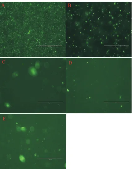
For the observation of fluorescent bacteria, trans-formed bacteria were washed in PBS (pH = 7.5) and ob-served via direct fluorescence microscopy, and bacteria without pLEM415-gfp-p32 were used as a negative con-trol. The result showed that the transformed bacteria ex-pressed fluorescence, whereas no fluorescence was observed in the negative control (Figure 1). Furthermore, we demonstrated that GFP was detectable in clones of transformed lactobacilli after subculturing for 100 genera-tions without antibiotic. The results suggested that GFP could be stably expressed in the recombinant lactobacilliin vitro.
MonitoringL. plantarum-GFP in the gastrointestinal tract of goat
After 2, 6, 24, 48, and 72 h of inoculating goats with the transformant, the geometric means of log10 concentra-tions of bacteria in rumen, duodenum, and ileum were ana-lysed (Figure 2). At 2 h post-administration, transformed lactobacilli can be detectable in all luminal contents. In the rumen, the concentrations of bacteria initially decreased, reached minimum at 42 h, and then increased. In the duode-num, however, a constant decrease was observed. Com-pared with those in other gastrointestinal sections, the transformant colonized in the ileum were highest at any time but without regular changes.
Discussion
A breakthrough in the transformation of
Lactobacillus strains was made when Chassy and
Flickinger used electroporation to introduce plasmid and phage DNA intoL. casei(Bringelet al., 1989). Since then, methods for the transformation ofLactobacillushave been rapidly developed and widely applied (Lin and Savage, 1986; Bringel and Hubert, 1990; Posno et al., 1991; Bhowmick and Steele, 1993; Masonet al., 2005; Palomino
et al., 2010), although few studies have optimized this pro-cedure for lactobacilli. Numerous studies have shown that various parameters, including the competent cell prepara-tion, electrical parameters, and host specificity, can influ-ence the transformation efficiency of Lactobacillus
(Hashibaet al., 1990; Weiet al., 1995; Serroret al., 2002). At present, no standard protocol of competent cell preparation has been established. Numerous studies have shown that culture, suspension, and resuscitation medium of competent cells should be adjusted according to different species of lactic acid bacteria. When competent cells were established according to the procedure described by Mason
Figure 1- Fluorescent detection ofL. plantarum-GFP under fluorescence microscopy. A: fluorescentE. coliDH5acultured in LB B:L. plantarum-GFP cultured in MRS; C to E: contents of rumen, duodenum, and ileum from goat inoculated withL. plantarum-GFP.
Electrical parameters are another important factor that affects electrotransformation efficiency. The optimiza-tion of electrical condioptimiza-tions was inconsistent in a previous study (Teresaet al., 2004). Considering the different sus-pension used, electrical parameters need be optimized to obtain the highest transformation efficiency and a mini-mum of cell destruction at the same time. In this study, an optimum transformation efficiency was obtained at a pulse strength of 2.0 kV, 200Wparallel resistance, and 25mF ca-pacitance (Table 2).
Goryet al.(2001) demonstrated that the expression of GFP did not alterL. sakeigrowth and the GFP-L. sakei
strains can be monitored both at laboratory growth condi-tions and in dry sausage samples. Yuet al.(2007) orally in-oculated chicken with D17-GFP and found that D17-GFP could propagate and persist at a high level in the gas-tro-intestinal tract after 2 h. Wanget al.(2011) orally ad-ministered GFP-L. lactisWH-C1 to mice and found that thisLactococcusstrain could exist in all gastro-intestinal tracts for extended periods. The above tests both showed that the recombinantLactobacillusmainly colonized in the downstream of gastro-intestinal tract. By contrast, trends in the number of Lactobacillusin this study indicated that
Lactobacilluscould colonize in the rumen but not in the du-odenum. Whether lactobacilli could colonize in the ileum requires further studies in the future. Whether exogenous
Lactobacilluscan adhere in the intestinal mucosa has been considered as one of the selection criteria for probiotic strains (Klaenhamme, 1982; Collinset al., 1998). Thus, our findings indicate thatLactobacillusfrom rumen can per-form an important function in goats as probiotic or expres-sion and delivery vehicles for recombinant proteins.
In addition, the GFP expression ofLactobacilluswas successfully applied to animal testing because it offers a simple and rapid method to detect the colonization ability of LAB. This result will aid in better understanding a series of problems, such as the study of relationship between mi-croorganisms and intestinal epithelial cells at the cellular level and the effects on the resident microbiota. In addition, our study will enable us to better understand the distribu-tion and movement of microorganisms in the gastro-intestinal tract.
Acknowledgments
This research was supported by The National Special Research Fund for Non-profit Sector (Agriculture) (20130305905) and China Agriculture Research System (CARS-40-13). We are grateful to Lei Qu and Hailong Yan of Yulin University for their kind help and cooperation dur-ing the animal experiments.
References
Anderson DG, McKay LL (1983) Simple and rapid method for isolating large plasmid DNA fromlactic streptococci. Appl Environ Microbiol 46:549-552.
Azadnia P, Zamani MH, Shah AGet al.(2011) Isolation and Iden-tification of Thermophilic Lactobacilli from Traditional Yoghurts of Tribes of Kazerun. J Anim Vet Adv 10:774-776.
Bhowmick T, Steele JL (1993) Development of an electroporation procedure for gene disruption inLactobacillus helveticus
CNRZ 32. J Gen Microbial 139:1433-1439.
Biller JA, Katz AJ, Flores AFet al.(1995) Treatment of recurrent Clostridium difficile colitis withLactobacillus GG.J Pediatr Gastroenterol Nutr 21:224-226.
Bringel F, Frey L, Hubert JC (1989) Characterization, cloning, curing and distribution in lactic acid bacteria of pLP1, a plasmid fromLactobacillus plantarumCCM 1904 and its use in shuttle vector construction. Plasmid 22:193-202. Bringel F, Hubert JC (1990) Optimized transformation by
electro-poration of Lactobacillus plantarumstrains with plasmid vectors. Appl Microbial Biotechnol. 33:664-670.
Carmen Collado M, Hernández M (2007) Identification and dif-ferentiation of Lactobacillus, Streptococcus and
Bifidobacterium species in fermented milk products with
bifidobacterium. Microbiol Res 162:86-92.
Christensen HR, Frøkiaer H, Pestka JJ (2002) Lactobacilli differ-entially modulate expression of cytokines and maturation surface markers in murine dendritic cells. J Immunol 168:171-178.
Coeuret V, Dubernet S, Bernardeau Met al.(2003) Isolation, characterisation and identification of lactobacilli focusing mainly on cheeses and other dairy products. Dairy Science and Technology 83:269-306.
Collins JK, Thornton G, Sullivan GO (1998) Selection of pro-biotic strains for human application. Int Dairy J 8:487-490. Forestier C, Champs CD, Vatoux Cet al.(2001) Probiotic activi-ties ofLactobacillus casei rhamnosus: in vitro adherence to intestinal cells and antimicrobial properties. Res Microbiol 152:167-173.
Fuller R (1992) Probiotics: The Scientific Basis. Chapmann and Hall, London, pp. 398.
Gory L, Montel MC, Zagorec M (2001) Use of green fluorescent protein to monitor Lactobacillus sakeiin fermented meat products. FEMS Microbiol Lett 194:127-133.
Hashiba H, Takiguchi R, Iskii Set al.(1990) Transformation of
Lactobacillus helveticus subsp. jugurtiwith plasmid pLHR by electroporation. Agric Biol Chemistry 54:1537-1541. Jia Jinghua, Wang Yanping, Zhou Leiet al.(2006) Expression of
Pseudomon as aeruginosa Toxin ExoS effectively induces apoptosis in host cells. Infect Immun 74:6557-6570. Table 2- Transformation frequency of different lactobacilli.
Voltage (kV) Transformation frequencya(cfu/mg plasmid DNA)
Mason procedure Modified procedure
1.5 1.1 x 10² 4.3 x 10³
2.0 2.7 x 10² 8.6 x 10³
2.5 Not detectedb 3.4 x 10³
Kitts P, Adams M, Kondepudi Aet al.(1995) Green fluorescent protein (GFP): a novel reporter for monitoring gene expres-sion in living cells and organisms. Clontechniques 10:1-3.
Klaenhamme TR (1982) Microbial considerations in selection and preparation ofLactobacillusstrains for use as dietary adjuncts. J Dairy Sci 65:1339-1349.
Lee DY, Seo YS, Rayamajhi Net al.(2009) Isolation, character-ization, and evaluation of wild isolates of Lactobacillus reuteri from pig feces. The Journal of Microbiology 47:663-672.
Lin JHC, Savage DC (1986) Genetic transformation of rifampicin resistance in Lactobacillus acidophilus. J Gen Microbiol 132:2107-2111.
Mason CK, Collins MA, Thompson K (2005) Modified electro-poration protocol for Lactobacilli isolated from the chicken crop facilitates transformation and the use of a genetic tool. J Microbiol Methods 60:353-363.
Mercenier A, Dutot P, Kleinpeter Pet al.(1996) Development of lactic acid bacteria as live vectors for oral or local vaccines. Adv Food Sci 18:73-77.
Palomino MM, Allievi MC, Prado-Acosta Met al.(2010) New method for electroporation ofLactobacillusspecies grown in high salt. J Microbiol Methods 83:164-167.
Posno M, Leer RJ, Van Luijk Net al.(1991) Incompatibility of
Lactobacillus vectors with replicons derived from small crypticLactobacillusplasmids and segregational instability of the introduced vectors. Appl Environ Microbial 57:1822-1828.
Sambrook J, Fritsch EF, Maniatis T (1989) Molecular Cloning: A Laboratory Manual. 2nd edn. Cold Spring Harbor Labora-tory, Cold Spring Harbor, NY.
Scheppler L, Vogel M, Zuercher AWet al.(2002) Recombinant
Lactobacillus johnsoniias a mucosal vaccine delivery vehi-cle. Vaccine 20:2913-2920.
Serror P, Sasaki T, Ehrlich SDet al.(2002) Electrotransformation of Lactobacillus delbrueckii ssp. bulgaricus and L. delbrueckiissp. lactiswith various plasmids. Appl Environ Microbiol 68:46-52.
Servin AL (2004) Antagonistic activities of lactobacilli and
bifidobacteriaagainst microbial pathogens. FEMS Micro-bio Rev 28:405-440.
Teresa AM, Carmen RM, Mesas JM (2004) Transformation of
Lactobacillus plantarum by electroporation with in vitro modified plasmid DNA. FEMS Microbiol Lett 241:73-77. Van de Guchte M, Van der Vossen JMBM, Kok Jet al.(1989)
Construction of a lactococcal expression vector: expression of hen egg white lysozyme in Lactococcus lactissubsp. Lactis. Appl Environ Microbiol 55:224-228.
Wang YP, Wang JR, Dai WL (2011) Use of GFP to trace the colo-nization ofLactococcus lactisWH-C1 in the gastrointestinal tract of mice. J Microbiol Methods 86:390-392.
Wei MQ, Rush CM, Norman HM et al. (1995) An improved method for the transformation ofLactobacillusstrains using electroporation. J Microbiol Methods 21:97-109.
Yu QH, Dong SM, Zhu WYet al.(2007) Use of green fluorescent protein to monitorLactobacillusin the gastro-intestinal tract of chicken. FEMS Microbiol Lett 275:207-213.
Associate Editor: Susana Marta Isay Saad