UNIVERSIDADE NOVA DE LISBOA FACULDADE DE CIÊNCIAS MÉDICAS
TRANSCRIPTIONAL REGULATION OF THE
MANNOSYLTRANSFERASE-ENCODING GENE
PIGM
IN
INHERITED GLYCOSYLPHOSPHATIDYLINOSITOL (GPI)
DEFICIENCY
JOANA RODRIGUES SIMÕES DA COSTA
Tese para obtenção do grau de Doutor em Ciências da Vida na especialidade em Biologia Celular e Molecular
UNIVERSIDADE NOVA DE LISBOA FACULDADE DE CIÊNCIAS MÉDICAS
TRANSCRIPTIONAL REGULATION OF THE
MANNOSYLTRANSFERASE-ENCODING GENE
PIGM
IN
INHERITED GLYCOSYLPHOSPHATIDYLINOSITOL (GPI)
DEFICIENCY
JOANA RODRIGUES SIMÕES DA COSTA
TESE ORIENTADA PELO DR. ANTÓNIO ALMEIDA E PELO PROFESSOR ANASTASIOS KARADIMITRIS
Tese para obtenção do grau de Doutor em Ciências da Vida na especialidade em Biologia Celular e Molecular
Acknowledgements
I would like to thank both my supervisors, Professor Tassos Karadimitris and Dr. António Almeida. To Tassos, for giving me the opportunity to carry out my PhD research in his lab. His scientific guidance and particularly his trust in me were fundamental to the development of my project and for helping me to believe in myself as a researcher. I will never forget his inspirational words: “TFs are the masters of the Universe!”. To Dr. António Almeida for giving me the opportunity to undertake my PhD and for understanding and supporting my decisions on what was best for my research.
I also wish to thank Fundação para a Ciência e Tecnologia (FCT) for funding my PhD studies.
A special acknowledgement goes to Valentina Caputo. The number of words that I am allowed to write on this page are certainly not enough to express my appreciation for the person that helped me the most. Her ongoing support, motivation, sharing of knowledge, thoughts, ideas and friendship kept me going. All our discussion made me grow as a researcher and gave me belief in myself. Without her I couldn’t have given any “colour” to my research or completed this work. She was truly my greatest support in the lab and an inspiration as a post-doc. Gracias Vale. I’ll never forget..
I must acknowledge Professor Irene Roberts for her support. Special thanks also go to Dr. Ian Sudbery for his collaboration and assistance with the bioinformatics aspect of this project.
I would like to thank my friend and colleague Kelly, for helping me, even without knowing, to make the big decision to stay in London to carry out my research. Her joy and support in the lab were fundamental to my work and her confidence an inspiration.
For all her help and strong support I wish to thank Katerina. Special thanks go to David O’Connor, for his care, strong support and friendship (including sharing good music and the English lessons!). Thanks to Antonella for her help in the lab and enormous inspiration. Neha and Deena, thank you for your attention! To Luciana, for making each day in the lab fun and making sure things ran smoothly. To Maria..for listening me so many times…
caring and for always being so supportive. To Elena and Gigi, for their help and support. A special acknowledgement goes to Ilaria Marigo for understanding me so well and for all the advice.
Gostaria de agradecer à Jacinta por toda a ajuda científica e pessoal, incluindo os conselhos e palavras de ânimo e de apoio, desde o inicio até ao final do meu doutoramento. Obrigada Jace!
Ao Bruno e ao Hélio pelo apoio e à Gabriela por tudo o que me ensinou no laboratório. À Sofia, pela inalterável amizade, pela capacidade de mesmo à distancia me ter ouvido e apoiado tantas vezes e por ter contribuído para que este desafio tivesse sido mais leve. À Cristiana (Flatinha), à Rita D. e ao Jaime, pelo apoio, pela amizade e por todos os momentos especiais de boa disposição. Às LHO, por estarem sempre presentes, quer nos bons quer nos maus momentos . Martinha, Johny, Lara e Ju. Obrigada. À Cheila e ao Bruno, pela ajuda e partilha. Obrigada também aos restantes elementos do CIPM. E às minhas amigas de faculdade, Telma, Débora e Teresa.
Gostaria de agradecer aos meus tios Nita e Quim por ouvirem os meus desabafos e ainda que nas minhas poucas palavras consigam sempre compreender-me tão bem. O vosso apoio durante o meu percurso tem sido fundamental.
E finalmente aos meus pais e à minha irmã. Por me terem encorajado sempre a seguir o meu caminho e apoiado em todas as decisões mesmo que isso tenha significado a minha ausência.. Pela confiança que depositaram em mim, por me ouvirem sempre que eu mais preciso e por me fazerem sorrir!
Quero agradecer especialmente à minha mãe por ser o meu maior suporte (e o de todos nós).
List of Publications
Caputo VS, Costa JR, Makarona K, et al. Mechanism of Polycomb recruitment to CpG islands revealed by inherited-disease-associated mutation. Human Molecular Genetics. 2013, 22 (16), pp.3187-3194.
Makarona, K, Caputo VS, Costa JR, et al. Transcriptional and epigenetic basis for restoration of G6PD enzymatic activity in human G6PD-deficient cells. Blood. 2014, 124 (1), pp.134-141.
List of Figures and Tables ... i
List of abbreviations ... v
Resumo ... ix
Abstract ... xi
Chapter 1 – Introduction ... 1
1.1 Regulation of gene expression ... 1
1.1.1 Transcriptional regulation of gene expression ... 1
1.1.2 The transcriptional machinery ... 3
1.1.3 Epigenetic control of transcription... 5
1.1.4 Transcription factors and DNA-binding specificity ... 6
1.1.5 Deregulation of transcription in disease ... 8
1.2 The Glycosylphosphatidylinositol (GPI) biosynthetic pathway ... 9
1.2.1 GPI-mediated anchoring of proteins in the cell membrane ... 9
1.2.2 Biosynthesis of the GPI-anchor moiety ... 13
1.2.3 Acquired-GPI deficiency ... 15
1.2.4 Inherited-GPI deficiency (IGD) ... 16
1.2.4.1 PIGM-associated IGD... 17
1.2.4.2 Deregulation of PIGM transcription ... 23
1.3 The Specificity Protein (Sp) transcription factor family ... 24
1.4 Haematopoiesis ... 28
1.4.1 Erythropoiesis ... 30
1.4.2 Regulation of erythropoiesis by GATA-1 and KLF1 Transcription Factors ... 32
1.5. Aims and Hypothesis ... 35
Chapter 2 - Materials and Methods ... 37
2.1 Cell lines, primary cells and treatment... 37
2.4 Cytospins preparations ... 39
2.5 Chromatin Immunoprecipitation (ChIP) assays ... 40
2.6 Circular chromosome conformation capture (4C) ... 42
2.7 DNA sequencing... 43
2.8 Flow-activated cell sorting (FACS) ... 44
2.9 Generation of FRT stable cell lines ... 44
2.10 In vitro erythroid differentiation and cytospin staining ... 45
2.11 Micrococcal nuclease (MNase) protection assay ... 46
2.12 Plasmids and cloning ... 47
2.13 Rapid amplification of cDNA ends (RACE) ... 49
2.14 Reporter assays ... 50
2.15 RNA extraction, cDNA synthesis and quantitative reverse transcriptase-PCR (qRT-PCR) ... 51
2.16 Transfections ... 52
2.17 Viral production and transduction ... 53
2.18 Western-blot ... 53
2.19 Statistical analysis ... 54
Chapter 3 – Results ... 55
3.1 Elucidating the role of PIGM transcription in PIGM-associated IGD ... 55
3.1.1 Introduction ... 55
3.1.2 Experimental design... 56
3.1.3 Results ... 57
3.1.4 Discussion ... 76
4.1 Exploring the role of the erythroid-lineage affiliated TFs GATA-1 and KLF1 in PIGM-associated IGD ... 81
4.1.1 Introduction ... 81
4.1.3 Results ... 82
4.1.4 Discussion... 97
5.1 Exploring the role of the generic family of Sp transcription factors in PIGM-associated IGD ... 101
5.1.1 Introduction ... 101
5.1.2 Experimental design ... 102
5.1.3 Results ... 102
5.1.4 Discussion... 113
6.1 Investigating the genomic interactions of PIGM ... 117
6.1.1 Introduction ... 117
6.1.2 Experimental design ... 118
6.1.3 Results ... 119
6.1.4 Discussion... 122
Chapter 4 – Discussion and concluding remarks ... 124
Appendix A ... 133
Appendix B – Inherited GPI Deficiency ... 139
List of Figures and Tables
Figures
Fig. 1 – Schematic representation of a typical gene regulatory region ……….. 2
Fig. 2 – Levels of structural organization of the DNA and histone proteins……... 5
Fig. 3 - Structure of the GPI anchor common core ……… 12
Fig. 4 – Biosynthetic pathway of the Glycosylphosphatidylinositol (GPI) anchor ... 13
Fig. 5 – Predicted Sp1 binding sites at the PIGM promoter ……….. 19
Fig. 6 – Schematic representation of Sp1 ……….. 26
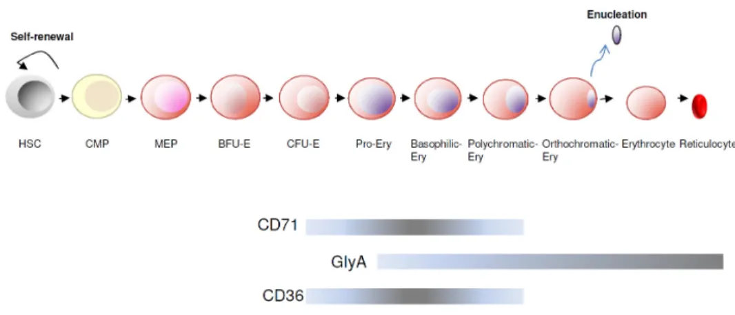
Fig. 7 – Haematopoietic hierarchy ... 29
Fig. 8 – Erythropoiesis ………... 31
Fig. 9 – Family trees of children with inherited GPI deficiency.………... 38
Fig. 10 – Schematic representation of the FRT system ……… 45
Fig. 11 – Schematic representation of 5- Rapid Amplification of cDNA-Ends (RACE) ……….. 50 Fig. 12 – Flow cytometric profile of the red blood cells (RBC) ……… 55
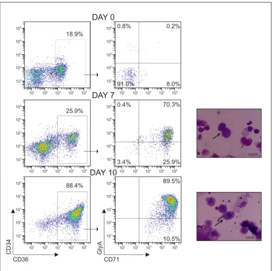
Fig. 13 - Isolation of the haematopoietic cells derived from peripheral blood of a normal donor and assessment of GPI expression ……….. 59 Fig. 14 - In vitro erythroid differentiation from a peripheral blood sample ……….. 60
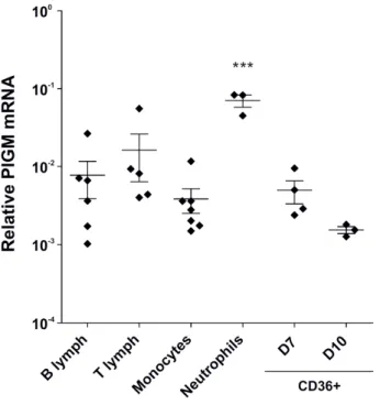
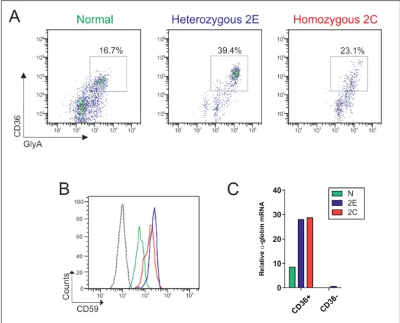
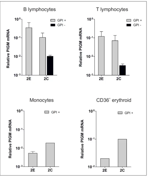
Fig. 15 - PIGM mRNA expression in haematopoietic primary cells derived from normal donors ……… 61 Fig. 16 - RNA-seq analysis in mouse fetal liver erythroid precursor cells ………… 62 Fig. 17 - GPI expression profile of haematopoietic cells isolated from individuals of Family 2………..
63 Fig. 18 - In vitro erythroid differentiation from PBMC of individuals from Family 2 and a normal control, at day 7 ……….
64 Fig. 19 - PIGM mRNA expression in haematopoietic cells isolated from PBMC of individuals from Family 2 ………..
Fig. 21 - PIGX mRNA expression levels in LBCLs ……… 69
Fig. 22 - Luciferase reporter assay in K562 (A) and HeLa (B) cells ……… 70
Fig. 23 - Effect of nucleosome occupancy on PIGM transcription ………... 72
Fig. 24 – Gel electrophoresis of the 5’-RACE-PCR ...………... 74
Fig. 25 - Schematic representation of the TSSs mapped at the PIGM promoter in K562 (K) and HeLa (H) cells ………. 75 Fig. 26 - CAGE analysis of PIGM (strand -1) in the B cell line GM12878 (red) and in K562 (blue) retrieved from the ENCODE database ……….. 76 Fig. 27 - Bioinformatic analysis of the PIGM promoter ……… 83
Fig. 28 - ChIP-seq analysis at the PIGM promoter ……… 84
Fig. 29 - Schematic representation of the PIGM promoter ……… 85
Fig. 30 - GATA-1 mRNA expression levels in cell lines ………... 86
Fig. 31 - GATA-1 occupancy at the length of the PIGM promoter in K562 ……… 87
Fig. 32 - GATA-1 occupancy at the length of the PIGM promoter in primary erythroid precursor cells ……… 87 Fig. 33 - K562 cells transduced with short hairpin (sh) RNA targeting GATA-1 and control scramble ……… 90 Fig. 34 - mRNA expression in K562 scramble and K562 GATA-1 knock-down cells ……… 91 Fig. 35- Gel electrophoresis of the PCR products of parental and recombinant 293-FRT amplified genomic DNA ………... 92 Fig. 36 - FACS analysis of 293T PIGM-promoter GFP recombinant cells ………... 93
Fig. 37 - 293T PIGM promoter-GFP recombinant cells ……… 94 Fig. 38 - mRNA expression in 293T PIGM promoter-GFP recombinant cells transiently transfected with MigR1-ratCD2 (control) or MigR1-hGATA-1 plasmids ...
95
Fig. 39 - KLF1 occupancy at the length of the PIGM promoter in erythroid precursor cells ………
scramble ……… 103 Fig. 41 - mRNA expression in the heterozygous LBCL scramble and LBCL Sp1 knock-down cells ………...
104 Fig. 42 - mRNA expression in K562 cells transduced independently with two shRNAs targeting Sp1 ………
105 Fig. 43 - Sp1 mRNA expression in K562 and WT LBCL (N2) cells ……… 105 Fig. 44 - mRNA expression in K562 cells transduced with MigR1-GFP (control) or MigR1-Sp1DN-GFP retroviruses ………..
106
Fig. 45 – Effect of MitA on gene expression in the -270C>G heterozygous LBCL 2D (A) and K562 cells (B) ……….
108 Fig. 46 - Sp1 occupancy at the length of the PIGM promoter in K562 and WT LBCL - N2 ……….
110 Fig. 47 - Sp1 occupancy at the length of the PIGM promoter in primary erythroid precursor cells ………
111 Fig. 48 - Sp2 occupancy at the Sp2 locus and at the length of the PIGM promoter .. 112 Fig. 49 - Schematic representation of the 4C assay ………... 119 Fig. 50 - Heat map showing the average contact intensities of PIGM ……….. 121 Fig. 51 – Proposed mechanism of transcriptional regulation of PIGM in erythroid and B cells ………..
131 Fig. A1 - pcDNA5/FRT vector used in the stable luciferase reporter assays ……… 132 Fig. A2 - MigR1 vector used for overexpression ……… 132 Fig. A3 - Mononuclease digestion ……….. 133 Fig. A4 – Optimization of the sonication conditions ………. 133 Fig. A5 - pLKO1-modified GFP vector used in the lentivirus transductions ……… 134 Fig. A6 - Digestion of the pLKO.1 shRNA plasmids ……… 134 Fig. A7 - Virus titration in 293T cells ……… 135 Fig. A8 - Western-Blot showing exogenous hGATA-1 expression in HeLa cells 135 Fig. A9 - Evaluation of the transfection efficiency of the MigR1-ratCD2 plasmid in Flp-In 293 cells by FACS ………
Tables
Table 1 - Examples of GPI-APs and respective function in mammalian cells …….. 11
Table 2 - Genetic abnormalities identified in the GPI biosynthetic genes and associated clinical features ………. 22 Table 3 - EBV-immortalised lymphoblastoid B cells (LBCLs) ……… 37
Table 4 - Antibodies for ChIP ……… 41
Table 5 - ChIP-RQ primers ……… 42
Table 6 - Primers used for sequencing ………. 44
Table 7 - MNase protection assay primers ……… 47
Table 8 - Primers used for cloning ……… 48
List of abbreviations
AA - Aplastic anaemia ALP - Alkaline phosphatase
BFU-E - Burst forming unit-erythroid bp - Base pair
BMF - Bone marrow failure BRE - TFIIB recognition element CFU-E - Colony-forming unit-erythroid CGI - CpG island
ChIP - Chromatin immunoprecipitation CLP - Common lymphoid progenitor CMP - Common myeloid progenitor DBD - DNA binding domain DHSs - DNase I hypersensitive sites DN - Dominant negative
DPE - Downstream core promoter element ER - Endoplasmic reticulum
EPO - erythropoietin Ery - Erythroblast
FLAER - Fluorescein-labeled proaerolysin GPI - Glycosylphosphatidylinositol GPI-APs - GPI -anchored proteins GPI-ET - GPI-ethanolamine
HATS - Histone Acetyltransferases HDACs - Histone Deacetylases
HPMR - Hyperphosphatasia mental retardation HSC - Haematopoietic stem cell
IGD - Inherited GPI Deficiency INR - Initiator
kd - Knock-down
KLF - Krüppel-like factor
LBCL - Lymphoblastoid B cell line LCR - Locus control region
MEP - megakaryocyte/erythroid progenitor min - Minute
MitA - Mithramycin A
MNase - Micrococcal nuclease NaBu - Sodium butyrate
NDR - Nucleosome-depleted region O/N - Overnight
PcG - Polycomb
PRC - Polycomb-repressive complex PolIII - RNA Polymerase III
PNH - Paroxysmal nocturnal haemoglobinuria RBC - Red blood cells
REAA - Restriction enzyme accessibility assay RNA PolII - RNA Polymerase II
SCF - Stem cell factor
SNP - Single nucleotide polymorphism Sp - Specificity protein
TAD - Transactivation domain TF - Transcription factor
TFBS - Transcription factor binding site TBP - TATA-binding protein
TSS - Transcription start site
Resumo
O glicosilfosfatidilinositol (GPI) é um complexo glicolipídico utlizado por dezenas de proteínas, o qual medeia a sua ancoragem à superfície da célula. Proteínas de superfície celular ancoradas a GPI apresentam várias funções essenciais para a manutenção celular. A deficiência na síntese de GPI é o que caracteriza principalmente a deficiência hereditária em GPI, um grupo de doenças autossómicas raras que resultam de mutações nos genes PIGA, PIGL, PIGM, PIGV, PIGN, PIGO e PIGT, os quais sao indispensáveis para a biossíntese do GPI.
Uma mutação pontual no motivo rico em GC -270 no promotor de PIGM impede a ligação do factor de transcrição (FT) Sp1 à sua sequência de reconhecimento, impondo a compactação da cromatina, associada à hipoacetilação de histonas, e consequentemente, impedindo a transcrição de PIGM. Desta forma, a adição da primeira manose ao GPI é comprometida, a síntese de GPI diminui assim como as proteínas ligadas a GPI à superficie das células. Pacientes com Deficiência Hereditária em GPI-associada a PIGM apresentam trombose e epilesia, e ausência de hemólise intravascular e anemia, sendo que estas duas últimas características definem a Hemoglobinúria Paroxística Nocturna (HPN), uma doença rara causada por mutações no gene PIGA.
configuração dos nucleossomas no promotor de PIGM é mais compacta em células B do que em células eritróides e tal está correlacionado com os níveis de expressão de PIGM, isto é, inferior nas células B. A presença de vários motivos de ligação para o FT específico da linhagem megacariocítica-eritróide GATA-1 no promotor de PIGM sugeriu que GATA-1 desempenha um papel regulador na sua transcrição. Os resultados mostraram que muito possivelmente GATA-1 desempenha um papel repressor em vez de activador da expressão de PIGM. Resultados preliminares sugerem que KLF1, um factor de transcrição restritamente eritróide, regula a transcrição de PIGM independentemente do motivo -270GC.
Em segundo lugar, a investigação do papel dos FTs Sp demonstrou que Sp1 medeia directamente a transcrição de PIGM em ambas as células B e eritróide. Curiosamente, ao contrário do que acontece nas células B, em que a transcrição de PIGM requer a ligação do FT geral Sp1 ao motivo -270GC, nas células eritróides Sp1 regula a transcrição de PIGM ao ligar-se a montante e não ao motivo -270GC. Para além disso, demonstrou-se que Sp2 não é um regulador directo da transcrição de PIGM quer nas células B quer nas células eritróides.
Estes resultados explicam a ausência de hemólise intravascular nos doentes com IGD associada a PIGM, uma das principais características que define a HPN.
Por último, resultados preliminares mostraram que a repressão da transcrição de PIGM devida à mutação patogénica -270C>G está associada com a diminuição da frequência de interacções genómicas em cis entre PIGM e os seus genes “vizinhos”, sugerindo adicionalmente que a regulação de PIGM e desses genes é partilhada.
Glycosylphosphatidylinositol (GPI) is a complex glycolipid used by dozens of proteins for cell surface anchoring. GPI-anchored proteins have various functions that are essential for the cellular maintenance. Defective GPI biosynthesis is the hallmark of inherited GPI deficiency (IGD), a group of rare autosomal diseases caused by mutations in PIGA, PIGL, PIGM, PIGV, PIGN, PIGO and PIGT, all genes indispensable for GPI biosynthesis.
A point mutation in the -270GC-rich box in the core promoter of PIGM disrupts binding of the transcription factor (TF) Sp1 to it, imposing nucleosome compaction associated with histone hypoacetylation, thus abrogating transcription of PIGM. As a consequence of PIGM transcriptional repression, addition of the first mannose residue onto the GPI core and thus GPI production are impaired; and expression of GPI-anchored proteins on the surface of cells is severely impaired. Patients with PIGM-associated IGD suffer from life-threatening thrombosis and epilepsy but not intravascular haemolysis and anaemia, two defining features of paroxysmal nocturnal haemoglobinuria (PNH), a rare disease caused by somatic mutations in PIGA.
Although the disease-causing mutation in IGD is constitutional and present in all tissues, the degree of GPI deficiency is variable and differs between cells of the same and of different tissues. Accordingly, GPI deficiency is severe in granulocytes and B cells but mild in T cells, fibroblasts, platelets and erythrocytes, hence the lack of intravascular haemolysis.
The transcriptional events underlying differential expression of GPI in the haematopoietic cells of PIG-M-associated IGD are not known and constitute the general aim of this thesis.
repressor rather than an activator of PIGM expression. Preliminary data suggested that KLF1, an erythroid-specific TF, regulates PIGM transcription but independently of the -270GC motif.
Secondly, investigation of the role of the Sp TFs showed that Sp1 directly mediates PIGM transcriptional regulation in both B and erythroid cells. However, unlike in B cells in which active PIGM transcription requires binding of the generic TF Sp1 to the -270GC-rich box, in erythroid cells, Sp1 regulates PIGM transcription by binding upstream of but not to the -270GC-rich motif. Additionally, I showed that Sp2 is not a direct regulator of PIGM transcription in B and erythroid cells.
These findings explain lack of intravascular haemolysis in PIGM-associated IGD, a defining feature of PNH.
Lastly, preliminary work shows that transcriptional repression of PIG-M by the pathogenic -270C>G mutation is associated with reduced frequency of in cis genomic interactions between PIGM and its neighbouring genes, suggesting a shared regulatory link between these genes and PIGM.
Chapter 1 – Introduction
1.1 Regulation of gene expression
Gene expression is the fundamental process by which the genetic information is converted to messenger RNA (mRNA) that is ultimately translated into functional proteins essential to the cell.
Control of gene expression has to be dynamic and highly regulated in order that spatial and temporal patterns that ensure cell and tissue development and function are achieved. For this purpose, gene expression is fine-tuned at different levels starting with transcription. RNA Polymerase II (RNA PolII) binding and active or repressed gene transcription is regulated by the integration of in cis DNA sequence information (promoters, enhancers and insulators), DNA elements with processes that determine chromatin packaging, histone and DNA modification (epigenetic marks). Next, I will provide an overview of transcriptional regulation of gene expression.
1.1.1 Transcriptional regulation of gene expression
Fig. 1 - Schematic representation of a typical gene regulatory region (adapted from Maston, G.A. et al, 2006). The promoter region comprises the proximal and the core promoter and usually spans ≤ 1Kb. The core promoter normally encompasses the transcription start site (TSS), from where transcription is initiated (black arrow).
Whereas the core promoter directs the initiation of transcription at precise sites by nucleating the Pre-initiation complex (PIC) and the RNA PolII, the proximal promoter and distal regulatory elements can activate or repress transcription. Distal regulatory elements, like enhancers or LCR, dispersed over tens to thousands of kilobases from the transcription start site (TSS), regulate gene transcription by forming DNA loops, in which distal DNA-bound proteins are nucleated to the core promoter. A very well-studied example is the β-globin locus where the distal elements - deoxyribonuclease I (DNase I) hypersensitive sites (DHSs) – in the LCR physically interact with the β-globin gene, HBB, by chromosomal looping (1, 2). This mechanism of gene regulation has also been demonstrated in transitional states of α-globin expression during erythroid differentiation (3).
Alterations in DNA regulatory elements have been associated with human diseases and in fact, it is estimated that between 1 to 2% of disease-causing point mutations are in regulatory regions of the genome, the majority located within 1Kb of the TSS (8). Nevertheless, cis-acting mutations do not necessarily affect the nearby gene. In agreement, a genome-wide association study (GWAS) has recently linked single nucleotide polymorphisms (SNPs) within DHSs not only to adjacent genes but also to new distant gene targets. Interestingly, most SNPs within DHSs overlapped with a transcription factor recognition sequence further suggesting the disruption of common transcriptional networks in disease (9).
As reviewed by Butler J. et al (10), the core promoter is the minimal DNA sequence encompassing the TSS (extending approximately 35 nucleotides upstream and downstream), that is sufficient to direct initiation of transcription by “docking” the basal transcriptional machinery; the proximal promoter, immediately upstream of the core promoter, spans a few hundred nucleotides. Proximal promoters, as well as distal regulatory elements, contain short DNA sequences known to anchor specific transcription factors (TFs), proteins that bind DNA and modulate transcription. TFs can be subdivided into two groups: generic and cell-type specific. Generic TFs are ubiquitously expressed in all cell types whereas cell-type specific TFs have restricted expression.
The DNA sequences bound by TFs, called transcription factor binding sites (TFBS), are typically constituted by 6 to 12 nucleotides although binding specificity is usually determined by 4 to 6 positions within the site (11).
1.1.2 The transcriptional machinery
derived from binding of the general factors and RNA PolII at the core promoter - basal transcription -, is usually low but can be potentiated by transcription factors that once recruited stabilize the transcriptional machinery. Accordingly, transcriptional activity is greatly stimulated or repressed by activators or co-repressors, respectively, localised upstream at the core promoter. Co-factors lack DNA binding properties and therefore interact physically with the DNA through bound TFs. For instance, Mediator, a multiprotein complex, is a co-activator known to target RNA PolII and recently, various crucial aspects of transcriptional regulation have been assigned to it (12). RNA PolII core promoters are functionally and structurally diverse. A small proportion contains a TATA box located 30 base pairs (bp) upstream of the TSS that guides RNA PolII to initiate transcription (“sharp” promoters). Usually, TATA promoters exhibit a low GC dinucleotide content and are associated with tissue-specific transcription. On the other hand, the majority of the promoters contain long (on average 1000bp) GC-rich DNA sequences called CpG island (CGIs) and correlate with the presence of multiple TSSs (“broad” promoters) (13-15). CGIs-associated promoters have been linked for long to housekeeping (ubiquitously expressed) genes, but is now clear that tissue-specific genes also contain CGI promoters (16, 17). Interestingly, some promoters contain both a TATA box and a CpG island, like in the erythropoietin gene (18, 19) and new studies have suggested that promoters of this type are capable of both TATA-dependent and TATA-independent transcriptional initiation (20). Therefore, the initial distinction between high and low-CpG promoters tends to fall in disuse and as suggested by Lenhard, B et al, 2012, (21) dividing promoters into “sharp” and “broad” may provide a better functional classification.
Apart from the TATA box, core promoters also contain other functional elements including initiator (INR), TFIIB recognition element (BRE), downstream core promoter element (DPE), amongst others (10).
isoforms that might generate distinct protein products and moreover, it is associated with patterns of gene expression, i.e., ubiquitous or tissue-specific/developmental stage-specific (22). There are examples in the literature in which the generation of a new promoter element causes disease by interfering with normal gene expression (23).
1.1.3 Epigenetic control of transcription
TFs usually are part of multiprotein complexes. Beyond recruiting co-activators and co-repressors, TFs also recruit histone-modifying enzymes and remodelling complexes that modify the nucleosomes, the basic unit of chromatin, in a dynamic manner. The term Epigenetics refers transferable regulatory information that controls gene transcription by processes other than changes in the underlying DNA sequence.
The nucleosome, the basic unit of chromatin, is composed by an octamer of four core histones H2A, H2B, H3 and H4, wrapped around a string of 147 base pairs of DNA. Consecutive nucleosomes are regularly arranged and connected by the DNA linker histone H1 and once compacted in higher-order structures originate the chromosome (Fig. 2).
packaging of the chromatin fiber originates the chromosome. (Adapted from image on the National Human Genome Research Institute website; author - Darryl Leja).
In the genome, the type and positioning of the nucleosomes is determined by a variety of factors including, DNA sequence, activators, nucleosome remodelers and the RNA PolII transcription machinery (24).
The histones that constitute the nucleosomal unit are proteins with a globular domain and an N-terminal “tail”. Whilst the globular domains interact with each other in order to form the octameric core, the histone “tails” undergo post-translational modifications (PTM) at specific aminoacid residues. Those modifications, mediated by histone-modifying enzymes, include acetylation, methylation, phosphorylation, ubiquitylation, sumoylation, ADP ribosylation, deamination and proline isomerization. Acetylation of histone “tails” is one of the best studied modifications. Usually, acetylation of histone “tails” by histone acetyltransferases (HATs) is associated with active gene expression whereas hypoacetylation at specific promoters by histone deacetylases (HDACs) is associated with repressed genes (25). Nevertheless, HDACs have been also found at active genes suggesting that gene expression results from a dynamic, balanced process between acetylation and deacetylation (26).
Another epigenetic mark that modulates gene expression is DNA methylation. DNA methylation occurs at the cytosine nucleotides that constitute the CpG modules and it is typically associated with gene silencing (27).
1.1.4 Transcription factors and DNA-binding specificity
It is estimated that there are approximately 1,400 transcription factors containing sequence specific DNA-binding properties (28).
contain a transactivation domain (TAD) which by recruiting the other elements of the transcriptional machinery stimulates transcription.
Whereas the DBD of TFs has been studied in vitro for many decades by techniques such as Electrophoretic Mobility Shift Assay (EMSA), full-length transcription factor occupancy has been studied in vivo only in the past few years by methods including Chromatin Immunoprecitipation (ChIP) and its derivatives (ChIP-chip, ChIP-seq and ChIP-exo) and DamID. DNase I hypersensitivity and Formaldehyde-Assisted Isolation of Regulatory Elements (FAIRE) have been also extensively used to investigate chromatin structure. The former is based on the principle that TF binding to the DNA generates a remodelled chromatin state more permissive to nuclease digestion. Studies have recently suggested the use of DHSs profile as quantitative benchmark in lineage specification (29-31).
Across the genome, only a small fraction of predicted consensus motifs is occupied by TFs (32, 33) suggesting that the presence of a consensus motif is not always sufficient to explain TF binding. Understanding what determines TF occupancy at particular consensus motifs is a very interesting and emerging area of research in the field of regulation of gene expression.
The sequence flanking the core motif (34), cofactor binding (35) and the chromatin landscape (DHSs and histone marks) (36, 37) are known to influence TF binding and consequently gene expression. Additionally, spacing and orientation between dimeric sites also influence TF binding specificity (34). Conversely, TF binding also influences the chromatin landscape (29, 38).
chromatin accessibility (39). Another study, using as a model the estrogen receptor (ER) compared, in multiple cell types, the properties of the cell-type specific and shared TF binding sites. Most shared sites contained high-affinity cognate sequences and did not show a dependency on open chromatin. On the other hand, cell specific sites harboured low-affinity sequence elements and were associated with chromatin accessibility (40). Other investigations suggest that the quantitative difference in TF-binding between cell types, rather than an ON/OFF event, should be taken into account when studying TF binding properties (41).
Together, these studies highlight the importance of the underlying DNA sequence in TF binding specificity. They reveal the complexity of TF binding and the importance in understanding DNA binding properties of individual TFs in a cell-type specific context in vivo.
Overall, the functional importance of differential and cell type-specific preferences of TF binding to regulatory DNA at a single gene level in health and in disease has not been determined. In my PhD project, I took advantage of a pathogenic promoter mutation that causes Inherited Glycosylphosphatidylinositol (GPI) Deficiency, i.e, IGD, to address these issues.
1.1.5 Deregulation of transcription in disease
Many diseases and syndromes have been linked to alterations in components of transcriptional regulation. Apart from mutations in regulatory elements, mutations in transcription factors, co-factors, chromatin regulators, and non-coding RNAs have been also reported.
The majority of the alterations in TFs relate by far with loss of function of the protein. Some others relate with generation of a fusion protein or de-regulated expression due to translocations, mutations in promoters or enhancers and very few are associated with gain of function or amplification (reviewed by Lee and Young, 2013 (52)).
Changes in gene regulatory regions normally alter transcription either by gaining or loss of a transcription factor binding site. For instance, a SNP in the α-globin cluster generates a new GATA-1 binding site, which by recruiting an erythroid-specific complex to the new promoter causes α-thalassemia (23). In the Pierre Robin syndrome, a heterozygous point mutation alters binding of the TF MSX1, abrogating the enhancer function, thus causing cleft palate (53).
One of the best studied Mendelian disorders caused by a mutation affecting TF binding is the PIGM-associated Inherited GPI Deficiency. It is caused by a hypomorphic C>G point mutation in the core promoter of the GPI mannosyltransferase I (GPI-MTI), PIGM, abrogating binding of the TF Sp1 (38).
In the subsequent sections, first, I will give an overview of the GPI biosynthetic pathway and the diseases associated with it followed by a more extensive description of the clinical, cellular, biochemical and ‘transcriptional’ phenotype of PIGM-associated GPI deficiency.
1.2 The Glycosylphosphatidylinositol (GPI) biosynthetic pathway
1.2.1 GPI-mediated anchoring of proteins in the cell membrane
Whereas a large number of proteins are embedded in the phospholipid bilayer of the cell membrane through a transmembrane arrangement, some surface proteins are anchored on the cell membrane through covalent linkage to GPI (54).
Table 1 – Examples of GPI-APs and respective function in mammalian cells [adapted from (55)]
GPI-anchored protein Function
Alkaline phosphatase 5’-nucleotidase Acetylcholinesterase Dipeptidase
Enzymes
LFA-3 NCAM
Cell-cell interaction and adhesion
CD55 (DAF)
CD59 Complement regulation
Thy-1 CD14
Carcinoembryonic antigen (CEA)
CD52
Mammalian antigens
CD16b
Folate-binding protein Others
Fig. 3 – Structure of the GPI anchor common core. Adapted from Mayor and Riezman, 2004 (56).
Despite their common core, there is variability in both glycan and lipidic components of the GPI anchors, which might occur either before or after attachment to the proteins (56). Those variations are believed to have a biological significance, namely, in the lateral mobility of GPI-APs and in trafficking. However, the evolutionary purpose of this structurally diverse manner of expressing proteins, as well as the functional role of GPI itself, beyond the physical anchoring, remain unknown. The importance of GPI anchoring in mammals has been highlighted by the fact germ line disruption of GPI biosynthesis is embryonic lethal. For instance, Pig-a knock out mice presented neurodevelopmental defects and failed to develop beyond the ninth day of gestation (57).
1.2.2 Biosynthesis of the GPI-anchor moiety
The biosynthesis of the GPI-anchor moiety occurs at least in eleven sequential steps in the endoplasmic reticulum (ER) by a series of more than 20 enzymes located in its membrane (Fig.3)
Fig. 4 – Biosynthetic pathway of the Glycosylphosphatidylinositol (GPI) anchor. Assembly of the GPI occurs in the Endoplasmic Reticulum and once assembled the anchor is attached to a protein containing the appropriate GPI attachment signal. The complex GPI-anchor protein (GPI-AP) is then exported to the cell surface via Golgi. Mutations in genes highlighted in red cause Inherited GPI Deficiency.
The steps in the biosynthesis of GPI are as follow:
Step 1: The synthesis of GPI is initiated by the addition of N-acetylglucosamine (GlcNAc) from UDP-GlcNAc to free PI on the cytosolic face of the ER. This reaction is mediated by GPI-GlcNAc, a large enzymatic complex constituted by PIGA (58-60), PIGC (61), PIGH (62), PIGQ (63), PIGY (64), PIGP (65) and two other additional components, PIGP and DPM2 (65). PIGA is the catalytic subunit of the complex.
Step 3: GlcN-PI is flipped to the luminal side of the ER by mediation of a yet unidentified enzyme, generally called “flippase”.
Step 4: An acyl chain is transferred from an acyl-CoA to the 2-position of the inositol by PIGW, originating GlcN-(acyl)PI. The acylation of the inositol is indispensable for the attachment of the Etn-P to the terminal mannose later in the pathway (step 9) (67).
Steps 5, 6 and 8 – Constitute the addition of mannose residues. Three mannose residues from dolichol-phosphate-mannose (DPM) are subsequently added to GlcN-(acyl)PI through the action of specific GPI mannosyltransferases (GPI-MT), namely PIGM/PIGX (68, 69), PIGV and PIGB. GPI-MTI consists of two subunits: the catalytic subunit, PIGM, and the subunit required for its stabilisation, PIGX. The second and the third mannose residues are transferred by the GPI- MTII and the GPI-MTIII, PIGV and PIGB, respectively. A fourth mannose residue can also be added to the GPI intermediate by PIGZ (also called SMP3), depending on the mammalian cell type and tissue (68, 70-72).
Steps 7, 9 and 10 – Comprise Etn-P modification of mannose residues. The three mannose residues are modified by the addition of three Etn-Ps from phosphatidylethanolamine. The enzymes responsible for these modifications are the GPI-ethanolamine (GPI-ET) phosphate transferases. PIGF constitutes the stabilising element whereas PIGN, PIGO and PIGG have catalytic activity (73). Only the modification in the terminal mannose (Man-3) is essential for GPI expression in mammals (74).
Once assembled, GPI-APs are transported to the Golgi via secretory vesicles. Whereas in some circumstances of GPI deficiency, precursor proteins are retained in the ER and undergo ER-associated degradation, in others, even if they form an intermediate complex with GPI transamidase, they are hydrolysed and secreted in its soluble form. One such example is the secretion of free alkaline phosphatase (ALP) seen in patients with hyperphosphatasia mental retardation syndrome (HPMR) (78).
Recently, new sequencing technologies such as whole-exome sequencing analysis have allowed high throughput screening of potential disease-associated genes (79) and as a result, several novel diseases associated with GPI deficiency have been identified and characterised.
GPI- associated diseases comprise acquired- and inherited- GPI deficiency. Whereas acquired deficiency results from mutations in somatic cells, inherited deficiency is caused by germline mutations. By definition, a mutation is a pathogenic alteration in the DNA sequence typically with a population frequency of < 1% (80).
1.2.3 Acquired-GPI deficiency
In PNH, the most dramatic effect is seen in the erythrocytes, where deficiency in the GPI-anchored complement inhibitors CD55 and CD59 triggers an unopposed activation of the complement cascade, leading to red cell destruction manifesting as intravascular haemolysis. The other defining features of PNH are venous thrombosis and bone marrow failure (BMF). The reason for bone marrow failure still remains unknown but various interesting findings have been made. The close association between PNH and aplastic anaemia (AA), in which stem cells in the bone marrow are destroyed by the autoimmune system has suggested that both diseases share a similar mechanism. Based on this hypothesis, selective targeting of GPI+ HSCs by autoreactive T –cells would permit GPI- HSCs to escape the immunological attack and to expand. In line with this, the analysis of the T-cell repertoire of PNH patients showed a high usage of the T-cell antigen receptor BV when compared to normal, age-matched controls (85) and more recently, Gargiulo, L. and collaborators, have shown the presence in PNH patients at frequencies higher than controls of a novel CD1d-dependent, GPI-reactive T-cell population, enriched in an invariant TCRα chain (86).
These findings support the hypothesis that GPI itself is a target of autoreactive T-cells in PNH and help to explain the pathogenetic mechanism driving BMF in PNH.
Defective GPI biosynthesis also constitutes the hallmark of inherited GPI deficiency (IGD), a group of rare autosomal recessive diseases caused by germline mutations in PIGM, PIGA, PIGL, PIGV, PIGN, PIGO and PIGT (Fig. 4).
1.2.4 Inherited-GPI deficiency (IGD)
1.2.4.1 PIGM-associated IGD
PIGM-associated IGD was identified in two unrelated consanguineous families of Middle Eastern and Turkish origin (see Fig. 9), in which the probands presented in infancy with abdominal vein thrombosis and later developed absence seizures. Haematological investigations showed a normal thrombophilia screen in all affected children apart from an intermittently low-positive Ham test, indicating some degree of CD59 absence from the erythrocyte surface. Intriguingly, none of the affected children exhibited clinical evidence of significant intravascular haemolysis or bone marrow failure, defining features of PNH. Moreover, an unusual expression pattern of GPI-linked proteins and GPI itself on blood cells was observed. Whereas in PNH, distinct populations of cells resulting from normal, complete or intermediate loss of GPI are observed, in PIGM-associated IGD the expression pattern of GPI-anchored proteins varied according to the cell type. Red blood cells of the affected children presented near normal expression of CD59, with a small proportion of cells (<5%) being variably deficient while 30-50% of platelets were CD59 negative. Granulocytes were mostly GPI negative as assessed by fluorescein-labeled proaerolysin (FLAER), CD59 and CD24 antibodies. The constitutional nature of the disease was demonstrated by staining primary fibroblasts from one affected child with CD59 antibody and FLAER, in which there was a clear reduction in the expression of GPI.
By employing homozygosity mapping and genome-wide linkage search using SNP in both families, PIGM was identified as the only common candidate gene. As described above, PIGM along with PIGX, constitute the enzymatic complex GPI-MTI which is responsible for transferring the first mannose to GPI on the luminal side of the ER (68).
The block in GPI biosynthesis at the biochemical level was directly demonstrated by substrate labelling of patient-derived EBV-transformed lymphoblastoid B cell lines (LBCLs), followed by glycolipid extraction and high performance thin layer chromatography. Whereas the first steps of the biosynthetic pathway were intact, incorporation of H-D-mannose in GPI-enriched glycolipids was impaired in both families.
confirmed in rescue experiments in which biosynthesis and expression of GPI at the cell surface were restored after transfection of PIGM-deficient LBCLs with PIGM WT cDNA.
Genetic defect in PIGM-associated IGD and its impact on transcription
In order to investigate the origin of the defect at the sequence level, Sanger sequencing analysis covering the single-exon of PIGM, its 3’- untranslated region (UTR) and 2kb region upstream of the ATG was performed. Sequencing analysis identified a single nucleotide substitution (C>G) at position -270 from ATG which was the cause of drastic reduction of PIGM mRNA levels in PIGM-deficient LBCLs (1% of normal controls). In heterozygous parental LBCLs, PIGM mRNA expression was about half of the normal levels which was consistent with a typical recessive trait.
Fig. 5 - Predicted Sp1 binding sites at the PIGM promoter. Apart from the -270GC box, four putative Sp1binding sites were predicted at the promoter. -270C>G mutation (red arrowhead) abrogates Sp1 binding. Black arrow indicates the putative TSS.
The impact of the Sp1-binding mutation on promoter activity of PIGM was directly assessed in reporter assays. By transfecting PIG-M-deficient and heterozygous LBCLs with plasmids containing either the WT or the MUT promoter upstream of a luciferase reporter gene, a decrease of 80 to 90% in PIGM promoter activity was observed in the presence of the mutation (87).
Altogether, these findings identified the biochemical and genetic defect in PIGM -associated IGD and very importantly, they showed the importance of precise transcription factor binding and regulation of gene expression.
Sp1 belongs to the Sp/Kruppel family of transcription factors which are critical regulators of gene expression of many housekeeping and inducible genes (88-90). Mutations generating or abolishing Sp1 binding sites in regulatory regions are not novel and they have been reported in several human diseases (45, 91-94).
From molecular characterisation to targeted therapy
Apart from interacting with the basal transcriptional machinery (95, 96) , Sp1is also known to influence transcription by recruiting and stimulating histone modifying enzymes, such as HATs (97, 98) and HDACs (99-101) to the gene promoter regions. The regulatory role of Sp1/Sp3 associated with the effect of the HDACs in promoting histone deacetylation and preventing high-order organization of the chromatin and the importance of the acetylation status during development prompted the authors to investigate whether PIGM promoter would contain responsive elements to butyrate and if so, how transcription of PIGM would be modulated.
By chromatin immunoprecipitation (ChIP) it was shown that in the presence of the -270 C>G mutation, histone 4 (H4) was hypoacetylated at the promoter in PIGM-deficient LBCLs and acetylated in heterozygous LBCLs. Treatment with 3mM sodium butyrate (NaBu), a pan- HDAC inhibitor, resulted in restoration of H4 acetylation at the MUT promoter, a 20-fold increase of the PIGM mRNA levels and restoration of GPI expression at the cell surface, as measured by FLAER. More importantly, in a child treated with sodium phenylbutyrate, peripheral blood cell PIGM mRNA levels drastically increased (from 0.15% before treatment to 60.77% after treatment), cell-surface GPI expression in granulocytes was restored to nearly normal levels and strikingly the child became seizure-free (102).
Altogether these results demonstrated that the -270C>G mutation disrupts a Sp1-dependent butyrate responsive element which is associated with histone hypoacetylation at the PIGM promoter. Further investigations regarding the molecular mechanism of transcriptional regulation in PIGM-associated IGD will be summarised in the next section.
strengthen the importance of genetic screening and biochemical analysis of genes associated with glycosylation and in particular with the biosynthesis of GPI.
Interestingly, in this group of rare inherited disorders the severity of the disease not only correlates with the type of mutation but also with the location of the gene in the biosynthetic pathway. Accordingly, mutations in PIGO are more severe than mutations in PIGV since they cause a more pronounced growth delay in patients (103). Other very interesting evidence in this group of disorders is the absence of haemolysis in patients, suggesting normal GPI expression in the surface of the erythrocytes. Understanding how those genetic abnormalities contribute to different phenotypic patterns in the haematopoietic cells still remains to be elucidated. The contribution of somatic events in this group of rare diseases should be also start taken into account.
Table 2 – Genetic abnormalities identified in the GPI biosynthetic genes and associated clinical features
Gene Mutation Disease Clinical features References
PIGM Homozygous g.-270G>C
PIGM
-associated IGD
Epilepsy, thrombosis of the
hepatic veins (87) (102)
PIGA c.1234C>T X-linked disease
Cleft palate, neonatal seizures, contractures and central nervous system (CNS) malformations
(104)
PIGL
Compound heterozygous c.500T>C + c.274delC, c.652C>T, c.427-1G>A or del17p12-p11.2
CHIME
Coloboma, heart defects, ichthyosiform dermatosis, mental retardation and ear anomalies with hearing loss
(105) (106) (107) (108) (109) PIGV Homozygous c.1022C>A or c.766C>A; compound heterozygous c.1022C>A+c.1154A>C; c.1022C>A+c.1022C>T
HPMR Mental retardation and elevated alkaline phosphatase (ALP) levels
(78) (110) (111,
112)
PIGN c.2126G>A
MCAHS (multiple- congenital- anomalies- hypotonia-seizures syndrome)
Developmental delay, cardiac, genitourinary and
gastrointestinal anomalies, severe neurological impairment with chorea and seizures
(113)
PIGO
Compound heterozygous c.2869C>T+ c.2361dup; c.2869C>T+
c.3069+5G>A
-
Mental retardation, elevated alkaline phosphatase TNAP gene, intractable seizures
Growth delay, malformations of the urinary system and heart
(103)
PIGT
Homozygous c.547A>C
Compound
heterozygous:c.1401-2A>G; 8-MB deletion
PNH
Distinct facial features, intellectual disability, hypotonia and seizures, abnormal skeletal, endocrine and ophthalmologic findings
Hemolytic crisis, abdominal pain, diarrhea, headache, arthralgia, dyspnea and fatigue
(114)
1.2.4.2 Deregulation of PIGM transcription
Gene silencing of CGI promoters is achieved through dense CpG methylation as mentioned before, or Polycomb (PcG) recruitment. Whether CpG methylation is the primary event in gene silencing is still a matter of debate. The PcG complex in mammals comprises polycomb-repressive complex 1 (PRC1) and 2 (PRC2). PRC2 is recruited to CGIs and then trymethylates H3K27 which is recognised by PRC1 complexes that impede transcriptional elongation, thus repressing gene expression by further modifying the histone tails (H2A-K119 ubiquitination) (116-118). The hierarchy of functional regulators that is responsible for Polycomb recruitment is still under investigation. Most results have arisen from studies in embryonic stem (ES) cells in which gene expression activation and repression has to be properly controlled.
Taking advantage of the -270C>G mutation at the CGI-PIGM promoter, the transcriptional mechanism driving repression of the housekeeping gene was investigated in more detail.
Sp1 binding and recruitment of HATs were required for nucleosome relaxation and normal transcription.
Repression of gene expression by nucleosome compaction is usually associated with DNA and histone methylation. Accordingly, the MUT promoter was enriched for the H3K27me3 mark, characteristic of developmentally regulated/poised genes, and suggestive of PcG recruitment and transcriptional repression. Further analysis showed that the CGI promoter was not methylated. Surprisingly, the co- presence of the active mark, H3K4me3 in the MUT promoter suggested the presence of a bivalent chromatin state. Altogether, the findings indicated that active TF binding “protects” against silencing by PcG, instructing a permissive chromatin state. Contrary, absence of active TF binding in CGIs directs Polycomb recruitment and deposition of bivalent histone methylation marks thus explaining the repression, yet reversible, of PIGM transcription and GPI deficiency in B cells (38).
Understanding the mechanism by which erythroid cells overcome the pathogenic C>G mutation and express near normal levels of GPI and GPI-APs demanded further investigation.
Below, as a prelude to the experimental work I plan to address this aspect of the molecular pathogenesis of PIGM-associated IGD, I provide a brief overview of the erythropoiesis and its transcriptional control. Before, a summary of the role of Sp TFs in transcriptional regulation is presented.
1.3 The Specificity Protein (Sp) transcription factor family
The Specificity Protein/Krüppel-like Factor (Sp/KLF) transcription factor family shares a highly conserved DNA binding domain of C2H2-type zinc fingers.
Members of this family bind to CACCC/GC/GT boxes, which are very common in the regulatory regions of many cellular genes.
in abnormalities more prominent in the nervous system (122). In fact, alterations in the Sp4 and Sp1 levels have been associated to neuropsychiatric disorders (123) (124) and a very recent study proposes these proteins as potential biomarkers of early-stages of psychotic diseases (125).
Depending on the promoter architecture and cellular context, Sp1 and Sp3 compete for DNA binding and it has also been suggested that Sp1 and Sp3 are functional redundant. However, results derived from developmental studies in mice are not in agreement and suggest they might have functional redundant roles only in early stages of development. Accordingly, Sp1 knockout mice are not viable and die around embryonic day 11 (126) whereas Sp3 knockout mice are viable and die from respiratory abnormalities (127).
Sp2 contains the least conserved DNA binding domain within the Sp family and mainly recognises GC box promoter elements. A recent study has identified various Sp2 gene targets in mouse embryonic fibroblasts (MEFs) and human embryonic kidney (HEK) 293 cells, defining Sp2 as a major regulator of genes involved in various cellular pathways (128).
Sp1 was the first identified member of the Sp family and many of its functions have been revealed by in vitro experiments. Sp1 has two isoforms and it is constituted by four domains. Two transactivation domains, that are localised in the N-terminus and are required for Sp1 transactivation potential; an internal domain and a carboxyl-terminus domain required for synergistic transactivation (Fig. 6). The N-terminus domains directly contact elements of the transcriptional machinery, such as TBP (TATA-binding protein)/TFIID (95, 96), thus activating transcription initiation. It is possible that in this way Sp1 directly recruits TFIID that potentiates Sp1-mediated transcription of TATA-less genes. Interestingly, Sp1 also has the ability to regulate gene expression by mediating chromatin loops between enhancer and promoter regions (129).
Fig. 6 – Schematic representation of Sp1. Sp1 is constituted by four domains (A, B, C and D). Domains A and B comprise the transactivation domains (TADs). Sp1 binds to the DNA through their zinc-fingers (Zn).
Sp1 is a very versatile protein, regulating expression of genes involved in various cellular processes, such as cell differentiation (130), apoptosis (131), angiogenesis and cancer (132). The posttranslational modifications of Sp1 affect its transcriptional activity and stability and therefore contribute for the complexity of its regulatory functions (133). Among those modifications are phosphorylation, acetylation, sumoylation, ubiquitylation and glycosylation.
For a long time, Sp1 was recognised as an activator of transcription of housekeeping and TATA-less genes. However, it is now known that Sp1 can also activate transcription of tissue-specific genes by interacting with tissue specific TFs such as GATA-1 (134, 135). Interactions with signal-induced factors, like SREBP-1a have also been reported (136).
As mentioned before, Sp1 interacts with HATs and HDACs.
Mutations in DNA sequences causing disruption of Sp binding sites have been reported in a growing body of literature. Notably, these mutations have an impact in normal gene expression and are in the basis of various human diseases.
Sp1 and impairs promoter activity causing erythropoietic protoporphyria (EPP) (93).
Abrogation of Sp1 binding in the regulatory region of the NME4 gene impairs binding of the erythroid specific TF SCL/Tal1 localised 55bp apart. Consequently, NME4 expression is altered in erythroid cells but is not affected in nonerythroid cells (139).
1.4 Haematopoiesis
Haematopoiesis is the biological process in which a pluripotent haematopoietic stem cell (HSC) gives rise to haematopoietic progenitor cells which in turn differentiate in different blood cell types. In mammals, haematopoiesis occurs sequentially in the yolk sack, fetal liver and bone marrow.
A pluripotent HSC is highly proliferative and capable of self-renewal, or in other words, to produce new HSCs. After differentiation, a long-term HSC ((LT)-HSC) originates an HSC with lower self-renewal potential, designated short term (ST)-HSC.
(ST)-HSC has the ability to differentiate into a multipotent progenitor cell–MPP, which in turn differentiates into a common myeloid progenitor (CMP) or common lymphoid progenitor (CLP) cell. CMP then differentiates into megakaryocyte/erythroid progenitor, called MEP or into granulocyte/macrophage progenitor, GMP; CLP originates B and T lymphocytes. MEPs give rise to red blood cells (RBC) and megakaryocytes and GMPs give rise to mast cells, eosinophils, neutrophils and monocytes/macrophages (Fig. 7). The specific pathway that gives rise to red blood cells is called erythropoiesis and it will be described next. Specific markers at the cell surface of the different haematopoietic cell types allow their identification and selection. For instance, CD34, c-kit, IL-6R, Thy-1 and CD45RA constitute cell surface markers of HSCs (140).
External signals such as cytokines (SCF, TPO, Flt3 and GM-CSF) produced by stromal bone marrow cells, are known to play a critical role in the status of HSC and therefore influence haematopoiesis.
During the haematopoietic development, TFs play a crucial role by influencing the transcriptional programmes in both the formation of the HSC and maintenance of its function and in the blood cell lineage specification. In addition to binding to DNA, haematopoietic TFs interact with each other and also interact with chromatin-associated factors.
MLL (mixed lineage-leukemia gene), RunX1, TEL/ETV6, SCL/Tal-1 and LMO2 (141). SCL/Tal-1 and LMO2 are essential in the early stages of haematopoiesis as mice null embryos die from the complete absence of embryonic blood (142).
In what respects lineage specification, TFs play a dual role. They promote lineage differentiation and at the same time they act against factors favouring other choices. Accordingly, PU.1, a key TF for the monocytic and B lymphoid commitment, interacts and antagonizes GATA-1, which is essential in the megakaryocytic and erythroid development (143); the contrary is also true (144). Direct evidence of the importance of TFs in haematopoiesis has arisen from genetic studies using knock-out mice. One such example is the master regulator of erythropoiesis, GATA-1. Disruption of GATA-1 in a murine embryonic stem cell line failed to give rise to red blood cells in chimaeric mice (145) and in its absence, erythroid precursor cells arrest at the proerythroblast stage and undergo apoptosis (146).
Forced expression studies and reprogramming, also revealed the importance of master TFs in lineage specification. For instance, exogenous expression of GATA-1 in cell lines of monocytes induced the expression of erythroid-megakaryocyte lineage markers and down-regulation of monocytic markers (147, 148). Overexpression of C/EBP-α, was also shown to commit B- and T-cell progenitors into functional macrophages (149, 150).
Not surprisingly, de-regulation of TFs and chromatin-modifying enzymes in the haematopoietic cells is largely associated with the onset of a variety of haematological malignancies. For instance, heterozygous mutations in PU.1 have been linked to acute myeloid leukemia (151) and overexpression of Tal-1, and its consequent abnormal auto-regulation cause T- cell acute lymphoblastic leukemia (T-ALL) (152).
1.4.1 Erythropoiesis
marrow. In adult life, erythroid precursors are mainly produced in the bone marrow and once they mature, they expel their nucleus and enter the bloodstream. The growth of burst forming unit-erythroid (BFU-E), the more immature erythroid progenitors, depends of cytokines such as stem cell factor (SCF), thrombopoietin (TPO), interleukin 3 (IL3) and 11 (IL11), and Flt-3 ligand. Under normal conditions these cells differentiate into colony-forming unit-erythroid (CFU-E) cells which are highly dependent of erythropoietin (EPO) and give rise to the erythroid precursors. At this stage, pro-erythroblasts undergo four to five cell divisions and sequentially originate morphologically distinct populations - basophilic, polychromatic and orthochromatic erythroblasts -, through acquisition of the surface markers, CD36, transferrin receptor protein 1 (CD71) and glycophorin A (GPA). CD71 loss anticipates enucleation, giving rise to a more mature and reduced in size cell, the reticulocyte (Fig. 8). Reticulocytes then enter the blood circulation where they mature into erythrocytes. The life-span of erythrocytes is 120 days (153). Erythroid gene regulation not only depends of the antagonising effect between haematopoietic TFs, but also depends of chromatin changes mediated by histone post-translational modifications (154).
1.4.2 Regulation of erythropoiesis by GATA-1 and KLF1 Transcription Factors
During haematopoietic differentiation, lineage specification is largely determined by the coordinated expression of specific TFs, in a quantitative and temporal manner.
The GATA family of TFs has been implicated in the specification of erythroid/megakaryocytic cells as well as in the transcriptional program of T cells (155). The GATA family comprises the haematopoietic and non-haematopoietic subfamily. The haematopoietic sub-family is constituted by GATA-1, GATA-2 and GATA-3 and their expression is restricted to the haematopoietic system (156); the non-haematopoietic sub-family comprises GATA-4, GATA-5 and GATA-6 which are expressed in several tissues (157).
GATA-1 constitutes the master regulator of erythropoiesis as it is responsible for survival and proliferation of erythroid cells. It is also expressed in megakaryocytes, eosinophils and mast cells. GATA-1 has been extensively studied both in mouse and human erythroid cells. GATA-1 deficient embryonic stem cells arrest at the proerythroblast stage and GATA-1 null mice show complete ablation of erythropoiesis and die from severe anaemia (158).
GATA-1 is constituted by two zinc-fingers: a C-terminal zinc-finger that binds DNA with high-affinity and an N-terminal zinc finger responsible for stabilizing the interaction (159, 160). GATA-1 regulates transcription of erythroid-specific genes through binding to its consensus motif (T/A)GATA(A/G) in gene regulatory regions. Recently, ChIP-seq data in a mouse-erythroid cell line identified more than 2,000 GATA-1 targets spanning more than 15,000 binding sites (161). It usually regulates gene expression in association with FOG-1 (162), Scl/Tal-1 (163), LMO2 (164) and others. It also interacts with TFs like KLF1 (165) and proteins with acetyltransferase properties like CBP/p300 (166, 167).
![Table 1 – Examples of GPI-APs and respective function in mammalian cells [adapted from (55)]](https://thumb-eu.123doks.com/thumbv2/123dok_br/15745936.637157/37.918.266.694.176.700/table-examples-gpi-respective-function-mammalian-cells-adapted.webp)