Alterations in gene expression profiles correlated with cisplatin cytotoxicity
in the glioma U343 cell line
Patricia Oliveira Carminati
1, Stephano Spano Mello
1, Ana Lucia Fachin
1, Cristina Moraes Junta
1,
Paula Sandrin-Garcia
1, Carlos Gilberto Carlotti
2, Eduardo Antonio Donadi
3, Geraldo Aleixo Silva Passos
1,4and Elza Tiemi Sakamoto-Hojo
1,51
Departamento de Genética, Faculdade de Medicina de Ribeirão Preto, Universidade de São Paulo,
SP, Brazil.
2Departamento de Cirurgia e Anatomia, Faculdade de Medicina de Ribeirão Preto,
Universidade de São Paulo, Ribeirão Preto, SP, Brazil.
3
Departamento de Clínica Médica, Faculdade de Medicina de Ribeirão Preto,
Universidade de São Paulo, Ribeirão Preto, SP, Brazil.
4Faculdade de Odontologia de Ribeirão Preto, Universidade de São Paulo, Ribeirão Preto, SP, Brazil.
5Departamento de Biologia, Faculdade de Filosofia, Ciências e Letras de Ribeirão Preto,
Universidade de São Paulo, Ribeirão Preto, SP, Brazil.
Abstract
Gliomas are the most common tumors in the central nervous system, the average survival time of patients with glioblastoma multiforme being about 1 year from diagnosis, in spite of harsh therapy. Aiming to study the transcriptional profiles displayed by glioma cells undergoing cisplatin treatment, gene expression analysis was per-formed by the cDNA microarray method. Cell survival and apoptosis induction following treatment were also evalu-ated. Drug concentrations of 12.5 to 300mM caused a pronounced reduction in cell survival rates five days after treatment, whereas concentrations higher than 25mM were effective in reducing the survival rates to ~1%. However, the maximum apoptosis frequency was 20.4% for 25mM cisplatin in cells analyzed at 72 h, indicating that apoptosis is not the only kind of cell death induced by cisplatin. An analysis of gene expression revealed 67 significantly (FDR < 0.05) modulated genes: 29 of which down- and 38 up-regulated. These genes belong to several classes (me-tabolism, protein localization, cell proliferation, apoptosis, adhesion, stress response, cell cycle and DNA repair) that may represent several affected cell processes under the influence of cisplatin treatment. The expression pattern of three genes (RHOA, LIMK2 and TIMP2) was confirmed by the real time PCR method.
Key words:apoptosis, cisplatin, gene expression, glioma.
Received: May 20, 2009; Accepted: August 24, 2009.
Introduction
Malignant gliomas are the most common primary ma-lignancies in the brain, comprising more than 60% of pri-mary brain tumors (Huanget al., 2002; Iwadateet al., 1996; Kunwaret al., 2001). In an adult population, this type of tu-mor accounts for about 1% of all cancers, with tu-more than 2% of deaths being attributed to malignant gliomas (Wong
et al., 2007). The average survival time of patients with the most malignant type, glioblastoma multiforme, is about 1 year after diagnosis, irrespective of the aggressive combi-nation of surgery, radiotherapy and chemotherapy.
Progno-sis, in the case of malignant astrocytic gliomas, is dismal, due to their ability to diffusely infiltrate into the normal brain parenchyma. In cell cultures, malignant glioma cells proved to be very resistant to apoptosis induced by various anticancer agents (Bogler and Weller, 2002; Iwamaruet al., 2007; Lefrancet al., 2005). In spite of advances in anti-cancer therapies, the prognosis for glioma patients is still very discouraging (Ohgaki, 2005).
Cisplatin is a DNA-damaging agent used in first-line chemotherapy against epithelial malignancies of the lungs, ovaries, bladder, testis, head, neck, esophagus, gut, colon and pancreas, as also in second- and third-line treatment against a number of metastatic malignancies, including breast and prostate cancer, melanomas, malignant gliomas and others (Boulikas and Vougiouka, 2004). Cisplatin forms primarily 1,2-intrastrand crosslinks between
adja-Send correspondence to Elza Tiemi Sakamoto-Hojo. Departamen-to Biologia, Faculdade de Filosofia, Ciências e Letras de Ribeirão Preto, Universidade de São Paulo, Av. Bandeirantes 3900, 14040-901 Ribeirão Preto, SP, Brazil. E-mail: [email protected].
cent purines in DNA and also introduces DNA 1,3-intras-trand crosslinks and, to a lesser extent, inters1,3-intras-trand crosslinks. The mechanisms of cisplatin-induced cytotoxi-city are not completely understood yet. However, it has been reported that the antitumoral activity of cisplatin is probably due to the formation of DNA adducts that block DNA replication and transcription, thereby triggering cel-lular responses, including apoptosis (Brabec and Kaspar-kova, 2005; Torigoeet al., 2005; Zhanget al., 2006).
The development of cDNA microarrays technology has facilitated the analysis of gene expression profiles that can generate a large body of information on genes and path-ways related to the response to several antitumoral drugs (Liet al., 2007).
In order to investigate how glioma cells respond to antitumoral cisplatin, we measured cell survival and apop-tosis induction, in addition to analysis of gene expression displayed by cisplatin-treated compared with untreated U343 cells, by using the cDNA microarray technique. This approach was propitious for registering significantly modu-lated genes that play important roles in the innumerous sig-naling pathways involved in cisplatin-treated glioma cells. While providing a general characterization of cisplatin cytotoxicity in U343 cells, we showed that at conditions of moderate to high drug cytotoxicity (25mM cisplatin), capa-ble of inducing a significant reduction in survival rates after 5 days of treatment, transcriptional changes involved the modulation of several genes belonging to diverse func-tional categories. The main biological processes associated with these modulated genes were metabolism, cell prolifer-ation, apoptosis, cell adhesion, stress response, cell cycle control and DNA repair.
Material and Methods
Cell culture conditions and reagents
Human glioma cell line U343 was kindly provided by Dr. James T. Rutka (The Arthur and Sonia Labatt Brain Tu-mour Research Center, Canada). MRC-5 (SV-40 trans-formed fibroblast cell line) was provided by Dr. Carlos F. M. Menck (ICB-USP, São Paulo, Brazil). Cells were rou-tinely grown in Dulbecco’s modified Eagle’s medium (DMEM) + F10 (1:1) (Sigma Aldrich, St. Louis, MO, USA), supplemented with 15% fetal bovine serum (Cul-tilab, Campinas, SP, Brazil), ciprofloxacin and kanamicin in 25 cm2culture flasks (Corning, NY, USA). The cell cul-tures were kept at 37 °C in a humidified atmosphere of 5% CO2. Cisplatin (Sigma Aldrich, St. Louis, MO, USA) was dissolved in sterile water just before use.
Cell survival
Cells were treated with 12.5; 25; 50; 75; 150 and 300mM cisplatin and harvested at 24 h and 5 days later. Cell survival after cisplatin treatment was measured by us-ing the Cell Proliferation Kit II (Roche) containus-ing the
tetrazolium salt XTT. Surviving cells with active mito-chondria are capable of cleaving the XTT substrate into an orange formazan dye. After 1 h incubation, the amount of formazan dye can be measured by spectrophotometry (Amersham Biosciences, England, UK) analysis performed at optical densities (OD) of 492 and 690 nm. Cell survival was calculated as the percentage of absorbance displayed by cisplatin treated cells compared to untreated cells. Each experiment was repeated at least three times.
Apoptosis detection
Cisplatin-induced apoptosis was determined using a mixture of propidium iodide (5mg/mL), fluorescein dia-cetate (15mg/mL) and Hoechst 33342 (2mg/mL) (all from Sigma Aldrich, St. Louis, MO, USA). Cells treated with several concentrations of cisplatin (12.5; 25 and 50mM) were harvested at different times (24, 48 and 72 h) after treatment. Floating and adherent cells were collected and stained. 500 cells per treatment were examined through a fluorescence microscope (Axiophot, Zeiss) to score apop-totic cells. At least three independent experiments were car-ried out.
RNA extraction and gene expression
Experiments for gene expression analysis were car-ried out on U343 cells treated with 25mM cisplatin for 48 h. A total of four independent experiments were made, and RNA extraction was performed at 48 h following cisplatin treatment. Total RNA was isolated from cultured cells us-ing the Trizol reagent accordus-ing to manufacturer’s instruc-tions. The integrity of RNA samples was evaluated by denaturing agarose gel electrophoresis under standard con-ditions.
cDNA microarrays
For gene expression analysis, 936 cDNA clones from the IMAGE consortium were used to construct the micro-array on nylon membrane. These clones, kindly provided by Dr Catherine Nguyen (INSERM, Marseille, France), were amplified on 96-well plates by polymerase chain reac-tion (PCR). The PCR products were purified and spotted in duplicate onto Hybond N+ membranes (Amersham Phar-macia Biotech, Buckinghamshire, UK), using the Genera-tion III Microarray Spotter device (Amersham Pharmacia Biotech, Buckinghamshire, UK).
cDNA probe labeling and hybridization
ex-posed to radiation-sensitive imaging plates for 24 h. Hy-bridization signals were detected in a phosphor image device (Cyclone, Packard Instruments, USA).
The complex probe was prepared with 10mg of total RNA and 8mg of oligo(dT)25. The reaction mixture was in-cubated for 8 min at 70 °C, and then cooled to 42 °C. This process improves long polyA tail saturation. Reverse tran-scription was performed in a reaction mixture containing 1mL of RNasin (Promega, 40U/ul), 6mL of buffer 5x, 2mL of DTT 0.1 M, 0.6mL of dATP 20 mM, 0.6mL of dTTP 20 mM, 0.6mL of dGTP 20 mM, 0.6mL of dCTP 120mM, 3 mL of a 33P-dCTP, 1 mL of reverse transcriptase (SUPERSCRIPT RNase H free RT, Invitrogen, 200 U/mL), and 2.8mL of sterile water. After 1 h at 42 °C, 1mL of re-verse transcriptase was added and the mix was incubated for 1 h at 42 °C. Subsequently, 1mL of SDS 10%, 1mL of EDTA 0.5 M and 3mL of NaOH 3 M were added to the mixture in order to degrade mRNA and rRNA templates. The reaction mixture was incubated for 30 min at 68 °C and then for 15 min at room temperature. Finally, 10mL of Tris 1 M, 3mL of HCl 2N were added to neutralize the reaction. The volume was completed to 100mL, and the probe puri-fied on a Sephadex G-50 column.
Membranes were placed into hybridization flasks, and hybridization with the complex probe was performed at 65 °C for 48 h, followed by washes with 0.1x SSC, 0.1% SDS at 68 °C for 3 h, and exposure to radiation-sensitive imaging plates for 48 h. Images were captured in a phos-phor image device (Cyclone, Packard Instruments, USA). Thereafter, numerical values obtained for hybridization signals were quantified by using the BZScan software (Rougemont and Hingamp, 2003).
Analysis of microarray data
Data obtained by using the BZScan software were normalized through the following steps: background sub-traction; normalization of the amount of spotted cDNA by oligo-vector labeling values and correlation-based filtering of array elements, which indicated unreliable elements with low correlation. A global normalization procedure was per-formed, which consisted of dividing all the individual spot values obtained in one experiment by the median value cal-culated for the whole experiment (Quackenbush, 2002). The normalized data were analyzed by MEV software. Sta-tistical analysis by t-test and SAM (Significance Analysis of Microarrays) method (Tusher et al., 2001) were per-formed in order to select significantly modulated genes at a FDR (False Discovery Rate) < 0.05. In order to search for gene functions, the data were submitted to S.O.U.R.C.E. (Stanford Online Universal Resource for Clones and ESTs), NCBI and DAVID-NIH (Denniset al., 2003). For three genes, the transcriptional profiles were confirmed by the real-time PCR method.
Real time PCR method
A quantitative real time PCR (qPCR) method was used to confirm gene expression profiles for three genes,
TIMP2,RHOAandLIMK2. RNA samples used in cDNA microarrays were submitted to decontamination of DNA traces by the treatment with the Deoxyribonuclease I, Am-plification Grade kit (Invitrogen), according to manufac-turer’s instructions. The reverse transcription step was carried out with the Superscript III Reverse Transcriptase kit (Invitrogen) according to manufacturer’s instructions, using DNAse-treated RNA samples as a template. The integrity of the obtained cDNA samples was tested by am-plification of the endogenous actin-b(ACTB) gene, and vi-sualization by agarose gel electrophoresis. qPCR was carried out using SYBR green master mix (Applied Bio-systems) and the DDCt method (Livak and Schmittgen, 2001). Each reaction had a total volume of 15mL, contain-ing 5.4 mL of water, 7.5 mL of SYBR Green, 0.75 mL (10mM stock) of each forward and reverse primers (manu-factured at Integrated DNA Technologies, USA) and 0.6mL of cDNA obtained from RT-PCR reactions, for each sample. The reactions were mounted in 96 wells poly-propylene plates covered with microplate adhesives. The reactions were carried out in an Applied Biosystems 7500 Real-Time PCR System (Applied Biosystems, UK) using the primer setsTIMP2: forward 5’ - TTC CCT CCC TCA AAG ACT GA - 3’, reverse 5’ - CGT CTG GCT AAT TGC ATC CT - 3’;RHOA: forward 5’- GAG TTG GCT TTG TGG GAC AC - 3’, reverse 5’ - ACT ATC AGG GCT GTC GAT GG - 3’;LIMK2:forward 5’ - TGC ACA TCA GTC CCA ACA AT - 3’, reverse 5’ - CGT CTG GCT AAT TGC ATC CT - 3’;ACTB: forward 5’- TTG CCG ACA GGA TGC AGA AGG A - 3’, reverse 5’- AGG TGG ACA GCG AGG CCA GGA T- 3’, with an annealing temperature near 60 °C and an amplicon of 100-150 bp. PCR conditions were: 50 °C for 2 min, 10 min at 95 °C, followed by 40 cy-cles at 95 °C for 15 s, and at 60 °C for 60 s. Dissociation curves were set up as follows: 95 °C for 15 s, 60 °C for 20 s and 95 °C for 15 s.
Statistical analysis
Statistical analyses for survival and apoptosis induc-tion assays were performed by using the Student’sttest, and a value of p£0.05 was considered as significant.
Results
sur-vival fractions after 24 h of cisplatin treatment similarly as U343 cells (Figure 1A), although after 5 days (Figure 1B), this was only slight when compared to U343 cells.
Cisplatin-induced apoptosis occurred in U343 cells after treatment with 12.5, 25 and 50mM for 24, 48 and 72 h (Figure 2). Analysis of cell morphology revealed apoptotic cells even after 24 h of treatment (3%), with higher frequen-cies after 48 (8%) and 72 h (20.4%) for 25mM cisplatin. The apoptosis frequency displayed by MRC-5 cells after cisplatin treatment (Figure 3) was very low (4%). Thus, on the basis of these results, the U343 glioma cell line proved to be more sensitive to cisplatin than the normal fibroblast cell line (MRC-5) under similar conditions. On the con-trary, the T98G glioma cell line was very resistant to cis-platin treatment at increasing concentrations (data not shown).
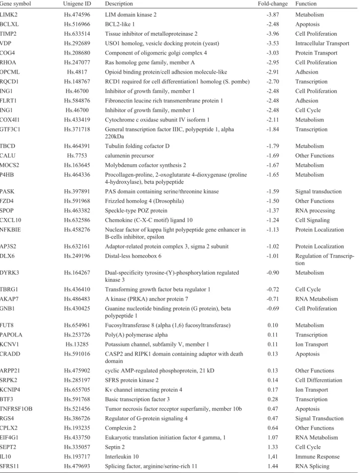
Alterations in gene expression were evaluated in U343 cells treated with 25mM cisplatin, and RNA extrac-tion was performed after 48 h. Statistical analysis was car-ried out by the SAM method, which indicated a total of 67 differentially expressed genes: 29 down-regulated and 38 up-regulated genes at a FDR < 0.05 (Table 1). Regarding to biological functions attributed to the set of significant
genes, the most frequent categories (represented by a vari-able number of genes) were related to metabolism, ubi-quitin-proteasome, cell proliferation, adhesion, apoptosis, cell cycle and DNA repair.
By applying the real time PCR method, we confirmed the down-regulation ofRHOA,LIMK2andTIMP2 genes,
by using the same remaining RNA samples as those em-ployed in the microarray experiments. The results indicated similar gene expression patterns obtained by both methods (Figure 4). These genes were selected based on their func-tions associated with glioma cells. There were certain vari-ations regarding the magnitude of relative expression, although gene expression modulation occurred in the same direction.
Discussion
It is well known that cisplatin cytotoxicity is attrib-uted to the formation of various DNA adducts that trigger cellular responses culminating in cell death (Zhanget al., 2006). Studies on the quantitative and qualitative modula-tion of gene expression profiles under condimodula-tions of drug treatment is an interesting approach to characterize the
Figure 1- Cell survival. U343 and MRC-5 cell lines were treated with increasing concentrations of cisplatin (12.5; 25; 50; 75; 150 and 300mM). The cells were harvested 24 h (A) and 5 days (B) after treatment (mean±SD). Cell survival was measured by XTT assay.
Figure 2- Frequency of apoptotic cells in U343 cell cultures treated with different concentrations of cisplatin (12.5; 25 and 50mM). The results were obtained 24, 48 and 72 h after treatment. 500 cells were analyzed for each experiment (mean±SD).
Table 1- Genes differentially expressed in the U343 glioma cell line after cisplatin treatment (25mM for 48 h) selected through SAM analysis (FDR < 0.05).
Gene symbol Unigene ID Description Fold-change Function
LIMK2 Hs.474596 LIM domain kinase 2 -3.87 Metabolism
BCLXL Hs.516966 BCL2-like 1 -2.48 Apoptosis
TIMP2 Hs.633514 Tissue inhibitor of metalloproteinase 2 -3.96 Cell Proliferation
VDP Hs.292689 USO1 homolog, vesicle docking protein (yeast) -3.53 Intracellular Transport
COG4 Hs.208680 Component of oligomeric golgi complex 4 -3.03 Protein Transport
RHOA Hs.247077 Ras homolog gene family, member A -2.95 Cell Proliferation
OPCML Hs.4817 Opioid binding protein/cell adhesion molecule-like -2.91 Adhesion
RQCD1 Hs.148767 RCD1 required for cell differentiation1 homolog (S. pombe) -2.70 Transcription
ING1 Hs.46700 Inhibitor of growth family, member 1 -2.48 Cell Proliferation
FLRT1 Hs.584876 Fibronectin leucine rich transmembrane protein 1 -2.48 Adhesion
ING1 Hs.46700 Inhibitor of growth family, member 1 -2.48 Cell Cycle
COX4I1 Hs.433419 Cytochrome c oxidase subunit IV isoform 1 -2.11 Metabolism
GTF3C1 Hs.371718 General transcription factor IIIC, polypeptide 1, alpha 220kDa
-1.84 Transcription
TBCD Hs.464391 Tubulin folding cofactor D -1.79 Metabolism
CALU Hs.7753 calumenin precursor -1.69 Other Functions
MOCS2 Hs.163645 Molybdenum cofactor synthesis 2 -1.67 Metabolism
P4HB Hs.464336 Procollagen-proline, 2-oxoglutarate 4-dioxygenase (proline 4-hydroxylase), beta polypeptide
-1.65 Metabolism
PASK Hs.397891 PAS domain containing serine/threonine kinase -1.59 Signal transduction
FZD4 Hs.591968 Frizzled homolog 4 (Drosophila) -1.50 Other Functions
SPOP Hs.463382 Speckle-type POZ protein -1.37 RNA processing
CXCL10 Hs.632586 Chemokine (C-X-C motif) ligand 10 -1.24 Cell Signaling
NFKBIE Hs.458276 Nuclear factor of kappa light polypeptide gene enhancer in B-cells inhibitor, epsilon
-1.13 Protein Localization
AP3S2 Hs.632161 Adaptor-related protein complex 3, sigma 2 subunit -1.02 Protein Localization
DLX6 Hs.249196 Distal-less homeobox 6 -1.01 Regulation of
Transcrip-tion
DYRK3 Hs.164267 Dual-specificity tyrosine-(Y)-phosphorylation regulated kinase 3
-0.90 Metabolism
TBRG1 Hs.436410 Transforming growth factor beta regulator 1 -0.72 Cell Cycle
AKAP7 Hs.486483 A kinase (PRKA) anchor protein 7 -0.71 RNA Metabolism
GNB1 Hs.430425 Guanine nucleotide binding protein (G protein), beta polypeptide 1
-0.69 Cell Proliferation
FUT8 Hs.654961 Fucosyltransferase 8 (alpha (1,6) fucosyltransferase) 0.10 Metabolism
PAPOLA Hs.253726 Poly(A) polymerase alpha 0.11 Transcription
KCNV1 Hs.13285 Potassium channel, subfamily V, member 1 0.11 Ion Transport
CRADD Hs.591016 CASP2 and RIPK1 domain containing adaptor with death domain
0.13 Apoptosis
ARPP21 Hs.475902 cyclic AMP-regulated phosphoprotein, 21 kD 0.13 Other Functions
SRPK2 Hs.285197 SFRS protein kinase 2 0.14 Cell Differentiation
KCNIP4 Hs.655705 Kv channel interacting protein 4 0.17 Ion Transport
BTF3 Hs.591768 Basic transcription factor 3 0.28 Transcription
TNFRSF1OB Hs.521456 Tumor necrosis factor receptor superfamily, member 10b 0.47 Apoptosis
RGS4 Hs.386726 Regulator of G-protein signaling 4 0.47 Signal Transduction
CPLX2 Hs.193235 Complexin 2 0.64 Other Functions
EIF4G1 Hs.433750 Eukaryotic translation initiation factor 4 gamma, 1 1.07 RNA Metabolism
SEPT2 Hs.335057 Septin 2 1.33 Cell Cycle
IL10 Hs.193717 Interleukin 10 1,41 Immune Response
mechanisms by which chemotherapeutic agents act on can-cer cells. Although cisplatin has been used for a long time, the molecular mechanisms of cell responses associated to
its cytotoxic activity are poorly clarified. In the present work, we first studied the potential of cisplatin to induce cell death in the glioma U343 cell line. When compared to the SV40 transformed fibroblast cell line (MRC-5), U343 cells proved to be the more sensitive to cisplatin.
Survival experiments carried out with increasing drug concentrations confirmed the high potential of cisplatin to induce cytotoxic effects, as well as apoptosis, in U343 cells. Furthermore, a strong residual cytotoxic effect could still be observed several days following drug treatment. Sur-vival analysis performed after 5 days demonstrated a sig-nificant reduction in the survival rates following drug treatment (12.5 to 300mM), and a pronounced effect was observed at concentrations higher than 25mM. The analysis of apoptosis showed that 25mM cisplatin induced 20.4% of apoptotic cells following 72 h, indicating that some consid-erable proportion of cells died by apoptosis. However, damaged cells can also be effectively eliminated by other processes, such as necrosis, mitotic catastrophe, autophagy,
Figure 4- Gene expression levels determined by the cDNA microarray and real time PCR methods forRHOA,LIMKand TIMP2. The same RNA samples were used in both methods. TheDDct-values represent the log ra-tio (base 2).
Table 1 (cont.)
Gene symbol Unigene ID Description Fold-change Function
APIP Hs.447794 APAF1 interacting protein 1.45 Apoptosis
ADAMTS1 Hs.643357 ADAM metallopeptidase with thrombospondin type 1 mo-tif, 1
1.61 Cell Proliferation
TAF4 Hs.18857 TAF4 RNA polymerase II, TATA box binding protein
(TBP)-associated factor, 135kDa
1.76 Regulation of Biological Process
ULK2 Hs.168762 Unc-51-like kinase 2 (C. elegans) 1.79 Metabolism
STAM Hs.441498 Signal transducing adaptor molecule (SH3 domain and ITAM motif) 1
1.80 Signal Transduction
GPR108 Hs.167641 G protein-coupled receptor 108 1.80 Other Functions
MSX1 Hs.424414 Msh homeobox 1 1.97 Other Functions
RAB37 Hs.592097 RAB37, member RAS oncogene family 2.23 Signal Transduction
NFRKB Hs.530539 Nuclear factor related to kappaB binding protein 2.52 Response to Stress
USP38 Hs.480848 Ubiquitin specific peptidase 38 2.63 Ubiquitin-Proteasome
PCDH17 Hs.106511 Protocadherin 17 2.79 Adhesion
MECP2 Hs.200716 Methyl CpG binding protein 2 (Rett syndrome) 2.82 Transcription
TIAM1 Hs.517228 T-cell lymphoma invasion and metastasis 1 2.92 Adhesion
TUSC4 Hs.437083 Tumor suppressor candidate 4 3.06 Cell Cycle
POLR2K Hs.351475 Polymerase (RNA) II (DNA directed) polypeptide K, 7.0kDa
3.07 Transcription
PSMA1 Hs.102798 Proteasome (prosome, macropain) subunit, alpha type, 1 3.20 Ubiquitin-Proteasome
SEMA6A Hs.156967 Sema domain, transmembrane domain (TM), and cytoplas-mic domain, (semaphorin) 6A
3.35 Apoptosis
CDH13 Hs.654386 Cadherin 13, H-cadherin (heart) 3.38 Adhesion
NEK8 Hs.448468 NIMA (never in mitosis gene a)- related kinase 8 3.41 Other Functions
RAD51C Hs.412587 RAD51 homolog C (S. cerevisiae) 3.73 DNA Repair
P2RX4 Hs.321709 Purinergic receptor P2X, ligand-gated ion channel, 4 3.80 Apoptosis
TNFAIP1 Hs.76090 Tumor necrosis factor, alpha-induced protein 1 (endothelial) 4.01 Immune Response
INSM1 Hs.89584 Insulinoma-associated 1 4.74 Cell differentiation
as well as premature senescence, which irreversibly arrests cell division (Brown and Attardi, 2005).
We also tested temozolomide against a panel of glio-ma cell lines, viz., U343, U87, U251, U138 and T98G, in the laboratory, and only T98G cells were found to be sensi-tive to various concentrations of temozolomide (data not shown). According to other authors, cisplatin decreased the viability of A172 glioma cells in a time- and dose-depen-dent manner. Furthermore, cisplatin induced cytotoxicity in A172 cells showed characteristics related to apoptosis (Parket al., 2006). Apoptosis is a common response of cells to platinum compounds (Sorensonet al., 1990), and ac-cordingly, in the present study we observed apoptosis as the primary effect of cisplatin on glioma cells.
Evaluation of gene expression can provide informa-tion on regulatory mechanisms, biochemical pathways and potential targets for clinical intervention and therapies in a variety of diseases (Zhanget al., 2006). The expression profiles of drug-treated cells can be readily compared with untreated control cells to reveal sets of genes that have un-dergone alterations at the transcriptional level in response to drug treatment (Dualeet al., 2007). In the present study, the findings concerning gene expression profiles disclosed 67 significantly modulated genes in U343 cells treated with 25mM cisplatin for 48 h. The experimental conditions of drug treatment were chosen on the basis of results from sur-vival and apoptosis experiments. The statistical analysis carried out by SAM was applied to identify those gene sig-natures whose mRNA levels were significantly and differ-entially expressed between cisplatin- treated and untreated U343 cells. The quantitative results of gene expression in-dicated a set of up- and down-regulated genes, mainly related to metabolism, ubiquitin-proteasome, cell prolifera-tion, adhesion, apoptosis, cell cycle control and DNA re-pair. Among the exclusively modulated genes, only a few were selected for discussion, and this was mainly due to their biological relevance. In the case of three genes (RHOA,LIMK2 andTIMP2), the expression pattern was confirmed by the real time PCR technique, and was com-patible with the results obtained by the microarray method.
In the set of genes modulated by cisplatin, the most frequent category was related to metabolism, represented by two up-regulated (FUT8 and ULK2) and six down-regulated genes (COX4I1, DYRK3, TBCD, LIMK2,
MOCS2andP4HB).
Some of these, such asDYRK3, play a role in cell growth and development in the glioma cell line (Yamanaka
et al., 2006), whereasLIMK2is involved in stress fiber and focal adhesion formation and membrane blebs during the apoptotic process. The down-regulation of LIMK2, also demonstrated by the real time PCR method, may affect sev-eral functions, including apoptosis induction. In fibrosar-coma, the reduced expression inLIMK2protein was found to restrict the metastatic potential (Suyamaet al., 2004). Some modulated genes, such asUSP38andPSMA1, were
related to the proteasome system. The ubiquitin-protea-some system is responsible for the degradation of both damaged proteins and regulators of growth and stress re-sponse. Alterations in this proteolytic system are associated with various forms of human pathologies (Deng et al., 2007). Ubiquitin specific proteases (USPs) belong to a complex family of deubiquitinating enzymes that specifi-cally cleave ubiquitin conjugates in a great variety of sub-strates, thereby regulating the production and recycling of ubiquitin itself, and are critically involved in the control of cell growth, differentiation, and apoptosis (Ovaa et al., 2004; Rolenet al., 2006).
U343 cells treated with cisplatin also showed up-regulated (ADAMTS1 and CDH13), and down-regulated genes (GNB1,TIMP2,RHOAandING1) related to cell pro-liferation. ADAMTS1 negatively regulates tumor growth and metastasis (Vazquezet al., 1999; Luqueet al., 2003; Choiet al., 2008) , whereasTIMP2takes part in degrading ECM (extracellular matrix) and regulating the invasion process (Luet al., 2004), considered the root cause of the high recurrent incidence in glioblastoma (Kong et al., 2007). TIMPs have also been shown to exert pluripotential effects on cell growth, apoptosis and differentiation (Baker
et al., 2002; Jiang et al., 2002). Similar to TIMP2, the
RHOAgene was also down-regulated in cisplatin-treated glioma cells, and the decreased expression levels were also confirmed through real time PCR analysis. The protein en-coded byRHOAis involved in cell proliferation/stress re-sponse, and belongs to the Rho GTPases family which participates in cell growth, lipid metabolism cytoarchi-tecture, membrane trafficking, transcriptional regulation and apoptosis in response to genotoxic agents. They trigger specific signals that lead to uncontrolled cell growth, en-hanced angiogenesis, inhibition of apoptosis and genetic instability, thus resulting in tumor development (Aznar and Lacal, 2001; Luet al., 2009). In astrocytomas,RHOA ex-pression positively correlates with the degree of malig-nancy (Yanet al., 2006).
One of the most distinct features of gliomas is the in-vasive growth pattern, which prevents total surgical resec-tion. Their ability to infiltrate into normal brain parenchyma is associated to the process of cellular adhe-sion (Gieseet al., 1994). In the present work, we found five modulated genes under cisplatin treatment, which are clo-sely related to adhesion. Among these,CDH13,TIAM1and
PCDH17 were up- and FLRT1 and OPCML down-regulated, thus indicating that the invasion capacity of glioma cells can be altered by cisplatin treatment.
Tumor cell invasion involves complex interactions be-tween normal and malignant cells. It is well established that this dynamic process requires the concerted effects of vari-ous molecules including proteolytic enzymes, growth fac-tors, adhesion molecules and extracellular matrix molecules (Cuiet al., 2008).
Cell response to induced DNA damage is a highly complex event that is orchestrated by a multitude of pro-teins and signaling pathways operating together in a cell context to activate mechanisms of DNA repair, cell cycle arrest and apoptosis, all depending on the extent of the DNA damage. In the present work, we analyzed gene ex-pression profiles under conditions of apoptosis induction by cisplatin in the U343 cell line. Several modulated genes were related to apoptotic cell death (TNFRSF10B,BCL-XL,
APIP,SEMA6A,CRADDandP2RX4). These findings sug-gest that the altered expression pattern of apoptosis related genes caused by cisplatin may be involved in chemosen-sitivity, as observed in survival assaying and in the fre-quency of induced apoptosis. Dualeet al. (2007) found several apoptosis related genes in testicular germ cell tu-mors after cisplatin exposure (including BCL-2 family genes), suggesting the sensitivity of these cell lines to chemotherapeutic agents.
Some other cisplatin-modulated genes were related to cell cycle control (TBRG1,SEPT2,ING1andTUSC4) and DNA repair (RAD51C). Septins are involved in several pro-cesses, including membrane dynamics, vesicle trafficking, apoptosis, infection and cytoskeletal remodeling (Hall et al., 2005). SEPT2 is a cell cycle-regulated protein, essential for cytokinesis in human astrocytoma cells (Kim et al., 2004). Kremeret al.(2007) demonstrated a link between septins, the actin cytoskeleton and DNA damage check-point response.
ING proteins play a significant role in several impor-tant cellular processes, such as growth regulation, senes-cence, apoptosis, DNA repair and cell migration (Ythieret al., 2008; Shahet al., 2009)). TP53 target genes such as
p21WAF1 and BAX, have previously been identified as downstream targets ofp33ING1andp32ING2(isoforms of the ING family) (Fenget al., 2006). LN229 glioblastoma cells differentially up-regulatedp47ING1a in response to cisplatin, this possibly representing a protective response against drug-induced DNA damage (Tallenet al., 2008).
The HRR (Homologous Recombination Repair) pathway is critically important in the repair of DNA dam-age induced by crosslink dam-agents, such as cisplatin (Golding
et al., 2004; Jayathilakaet al., 2008). However, only the
RAD51C gene was induced in cisplatin-treated glioma cells, probably due to the high level of drug cytotoxicity at the conditions tested.RAD51plays a role in the strand inva-sion and exchange between a free DNA-end proximal to the damaged site and a homologous double stranded DNA (Kuznetsovet al., 2009).
In U373 glioblastoma cells undergoing cisplatin treatment, several genes were modulated, including those encoding proteins involved in transcriptional regulation, stress response, signal transduction, metabolism, cell struc-ture and adhesion, apoptosis and survival, inflammation and immune responses, and other processes (Ma et al., 2006). Liet al.(2007) encountered altered expression in several genes involved in DNA repair, apoptosis, cell cycle control and metabolism in ovarian cancer cells that had been exposed to cisplatin for several hours, whereas Bassi
et al.(2008) also came upon genes connected with DNA re-pair modulated in response to ionizing radiation in U343 glioma cells.
In conclusion, cisplatin-treated U343 cells showed transcriptional changes that reflect several biological pro-cesses that were affected in consequence of drug treatment. These processes are related to the extensive DNA damage caused by cisplatin treatment, visualized through the amount of induced cell death. These findings highlight the complexity of cellular responses and the signaling path-ways ultimately leading to cell death in glioma cells.
Acknowledgments
The authors would like to thank Dr. Catherine Nguyen (INSERM-Marseille, France) for kindly providing cDNA clones, Flavia S. Donaires for help with bioinfor-matics analysis, and Sueli A. Neves and Luiz A. da Costa Jr for technical assistance. This work was supported by FA-PESP (Fundação de Amparo à Pesquisa do Estado de São Paulo [99/12135-9, 04/15611-6]) and CNPq (Conselho Nacional de Desenvolvimento Científico e Tecnológico).
References
Aznar S and Lacal JC (2001) Rho signals to cell growth and apoptosis. Cancer Lett 165:1-10.
Baker AH, Edwards DR and Murphy G (2002) Metalloproteinase inhibitors: Biological actions and therapeutic opportunities. J Cell Sci 115:3719-3727.
Bassi C, Mello SS, Cardoso RS, Godoy PD, Fachin AL, Junta CM, Sandrin-Garcia P, Carlotti CG, Falcão RP, Donadi EA, et al.(2008) Transcriptional changes in U343 MG-a glio-blastoma cell line exposed to ionizing radiation. Hum Exp Toxicol 27:919-929.
Bogler O and Weller M (2002) Apoptosis in gliomas, and its role in their current and future treatment. Front Biosci 7:e339-353.
Boulikas T and Vougiouka M (2004) Recent clinical trials using cisplatin, carboplatin and their combination chemotherapy drugs (review). Oncol Rep 11:559-595.
Brabec V and Kasparkova J (2005) Modifications of DNA by platinum complexes. Relation to resistance of tumors to platinum antitumor drugs. Drug Resist Updat 8:131-146. Brown JM and Attardi LD (2005) The role of apoptosis in cancer
development and treatment response. Nat Rev Cancer 5:231-237.
ADAMTS1 in non-small cell lung cancer. Cancer Genet Cytogenet 187:80-84.
Cui Y, Ying Y, van Hasselt A, Ng KM, Yu J, Zhang Q, Jin J, Liu D, Rhim JS, Rha SY,et al.(2008) OPCML is a broad tumor suppressor for multiple carcinomas and lymphomas with frequently epigenetic inactivation. PLoS ONE 3:e2990. Deng S, Zhou H, Xiong R, Lu Y, Yan D, Xing T, Dong L, Tang E
and Yang H (2007) Over-expression of genes and proteins of ubiquitin specific peptidases (USPs) and proteasome sub-units (PSs) in breast cancer tissue observed by the methods of RFDD-PCR and proteomics. Breast Cancer Res Treat 104:21-30.
Dennis Jr G, Sherman BT, Hosack DA, Yang J, Gao W, Lane HC and Lempicki RA (2003) DAVID: Database for Annotation, Visualization, and Integrated Discovery. Genome Biol 4:P3. Duale N, Lindeman B, Komada M, Olsen AK, Andreassen A, Soderlund EJ and Brunborg G (2007) Molecular portrait of cisplatin induced response in human testis cancer cell lines based on gene expression profiles. Mol Cancer 6:53. Feng X, Bonni S and Riabowol K (2006) HSP70 induction by
ING proteins sensitizes cells to tumor necrosis factor alpha receptor-mediated apoptosis. Mol Cell Biol 26:9244-9255. Giese A, Rief MD, Loo MA and Berens ME (1994) Determinants
of human astrocytoma migration. Cancer Res 54:3897-3904.
Golding SE, Rosenberg E, Khalil A, McEwen A, Holmes M, Neill S, Povirk LF and Valerie K (2004) Double strand break re-pair by homologous recombination is regulated by cell cy-cle-independent signaling via ATM in human glioma cells. J Biol Chem 279:15402-15410.
Hall PA, Jung K, Hillan KJ and Russell SE (2005) Expression pro-filing the human septin gene family. J Pathol 206:269-278. Huang ZY, Baldwin RL, Hedrick NM and Gutmann DH (2002)
Astrocyte-specific expression of CDK4 is not sufficient for tumor formation, but cooperates with p53 heterozygosity to provide a growth advantage for astrocytesin vivo. Oncogene 21:1325-1334.
Iwadate Y, Fujimoto S, Tagawa M, Namba H, Sueyoshi K, Hirose M and Sakiyama S (1996) Association of p53 gene mutation with decreased chemosensitivity in human malignant glio-mas. Int J Cancer 69:236-240.
Iwamaru A, Szymanski S, Iwado E, Aoki H, Yokoyama T, Fokt I, Hess K, Conrad C, Madden T, Sawaya R,et al.(2007) A novel inhibitor of the STAT3 pathway induces apoptosis in malignant glioma cells bothin vitroandin vivo. Oncogene 26:2435-2444.
Jayathilaka K, Sheridan SD, Bold TD, Bochenska K, Logan HL, Weichselbaum RR, Bishop DK and Connell PP (2008) A chemical compound that stimulates the human homologous recombination protein RAD51. Proc Natl Acad Sci USA 105:15848-15853.
Jiang Y, Goldberg ID and Shi YE (2002) Complex roles of tissue inhibitors of metalloproteinases in cancer. Oncogene 21:2245-2252.
Kim DS, Hubbard SL, Peraud A, Salhia B, Sakai K and Rutka JT (2004) Analysis of mammalian septin expression in human malignant brain tumors. Neoplasia 6:168-178.
Kong L, Li Q, Wang L, Liu Z and Sun T (2007) The value and cor-relation between PRL-3 expression and matrix metallo-proteinase activity and expression in human gliomas. Neuropathology 27:516-521.
Kremer BE, Adang LA and Macara IG (2007) Septins regulate actin organization and cell-cycle arrest through nuclear ac-cumulation of NCK mediated by SOCS7. Cell 130:837-850. Kunwar S, Mohapatra G, Bollen A, Lamborn KR, Prados M and
Feuerstein BG (2001) Genetic subgroups of anaplastic astro-cytomas correlate with patient age and survival. Cancer Res 61:7683-7688.
Kuznetsov SG, Haines DC, Martin BK and Sharan SK (2009) Loss of Rad51c leads to embryonic lethality and modulation of Trp53-dependent tumorigenesis in mice. Cancer Res 69:863-872.
Lefranc F, Brotchi J and Kiss R (2005) Possible future issues in the treatment of glioblastomas: Special emphasis on cell mi-gration and the resistance of migrating glioblastoma cells to apoptosis. J Clin Oncol 23:2411-2422.
Li J, Wood 3rd WH, Becker KG, Weeraratna AT and Morin PJ (2007) Gene expression response to cisplatin treatment in drug-sensitive and drug-resistant ovarian cancer cells. Oncogene 26:2860-2872.
Livak KJ and Schmittgen TD (2001) Analysis of relative gene ex-pression data using real-time quantitative PCR and the 2(-Delta Delta C(T)) Method. Methods 25:402-408. Lu Q, Longo FM, Zhou H, Massa SM and Chen YH (2009)
Sig-naling through Rho GTPase pathway as viable drug target. Curr Med Chem 16:1355-1365.
Lu W, Zhou X, Hong B, Liu J and Yue Z (2004) Suppression of in-vasion in human U87 glioma cells by adenovirus-mediated co-transfer of TIMP-2 and PTEN gene. Cancer Lett 214:205-213.
Luque A, Carpizo DR and Iruela-Arispe ML (2003) ADAMTS1/METH1 inhibits endothelial cell proliferation by direct binding and sequestration of VEGF165. J Biol Chem 278:23656-23665.
Ma Y, Yuan RQ, Fan S, Hu C, Goldberg ID, Laterra JJ and Rosen EM (2006) Identification of genes that modulate sensitivity of U373MG glioblastoma cells to cis-platinum. Anticancer Drugs 17:733-751.
Ohgaki H (2005) Genetic pathways to glioblastomas. Neuro-pathology 25:1-7.
Ovaa H, Kessler BM, Rolen U, Galardy PJ, Ploegh HL and Masucci MG (2004) Activity-based ubiquitin-specific pro-tease (USP) profiling of virus-infected and malignant human cells. Proc Natl Acad Sci USA 101:2253-2258.
Park CM, Park MJ, Kwak HJ, Moon SI, Yoo DH, Lee HC, Park IC, Rhee CH and Hong SI (2006) Induction of p53-mediated apoptosis and recovery of chemosensitivity through p53 transduction in human glioblastoma cells by cisplatin. Int J Oncol 28:119-125.
Quackenbush J (2002) Microarray data normalization and trans-formation. Nat Genet (Suppl) 32:496-501.
Reed JE, Dunn JR, du Plessis DG, Shaw EJ, Reeves P, Gee AL, Warnke PC, Sellar GC, Moss DJ and Walker C (2007) Ex-pression of cellular adhesion molecule ‘OPCML’ is down-regulated in gliomas and other brain tumours. Neuropathol Appl Neurobiol 33:77-85.
Rougemont J and Hingamp P (2003) DNA microarray data and contextual analysis of correlation graphs. BMC Bioinfor-matics 4:15.
Sellar GC, Watt KP, Rabiasz GJ, Stronach EA, Li L, Miller EP, Massie CE, Miller J, Contreras-Moreira B, Scott D,et al. (2003) OPCML at 11q25 is epigenetically inactivated and has tumor-suppressor function in epithelial ovarian cancer. Nat Genet 34:337-343.
Shah S, Smith H, Feng X, Rancourt DE and Riabowol K (2009) ING function in apoptosis in diverse model systems. Bio-chem Cell Biol 87:117-125.
Sorenson CM, Barry MA and Eastman A (1990) Analysis of events associated with cell cycle arrest at G2 phase and cell death induced by cisplatin. J Natl Cancer Inst 82:749-755. Suyama E, Wadhwa R, Kawasaki H, Yaguchi T, Kaul SC,
Naka-jima M and Taira K (2004) LIM kinase-2 targeting as a pos-sible anti-metastasis therapy. J Gene Med 6:357-363. Tallen UG, Truss M, Kunitz F, Wellmann S, Unryn B, Sinn B,
Lass U, Krabbe S, Holtkamp N, Hagemeier C,et al.(2008) Down-regulation of the inhibitor of growth 1 (ING1) tumor suppressor sensitizes p53-deficient glioblastoma cells to cis-platin-induced cell death. J Neurooncol 86:23-30.
Torigoe T, Izumi H, Ishiguchi H, Yoshida Y, Tanabe M, Yoshida T, Igarashi T, Niina I, Wakasugi T, Imaizumi T,et al.(2005) Cisplatin resistance and transcription factors. Curr Med Chem Anticancer Agents 5:15-27.
Tusher VG, Tibshirani R and Chu G (2001) Significance analysis of microarrays applied to the ionizing radiation response. Proc Natl Acad Sci USA 98:5116-5121.
Vazquez F, Hastings G, Ortega MA, Lane TF, Oikemus S, Lom-bardo M and Iruela-Arispe ML (1999) METH-1, a human ortholog of ADAMTS-1, and METH-2 are members of a
new family of proteins with angio-inhibitory activity. J Biol Chem 274:23349-23357.
Wong ML, Kaye AH and Hovens CM (2007) Targeting malignant glioma survival signalling to improve clinical outcomes. J Clin Neurosci 14:301-308.
Yamanaka R, Arao T, Yajima N, Tsuchiya N, Homma J, Tanaka R, Sano M, Oide A, Sekijima M and Nishio K (2006) Identi-fication of expressed genes characterizing long-term sur-vival in malignant glioma patients. Oncogene 25:5994-6002.
Yan B, Chour HH, Peh BK, Lim C and Salto-Tellez M (2006) RhoA protein expression correlates positively with degree of malignancy in astrocytomas. Neurosci Lett 407:124-126. Ythier D, Larrieu D, Brambilla C, Brambilla E and Pedeux R
(2008) The new tumor suppressor genes ING: Genomic structure and status in cancer. Int J Cancer 123:1483-1490. Zhang P, Zhang Z, Zhou X, Qiu W, Chen F and Chen W (2006)
Identification of genes associated with cisplatin resistance in human oral squamous cell carcinoma cell line. BMC Cancer 6:224.
Internet Resources
MEV software: http://tm4.org/mev.html (last date of access: No-vember 1, 2009).
S.O.U.R.C.E. (Stanford Online Universal Resource for Clones and ESTs): source.stanford.edu (last date of access: October 30, 2009).
NCBI: http://www.ncbi.nlm.nih.gov/.
Associate Editor: Carlos F.M. Menck