RUTE ISABEL PAULO MARTINS
POST-TRANSCRIPTIONAL REGULATION OF HFE
GENE EXPRESSION
LISBOA
nº de arquivo
RUTE ISABEL PAULO MARTINS
POST-TRANSCRIPTIONAL REGULATION OF HFE
GENE EXPRESSION
LISBOA
2010
Thesis presented to obtain the Ph.D.
degree in Biology (Molecular Genetics),
by the Universidade Nova de Lisboa,
Agradecimentos
Agradecimentos
Ao concluir este trabalho gostaria de expressar o meu reconhecimento a todos aqueles que
de algum modo contribuíram para a sua concretização.
Esta tese de Doutoramento é o resultado de um trabalho de investigação realizado no
Departamento de Genética do Instituto Nacional de Saúde Dr. Ricardo Jorge (INSA), em
Lisboa, entre Janeiro de 2006 a Dezembro de 2009, onde foram disponibilizadas as
condições essenciais para o seu desenvolvimento. Ao Presidente do Conselho Directivo do
INSA, Professor Doutor José Pereira Miguel, manifesto o meu apreço.
Agradeço à Doutora Maria Guida Boavida e ao Doutor João Lavinha por me terem recebido
no então Centro de Genética Humana, permitindo a realização do meu trabalho, e por todo
o interesse que demonstraram pelo mesmo.
Este trabalho foi, na sua maioria, financiado pela Fundação para a Ciência e a Tecnologia no
âmbito do projecto de investigação PTDC/SAU/GMG/64494/2006, através do Programa de
Financiamento Plurianual do Centro de Investigação em Genética Molecular Humana e sob a
forma de uma Bolsa de Doutoramento com a referência SFRH/BD/21340/2005.
À minha orientadora Doutora Paula Faustino estou reconhecida pela oportunidade inicial de
me integrar no seu grupo de investigação, cujo acompanhamento ao longo destes oito anos
tem sido crucial para a minha formação enquanto cientista. Pela transmissão de
conhecimentos, pelo exemplo de rigor científico, pela disponibilidade, mas também pelo
companheirismo e amizade com que sempre me distinguiu, muito tenho a agradecer-lhe.
À minha orientadora Doutora Luísa Romão agradeço os seus ensinamentos, estímulo e rigor
científico, que foram essenciais ao longo deste período de quatro anos, sempre me
relembrando da importância da perseverança e da dedicação ao longo deste árduo percurso.
Saliento ainda a amizade demonstrada e os conselhos que sempre me transmitiu.
Ao Doutor Pedro Baptista, como elo de ligação da Faculdade de Ciências e Tecnologia da
Universidade Nova de Lisboa agradeço toda a disponibilidade, assim como o interesse
Agradecimentos
A todos os meus colegas do Departamento de Genética, e muitos deles agora amigos,
agradeço todo o apoio, cumplicidade e amizade com que sempre me distinguiram. A sua
companhia, ao trabalhar num espírito tão agradável, tornou-se essencial para a minha
motivação diária ao longo da execução deste trabalho. Em particular, agradeço ao Bruno
Silva e à Daniela Proença pelo contributo em vários aspectos práticos deste trabalho, à Ana
Luísa Silva por todos os ensinamentos e ideias profícuas e à Ana Morgado pela assistência na
execução e revisão deste texto.
Gostaria de agradecer ainda a paciência e apoio de todos aqueles que discutiram este
trabalho e reviram este texto, absolutamente fundamentais para a sua execução e
conclusão.
Aos meus amigos e familiares serei eternamente grata pela sua compreensão das minhas
dificuldades de disponibilidade inerentes à condição de estudante de Doutoramento,
apoiando-me sempre e demonstrando o maior dos companheirismos e amizade.
Aos meus Pais, agradeço o seu amor, carinho, confiança e apoio incondicionais,
incentivando sempre as minhas decisões e por estarem, de um modo ou outro, sempre
presentes.
Finalmente agradeço ao Miguel, pelo apoio incondicional e paciência infinita, pelo incentivo
e pelo amor, acreditando em mim muito mais que eu própria. O que lhe reconheço jamais
Sumário
Sumário
O ferro é um elemento essencial em diversos processos metabólicos celulares. O desafio que
se coloca para a maioria dos organismos prende-se com o controlo do ferro absorvido de
modo a suprir as necessidades destes processos evitando, no entanto, os danos causados
pelo ferro livre. Na realidade, algumas das doenças humanas mais comuns estão
relacionadas com a perturbação da homeostase do ferro. Entre estas, encontra-se a
hemocromatose hereditária que, estando maioritariamente associada a mutações no gene
HFE, origina a acumulação de ferro em vários órgãos. A proteína HFE actua na homeostase
do ferro através da regulação da expressão da hepcidina no fígado. O principal transcrito
HFE apresenta baixos níveis de expressão numa série de tecidos humanos, tendo sido
descritos diversos transcritos adicionais.
O trabalho aqui apresentado aborda a caracterização dos transcritos alternativos de HFE, os
mecanismos envolvidos na sua génese, assim como o seu possível papel fisiológico e
regulação. A análise de diversos tecidos humanos permitiu identificar vários transcritos HFE
resultantes de splicing alternativo. O estudo funcional de algumas proteínas
correspondentes demonstrou que o processo de splicing alternativo pode gerar variantes
não funcionais ou produzir uma variante HFE solúvel que é secretada pelas células associada
à beta2-microglobulina. Esta proteína poderá desempenhar um papel crucial na homeostase
do ferro, actuando como um agonista ou antagonista da HFE full length. Além disso, foi
demonstrado que a expressão do transcrito HFE principal é fisiologicamente regulada pelo
mecanismo de nonsense-mediated mRNA decay (NMD), dado que os seus níveis aumentam
quando este mecanismo é inibido. A pesquisa realizada em tecidos humanos permitiu
verificar que a expressão do mRNA HFE resulta da utilização de quatro locais de clivagem e
poliadenilação alternativos. Este padrão de poliadenilação alternativa específico de tecido
aparenta responder a estímulos de ferro, actuando coordenadamente com o NMD no
ajustamento dos níveis de expressão de HFE.
Esta dissertação demonstra que a regulação da expressão do gene HFE é influenciada
pós-transcricionalmente pelos mecanismos de splicing alternativo, poliadenilação alternativa e
NMD. Este conhecimento poderá conduzir a novas perspectivas de investigação na área do
metabolismo do ferro e contribuir para o delinear de novas estratégias terapêuticas a aplicar
Abstract
Abstract
Iron is a key element for numerous metabolic processes in living cells. The challenge for
most organisms is to acquire the adequate amounts of iron for these processes yet avoiding
the toxicity associated with free iron. In fact, disruptions of iron homeostasis account for
some of the most common human diseases. Amongst these, lays hereditary
hemochromatosis, which is mainly associated with mutations in the HFE gene, leading to
iron overload in specific organs. HFE protein acts in iron homeostasis by regulating the
expression of hepcidin in the liver. Besides the major HFE transcript, which is expressed at
low levels in a wide range of human tissues, several additional alternative HFE transcripts
have been described.
The work presented in this dissertation addresses the characterization of HFE alternative
transcripts, the biological mechanisms involved in their genesis as well as their physiological
significance and regulation. A variety of human tissues was analysed and shown to express
several alternatively spliced HFE transcripts. Functional analysis of the corresponding
proteins revealed that alternative splicing can either generate non-functional HFE protein
variants or produce a soluble HFE variant that is secreted by cells associated with
beta2-microglobulin. This soluble HFE may have a vital role in iron homeostasis by acting as an
agonist or antagonist of the full length HFE. Furthermore, HFE transcripts were found to be
physiologically regulated by the nonsense-mediated mRNA decay (NMD), since its levels are
significantly increased when depleting human cells from a key NMD effector. Through the
analysis of several human tissues, it is shown that HFE mRNA expression results from
alternative cleavage and polyadenylation at four different sites. This tissue-specific
polyadenylation pattern seems to respond to cellular iron status, acting coordinately with
NMD to fine-tune HFE’s expression levels.
The regulation of HFE gene expression is here shown to be post-transcriptionally influenced
by alternative splicing, alternative polyadenylation and nonsense-mediated mRNA decay
mechanisms. These findings may hint future directions in the active field of iron biology
research and provide interesting cues that could be translated into new therapeutics for iron
Abbreviations
Abbreviations
A adenosine
apo-Tf iron-free transferrin
bp base pairs
BMP bone morphogenetic protein
C cytidine
CBP cap-binding protein
CD cluster of differentiation of leukocytes
cDNA mRNA complementary DNA
C/EBP CCAAT/enhancer binding protein
Cyto cytoplasmic tail
C-terminal carboxyl-terminal
DAPI 4,6‐diamidino‐2‐phenylindole
DcytB duodenal cytochrome B
DMEM Dulbecco’s modified Eagle medium
DMT1 divalent metal transporter 1
DNA deoxyribonucleic acid
DNase deoxyribonuclease
DSE downstream sequence element
EJC exon junction complex
Epo-R erythropoietin receptor
ER endoplasmic reticulum
eRF eukaryotic release factor
ERK 1/2 extracellular signal-regulated kinases 1/2 ESE exonic splicing enhancer
ESS exonic splicing silencer
FBS fetal bovine serum
Fe2-Tf diferric-iron-loaded transferrin
G guanosine
GDF15 growth differentiation factor 15 GFP green fluorescent protein GPI glycosylphosphatidylinositol
GPS1 G protein pathway suppressor 1
HCP1 heme carrier protein 1
HFE high Fe
HIF-1 hypoxia inducible factor-1
HIV human immunodeficiency virus
HH hereditary hemochromatosis
Abbreviations
hnRNP heterogenous nuclear RPN
HO1 heme oxygenase 1
holo-Tf iron-loaded Tf
IL interleukin
IP immunoprecipitation
IRE iron responsive element
IRP iron regulatory protein ISE intronic splicing enhancer ISS intronic splicing silencer
JAK Janus kinase
kb kilo bp
kDa kilo dalton
MAPK mitogen-activated protein kinase MHC major histocompatibility complex
mRNA messenger ribonucleic acid
Nef negative factor
NMD nonsense mediated mRNA decay
nt nucleotide
NTBI non-transferrin bound iron
OMIM Online Mendelian Inheritance in Men
ORF open reading frame
PABPC1 cytoplasmic poly(A)-binding protein 1
PBMC peripheral blood mononuclear cell
PBS phosphate buffered saline
PCR polymerase chain reaction
PCNA proliferating cell nuclear antigen
Poly(A) polyadenylation
PVDF polyvinylidene fluoride
PTC premature termination codon
qPCR quantitative real-time PCR
RACE rapid amplification of cDNA ends RES reticuloendothelial system
RNAP II RNA polymerase II
RNP ribonucleoprotein
RNase ribonuclease
RPMI Roswell Park Memorial Institute
ROS reactive oxygen species
RT reverse transcription
SDS-PAGE sodium dodecyl sulfate‐polyacrylamide gel electrophoresis
Abbreviations
siRNA short interfering RNA
Steap3 six-transmembrane epithelial antigen of the prostate 3
sTfR1 serum TfR1
SMAD this acronym results from the combination of two proteins: the Caenorhabditis elegans protein Sma (designated as mutations in this gene causes animals to be
small) and the drosophila protein MAD (“mothers against decapentaplegic”)
SMG suppressor with morphogenetic effects on genitalia
snRNP small nuclear RNP
ss splice site
STAT3 signal transducer and activator of transcription 3 TBST tris‐buffered saline with triton
T thimidine
Tf transferrin
TfR transferrin receptor
TGF- transforming growth factor
Tm transmembrane domain
Tris tris(hydroxymethyl)aminomethane
TWSG1 twisted gastrulation 1
U uridine
uORF upstream ORF
UPF up-frameshift
UTR untranslated region
WT wild type
Zip14 Zrt-Irt-like protein 14
Index
Contents
Agradecimentos ... iii
Sumário ... v
Abstract ... vii
Abbreviations ... ix
Contents ... xiii
Figure Index ... xvii
Table Index ... xvii
Chapter 1 - General Introduction ...19
I. General overview of iron metabolism ... 21
I.1. Iron distribution, utilization and recycling ... 21
I.2. Iron acquisition, transport and storage ... 23
I.2.1. Iron absorption ... 23
I.2.2. Cellular iron uptake ... 24
I.2.2.1. Transferrin bound iron ... 24
I.2.2.2. Non-transferrin bound iron ... 26
I.2.3. Iron storage ... 27
II. Regulation of iron homeostasis... 27
II.1. Molecular mechanisms involved in the expression of iron-related genes ... 28
II.1.1. Transcriptional regulation ... 28
II.1.2. Post-transcriptional regulation... 29
II.1.2.1. Iron regulatory proteins ... 29
II.1.2.2. Alternative splicing ... 30
II.1.3. Post-translational regulation ... 32
II.2. Systemic regulation of iron homeostasis by hepcidin ... 33
II.2.1. Hepcidin regulation by erythropoiesis, hypoxia and inflammation ... 34
II.2.2. Hepcidin regulation by iron status ... 37
II.2.2.1. Hemojuvelin, BMPs and matriptase-2 ... 37
II.2.2.2. HFE and transferrin receptors (TfR1 and TfR2) ... 39
II.2.2.2.1. HFE biology and function ... 42
II.2.2.2.2. HFE molecular genetics and expression ... 47
II.3. Disorders of disrupted iron homeostasis ... 50
Index
III. Post-transcriptional regulation of gene expression ... 53
III.1. General aspects of the gene expression pathway ... 54
III.2. Constitutive and alternative splicing ... 55
III.2.1. Alternative splicing and gene expression diversity ... 56
III.3. Nonsense-mediated messenger RNA decay ... 58
III.3.1. NMD rule and activation ... 58
III.3.2. Gene expression regulation by NMD ... 61
Chapter 2 - Differential HFE gene expression regulation by alternative splicing in human tissues ... 65
Author’s note ... 67
I. Abstract ... 69
II. Introduction ... 70
III. Materials and Methods ... 72
III.1. First strand cDNA synthesis and polymerase chain reaction ... 72
III.2. Quantitative real-time PCR ... 73
III.3. Plasmid constructs ... 73
III.4. Cell culture and transient transfections ... 74
III.5. Immunofluorescence assays ... 74
III.6. Immunoprecipitation assays ... 75
III.7. Western blot analysis ... 75
IV. Results ... 76
IV.1. HFE mRNA is alternatively spliced in different human tissues ... 76
IV.2. Absolute and relative quantification of HFE exon 2 skipping and intron 4 inclusion transcripts reveal a tissue-specific pattern ... 78
IV.3. HFE variants present distinct subcellular localization ... 80
IV.4. Immunoprecipitation assays reveal a soluble and secreted HFE protein isoform ... 82
V. Discussion ... 84
VI. Acknowledgments ... 86
Chapter 3 - Alternative polyadenylation and nonsense-mediated decay act in concert to fine-tune HFE mRNA levels in response to the cellular iron status ... 87
Author’s note ... 89
I. Abstract ... 91
II. Introduction ... 92
III. Materials and Methods ... 95
III.1. 3’ Rapid amplification of cDNA ends ... 95
Index
III.3. Cell culture and transfections ... 97
III.4. Transient transfections of siRNAs ... 98
III.5. RNA isolation ... 98
III.6. First strand cDNA synthesis and quantitative real-time PCR ... 99
III.7. Western blot analysis ... 99
III.8. Statistical analysis ... 99
IV. Results ... 100
IV.1. Usage of two novel alternative polyadenylation sites in the human HFE transcripts located at exons 6 and 7 ... 100
IV.2. The physiological human HFE mRNA is a natural NMD-target ... 102
IV.3. The HFE transcripts carrying a nonsense mutation are also committed to NMD ... 105
IV.4. Holo-transferrin seems to induce preferential recognition of a poly(A) signal at exon 6, shortening the 3’ UTR, and thus it mitigates the effect of NMD on HFE gene expression regulation ... 108
V. Discussion ... 111
VI. Acknowledgments ... 115
Chapter 4 - General Discussion ... 117
I. General discussion and concluding remarks ... 119
II. Future perspectives ... 127
Figure and Table Index
Figure Index
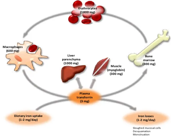
Figure 1.1. Distribution of iron within the body. ...22
Figure 1.2. Iron acquisition, transport and storage. ...25
Figure 1.3. Transcriptional regulation of hepcidin expression. ...35
Figure 1.4. Schematic representation of the HFE protein at cell surface. ...43
Figure 1.5. Schematic representation of the HFE gene. ...47
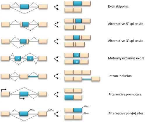
Figure 1.6. Elementary alternative splicing events. ...57
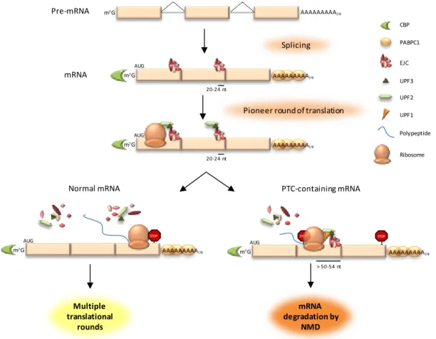
Figure 1.7. Premature stop codon recognition in mammals. ...60
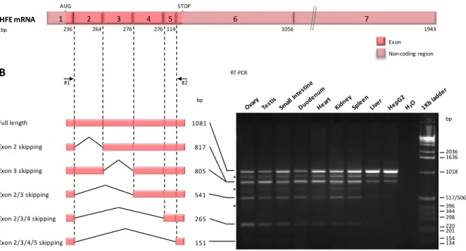
Figure 2.1. Splicing forms of HFE gene in several human tissues. ...76
Figure 2.2. Expression of the intron 4 inclusion HFE splice transcript in several human tissues. ...78
Figure 2.3. Absolute and relative quantification of the exon 2 skipping and intron 4 inclusion HFE splice transcripts. ...80
Figure 2.4. Cellular localization of HFE splice variants by immunofluorescence analysis. ...81
Figure 2.5. Immunoprecipitation assays of transfected HFE splice variants. ...83
Figure 3.1. Usage of four alternative poly(A) sites for 3’ end cleavage and polyadenylation of human HFE transcripts. ... 101
Figure 3.2. Downregulating UPF1 from HeLa or HepG2 cells results in an upregulation of the endogenous HFE transcripts indicating that the physiological HFE mRNA is a natural NMD-target. ... 104
Figure 3.3. Normal and nonsense-mutated human HFE transcripts show low levels of expression when compared to those obtained for an NMD-resistant HFE transcript. ... 107
Figure 3.4. Holo-transferrin treatment of HepG2 cells increases endogenous HFE mRNA levels by inducing a preferential recognition of a poly(A) signal at exon six, which makes the transcripts NMD-resistant. ... 110
Table Index
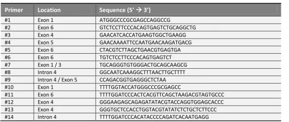
Table 2.1. DNA oligonucleotides used in the current work...73CHAPTER 1
General Introduction
I.
General overview of iron metabolism
Iron is crucial for life, being involved in a diversity of cellular processes such as oxygen
transport, electron transfer and DNA synthesis. The biological relevance of iron is largely
attributable to its chemical properties as a transition metal. This capability of readily
engaging one electron, allows iron to coexist either in an oxidized insoluble (Fe3+) or a
reduced soluble (Fe2+) form. The dark side of this element is that, in excess, free iron
catalyzes the production of oxidative radicals that are able to damage the macromolecular
components of cells. On the other hand, cellular iron deficiency arrests cell growth leading to
cell death [Halliwell and Gutteridge 1984].
Iron is the fourth most abundant component in the Earth’s crust, but most of the environmental iron exists in the ferric (Fe3+) form, which is almost insoluble in water at
neutral pH, severely compromising its biological utility. Being an essential trace element
required by virtually all organisms (except for a few species of bacteria), they have
developed complex systems of iron transport and management to deal with its poor
bioavailability [Chua et al. 2007]. Even so, iron balance is tenuous, as both iron deficiency and
iron overload are deleterious. These disorders of iron homeostasis are amongst the most
common diseases in humans, affecting up to one-quarter of the world's population [McLean et
al. 2009]. To get a glimpse of how iron balance is accomplished, the molecular mechanisms
involved in the regulation of iron homeostasis will be exposed in the course of this
dissertation.
I.1.
Iron distribution, utilization and recycling
One astonishing feature of iron metabolism is the extent to which body iron is conserved.
Although the adult human organism contains 3 to 5 g of iron, only 1-2 mg enters and leaves
the body on a daily basis (Figure 1.1). Iron excretion is a rather unregulated pathway, as it is
the result of mandatory losses through menstruation, sloughing of epithelial cells from the
skin and from the mucosal cells of the gastrointestinal, biliary and urinary tracts [Cook et al.
1973; Andrews 1999]. Conversely, all cells require a small amount of iron but the precursors of
red blood cells, the erythroblasts, are by far the most demanding. Under normal
Chapter 1
heme biosynthesis in the bone marrow to fulfill the production of more than 200 billion
erythrocytes [Hentze et al. 2004; Chua et al. 2007]. In fact, more than two thirds (60-70%) of the
total body iron content is present as hemoglobin in erythrocytes, whilst another 10% is
contained in myoglobin, iron-containing enzymes and cytochromes. The remaining 20-30% is
stored in the liver and macrophages as ferritin and hemosiderin [Cook et al. 1973; Andrews 1999].
The major source of iron for the erythroid precursors is plasma iron-transferrin (Fe2-Tf). But
the circulating Fe2-Tf pool is 10 times smaller than the daily iron requirements, so a high
turnover rate is necessary to ensure the adequate supply of iron to the bone marrow
[Andrews 1999; Nemeth 2008]. This recycling process is carried out by macrophages of the
reticuloendothelial system (RES) present in the spleen, liver and bone marrow. Through the
phagocytosis of senescent erythrocytes, the iron within is recovered, transferred to the bone
marrow and re-incorporated during the synthesis of new red blood cells [Knutson and
Wessling-Resnick 2003].
Figure 1.1. Distribution of iron within the body. In a balanced state, about 1-2 mg of iron is daily absorbed and a similar amount is lost. Most of the iron that circulates in the plasma is incorporated into hemoglobin in erythroid cells. As only about 0.1% of the total body iron content is found in the plasma, the recycling of the iron present in the senescent erythrocytes by the macrophages is crucial to meet the erythropoietic demands. The major iron storage compartments are the liver, the macrophages and the muscles. (Adapted from Pietrangelo 2004).
Bone marrow (300 mg) Erythrocytes
(1800 mg)
Macrophages (600 mg)
Sloughed mucosal cells Desquamation Menstruation
Liver parenchyma
(1000 mg)
Plasma transferrin
(3 mg)
Dietary iron uptake (1-2 mg/day)
Muscle (myoglobin)
(300 mg)
General Introduction
I.2.
Iron acquisition, transport and storage
I.2.1.
Iron absorption
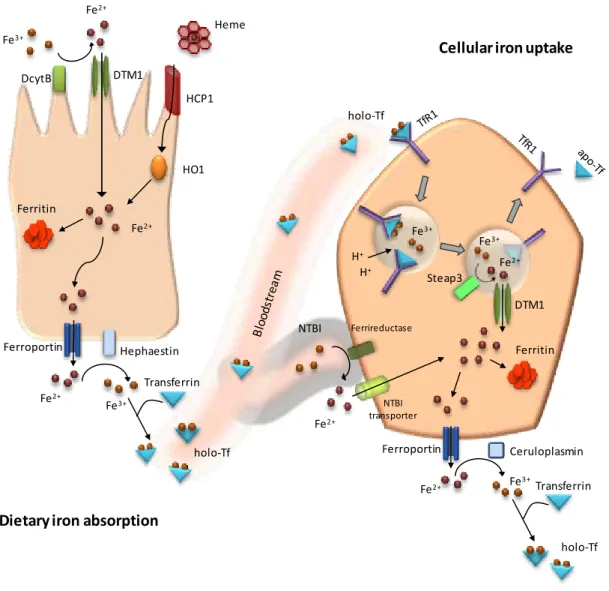
Dietary iron absorption is achieved in the duodenum, where iron must traverse the apical
and basolateral membranes of the absorptive cells (enterocytes) in order to reach the
bloodstream (Figure 1.2). Iron is present in two forms in the diet: heme iron (derived from
hemoglobin and myoglobin) and non-heme iron (present as iron hydroxides, salts and
iron-containing proteins such as ferritin) [Carpenter and Mahoney 1992; Lopez and Martos 2004]. Heme
and non-heme iron pass from the intestinal lumen to the enterocyte by distinct pathways,
but once within the cell, iron from each source will be part of a common intracellular pool
that can either be stored in the form of ferritin or transported into the bloodstream [Hentze et
al. 2004; Chua et al. 2007].
Although heme iron is more efficiently absorbed than non-heme iron, this only accounts for
about 10 to 15% of daily iron intake [Carpenter and Mahoney 1992]. The mechanism by which
heme is taken up by duodenal enterocytes remains controversial. A candidate brush border
heme transporter was described, the heme carrier protein 1 (HCP1), but it was further
demonstrated that this protein transports folate far more efficiently than heme [Shayeghi et al.
2005; Qui et al. 2006]. After crossing the apical membrane, iron is excised from the heme
porphyrin ring, under the action of heme oxygenase 1 (HO1), becoming part of the cytosolic
iron [Raffin et al. 1974].
The majority of non-heme iron enters the gastrointestinal tract in the ferric form that must
be converted into the ferrous form for bioavailability. Numerous dietary components are
capable of reducing ferric iron, but the enterocytes have endogenous reducing activity [Lopez
and Martos 2004]. It is currently accepted that this is achieved by the duodenal cytochrome B
(DcytB) reductase that is expressed on the apical membrane [McKie et al. 2001]. However, the
absence of an abnormal phenotype in DcytB knockout mice, suggests the presence of other
brush border ferrireductases [Gunshin et al. 2005]. Once Fe2+ is formed, it becomes a substrate
for the divalent metal transporter 1 (DMT1; also known as DCT1 or Nramp2), the intestinal
iron importer, for transport across the membrane into the cytoplasm [Gunshin et al. 1997;
Fleming et al. 1997]. The role of this molecule is supported by the animal models, where a
defective DMT1 gene leads to ineffective iron uptake and microcytic anemia [Fleming et al.
Chapter 1
Once within the enterocyte, iron has two possible fates, depending on iron requirements. If
the iron demand is low, it can be stored as ferritin, being eventually lost by sloughing of the
villus tip [Geyer 1979]. Conversely, if there is a requirement to replenish the stores or an
increased metabolic demand, iron will be transported across the basolateral membrane into
the circulation [Abboud and Haile 2000; Donovan et al. 2000; McKie et al. 2000]. This transport is
assured by ferroportin (also known as IREG1, MTP1 or SLC40A1). Selective inactivation of the
murine ferroportin in intestinal cells confirms that ferroportin is the major, and most
probably, the only iron exporter [Donovan et al. 2005]. As ferroportin is selective for Fe2+, the
iron export depends on a multicopper oxidase to convert Fe2+ to Fe3+ for incorporation of
iron into transferrin, the serum iron carrier protein [Schade and Caroline 1946]. Hephaestin is a
membrane-bound homologue of the serum multicopper oxidase ceruloplasmin and most
likely the responsible for the release of oxidized iron into the bloodstream [Vulpe et al. 1999]. It
was also shown that ceruloplasmin may carry out the oxidase function at the basolateral
membrane of the enterocyte [Cherukuri et al. 2005]. This hypothesis arose from the evidence
that iron accumulation in enterocytes of the mouse model for sex-linked anemia is resolved
after the neonatal period, suggesting that hephaestin is needed for iron stores during rapid
growth, while ceruloplasmin may be required in adult stages [Edwards and Bannerman 1970;
Cherukuri et al. 2005].
I.2.2.
Cellular iron uptake
I.2.2.1.
Transferrin bound iron
The major iron source for most tissues is transferrin bound iron. Transferrin (Tf) has two
iron-affinity binding sites, keeping iron non-reactive in circulation and extravascular fluid,
delivering it to cells bearing specific receptors [Bailey et al. 1988]. The classic transferrin
receptor 1 (TfR1) is expressed in most cells, presenting a higher expression in rapid
proliferating cells, activated lymphocytes and erythroid precursors [Ponka and Lok 1999; Ned et
al. 2003]. The almost ubiquitous expression of this receptor reveals the importance of a
constitutive pathway for iron acquisition by receptor-mediated endocytosis, the so-called
transferrin cycle, which has become a paradigm in cell biology. Briefly, to initiate the cycle,
diferric transferrin (or holo-transferrin; holo-Tf) binds to TfR1 at cell surface that will
General Introduction
promoting iron release from Tf and Fe3+ will be reduced by the ferrireductase Steap3
(Six-transmembrane epithelial antigen of the prostate 3), allowing (Six-transmembrane transport by
DMT1 [Dautry-Varsat et al. 1983; Fleming et al. 1998; Ohgami et al. 2005]. The iron is then utilized by
the cell or stored as ferritin. The Tf cycle is completed when the endosome returns and fuses
with the cell membrane, where the receptor becomes accessible and apo-Tf (iron-free
transferrin) is released to circulation, allowing both molecules to start the cycle all over
again.
Figure 1.2. Iron acquisition, transport and storage. The absorption of dietary iron (heme and non-heme) is achieved by the enterocytes of the duodenum (on the left). Once within the enterocyte, iron may be stored or transported to the bloodstream. Then, iron is acquired by almost all human cells, so a generic cell is depicted on the right. Uptake of transferrin bound iron occurs through the transferrin cycle, whereas non-transferrin bound iron is mediated by specific transporters. See text for details.
Fe3+ Fe2+ DTM1 DcytB Heme HCP1 HO1 Fe2+ Ferritin Hephaestin Ferroportin Fe2+ Fe3+ Transferrin H+ H+ Fe3+ Fe3+ Fe2+ DTM1 Steap3 Ferritin Ferroportin
Fe2+ Fe3+Transferrin
Ceruloplasmin holo-Tf holo-Tf holo-Tf NTBI Fe2+ Ferrireductase NTBI transporter
Dietary iron absorption
Chapter 1
It is currently accepted that there is an important molecule liable to affect the transferrin
cycle, the HFE (high Fe) protein. The membranar association of HFE with TfR1, as well as its
ability to compete with transferrin for binding to the receptor, brought new insights to the
iron metabolism field [Parkkila et al. 1997a; Feder et al. 1998; Lebron et al. 1998]. A second transferrin
receptor, TfR2, is strongly expressed in liver hepatocytes, but has a lower binding affinity for
holo-Tf than TfR1 [Kawabata et al. 1999]. TfR2 is capable of mediating the internalization and
recycling of Tf by a similar mechanism to that described for TfR1 [Kawabata et al. 1999; Graham et
al. 2008]. Recent evidence suggests that both HFE and TfR2 are involved in a specific pathway
with strong interactions with the uptake of iron by the TfR1-mediated endocytosis, which
will be further explored in this thesis.
I.2.2.2.
Non-transferrin bound iron
Iron can also be present in the plasma in a free form, generally designated as non-transferrin
bound iron (NTBI). It actually consists in iron bound to low affinity molecules, with the major
component identified as ferric citrate [Grootveld et al. 1989]. The concentration of NTBI is
normally low but it increases when the binding capacity of transferrin becomes saturated.
Since it easily penetrates into cells, particularly in the liver and heart, NTBI has great
pathophysiological importance in iron overload disorders [Breuer et al. 2000; Chua et al. 2004].
The uptake of NTBI by the cells is still far from understood (Figure 1.2). It likely involves cell
surface reduction by an unidentified ferrireductase to dissociate iron from its ligand, possibly
by Steap3 [Chua et al. 2007]. Then, iron is delivered into the cell by a transporter [Trinder and
Morgan 1998]. Several plausible candidate transporters for NTBI have emerged, including
DMT1, Zip14 (Zrt-Irt-like protein 14) and calcium channels [Oudit et al. 2003; Chua et al. 2004; Liuzzi
et al. 2006; Shindo et al. 2006]. The relative contribution of these transporters to NTBI uptake is
poorly characterized, but it is likely that more than one of these transporters is involved
General Introduction
I.2.3.
Iron storage
Following delivery to the cells, iron enters an intermediate intracellular labile iron pool,
where it can be incorporated into ferritin or heme, associated with other non-heme iron
proteins in the cytosol or exchanged between the intracellular endosomal, lysosomal and
mitochondrial compartments [Mulligan et al. 1986; Chua et al. 2007].
In general, the excess of iron will be stored in the form of ferritin, a water soluble molecule
consisting of 24 subunits, capable of sequestering up to 4,500 atoms of iron (Figure 1.2)
[Harrison 1977]. As the amount of iron in the cells increases, a larger percentage deposits in
hemosiderin, an insoluble molecule thought to be a by-product of ferritin degradation [Munro
and Linder 1978]. This has been suggested as a protective mechanism against oxidative
damage, since iron stored in hemosiderin is more inaccessible and less effective in producing
free radicals than iron stored in ferritin *O’Connell et al. 1986+.
The main sites for body iron storage are the hepatic parenchyma (or hepatocytes) and the
macrophages of the reticuloendothelial system [Cook et al. 1973; Andrews 1999]. In fact, iron
accumulation in the reticuloendothelial cells of the liver, spleen and bone marrow, occurs
when body iron stores are replete. Iron in the RES is a secondary accumulation due to the
catabolism of the red cell heme acquired via erythrophagocytosis [Knutson and Wessling-Resnick
2003]. Stored iron in hepatocytes and macrophages can be mobilized to meet erythropoietic
and cellular demands, when body iron stores are low [Andrews 1999; Hentze et al. 2004; Chua et al.
2007].
II.
Regulation of iron homeostasis
Since iron loss is essentially an unregulated process, a tight balance between iron
absorption, uptake, transport, storage and utilization is essential to maintain iron
homeostasis. Among these compartments, a constellation of factors directly or indirectly
related with iron regulation (the so-called iron-related genes) must be extensively controlled
Chapter 1
II.1.
Molecular mechanisms involved in the expression of
iron-related genes
II.1.1.
Transcriptional regulation
The transcriptional regulation of iron-related genes has been shown to respond to several
stimuli like iron status, erythropoietic activity, inflammation and hypoxia. In fact, the major
player in the regulation of iron homeostasis is the liver-derived hormone hepcidin, which is
transcriptionally controlled by all the stimuli above indicated [Nicolas et al. 2002a; Nicolas et al.
2002b; Nemeth et al. 2003; Pinto et al. 2008]. It is known that the hepcidin promoter contains
binding motifs for several recognized transcriptional factors [Courselaud et al. 2002; Truksa et al.
2007; Weizer-Stern et al. 2007; Casanovas et al. 2009; Truksa et al. 2009]. The expression of hepcidin is
indeed regulated by a range of upstream molecules that culminates in the binding of factors
to the hepcidin promoter and these pathways will be further exposed in this thesis.
Cytokines, such as interleukin-6 (IL-6), interleukin-1 (IL-1) and interferon-γ, have been shown to affect the messenger RNA (mRNA) expression of several iron-related genes. Such is the
case of H-ferritin [each ferritin is composed by two chains, heavy (H) and light (L)],
transferrin receptor 1, hepcidin and ferroportin genes [Wei et al. 1990; Fahmy and Young 1993; Tran
et al. 1997; Lee et al. 2004; Nemeth et al. 2004a; Lee et al. 2005].
Hypoxia is intimately related with erythropoiesis. Under low oxygen tension, the
transcription factor HIF-1 (hypoxia inducible factor-1) is activated and will interact with
erythropoietin, increasing the iron required for erythropoiesis [Wang and Semenza 1995; Semenza
1999]. Consequently, hypoxia has been shown to affect the expression of transferrin, TfR1,
ceruloplasmin, ferroportin, DcytB and hepcidin genes [Rolfs et al. 1997; Lok and Ponka 1999;
Tacchini et al. 1999; McKie et al. 2000; Mukhopadhyay et al. 2000; McKie et al. 2001; Nicolas et al. 2002b].
As expected, iron levels may control the transcription of some iron-related genes. Indeed,
McKie et al. [2001] have shown that in iron-deprived mice, the expression of DMT1 mRNA
(with no iron responsive element) is increased. Interestingly, two reports have recently
shown that the expression of the hypoxia inducible factor HIF-2 in the intestine altered
both serum iron and tissue iron stores [Mastrogiannaki et al. 2009; Shah et al. 2009]. This is due to a
strong effect on the transcription of DMT1, DcytB and ferroportin within the enterocyte. It
was suggested that this HIF-2-mediated mechanism may override the hepcidin-ferroportin
General Introduction
The processes of erythrophagocytosis and the recycling of heme have shown to induce
changes in the macrophage gene expression, including variations in heme oxygenase 1,
ferroportin and ferritin. Regarding transcription, it was recently shown that ferroportin
expression is inhibited by Bach1 (btb and cnc homology 1) and activated by Nrf2 (nuclear
factor erythroid 2-related factor 2), in a heme-dependent mechanism involving an
MARE/ARE (Maf recognition elements/antioxidant response elements) sequence located 7
kb (kilo base pairs) upstream of the ferroportin promoter [Marro et al. 2010]. These authors
suggest that the iron released from hemoglobin by HO1 activity is unlikely to be involved in
this process since the transcription of ferroportin is activated by hemoglobin, hemin or the
protoporphyrin ring alone.
Heme was also proven to regulate the gene transcription of HO1 and ferritin (both heavy
and light) chains, through the transcriptional repressor Bach1 [Sun et al. 2002; Hintze and Theil
2005; Hintze et al. 2007; Marro et al. 2010].
II.1.2.
Post-transcriptional regulation
II.1.2.1.
Iron regulatory proteins
Iron-related genes display a specific mode of gene expression regulation. It involves the
interaction of cytosolic iron regulatory proteins (IRPs) with structural elements in mRNA
transcripts, designated iron responsive elements (IREs). The latter are conserved stem loop
structures present in either 5’ or 3’ untranslated regions (UTR) of several iron-regulated genes [Hentze et al. 1988; Muckenthaler et al. 2008; Hentze et al. 2010]. The IRPs act as sensors of the
cytoplasmic iron, controlling the expression of many of the proteins involved in iron
homeostasis, such as ferritin, TfR1, ferroportin and DMT1, among others. Under iron
depletion conditions, the binding of IRPs to IREs increases, resulting in an augmented mRNA
stability in transcripts with multiple IREs located at the 3’ UTR, such as TfR1 and DMT1 [Hentze and Kuhn 1996; Gunshin et al. 2001]. Conversely, the binding to single IREs in the 5’ UTR of an mRNA will block translation, as observed in ferritin L and H chains, erythroid
5-aminolevulinic acid synthase, mitochondrial aconitase and ferroportin mRNAs [Hentze et al.
1987; Hentze and Kuhn 1996; Muckenthaler et al. 1998; Abboud and Haile 2000; Donovan et al. 2000; McKie et
al. 2000; Eisenstein and Ross 2003]. On the contrary, when iron is abundant, IRPs are devoid of
Chapter 1
nucleases (3’ UTR IREs) or to translation complexes (5’ UTR IREs). This post-transcriptional mechanism is of extreme importance since it regulates the iron uptake via TfR1-Tf, a crucial
process for almost all cells [Theil 1994; Hentze and Kuhn 1996]. There are two recognized IRPs, 1
and 2, which are structurally and functionally similar. Although both are capable of mRNA
binding, only IRP1 possesses aconitase activity (ability to convert citrate to isocitrate). A
recent study provided experimental proof of the cellular iron transport regulation by the
IRE-IRP interaction through generating enterocyte-specific ablation of both IRE-IRP1 and 2 in mice.
The resulting animals developed intestinal iron malabsorption, as consequence of a strong
reduction in DMT1 expression and upregulation of ferroportin [Galy et al. 2008].
II.1.2.2.
Alternative splicing
The role of alternative splicing in generating proteomic diversity has been extensively
studied and considered the fail-safe mechanism by which organisms have survived and
evolved. This mechanism will be explored latter on in this thesis, but for now, examples of
how iron-related genes utilize alternative splicing forms to respond to certain stimuli will be
given.
The DMT1 gene expresses multiple isoforms with and without 3’ IREs [Gunshin et al. 1997; Fleming et al. 1998; Lee et al. 1998; Hubert and Hentze 2002]. These alternative transcripts result from
the combination of 5’ and 3’ exons (1A or 1B and IRE or non-IRE, respectively). The outcome of 4 DMT1 isoforms (1A/+IRE, 1A/-IRE, 1B/+IRE and 1B/-IRE) have implications on iron
regulation. Potentially, the two mRNA isoforms that are +IRE may be stabilized by an IRP. In
fact, it was shown by Hubert and Hentze [2002] that the main isoform that increases during
iron deficiency is the 1A/+IRE. But it was unclear if the transcription of the 1A form is
upregulated, if the +IRE RNA is stabilized, or both. On one hand, IRP ablation on enterocytes
was show to diminish +IRE DMT1 mRNA, suggesting that the IRE contributes to the
stabilization of this mRNA during iron deficiency [Galy et al. 2008]. On the other hand, the
overall gain in 1A isoforms in iron chelated Caco-2 cells is greater than the net gain in the
+IRE isoforms, favoring the transcriptional regulation of the 1A promoter during iron
deficiency [Hubert and Hentze 2002].
Ceruloplasmin is a multicopper oxidase present in plasma that promotes iron incorporation
General Introduction
essential role for this protein as a ferroxidase came with the identification of patients with
aceruloplasminemia, who develop diabetes, neurodegeneration and parenchymal iron
overload [Harris et al. 1995; Yoshida et al. 1995]. Studies in a murine model of aceruloplasminemia
reveal a physiologic role for ceruloplasmin in determining the rate of iron efflux from cells
with mobilizable iron stores [Harris et al. 1999]. There are now two recognized ceruloplasmin
proteins resulting from alternative splicing events that occur downstream of exon 18. The
secreted form includes solely the exon 19 to form the five C-terminal amino acids, whereas
the glycosylphosphatidylinositol (GPI)-anchored form will only include the exon 20, adding
30 amino acids that encode for GPI-anchor addition[Hellman and Gitlin 2002]. These isoforms
present distinct patterns of tissue expression. While serum ceruloplasmin is generally
considered as secreted by the liver (although extra-hepatic expression has also been
observed), the membrane-bound GPI-anchored ceruloplasmin is the predominant form in
the brain [Klomp and Gitlin 1996; Patel and David 1997; Hellman and Gitlin 2002; Banha et al. 2008].
As previously stated, TfR2 is a member of the transferrin receptor family capable of binding
transferrin, although with lower affinity than TfR1 [Kawabata et al. 1999]. TfR2 is expressed at
high levels in hepatocytes and at low levels in peripheral blood mononuclear cells (PBMCs),
spleen and erythroid progenitors [Kawabata et al. 1999; Kawabata et al. 2001; Forejtnikova et al. 2010].
Unlike TfR1, TfR2 has no IRE and it is not post-transcriptionally regulated by iron via the
IRE-IRP pathway [Kawabata et al. 1999]. Interestingly, the TfR2 gene encodes for two main
transcripts, a longer tissue-specific form (alpha) and a shorter one (beta), which utilizes a
putative start codon in exon 4 and is in frame with the major transcript. The beta isoform
lacks the intracellular and the transmembrane domains and is predicted to produce an
intracellular/secreted protein with a still unclarified function [Kawabata et al. 1999]. Indeed, a
recent work on murine models with a selective inactivation of the beta isoform, suggests a
specific splenic function for this isoform by targeting ferroportin expression since it may act
as a sensor of the iron recycled from erythropoiesis [Roetto et al. 2010]. Furthermore, the same
study reinforces the role of the hepatic alpha-TfR2 in the proposed TfR2-HFE complex,
whose formation is favored by increased diferric transferrin to activate hepcidin [Gao et al.
2009; Roetto et al. 2010].
Recently, an isoform of ferroportin lacking the IRE was identified in enterocytes and red
blood cell precursors. The expression of this isoform revealed the capability of these cells to
Chapter 1
5’ RACE (rapid amplification of cDNA ends) experiments performed during erythroid differentiation revealed multiple ferroportin transcripts, suggesting a tissue-specific
mechanism of iron export, but this requires further clarification [Cianetti et al. 2005].
Several other alternative transcripts generated by genes associated with iron metabolism
have been described, but their particular function and pattern of tissue expression remains
to be clarified. This is the case of HFE, hemojuvelin, transferrin, among others [Jeffrey et al.
1999; Rhodes and Trowsdale 1999; de Arriba Zerpa et al. 2000; Thenie et al. 2001; Papanikolaou et al. 2004].
II.1.3.
Post-translational regulation
Several mechanisms of post-translational regulation have been described in the iron
metabolism field. Among these, regulation of ferroportin by hepcidin plays a pivotal role in
controlling iron homeostasis. The hepcidin-ferroportin interaction induces the internalization
and degradation of ferroportin, resulting in a diminished iron release from cells [Nemeth et al.
2004b]. Due to its importance, this mechanism and upstream pathways leading to hepcidin
synthesis will be further developed in this thesis.
It is currently accepted that in hepatocytes, the membrane bound hemojuvelin acts to
stimulate the pathway leading to hepcidin expression, whereas its soluble form acts to
inhibit the same signaling pathway [Lin et al. 2005; Lin et al. 2008]. In fact, hemojuvelin itself has a
quite complex mode of self-regulation which is far from understood. The production of the
soluble form from the membrane-bound requires the action of the protease furin, whose
activity can be increased by iron deficiency and hypoxia [Silvestri et al. 2008a]. On the other
hand, in an iron loading situation, the inhibition of soluble hemojuvelin requires the
neogenin protein [Zhang et al. 2007]. Moreover, the serine protease matriptase-2 is able to
cleave membrane-bound hemojuvelin releasing peptides distinct from the soluble form(s),
which are thought to be secreted by an intracellular mechanism [Silvestri et al. 2008b].
Several studies have enlightened the role of glycosylation in HFE protein processing, which
allows proper intracellular trafficking and functional activity at the cell membrane [Gross et al.
1998; de Almeida et al. 2007a; Bhat et al. 2010].
Transferrin receptor 2 is regulated by transferrin saturation. In fact, diferric-Tf is a strong
General Introduction
surface stabilization of the receptor and by inhibiting its lysosomal degradation [Johnson and
Enns 2004; Robb and Wessling-Resnick 2004].
A study performed in Belgrade rats shows that the internalization of DMT1 protein in
duodenal enterocytes may be an acute regulatory mechanism to limit iron uptake [Yeh et al.
2000].
The depicted mechanisms are the outcome of an iron-driven regulation with an essential
role on systemic iron homeostasis. As stated before, the maintenance of systemic iron is
only achieved by an integration and coordination of a number of complex regulatory
pathways in which hepcidin is the central player.
II.2.
Systemic regulation of iron homeostasis by hepcidin
Hepcidin, a hormone synthesized mainly by hepatocytes and secreted to the plasma, has
been accepted as the key regulator of systemic iron homeostasis [Pigeon et al. 2001; Nicolas et al.
2001; Park et al. 2001]. Hepcidin production is stimulated by increased plasma iron and tissue
iron stores [Pigeon et al. 2001; Nicolas et al. 2002a; Nicolas et al. 2002b]. Hepcidin regulation of iron
occurs through its binding to ferroportin, the iron exporter required for iron efflux, present
in enterocytes, macrophages as well as in other iron exporting cells, including placental
syncytiotrophoblasts and hepatocytes [Abboud and Haile 2000; Donovan et al. 2000; McKie et al. 2000;
Donovan et al. 2005]. Upon reaching its target tissues, hepcidin binds to ferroportin present at
cell surface. It induces the phosphorylation of amino acids located at an intracellular loop of
ferroportin, triggering the internalization of the hepcidin-ferroportin complex. Within the
cell, ubiquitination of ferroportin and lysosomal degradation of both proteins will take place
[Nemeth et al. 2004b; De Domenico et al. 2007]. Decreased expression of ferroportin at cell surface
thereby reduces the iron efflux from cells into the plasma. In fact, hepcidin has been shown
to restrict intestinal iron absorption and macrophage iron release, by these means reducing
body iron stores and limiting the iron available for erythropoiesis [Laftah et al. 2004; Delaby et al.
2005].
The evidence that the role of hepcidin is fundamental was provided by both human
disorders and animal models. Mice in which the hepcidin gene was inadvertently inactivated
developed severe iron overload, whereas transgenic mice overexpressing hepcidin
Chapter 1
Since its discovery in the beginning of the new millennium, hepcidin has been placed as the
final target of diverse pathways. These regulatory pathways that control hepcidin gene
transcription have the common purpose of managing iron availability. Iron storage,
erythropoiesis, inflammation and hypoxia are the most extensively studied stimuli that
influence hepcidin expression, but only the coordinated action between these positive and
negative regulators will determine the net hepcidin level.
II.2.1.
Hepcidin
regulation
by
erythropoiesis,
hypoxia
and
inflammation
It has been established for quite some time that the erythropoiesis rate influences iron
absorption regardless of body iron stores, but only recent studies have disclosed the players
involved in this communication. A strong candidate for this activity was serum TfR1 (sTfR1)
levels, since it correlates well with erythropoietic mass and is responsive to iron deficiency
[Cazzola et al. 1999]. In fact, about 80% of sTfR1 is generated by the maturation of erythroid
cells [R’Zik et al. 2001]. Arguments against this hypothesis are given by the fact that sTfR1 is
produced even when erythroid cells no longer require iron for hemoglobin synthesis and by
the lack of response in iron absorption in mice overexpressing sTfR1 [Flanagan et al. 2006].
The hormone erythropoietin has been shown to be essential for erythroid differentiation,
but the direct relationship between erythropoietin and the suppression of hepcidin in liver
hepatocytes arose recently [Tan et al. 1992; Eckardt and Kurtz 2005; Fein et al. 2007; Pinto et al. 2008].
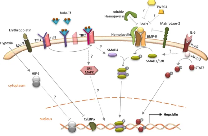
The erythropoietin receptor (Epo-R) belongs to the cytokine receptor superfamily and,
among many other tissues, is expressed at the cell surface of hepatocytes (Figure 1.3). Here,
erythropoietin may interact with Epo-R, triggering a decreased binding of the transcription
factor C/EBP (CCAAT/enhancer binding protein ) to a cognate site in the hepcidin
promoter [Courselaud et al. 2002; Pinto et al. 2008]. Alternatively, Huang et al. [2009] proposed that
erythropoietin can suppress hepcidin expression indirectly by the downregulation of the
signal transducer and activator of transcription 3 (STAT3) and SMAD4 [the name SMAD is a
combination of two proteins: the Caenorhabditis elegans protein Sma (designated as
mutations in this gene causes animals to be small) and the drosophila protein MAD
General Introduction
Recent research has considered the growth differentiation factor 15 (GDF15), a member of
the transforming growth factor (TGF-) superfamily, as the erythroid regulator of hepcidin
(Figure 1.3). This factor has increased expression and secretion during erythroid maturation
and is highly increased in patients with defective erythroid expansion [Tanno et al. 2007; Tamary
et al. 2008; Finkenstedt et al. 2009; Ramirez et al. 2009; Theurl et al. 2010]. Moreover, while in vitro
studies reveal the suppressive effect of GDF15 on hepcidin expression, GDF15 expression
itself was shown to be regulated by the iron status [Tanno et al. 2007; Lakhal et al. 2009]. Although
some skepticism may arise from the fact that GDF15 and hepcidin levels do not correlate in
patients undergoing hematopoietic stem cell transplant recovery (whereas other erythroid
markers correlate with hepcidin), the recent findings are consistent with previous proposals
in which erythropoiesis is positively related with iron absorption and mobilization [Kanda et al.
2008]. Moreover, they also reinforce the idea that erythropoiesis dominantly represses
hepcidin expression in spite of iron overload [Tanno et al. 2007; Lakhal et al. 2009].
Figure 1.3. Transcriptional regulation of hepcidin expression.There are several upstream stimuli that through signaling pathways determine the expression levels of hepcidin. Inflammatory status and iron overload act as positive regulators, whereas hypoxia and erythropoietic demand operate as repressors of hepcidin expression. See text for details. (Adapted from Anderson et al. 2009).
Chapter 1
The latest factor recognized as a putative erythroid regulator of hepcidin is a cytokine named
TWSG1 (twisted gastrulation 1) (Figure 1.3). Contrarily to GDF15, TWSG1 is produced during
the earlier stages of erythropoiesis [Tanno et al. 2009]. This study shows that TWSG1
suppresses hepcidin indirectly by inhibiting the bone morphogenetic proteins (BMPs)
signaling pathway. Here, it is proposed that TWSG1 and GDF15 might act together to
inappropriately inhibit hepcidin expression and deregulate iron homeostasis in thalassemia
syndromes.
Hypoxia is another negative regulator of hepcidin expression, independently of body iron
levels [Nicolas et al. 2002b; Choi et al. 2007; Peyssonnaux et al. 2007]. In fact, the transcriptional
hypoxia-inducible factor pathway was shown to regulate hepcidin expression in mice
[Peyssonnaux et al. 2007] (Figure 1.3). The liver-specific disruption of the von Hippel-Lindau
gene, which encodes for an essential component of the complex that degrades HIF, led to
decreased hepcidin mRNA levels in these mice. It has also been shown that HIF-1 is able to
bind the hepcidin promoter, suggesting a direct repression of hepcidin by HIF-1 [Peyssonnaux
et al. 2007]. Nevertheless, Choi and co-workers [2007] have brought disagreeing data, since
either HIF-1 overexpression or knockdown fail to alter hepcidin expression in HepG2 cells.
They have also shown that the increase in reactive oxygen species (ROS) in hypoxic cells
impaired the binding of C/EBP and STAT3 transcription factors to the hepcidin promoter,
with a negative effect on its expression [Choi et al. 2007]. Moreover, Volke et al. [2009] also
failed to find a direct transcriptional suppression of hepcidin by HIFs. So, whether or not HIFs
directly bind to the hepcidin promoter is currently controversial.
Inflammation is a robust inducer of hepcidin expression, evoking its function as an
antimicrobial peptide [Krause et al. 2000; Park et al. 2001; Nicolas et al. 2002b; Peyssonnaux et al. 2006;
Sow et al. 2007]. Under inflammatory conditions, iron absorption is reduced and iron is
sequestered in the macrophages, with a consequent hypoferremia in plasma [Nemeth et al.
2004a; Rivera et al. 2005]. A substantial body of evidence indicates that IL-6 is the predominant
cytokine involved in the inflammatory regulation of hepcidin, but IL-1 and IL-1 are also
able to stimulate hepcidin [Lee et al. 2004; Nemeth et al. 2004a; Lee et al. 2005] (Figure 1.3). This
induction has been shown to occur through the Janus kinase (JAK) 1/2 signal transducer and
STAT3 transcriptional mechanism [Wrighting and Andrews 2006; Verga Falzacappa et al. 2007; Truksa et
General Introduction
cooperative activity of the BMP signaling pathway, possibly through the TGF-/SMAD4
induction [Wang et al. 2005; Babbit et al. 2007; Yu et al. 2008].
Indeed, the BMP signaling pathway has a critical importance in the regulation of hepcidin
transcription activation and the cohort of proteins involved upstream this response will now
be exposed.
II.2.2.
Hepcidin regulation by iron status
Dissecting the elegant mechanisms that allow systemic iron homeostasis maintenance
through the modulation of hepcidin expression has been quite challenging. The study of
genetic disorders of iron status has brought to light some of the most important players that
take part of these pathways. This is the case of HFE, TfR2, hepcidin and hemojuvelin, among
others [Feder et al. 1996; Camaschella et al. 2000; Roetto et al. 2003; Papanikolaou et al. 2004; Finberg et al.
2008]. Nevertheless, other proteins such as the BMPs, matriptase-2 and transferrin have
been shown to be involved in the hepatic regulation of hepcidin driven by iron status.
Although the mechanisms by which TfR2 and HFE act are only recently beginning to be
untangled, the characterization of hemojuvelin has revealed a complex signal transduction
pathway that regulates hepcidin expression, the bone morphogenetic protein pathway.
II.2.2.1.
Hemojuvelin, BMPs and matriptase-2
Hemojuvelin’s pivotal role in iron homeostasis has been demonstrated by both clinical and animal studies. Whereas homozygosity or compound heterozygosity for mutations in the
human hemojuvelin gene are responsible for a juvenile form of hereditary
hemochromatosis, disruption of both hemojuvelin-homologue alleles in mice result in
marked iron deposition in the liver, heart and pancreas [Papanikolaou et al. 2004; Huang et al. 2005;
Niederkofler et al. 2005]. The severe downregulation of hepcidin in these cases despite the
presence of strong iron loading demonstrated that hemojuvelin is an essential upstream
regulator of hepcidin.
Hemojuvelin is a member of the repulsive guidance molecule family of proteins that function
as co-receptors of the bone morphogenetic proteins [Babitt et al. 2006]. Notably, recent studies
Chapter 1
thereby increasing hepcidin synthesis [Babitt et al. 2006; Truksa et al. 2006; Babitt et al. 2007; Yu et al.
2008] (Figure 1.3). In general, BMPs are a subfamily of cytokines that belong to the TGF-
superfamily [Heldin et al. 1997; Derynck and Zhang 2003]. Individual members of the BMP subfamily
are able to interact with type I and II receptors, therefore increasing the complexity of
hepcidin regulation by the BMP pathway [Derynck and Zhang 2003]. Activated BMP receptors
phosphorylate the SMAD1/5/8 protein complex which, in turn, will form a heteromeric
complex with the DNA binding protein SMAD4 [Babitt et al. 2007]. This complex translocates
into the nucleus and activates the transcription of target genes, such as hepcidin [Heldin et al.
1997; Derynck and Zhang 2003]. Evidence to support the BMP pathway in the regulation of
hepcidin expression came out by the study of mice with liver-specific disruption of SMAD4
gene, which developed severe iron overload with almost no hepcidin expression [Wang et al.
2005]. Moreover, these mice failed to respond to iron loading or IL-6 injection. Recent studies
on BMP6 knockout mice show the same iron overload phenotype as SMAD4 and
hemojuvelin knockout mice [Andriopoulos et al. 2009; Meynard et al. 2009]. This confirms previous
data according to which BMP6 expression is directly regulated by iron and essential for
hepcidin upregulation [Kautz et al. 2008; Yu et al. 2008].
The task of hemojuvelin in the activation of hepcidin through the BMP-SMAD pathway is far
from understood. In fact, hemojuvelin exists in at least two distinct forms: a transmembrane
glycosylphosphatidylinositol (GPI)-linked form, which stimulates hepcidin, and a soluble
form, which acts as an antagonist of the BMP signaling pathway [Lin et al. 2005; Zhang et al. 2005;
Kuninger et al. 2006; Babbit et al. 2007]. This soluble form appears to be released from the mature
hemojuvelin by furin or other pro-protein convertase [Silvestri et al. 2008a; Lin et al. 2008].
Evidence of the importance of this soluble hemojuvelin was given when its administration
lowered hepcidin expression in mice and cultured cells [Lin et al. 2005; Babitt et al. 2007]. This led
to the current model, in which soluble hemojuvelin can antagonize BMP signaling by binding
to the BMPs and impair their association with the heteromeric BMP type I/II receptors [Lin et
al. 2005; Babbit et al. 2007]. Importantly, the generation of soluble hemojuvelin was shown to be
increased by iron treatment and hypoxia thereby repressing hepcidin, most likely by the
increased activity of furin [Lin et al. 2005; Zhang et al. 2007; Silvestri et al. 2008a]. In parallel, work by
Zhang and co-workers [2005] revealed that hemojuvelin interacts with neogenin on the cell
membrane interfering with cellular iron levels. Recent studies confirm that neogenin may
have two functions in the regulation of hemojuvelin, one in promoting hemojuvelin shedding
General Introduction
the neogenin-hemojuvelin interaction [Zhang et al. 2007; Zhang et al. 2009b]. However, conflicting
data arise from two recent independent studies. Xia et al. [2008] have shown that the
knockdown or overexpression of neogenin fails to induce changes in the
hemojuvelin-induced BMP signaling and hepcidin expression, whereas Lee et al. [2010] indicate that
neogenin enhances BMP signaling resulting in hepcidin upregulation but stating that this
occurs by neogenin inhibition of hemojuvelin secretion.
The most recent partner shown to be involved in the hemojuvelin/BMP pathway is the
membrane-bound serine matriptase-2 (Figure 1.3). Its importance in systemic iron regulation
was firstly suggested by results obtained in two mouse models enclosing a mutated
matripase-2, in which a marked increase in hepcidin levels was concomitant with iron
deficiency anemia [Du et al. 2008; Folgueras et al. 2008]. These studies were promptly
corroborated by the clinical studies in patients with iron-refractory iron deficiency anemia
that were homozygous or compound heterozygous for mutations in matriptase-2 gene
[Finberg et al. 2008; Melis et al. 2008]. The proposed role for matriptase-2 is the cleavage of
membrane hemojuvelin into fragments, therefore inhibiting hepcidin expression activation
[Silvestri et al. 2008b]. Furthermore, recent findings by Finberg et al. [2010] suggest that the
involvement of matriptase-2 is required for the downregulation of the BMP/SMAD signaling,
thus contributing to the regulation of systemic iron homeostasis.
II.2.2.2.
HFE and transferrin receptors (TfR1 and TfR2)
It is now accepted that the iron present in the plasma and in the tissue stores enhances
hepcidin synthesis which, in turn, inhibits the release of iron from macrophages and
duodenal enterocytes to the plasma. The molecular details of this homeostatic loop are still
incompletely understood. In fact, the most likely candidates able to act as iron sensors
include the transferrin receptors, TfR1 and TfR2 (Figure 1.3). Although the molecular link
between these receptors was characterized almost fifteen years ago, when the HFE protein
was associated with hereditary hemochromatosis, the mechanism(s) by which HFE and the
transferrin receptors affect hepcidin expression have only recently emerged [Feder et al. 1996;
Schmidt et al. 2008; Gao et al. 2009; Ramey et al. 2009; Wallace et al. 2009; Gao et al. 2010; Poli et al. 2010].
Initial studies showed that HFE was associated with TfR1 at the cell membrane and that HFE