In vivo
evaluation of the mutagenic potential and phytochemical
characterization of oleoresin from
Copaifera duckei
Dwyer
Edson Luis Maistro1, José Carlos Tavares Carvalho1, Vera Cascon2and Maria Auxiliadora Coelho Kaplan2
1
Universidade José do Rosário Vellano, Faculdade de Farmácia, Alfenas, MG, Brazil. 2
Universidade Federal do Rio de Janeiro, Núcleo de Pesquisas de Produtos Naturais,Rio de Janeiro, RJ, Brazil.
Abstract
We characterized the chemical constituents ofCopaifera duckeioleoresin and used dermal application to Wistar rats to evaluated its possible mutagenic and cytotoxic activities on peripheral blood reticulocytes and bone marrow cells. Chemical characterization of the oleoresin revealed the presence of sesquiterpene hydrocarbons, an unidentified neutral diterpene and diterpene acids. To evaluate mutagenicity evaluation the rats were treated with 10, 25 and 50% of the LD50dose of the oleoresin for three consecutive days and peripheral blood collected after 0, 24, 48 and 72 h for
micronucleus analysis. The rats were humanly sacrificed 24 hours after the last treatment and chromosome prepara-tions made using standard techniques. At the three concentraprepara-tions and the three time intervals tested we found that there were no statistically significant differences in either the mean number of micronucleated reticulocytes (MNRETs) or the number of chromosomal aberrations as to the negative control. However, at 25 and 50% of the LD50
dose of the oleoresin there was a significant decrease in the mitotic index (MI) as compared to the negative control. Under our experimental conditions,C. duckeiV11 oleoresin produced no mutagenic effects on bone marrow cells or in peripheral reticulocytes as assessed by chromosome aberrations and the micronucleus test respectively, but showed cytotoxic activity at high doses.
Key words: Copaifera duckei(Caesalpinaceae), phytochemical characterization, micronucleus test, chromosome aberrations, cytotoxic effect.
Received: November 23, 2004; Accepted: March 24, 2005.
Introduction
The oleoresin obtained by tapping the trunk of trees of the genusCopaifera(Caesalpinaceae), is widely used in Brazilian popular medicine under the name ‘óleo de copaí-ba’ (copaiba oleoresin), predominantly as a healing, anti-septic and anti-inflammatory agent (Le Cointe, 1934; Pio Corrêa, 1984).
Copaiba oleoresins have been used as unique vegetal drugs despite the existence of more than 20 species of
Copaiferain Brazil (Dwyer, 1951) and the significant inter
and intra species differences in chemical composition (Cas-con and Gilbert, 2000) copaiba oleoresins have been used medicinally throughout Brazil. The oleoresin is a natural solution of diterpene acids in an essential oil composed mainly of sesquiterpenes and has been reported as being bactericidal (Maruzzela and Sicurella, 1960; Opdyke, 1976; Cascon et al., 2000; Tincusi et al., 2002),
anti-helminthic (Pellegrino, 1967; Gilbertet al., 1972),
analge-sic (Fernandes and Pereira, 1989), anti-inflammatory (Basileet al., 1988; Fernandeset al., 1992; Veiga-Junioret al., 2001) and gastro-protective (Paivaet al., 1998) as well
as showing antitumor (Ohsakiet al., 1994; Lima et al., 1998) and trypanocidal (Casconet al., 1998) activity. How-ever, in several of these evaluations commercial copaiba oleoresins were used, the chemical composition of which was either not given or only partially described.
There exists considerable interest in determining the risks that plant extracts may pose to health, since many of these extracts contain compounds known to cause diseases or even death to animals and humans by acting as natural mutagens and carcinogens (Panigrahi and Rao, 1982; Araú-joet al., 1999; Burimet al., 1999; Chaconet al., 2002). The objective of the study described in this paper was to
charac-terize the chemical constituents of Copaifera duckei
oleoresin and evaluate its mutagenic and cytotoxic poten-tial by applying the micronucleus test to peripheral blood and analyzing chromosomal aberrations in bone marrow cells of Wistar rats treated with this oleoresin.
www.sbg.org.br
Send correspondence to Edson Luis Maistro. Universidade José do Rosário Vellano, Laboratório de Genética, Caixa Postal 23, 37130-000 Alfenas, MG, Brazil. E-mail: edson.maistro@ unifenas.br.
Material and Methods
Plant material and chemical analysis
We collected 4.4 litres of Copaifera duckei Dwyer
(V11) oleoresin from trees growing at a site in Mazagão county in the Brazilian state of Amapá near the town of Macapá at 00°02’56” N; 051°44’46” W on the 7 of Decem-ber 1996. The collection of oleoresin and botanical material was made by Vera Cascon and Jonas de Oliveira Cardoso with the collaboration of the Amapá Institute of Scientific and Technological research (Instituto de Pesquisas Cientí-ficas e Tecnológicas do Estado do Amapá, IEPA). Botani-cal identification was made by Antônio Sérgio Lima da Silva, Museu Paraense Emílio Goeldi (MG), Belém, Pará, Brazil. The botanical material collection number was 031 deposited at 12/02/1998.
An equal volume of dichloromethane was added to the crude oleoresin which was esterified with diazomethane in ether and analyzed using gas chromatography - mass spectrometry (GC-MS) in a Hewlett Packard HP 6890 chromatograph (column 30 m x 250mm x 0,25mm) - HP 5 mass spectrometer (70 eV, mass selective detector 5972 A), using PFK as a reference. The temperature was started at 70 °C, rising by 2 °C per minute to 300 °C.
Both sesquiterpenes and methyl esters of diterpene acids were analyzed in the same sample and the majority of the compounds were characterized using the Wiley Li-brary/ Mass Spectra 275 and by comparison of retention times with data published by Braga (1994).
Animals and assay procedures
Experiments were carried out using six-week-old Wistar rats (Rattus norvegicus) weighing 90-110 g
ac-quired from Alfenas University animal house and kept in polyethylene boxes (n= 6) in a climate-controlled
environ-ment (25 ± 4 °C, 55 ± 5% humidity) with a 12h light/dark cycle (07:00h to 19:00h) and fed Labina-Purina (Agribrands Purina do Brasil Ltda, Paulínia, São Paulo, Brazil) and water ad libitum. The rats were divided into three experimental and two control groups each containing three females (F1to F3) and three males (M1to M3). Rats in
the experimental groups received 10%, 25% or 50% of the
LD50 dose (7.467 mg/kg body weight, Carvalho and
Cascon, 2003) ofCopaifera duckeioleoresin by dorsal der-mal injection for 3 consecutive days at 24 h intervals. The negative control group received 0.9% (w/v) NaCl by the same route as the experimental rats and the positive control group 30 mg of cyclophosphamide/kg body weight.
For the micronucleus test blood smears were col-lected using peripheral tail blood from experimental and control rats, the blood being collected before the first injec-tion (0 h) and at 24, 48 and 72 h after the first injecinjec-tion (Hayashiet al., 1990). A total of 8000 reticulocytes were analyzed per rat, 2000 for each collection time. All rats were humanly sacrificed 72 h after the first injection, each
rat being injected intraperitoneally with 0.5 mL of 0.16% (w/v) aqueous colchicine 90 min prior to euthanasia. Bone marrow was obtained at autopsy (t = 72 h) for the analysis of chromosome aberrations in metaphase cells using the method of Ford and Hamerton (1956). The UNIFENAS Animal Bioethical Committee approved the present study on 17thAugust 2003.
To detect micronuclei and chromosome aberrations slides were Giemsa stained and 100 metaphases per animal analyzed to determine the mean number of chromosomal aberrations in a blind test. Chromosomal aberrations were classified according to Savage (1976) as gaps, breaks, dele-tions, fragments, rings and dicentric chromosomes. Gaps were recorded but not included in the statistical analysis. The mitotic index was obtained by counting the number of mitotic cells in 1000 cells per animal. The data were sub-mitted to one-way analysis of variance (ANOVA) and the Tukey-Kramer multiple comparison test using the GraphPad Instat®software version 3.01 (GraphPad
Soft-ware, Inc., San Diego, USA). Results were considered sta-tistically significant at p < 0.05.
Results
Phytochemical characterization
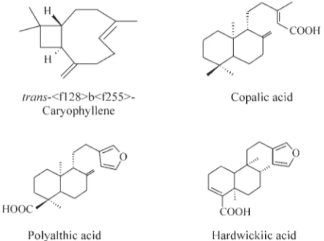
The analysis of the proportional distribution of ter-penes in the oleoresin showed the presence of 7.2% of sesquiterpene hydrocarbons, 1.8% of an unidentified neu-tral diterpene and 92.2% of diterpene acids (Figure 1). The main components of the oleoresin are the sesquiterpenes
trans-b-caryophyllene (4.5%), trans-a-bergamotene
(1.0%),a-humulene (0.7%), andb-bisabolene (1.0%) and
the diterpene copalic (3.7%), polyalthic (27.1%) and hardwickiic (59.3%) acids.
Mutagenic and cytotoxic evaluation
The results obtained in thein vivotest system are
pre-sented in Tables 1 and 2. The micronuclei assay showed no
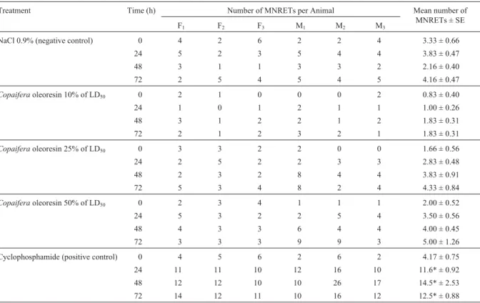
statistically significant differences in the mean number of micronuclei (MN) in peripheral blood reticulocytes (RETs) of the rats in any of the experimental groups as compared be-tween themselves or with the negative control group (Table 1). At the three concentrations tested, a small but statistically non significant increase was observed between the mean number of micronucleated reticulocytes (MNRETs) after 24, 48 and 72 h as compared with their respective 0 h controls. No sex differences were observed between any of the groups.
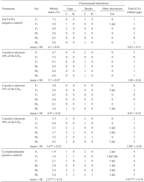
As compared to rats in the negative control group, the mitotic index (Table 2) of rats in the 10% LD50group was
not significantly different but rats in the 25% and 50% LD50
groups showed a significant decreases (p < 0.05 and p < 0.01 respectively).
There were no statistically significant differences in the mean number of chromosome aberrations between the three experimental groups and the negative control group (Table 2). In all treatments withCopaifera oleoresin the
most frequent chromosomal aberrations observed were chromatid breaks, followed by chromatid gaps, deletions and isochromatidic gaps.
Discussion
Thein vivorat micronuclei test and chromosome
ab-errations assay are two of the most frequently used and
sen-sitive tests for investigating the genotoxic profile of chemi-cals, these tests having been recommended for routine analysis because they produce results that are considered highly relevant in the human context (Moritaet al., 1997;
Prestonet al., 1987). TheCopaifera duckeioleoresin
ana-lyzed by us was very rich in diterpene acids and possessed moderate amounts of sesquiterpene hydrocarbons, con-firming the report by Cascon and Gilbert (2000) that there are differences in the chemical composition of the oleoresin produced by differentCopaiferaspecies.
Terpenes are abundant in superior plants and show a shared structure of isoprene units, the sesquiterpenes (C15
H24) having three such units and the diterpenes (C20H32)
four (Robbers et al., 1997). Some sesquiterpenes and
diterpenes are known to be cytotoxic and to inhibit tumors, with toxic sesquiterpenes generally containing one or more functional alkylating groups which suggests that they are possibly mutagenic and carcinogenic (Cassady and Baird, 1990; Wallet al., 1998).
Our data shows that treatmentC. duckeioleoresin
resulted in depression of mitotic activity and no statisti-cally significant increase in chromosome aberrations in bone marrow cells and in the mean number of MNRETs in the peripheral blood of Wistar rats. The dose related de-crease in mitotic index (Table 2) indicates thatC. duckei
Table 1- Number of micronucleated reticulocytes (MNRETs) observed in the peripheral blood cells of female (F1to F3) and male (M1to M3) Wistar rats treated withCopaifera duckeioleoresin. For each time period (0, 24, 48, 72 h) 2000 cells were analyzed, giving a total of 8000 cells per animal.
Treatment Time (h) Number of MNRETs per Animal Mean number of
MNRETs ± SE
F1 F2 F3 M1 M2 M3
NaCl 0.9% (negative control) 0 4 2 6 2 2 4 3.33 ± 0.66
24 5 2 3 5 4 4 3.83 ± 0.47
48 3 1 1 3 3 2 2.16 ± 0.40
72 2 5 4 5 4 5 4.16 ± 0.47
Copaiferaoleoresin 10% of LD50 0 2 1 0 0 0 2 0.83 ± 0.40
24 1 0 1 2 1 1 1.00 ± 0.26
48 3 1 2 2 1 2 1.83 ± 0.31
72 2 1 2 3 2 1 1.83 ± 0.31
Copaiferaoleoresin 25% of LD50 0 3 3 2 2 0 0 1.66 ± 0.56
24 2 5 2 2 3 3 2.83 ± 0.48
48 2 3 2 8 4 4 3.83 ± 0.91
72 5 3 4 8 2 4 4.33 ± 0.84
Copaiferaoleoresin 50% of LD50 0 2 3 4 1 1 1 2.00 ± 0.52
24 5 3 2 2 5 4 3.50 ± 0.56
48 4 3 3 6 4 4 4.00 ± 0.45
72 3 3 3 9 9 3 5.00 ± 1.26
Cyclophosphamide (positive control) 0 4 5 6 2 6 2 4.17 ± 0.75
24 11 11 10 12 16 10 11.6* ± 0.92
48 12 12 10 10 26 17 14.5* ± 2.53
72 14 12 11 10 16 12 12.5* ± 0.88
oleoresin depresses mitosis at high doses. Although not statistically significant, the dose related increase in the mean number of MNRETs observed at the two higher doses probably occurred due to cumulative effects of the resin because the rats were treated during three consecu-tive days.
Sena and Chen (1998) used the micronucleated cell assay and Swiss albino mice to evaluated thein vivobone marrow cell mutagenic potential of 25, 50 and 80% of the LD50 dose of orally administered Copaifera langsdorfii
oleoresin and demonstrated a statistically significant in-crease micronucleated cells (and hence mutagenic action)
Table 2- Mitotic Index and distribution of the different types of chromosomal aberrations (CA) observed in bone marrow cells of female (F1to F3) and male (M1to M3) Wistar rats treated with aCopaifera duckeioleoresin. For each treatment 100 cells per animal were analyzed, giving a total of n = 600 cells per treatment.
Chromosomal aberrations
Treatments Sex Mitotic
index (%)
Gaps Breaks Other aberrations Total (CA)
without gaps
C IC C IC OA
NaCl 0.9% (negative control)
F1 7.2 0 0 1 0 0 1
F2 5.8 1 0 0 0 1 del 1
F3 4.9 2 0 0 0 0 0
M1 5.6 0 0 2 0 0 2
M2 4.5 0 0 0 0 0 0
M3 8.6 0 0 1 0 0 1
mean ± SE 6.1 ± 0.63 0.83 ± 0.31
Copaiferaoleoresin 10% of the LD50
F1 4.7 0 0 2 0 0 2
F2 5.3 0 0 0 0 0 0
F3 4.1 0 0 1 0 0 1
M1 6.9 0 0 1 0 0 1
M2 6.6 0 0 1 0 0 1
M3 6.6 0 0 1 0 0 1
mean ± SE 5.7 ± 0.47 1.00 ± 0.26
Copaiferaoleoresin 25% of the LD50
F1 5.0 0 0 0 0 0 0
F2 5.0 0 0 0 0 2 del 2
F3 4.5 0 0 0 0 0 0
M1 4.4 0 0 0 0 0 0
M2 4.1 0 0 1 0 0 1
M3 3.6 1 0 0 0 1 del 1
mean ± SE 4.4* ± 0.22 0.67 ± 0.33
Copaiferaoleoresin 50% of the LD50
F1 3.3 3 0 2 0 0 2
F2 4.4 0 0 1 0 0 1
F3 3.1 2 2 0 0 2 del 2
M1 3.7 2 2 0 0 3 del 3
M2 3.2 2 0 1 1 0 2
M3 4.2 3 0 0 0 2 del 2
mean ± SE 3.6** ± 0.22 2.00* ± 0.26
Cyclophosphamide (positive control)
F1 1.9 1 0 2 0 2 del 4
F2 1.6 1 1 6 0 1 del/1dic 8
F3 2.1 1 0 1 0 3 del 4
M1 2.8 0 0 4 0 1 del 5
M2 2.6 3 2 6 0 2 del 8
M3 2.4 3 3 2 1 2 del 5
mean ± SE 2.2*** ± 0.18 5.67*** ± 0.76
Key: C = Chromatid; IC = isochromatid; OA = other aberrations; del = deletion, dic = dicentric; SE = standard error. *: Significantly different from the negative control at p < 0.05.
only at high doses, although these authors used a higher maximum dose than we did.
Donaldsonet al. (1994) extensively investigated the cytotoxicity of the antimitotic antitumor diterpene taxol de-rived from the yew treeTaxus brevifoliaand showed that the antimitotic effect of taxol classified it as an important anticancer agent. A genotoxicity study by Diaset al. (1997) showed that taxol had no radio-sensitizing effect on chro-mosomal aberrations induced by gamma radiation and also did not increase doxorubicin-induced chromosomal aberra-tions inin vitroChinese hamster ovary cells.
The active component of Eremanthus elaeagnus
wood oil is the sesquiterpene eremanthine, genotoxic eval-uationin vivoin rodents andin vitroin human lymphocytes having showed that low concentrations of eremanthine pro-duced no cytotoxic or clastogenic effects and that only doses of 400 mg Kg-1showed toxicity (Dias
et al., 1995).
Another sesquiterpene, Glaucolide B, isolated from
Vernonia eremophilaproduced no significant increase in
the frequency of chromosomal aberrations in mouse bone marrow cells but showed cytotoxic and clastogenic effects on human lymphocytesin vitro, indicating that caution is
needed in its medicinal use (Burimet al., 1999).
Available information about the evaluation of the mutagenic potential of copaiba oleoresin in different rodent species has shown that despite some qualitative differences between theCopaiferaoleoresins studied the toxic pattern
was similar, with cytotoxic and some genotoxic effects only occurring at high doses. Since there are about 20 dif-ferentCopaiferaspecies in Brazil and the oleoresins
ob-tained from these plants have been used in popular medicine, it is important to establish the relationship be-tween chemical composition and biological activity of au-thentic samples of the oleoresins in order to permit their validation as safe and effective phyto-medicines and to al-low adequate quality control.
The results of our study demonstrate that under the
experimental conditions employed Copaifera duckei
oleoresin presented cytotoxic effects at high doses but did not induce a statistically significant increase in the mean number of chromosome aberrations in the bone marrow cells or in the mean number of MNRETs from the periph-eral blood of Wistar ratsin vivo.
Acknowledgments
This investigation was supported by UNIFENAS, UFRJ and FAPEMIG (Rede Mineira de Ensaios Toxicológicos e FarmacoToxicológicos de Produtos Terapêuticos, EDT -1879/02). We are grateful to Jonas de Oliveira Cardoso for help in collecting the plant material and to Antônio Sérgio Lima da Silva for botanical identification.
References
Araújo MCP, Dias FL, Kronka SN and Takahashi CS (1999) Ef-fects of turmeric and its active principle, curcumin, on bleomycin-induced chromosome aberrations in Chinese hamster ovary cells. Genet Mol Biol 22:407-413.
Basile AC, Sertie JA, Freitas PCD and Zanini AC (1988) Anti-inflammatory activity of oleoresin from Brazilian
Copaifera.Journal of Ethnopharmacology 22:101-109. Braga WF (1994) Caracterização química dos constituintes do
óleo extraído deCopaifera cearensis. M. Sc. Thesis. Uni-versidade Federal do Rio de Janeiro, Rio de Janeiro. Burim RV, Canalle R, Lopes JLC and Takahashi CS (1999)
Genotoxic action of the sesquiterpene lactone glaucolide B on mammalian cellsin vitroandin vivo. Genet Mol Biol 22:401-406.
Carvalho JCT and Cascon V (2003) Fitoterápicos: Nova Opção Terapêutica de Antiinflamatórios (Aspectos Químicos, Far-macológicos e Aplicações Terapêuticas). Editora Robe, São Paulo, 630 pp.
Cascon V and Gilbert B (2000) Characterization of the chemical composition of oleoresins ofCopaifera guianensisDesf., Copaifera duckeiDwyer andCopaifera multijugaHayne.
Phytochemistry 55:773-778.
Cascon V, Fernandez-Ferreira E, Soares ROA, Gibaldi D, Gilbert B and Ribeiro-Santos R (1998) Avaliação da composição química e da atividade tripanosomicidain vitrode
óleo-re-sinas deCopaiferaspp. Resumos do XV Simpósio de Plan-tas Medicinais do Brasil, Águas de Lindoia, SP, pp 199. Cascon V, Gilbert B, Araújo GL, Rocha LM, Teixeira LA and
Carvalho ES (2000) Avaliação da atividade antimicrobiana de óleo-resinas deCopaiferaspp. Resumos do XVI Sim-pósio de Plantas Medicinais do Brasil, Recife, PE, pp 223. Cassady JM and Baird WM (1990) Natural products as a source of
potential cancer chemotherapeutic and chemopreventive agents. J Nat Prod 53:23-41.
Chacon DR, Libera AND, Cintra DEC, Carvalho JCT, Oliveira GA and Maistro EL (2002) Absence of genotoxic and anti-genotoxic effects of a standardized extract of the medicinal plant Solanum melongena on peripheral blood and bone
marrow cells of Wistar rats. Cytologia 67:417-422. Dias FL, Takahashi CS, Sakamoto-Hojo ET, Vichnewski W and
Sarti SJ (1995) Genotoxicity of the natural cercaricides “Sucupira” oil and Eremanthine in mammalian cellsin vitro
andin vivo. Environ Mol Mutagen 26:338-344.
Dias FL, Antunes LMG and Takahashi CS (1997) Effect of taxol on chromosome aberrations induced by gamma radiation or by doxorubicin in Chinese hamster ovary cells. Braz J Gen 20:389-395.
Donaldson KL, Goolsby GL and Wahl AF (1994) Cytotoxicity of the anticancer agents cisplatin and taxol during cell prolifer-ation and the cell cycle. Int J Cancer 57:847-855.
Dwyer JD (1951) The Central American, West Indian and South American species ofCopaifera(Caesalpinaceae). Brittonia 7:143-172.
Fernandes RM and Pereira NA (1989) Copalic acid analgesic ac-tivity in mice. Abstracts do Simpósio Brasil-China de Quí-mica e Farmacologia de Produtos Naturais, Rio de Janeiro, pp 248.
Fernandes RM, Pereira NA and Paulo LG (1992) Anti-inflam-matory activity of copaiba balsam (Copaifera cearensis
Gilbert B, Mors WB, Baker PM, Tomassini TCB, Goulart EG, Holanda JC, Costa JAR, Lopes JNG, Santos-Filho D, Sarti SJ, Turco AM, Vichnewski W, Lopes JLC, Thames AW, Pellegrino J and Katz N (1972) A atividade anti-helmíntica de óleos essenciais e de seus componentes químicos. Anais da Academia Brasileira de Ciências 44(supl.):423-428. Ford CE and Hamerton JL (1956) A colchicine, hypotonic citrate,
squash sequence for mammalian chromosomes. Stain Technol 31:247-251.
Hayashi M, Morita T, Kodama Y, Sofuni T and Ishidate Jr M (1990) The micronucleus assay with mouse peripheral blood reticulocytes using acridine orange-coated slides. Mutat Res 245:245-249.
Le Cointe P (1934) Árvores e Plantas Úteis: A Amazônia Brasi-leira (III). Livraria Clássica, Belém, 486 pp.
Lima SEM, Cascon V and Pereira NA (1998) Estudo dos efeitos da óleo-resina de copaíba sobre células de melanoma B16F10 em camundongos C57BL/6J. XIII Reunião Anual da Federação de Sociedades de Biologia Experimental (FESBE), Caxambu, pp 391-392.
Maruzzella JC and Sicurella NA (1960) Antibacterial activity of essential oil vapors. J Amer Pharmac Assoc 49:692-694. Morita T, Asano N, Awogi T, Sasaki YF, Sato S, Shimada H,
Sutou S, Suzuli T, Wakata A, Sofuni T and Hayashi M (1997) Evaluation of the rodent micronucleus assay in the screening of IARC carcinogens (Group 1. 2A and 2B). The summary report of the 6th collaborative study by CSGMT/JEMS MMS. Mutat Res 389:3-122.
Ohsaki A, Yan LT, Shigeru I, Edatsugi H, Iwata D and Komoda Y (1994) The isolation andin vivopotent antitumor activity of clerodane diterpenoid from the oleoresin of the Brazilian medicinal plant,Copaifera langsdorfiiDesfon. Bioorg Med
Chem Lett 4:2889-2892.
Opdyke DLJ (1976) Balsam copaiba. Food Cosmet Toxicol 14:687.
Paiva LAF, Rao VSN, Gramosa NV and Silveira ER (1998) Gastroprotective effect ofCopaifera langsdorffiioleoresin
on experimental gastric ulcer models in rats. J Ethnopharmac 62:73-78.
Pellegrino J (1967) Protection against human Schistosome cercariae. J Exper Parasitol 21:12.
Pio-Corrêa M (1984) Dicionário das Plantas Úteis do Brasil, e das Exóticas Cultivadas. Imprensa Nacional, Rio de Janeiro, v. I, pp 86-87; v. II, pp 370-375.
Panigrahi GB and Rao AR (1982) Chromosome-breaking ability of arecoline, a major betel-nut alkaloid, in mouse bone-marrow cellsin vivo. Mutat Res 103:197-204.
Preston RJ, Dean BD, Gallow S, Holden HE, Macfee AF and Shelby M (1987) Mammalian in vivo cytogenetic assay
analysis of chromosomal aberration in bone marrow cells. Mutat Res 189:157-165.
Robbers JE, Speedie MK and Tyler VE (1997) Farmacognosia e Farmacobiotecnologia. Editorial Premier, São Paulo, 372 pp.
Sena MA and Chen LC (1998) Avaliação da mutagenicidade do óleo de Copaíba (Copaifera langsdorfiiDesfon) em eritró-citos da medula óssea de camundongos. Genet Mol Biol 21(suppl):E.65.
Tincusi BM, Jiménez IA, Bazzocchi IL, Moujir LM, Mamami ZA, Barroso JP, Ravelo AG and Hernández BV (2002) Antimicrobial terpenoids from the oleoresin of the Peruvian medicinal plantCopaifera paupera. Planta Medica
68:808-812.
Veiga-Junior VR, Zunino L, Calixto LB, Patituti ML and Pinto AC (2001) Phytochemical and antiedematogenic studies of commercial copaiba oils available in Brazil. Phytot Res 15:476-480.
Savage JRK (1976) Classification and relationships of induced chromosomal structural changes. J Med Genet 13:103-122. Wall ME, Wani MC, Hughes TJ and Taylor H (1988) Plant antimutagenic agents. 1. General bioassay and isolation pro-cedures. J Nat Prod 51:866-873.