(1) Department of Microbiology and Immunology, Institute of Biological Sciences, São Paulo State University (UNESP), Botucatu, SP, Brazil. (2)Hematology Center, Medical School of UNESP, Botucatu, SP, Brazil.
Correspondence to: Alexandrina Sartori, Ph.D., Departamento de Microbiologia e Imunologia, Instituto de Biociências, UNESP, 18618-000 Botucatu, SP, Brasil. Fax number: 00-55-014-8213744. Phone number: 00-55-014-820-6058 E-mail: [email protected].
INCREASED NATURAL KILLER ACTIVITY DOES NOT PREVENT PROGRESSION
OF EXPERIMENTAL KALA-AZAR
Alexandrina SARTORI(1), Ramon KANENO(1), Nelson BARUZZI(2) & Maria Terezinha Serrão PERAÇOLI(1)
SUMMARY
Kala-azar is the visceral form of leishmaniasis and it is caused by intracellular parasites from the complex Leishmania
donovani. Golden hamster (Mesocricetus auratus) infected withLeishmania donovani develop a disease very similar to human
Kala-azar. There is conspicuous hipergammaglobulinaemia and their T cells do not respond to stimulation with parasite antigens. We used this experimental model to evaluate the natural killer (NK) activity during the initial phase of the disease. Outbred hamsters infected by intravenous route with 5.106 amastigotes of L. donovani 1S showed a concurrent increase in the spleen weight and in the spleen
cell number. Using the single cell assay we detected a significant increase in the percentage of NK effector cells on the 4th day of
infection. Imprints from spleen and liver showed at days 14 and 28 a significant increase in the parasite burden. These results show that the increased NK activity in the beginning of the infection was not able to restrain the progression of the disease in this experimental model.
KEYWORDS: Kala-azar; Leismania donovani; Natural killer activity.
INTRODUCTION
Kala-azar is the visceral form of leishmaniasis and it is caused by the intracellular parasites from the Leishmania donovanicomplex. The induction of Th1 effector cells capable of gamma IFN (IFNγ) production for activation of macrophages to a microbicidal state is necessary to eliminate these parasites22. The development of Th1 cells had been
initially associated with interleukin-12 (IL-12) in a murine model of listeriosis10. More recently it was demonstrated that IL-12 is produced
by macrophages in the early response to many other infectious agents, particularly bacteria and protozoa21,26. This IL-12 induces production of
IFNγ , first mainly by NK cells and then by T cells. This early response is important for the activation of the phagocytic cell system as a first line of defense against infection, but the IL-12 produced in this early phase, often acting in combination with the induced IFNγ, is also required for optimal generation of Th1 cells25. In addition to IFNγ production, NK
cells also act as effector cells of the natural resistance by direct lysis of the pathogen, as shown with Cryptococcus neoformans12 or by lysis of
infected cells as shown for Mycobacterium tuberculosis infected human monocytes28.
Active visceral leishmaniasis is associated with immune dys-regulation including very high levels of immunoglobulins7, splenomegaly,
negative skin test for Leishmania antigen15 and absence of proliferative
response by peripheral blood mononuclear cells stimulated with parasite antigens19. Indian kala-azar patients have normal numbers of peripheral
blood NK cells but impaired functional activity due to decreased binding and lysis of target cells13. In addition, plasma from patients with L.
donovani infection was shown to reduce the in vitro natural killer activity of normal human peripheral blood mononuclear cells14. On the other
hand NK cells from non-Leishmania- exposed individuals could respond
in vitro by proliferation and IFNγ production to L. aethiopica stimulation1.
In mice experimentally infected with L. donovani NK cells may contribute to parasite elimination11. Since the Syrian hamster is a good
model for the study of progressive visceral leishmaniasis caused by L. donovani, we used these animals to further investigate the involvement of NK cells, by assessing their lytic activity, during the initial phase of this infection.
MATERIAL AND METHODS
Animals: Female outbred hamsters (Mesocricetus auratus) 8-12 weeks old, were obtained from the Central Animal Facility at the São Paulo State University (UNESP), Botucatu, São Paulo. Pelleted food and water were available ad libitum.
Experimental infections: Twenty four hamsters were infected by intracardiac route with 5.106 amastigotes in 0.1 ml of phosphate-buffered
salt solution (PBS), pH 7.2. The same number of hamsters were kept infected to be used as controls. Groups of 4 infected and 4 non-infected animals were sacrificed at 1, 2, 4, 7, 14 and 28 days postino-culation and their spleens were used to evaluate the following parameters: spleen weight, number of splenic cells, NK cell activity and parasite burden. Liver was also collected for parasite burden evaluation.
Parasite burden: Parasite burden was determined by examining Giemsa-stained impression smears from livers and spleens as previously described4, and calculated according to the formula:
Total LDU = number of amastigotes x organ weight (mg) x 2 . 105
number of host-cell nuclei
Single cell cytotoxicity assay: The human erythroleukemia cell line K562 was maintained as a suspension culture in medium RPMI 1640 (Gibco Laboratories, Grand Island, NY) supplemented with 2 mM L-glutamine, 40 ug/ml gentamicin and 10% heat-inactivated fetal calf serum (complete medium). Cells were subcultured twice a week and at the day before the assay. Viability was assessed by trypan blue exclusion. K562 cells were previously tested and were highly susceptible to hamster spleen-cell cytotoxicity24.
Effector cells were prepared from spleen of normal and infected hamsters. The spleens were asseptically removed and cell suspensions prepared by teasing the material through a stainless-steel sieve in cold RPMI 1640 medium. After to be washed twice in cold RPMI cells were adjusted to 3.106/ml and adherent cells were depleted by incubation on
plastic Petri dishes (n.3003, Falcon, Oxnard, California, USA) at 37oC
for 60 min. Non-adherent spleen cells and K562 cells were used as effector and target cells, respectively. The single-cell cytotoxicity assay on poly-L-lysine (PLL) coated coverslips was performed as already described for the Syrian hamster17.
The number of target/binding cells (TBC) and cytotoxic effector cells were calculated as previously described29. Spontaneous target cell
death was determined on lymphocyte-free control coverslips by scoring the fraction of dead (trypan blue stained) target cells in 300 cells. The percentage of lymphocytes bound to target cells was determined by counting 500 lymphocytes (%TBC). The fraction of conjugates containing dead target cells was determined by scoring 100 conjugates. The fraction of target-cell binding lymphocytes that were cytotoxic (A) was calculated as A=B-(BxC), where B is the fraction of conjugates containing dead target cells and C is the fraction of spontaneously dead target cells. The percentage of NK-effector cells present in the samples was calculated as A x % TBC.
Statistical analysis: Student’s t-test was performed to determine the statistical significance of data.
RESULTS
Spleen weight alteration - Hamsters infected with 5.106 amastigotes
of L. donovani 1S showed, from the 7th day of infection on, increasing
spleen weight compared with non infected controls. The differences
(Figure 1a) were considered statistically significant on days 7, 14 and 28 of infection (p < 0.05). On the 28th day of infection, last point to be
evaluated, there was a striking difference, being the spleen from infected hamsters 3.8 times heavier than the correspondent controls.
Quantitation of splenic cells - Spleens from normal and infected hamsters were removed and cell suspensions prepared by teasing the material through a stainless steel sieve in RPMI medium. The total number of cells was counted in a Neubauer chamber. Figure 1b clearly
Fig 1. Spleen alterations during leishmanial infection. Spleen weight (a); splenic cell number (b). Control non infected hamsters (■), infected hamsters (o). Values represent the mean + standard error of the mean for four animals. Differences in spleen weight and spleen cell number between infected and non-infected hamsters were significant (p < 0.05) on days 7, 14 and 28 after infection.
0
0,1
0,2
0,3
0,4
0,5
0,6
0,7
S
p
le
e
n
w
e
ig
h
t (
g
)
Normal
Infected
0 50 100 150 200 250 300 350
1 2 4 7 14 28
Days of infection
S
p
le
n
ic ce
ll
n
u
m
b
e
r (
x 1
0
6
)
a
b
0.7
0.6
0.5
0.4
0.3
0.2
shows an increasing difference between the average number of cells from infected and normal hamsters. At the 14th and 28th days postinfection
these differences were very pronounced, being highly significant (p = 0.0013 and p = 0.01 respectively). The number of splenic cells at the 28th day was 7.8 times higher compared with the control animals.
Evaluation of NK cell activity - The results of NK cell activity, evaluated by the single cell assay, are shown on Table 1. The percentage of lymphocytes with the ability to bind NK-sensitive target cells did not undergo any significant change during the infection. However the percentage of conjugates in which lymphocytes killed the target cells and the percentage of NK-effector cells in the non-adherent splenic population were significantly higher on the 4th day of infection (p =
0.008). After the 4th day of infection the cells from infected animals kept
higher ability to form dead conjugates and higher NK-effector cells in comparison with control animals. These differences were, nevertheless, less expressive than the one observed at the 4th day of infection.
DISCUSSION
NK cells are able to respond rapidly, non-specifically, to the presence of infectious microorganisms. Together with phagocytes, NK cells represent the first line of defense against infection. This defense can take the form of direct lysis of the pathogen or involve lysis of infected cells. However, in many cases it is the production of IFNγ and the consequent IFNγ mediated macrophage microbicidal activity that is probably of central importance in resistance23.
In the present work we evaluated the NK activity in Syrian hamsters experimentally infected with L. donovani. This model seems to mimetize very well the progressive human visceral leishmaniasis16.
Hamsters infected by intracardiac route with 5.106 amastigotes
of L. donovani1S showed an increased spleen weight compared with
control non-infected animals. This alteration that begun on day 7th
postinfection, became more pronounced in the course of the disease and attained its maximum after 28 days of infection, the last period evaluated in this study. A very similar pattern was observed when the number of splenic cells was counted in the course of the infection. From the 7th day
on we detected an increasing number of cells that peaked on the 28th
day. This splenic hipertrophy is a classical description both in human and experimental kala-azar9 and could be attributed to both monocyte
recruitment20 and polyclonal activation of B cells due to parasite
components5. However, this impressive increase in the splenic cell number
through the infection was not associated to any alteration on the ability of splenic lymphocytes to recognize and bind NK-sensitive cells. As the single cell assay used in this study evaluates the percentage of non-adherent cells that bind to NK-sensitive cells, this finding could suggest that NK cells increased in number in a proportional way to the increase of the other non-adherent cells in the spleen. However, the percentage of conjugates in which lymphocytes had killed cells and the percentage of NK-effector cells in the infected animals were significantly higher after 96 h of infection. These results show an activation of NK cells in the beginning of the infection. Althought our work had not evaluated the cause of this activation, the cytokine IL-12 would be a good candidate Table 1
Frequency of lymphocytes forming conjugates and NK-effector cells in the spleen of hamsters experimentally infected with L. donovani
Days of Groups % of lymphocytes % of conjugates % of infection forming with dead NK-effector
conjugates* targets#
1 Control 9.16 (1.36) 19.95 (3.51) 1.80 (0.42)
Infected 8.86 (0.58) 22.69 (5.44) 2.0 (0.57)
2 Control 10.63 (1.46) 13.11 (2.01) 1.36 (0.07)
Infected 12.55 (4.23) 17.70 (8.71) 2.05 (0.72)
4 Control 10.53(2.36) 17.93 (5.26) 1.76 (0.29)
Infected 10.15 (1.57) 31.49 (3.89) 3.07 (0.45)
7 Control 10.15 (1.55) 16.23 (2.45) 1.61 (0.08)
Infected 11.33 (1.79) 21.58 (9.20) 2.42 (1.03)
14 Control 9.52 (2.28) 17.56 (3.16) 1.60 (0.20)
Infected 9.38 (0.92) 26.10 (6.85) 2.46 (0.80)
28 Control 11.29 (2.58) 16.98 (4.47) 1.79 (0.25)
Infected 9.18 (3.64) 26.81 (2.95) 2.48(1.10) (*)Calculated per 500 lymphocytes;
(#)Calculated per 100 conjugates. Spontaneously dead targets were always below 2%.Values represent the mean for four animals. In parentheses is shown the standard deviation.
0
500
1000
1500
2000
2500
3000
3500
4000
1
2
4
7
14
28
Days of infection
Tot
al
LD
U (
x
1
0
5
)
Liver
Spleen
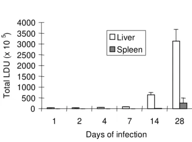
Fig 2. Parasite burden during leishmanial infection. Giemsa-stained impression smears from liver and spleen were microscopically assessed. Results represent the mean + standard error of the mean for four animals.
Splenic and hepatic parasite burden - To establish the extension of the parasitism in the liver and in the spleen, imprints from these organs were microscopically assessed. Figure 2 shows the parasite burden in the spleen and liver. After 1 day of infection only 1% (0.052 x 10⁶
parasites) of the total inoculum was retained by the spleen in comparison with 76% (3.8 x 10⁶ parasites) retained by the liver. The number of
due to its well established activation effect on NK cells6. In addition, a
recent report shows that IL-12 is required for NK cell activation and subsequent Th1 development in C3H mice experimentally infected with
L. major21. Also the ability of amastigotes to induce IL-12 production
from bone-marrow-derived macrophages in vitro was demonstrated by either direct measurement or indirectly through IFNγ release using SCID splenocytes18,27. From the 7th day to the 28th day of infection the NK
activity in the infected animals was still significantly higher compared with the control animals, even though not as much as on the 4th day. It is
possible that this increased NK cell activity had mediated an early resistance to the L.donovani infection, mainly in the spleen, keeping a low number of parasites, during the first two weeks of the disease. This increased NK activity, however, was not able to restrain the progression of the disease. The parasite burden from both spleen and liver increased significantly from the 14th to the 28th day of infection.
The progression of the disease, in spite of the significant increase in NK activity could suggest the development of an inefficient specific immune response. This could be the case if inhibitory cytokines such as IL-10 and TGF-β would be produced in excess. These two cytokines have been described as potent factors for macrophage deactivation3.
Besides, IL-10 and TGF-β have been reported as factors associated with the pathogenesis in human and experimental leishmaniasis respectively2,8.
In conclusion, our results show a clear activation of NK cells during the experimental leishmaniasis in the hamster. However, this increased NK activity was not able to restrain the progression of the disease in the experimental conditions used by us.
RESUMO
Atividade “natural-killer” aumentada não impede a progressão do Kala-azar experimental
O Kala-azar é a forma visceral da leishmaniose e é causado pelos parasitas do complexo Leishmania donovani. O hamster dourado
(Mesocricetus auratus) infectado com L. donovani desenvolve uma
doença bastante similar ao Kala-azar humano, apresentando hipergamaglobulinemia e supressão da resposta imune celular específica. Utilizamos este modelo experimental para avaliar a atividade natural killer (NK) na fase inicial da infecção. Hamsters não isogênicos infectados por via intravenosa com 5.106 amastigotas de L. donovani 1S
apresentaram aumento no peso do baço e no número de células esplênicas. Utilizando o “single cell assay” detectamos um aumento significativo no percentual de células NK efetoras no 4º dia de infecção. “Imprints” de baço e de fígado mostraram aumento significativo na carga parasitária após 14 e 28 dias de infecção. Os resultados mostram que o aumento da atividade NK, ocorrido no início da infecção, não foi capaz de bloquear a progressão da doença neste modelo experimental.
ACKNOWLEDGMENTS
The authors sincerely thank the Foundation for the development of UNESP (FUNDUNESP-199/92-DFP/F/CBS) for providing the financial assistance and Paulo Sérgio Ferreira for assistance with the manuscript.
REFERENCES
1. AKUFFO, H.; MAASHO, K. & HOWE, R. - Natural and acquired resistance to Leishmania: celular activation by Leishmania aethiopica of mononuclear cells from unexposed individuals is through the stimulation of natural killer (NK) cells.Clin. exp. Immunol., 94: 516-521, 1993.
2. BARRAL, A.; BARRAL-NETO, M.; YONG, E.C. et al. - Transforming growth factor β as a virulence mechanism for Leishmania braziliensis. Proc. nat. Acad. Sci. (Wash.), 90: 3442-3446, 1993.
3. BOGDAN, C.; PAIK, J.; VODOVOTZ, Y. & NATHAN, C. - Contrasting mechanisms for suppression of macrophage cytokine release by transforming growth factor-beta and interleukin-10. J. biol. Chem., 267: 23301-23308, 1992.
4. BRADLEY, D.J. & KIRKLEY, J. - Regulation of Leishmania population within the host. 1. The variable course of Leishmania donovani infections in mice. Clin. exp. Immunol., 30: 119-129, 1977.
5. BUNN-MORENO, M.M.; MADEIRA, E.D.; MENEZES, J.A. & CAMPOS-NETO, A. - Hypergammaglobulinaemia in Leishmania donovani infected hamsters: possible association with a polyclonal activation of B cells and with suppression of T cell function. Clin. exp. Immunol., 59: 427-434, 1985.
6. CHAN, S.H.; PERUSSIA, B.; GUPTA, J.W. et al. - Induction of interferon-γ production by natural killer cell stimulatory factor: characterization of the responder cells and sinergy with other inducers. J. exp. Med., 173: 869-879, 1991.
7. CHAVES, J. & FERRI, R.G. - Immunoglobulins in visceral leishmaniasis. Rev. Inst. Med. trop. S. Paulo, 8: 225-227, 1966.
8. GHALIB, H.W.; PIUVEZAM, M.R.; SKEIKY, Y.A. et al. - Interleukin 10 production correlates with pathology in human Leishmania donovani infections. J. clin. Invest., 92: 324-329, 1993.
9. HOMMEL, M. - The genus of Leishmania: biology of the parasites and clinical aspects.
Bull. Inst. Pasteur, 75: 5-102, 1978.
10. HSIEH, C.; MACATONIA, S.E.; TRIPP, C.S. et al. - Listeria-induced Th1 development in αβ-TCR transgenic CD4+ T cells occurs through macrophage production of IL-12. Science, 260: 547-549, 1993.
11. KIRKPATRICK, C.E. & FARREL, J.P. - Leishmaniasis in beige mice. Infect. Immun., 38: 1208-1216, 1982.
12. LEVITZ, S.M.; DUPONT, M.P. & SMAIL, E.H. - Direct activity of human T lymphocytes and natural killer cells against Cryptococcus neoformans. Infect. Immun., 62: 194-202, 1994.
13. MANNA, P.P.; BHARADWAJ, D.; BHATTACHARYA, S. et al. - Impairment of natural killer cell activity in Indian Kala-azar: restoration of activity by interleukin 2 but not by alpha or gamma interferon. Infect. Immun., 61: 3565-3569, 1993.
14. MANNA, P.P.; CHAKRABARTI, G.; BHATTACHARYA, S. et al. - Plasma of Indian Kala-azar patients suppresses natural killer cell activity in vitro. Trans. roy. Soc. trop. Med. Hyg., 88: 247-248, 1994.
15. MANSON-BAHR, P.E.C. - Immunity in Kala-azar. Trans. roy. Soc. trop. Med. Hyg., 55: 550-555, 1961.
16. NICKOL, A.D. & BONVENTRE, P.F. - Immunosuppression associated with visceral leishmaniasis of hamsters. Paras. Immunol., 7: 439-449, 1985.
18. REINER, S.L.; ZHENG, S.; WANG, Z.; STOWRING, L. & LOCKSLEY, R.M. -Leishmania promastigotes evade interleukin-12 (IL-12) induction by macrophages and stimulate a broad range of cytokines from CD4+ T cells during initiation of infection. J. exp. Med., 179: 447-456, 1994.
19. REZAY, H.R.; ARDEHALI, S.M.; AMIRHAKIMI, G. & KHARAZMI, A. -Immunological features of Kala-azar. Amer. J. trop. Med. Hyg., 27: 1979-1983, 1978.
20. RIDLEY, M.J. & RIDLEY, D.S. - Monocyte recruitment, antigen degradation and localization in cutaneous leishmaniasis. Brit. J. exp. Path., 67: 209-218, 1986. 21. SCHARTON-KERSTEN, T.; AFONSO, L.C.C.; WYSOCKA, M.; TRINCHIERI, G. &
SCOTT, P. - IL-12 is required for natural killer cell activation and subsequent T helper 1 development in experimental leishmaniasis. J. Immunol., 154: 5320-5330, 1995.
22. SCHARTON, T.M. & SCOTT, P. - Natural killer cells are a source of interferon-γ that drives differentiation of CD4+ T cell subsets and induces early resistance to Leishmania major in mice. J. exp. Med., 178: 567-577, 1993.
23. SHER, A.; OSWALD, J.; HIENY, S. & GAZZINELLI, R.T. - Toxoplasma gondii induces a T-independent IFN-γ response in NK cells which requires both adherent accessory cells and TNF-α. J. Immunol., 150: 3982-3998, 1993.
24. TEALE, D.M.; REES, E.C.; CLARK, A. & POTTER, C.W. - Detection and characterization of natural killer cells in Syrian golden hamster. Europ. J. Cancer clin. Oncol., 19: 537-545, 1983.
25. TRINCHIERI, G. - Interleukin-12: a proinflammatory cytokine with immunoregulatory functions that bridge innate resistance and antigen-specific adaptative immunity. Ann. Rev. Immunol., 13: 251-276, 1995.
26. TRIPP, C.S. & UNANUE, E.R. - Macrophage production of IL-12 is a critical link between the innate and specific immune responses to Listeria. Res. Immunol., 146: 515-519, 1995.
27. VARKILA, K.; CHATELAIN, R.; LEAL, L.M.C.C. & COFFMAN, R.L. - Reconstitution of C.B-17 scid mice with BALB/c T cells initiates a T helper type 1 response and renders them capable of healing Leishmania major infection. Europ. J. Immunol., 23: 262-268, 1993.
28. YOSHIMOTO, T. & PAUL, W.E. - CD4+ NK1.1+ T cells promptly produce interleukin 4 in response to in vivo challenge with anti-CD3. J. exp. Med., 179: 1285-1295, 1994.
29. WAHLIN, B.; ALSHEIKHLY, A.; PERLAMANN, P.; SCHREIBER, R.D. & MÜLLER-EBERHARD, H.J. - Enumeration and characterization of human killer and natural killer cells by a modified single cell assay. Scand. J. Immunol., 19: 529-539, 1984.