online | memorias.ioc.fiocruz.br
Rocio virus (ROCV) was first described in 1975 as the causative agent of an encephalitis epidemic in humans that occurred in the Ribeira Valley area of the southern coast of the state of São Paulo (SP), Brazil (Tiriba 1975, Tiriba et al. 1976, Lopes et al. 1978, Pinheiro et al. 1997). The prototypical viral strain was isolated in that same year from the cerebellum and spinal cord of a fatal human case of encephalitis (Karabatsos 1985). The epidemic last-ed seven years and more than 1,000 cases of encephalitis were reported, including 100 deaths and more than 200 cases of severe central nervous system (CNS) sequelae (Iversson 1988, Iversson et al. 1992). During the same pe-riod, five cases of encephalitis were reported in the north-eastern region of the state of Paraná in areas bordering the affected region of SP (Straatmann et al. 1997).
In May 1995, during a dengue virus type 2 epidemic in the city of Salvador, three patients who were initially suspected to be infected with dengue virus presented with IgM antibodies against ROCV. The presence of neutralising antibodies to ROCV was confirmed in two of these cases. These two patients had chronic
head-aches. The three patients reported no travel outside of the city of Salvador during the six months prior to infec-tion (Straatmann et al. 1997).
The biological cycle of ROCV is not completely un-derstood, but most likely involves an arthropod vector and susceptible vertebrate host, which would categorise the virus as an arbovirus in the genus Flavivirus, fam-ily Flaviviridae (Fauquet et al. 2005). There is strong evidence that the virus circulates among ornithophilic mosquitoes and wild birds (Mitchell et al. 1986).
Members of the genus Flavivirus have an important public health impact worldwide. More than 50% of the known flaviviruses have been associated with human diseases and can cause conditions ranging from haemor-rhagic fever, in the case of yellow fever virus, and dgue virus, to encephalitis, in the case of Saint Louis en-cephalitis virus, Japanese enen-cephalitis virus, West Nile virus and ROCV, among others (Travassos da Rosa et al. 1997, Vasconcelos et al. 1998, Fauquet et al. 2005).
Advances in modern virology have revealed that per-sistent viral infections are common among flaviviruses. To cause a persistent infection, the virus must actively reduce the antiviral immune response of the host. Thus, as an important part of persistent viral replication, the virus disrupts the homeostasis of the host by causing disease without destroying the infected cell (Oldstone 2005). Persistent infection with other flaviviruses has been reported in in vitro and in vivostudies and in case reports. Indeed, in an experimental study, Siirin et al. (2007) demonstrated that adult hamsters infected with
Financial support: CNPq (573739/2008-0, 301641/2010-2), CAPES, FAPESPA
+ Corresponding author: pedrovasconcelos@iec.pa.gov.br Received 27 October 2011
Accepted 2 February 2012
Persistence of experimental Rocio virus infection
in the golden hamster (
Mesocricetus auratus
)
Daniele Freitas Henriques1, Juarez Antonio Simões Quaresma2,3,
Helen Thais Fuzii2, Márcio Roberto Teixeira Nunes1, Eliana Vieira Pinto da Silva1,
Valéria Lima Carvalho1, Lívia Carício Martins1, Samir Mansour Moraes Casseb1,
Jannifer Oliveira Chiang1, Pedro Fernando da Costa Vasconcelos1,3/+
1Departamento de Arbovirologia e Febres Hemorrágicas, Instituto Evandro Chagas, Ananindeua, PA, Brasil 2Núcleo de Medicina Tropical,
Universidade Federal do Pará, Belém, PA, Brasil 3Departamento de Patologia, Universidade do Estado do Pará, Belém, PA, Brasil
Rocio virus (ROCV) is an encephalitic flavivirus endemic to Brazil. Experimental flavivirus infections have previously demonstrated a persistent infection and, in this study, we investigated the persistence of ROCV infection in golden hamsters (Mesocricetus auratus). The hamsters were infected intraperitoneally with 9.8 LD50/0.02 mL of ROCV and later anaesthetised and sacrificed at various time points over a 120-day period to collect of blood, urine and organ samples. The viral titres were quantified by real-time-polymerase chain reaction (qRT-PCR). The specimens were used to infect Vero cells and ROCV antigens in the cells were detected by immunefluorescence assay. The levels of antibodies were determined by the haemagglutination inhibition technique. A histopathologi-cal examination was performed on the tissues by staining with haematoxylin-eosin and detecting viral antigens by immunohistochemistry (IHC). ROCV induced a strong immune response and was pathogenic in hamsters through neuroinvasion. ROCV was recovered from Vero cells exposed to samples from the viscera, brain, blood, serum and urine and was detected by qRT-PCR in the brain, liver and blood for three months after infection. ROCV induced histopathological changes and the expression of viral antigens, which were detected by IHC in the liver, kidney, lung and brain up to four months after infection. These findings show that ROCV is pathogenic to golden hamsters and has the capacity to cause persistent infection in animals after intraperitoneal infection.
Saint Louis encephalitis virus continued to excrete the virus in urine for a prolonged period of time despite a robust immune response. These findings were similar to those reported for chronic infections with West Nile vi-rus (Tesh et al. 2005, Tonry et al. 2005).
Therefore, the objective of the present study was to investigate the possible occurrence of persistent ROCV infections in vivo using young golden hamsters ( Me-socricetus auratus) as the experimental model.
MATERIALS AND METHODS
Virus - ROCV strain SP H 34675 from the collection of the Section of Arbovirology and Hemorrhagic Fever, Evandro Chagas Institute (IEC), was selected for this study. This strain was obtained from the 10th passage of the virus in newborn Swiss albino mice and has an LD50 titre of 9.8/0.02 mL.
Animals - Forty-five two-three-week-old female golden hamsters (M. auratus) were obtained from the animal care facility of IEC. The use of these animals was approved by the Ethical Committee on Animal Experimentation/IEC/ Secretary of Health Surveillance/Health Ministry.
Study design - The ROCV suspension inoculated into the hamsters was prepared from the brains of new-born Swiss albino mice infected with the viral strain. The brains were macerated in phosphate buffered saline (PBS), pH 7.4, containing 0.75% bovine albumin frac-tion V and antibiotics. The suspension, which contained approximately 103 plaque-forming units (PFU)/0.1 mL based on a plaque assay in Vero cells, was inoculated in-traperitoneally into 30 hamsters. Fifteen non-inoculated animals served as negative controls. Three hamsters (2 animals infected with the viral strain and 1 animal from the control group) were anaesthetised and sacrificed at intervals of 24 h for seven days and then at intervals of 15 days over a period of four months (120 days) post-infection (p.i.) for the collection of blood, urine, liver, spleen, kidneys, lungs, heart and brain samples. Aliquots of the blood, serum and urine were stored at -70ºC prior to analysis for viraemia and the detection of antigens and/or antibodies. The viscera and brain were divided into two parts. One part was stored at -70ºC and used for the detection of viral antigens by an indirect fluorescent assay (IFA) and virus titration by real-time-polymerase chain reaction (RT-PCR). The other part was fixed in 10% buffered formalin and used for histopathological analysis and immunohistochemistry.
Serological tests - Specific antibodies against ROCV were detected in the sera of hamsters collected during the experiment by the haemagglutination inhibition (HI) as-say, as described by Clarke and Casals (1958) and adapted to microplates by Shope (1963). The antigen used was ex-tracted from the brains of newborn Swiss albino mice in-fected with ROCV by the sucrose-acetone method (Beaty et al. 1989). The hamster sera were tested against four haemagglutination units of ROCV antigen in an HI assay using two-fold serial dilutions from 1:20-1:5,120.
Virus assay - For in vitroculture, organ fragments, blood, serum and urine were inoculated into Vero cells
as described by Lennette (1995). The inocula were pre-pared from macerated organ fragments diluted 1:10 in PBS, pH 7.4, containing 0.75% bovine albumin fraction V and antibiotics. The suspension was centrifuged at 8,000 rpm for 10 min at 4ºC. The supernatant was then diluted 1:100 in maintenance medium for inoculation. The blood, serum and urine were directly diluted 1:50 in maintenance medium for inoculation. The inoculated specimens were examined daily under an inverted mi-croscope (Olympus CK-2) to detect cytopathic effects (CPE). Infection with the virus was confirmed by IFA according to the method of Tesh (1979).
RT-PCR - The virus was quantified in blood, brain fragments and liver collected during the experiment (3 months p.i. with ROCV) by reverse transcription fol-lowed by quantified RT-PCR (qRT-PCR) using the 7500 Real-Time PCR system (Applied Biosystems, USA). Af-ter extraction of viral RNA from the samples with Trizol LS (Invitrogen, USA), the reaction was performed with the SuperScript III Platinum SYBR Green One-Step qRT-PCR kit (Invitrogen). The reaction mixture con-tained 0.5 µL SuperScript III RT Platinum Taq Mix, 0.2 µM each of the ROCV-specific primers developed for this study [ROCV/NS5R (5’-GCT TCTGGAGTCCCT TTCCT–3’) and ROCV/NS5F (5’-GGCAAGGTTTCT TGAGTTCG-3’)], 12.5 µL 2x YBR Green and 5 µL ex-tracted RNA in a final volume of 25 µL. The amplifica-tion condiamplifica-tions were as follows: (i) reverse transcripamplifica-tion at 50ºC for 3 min, (ii) denaturation at 95ºC for 5 min and (iii) PCR consisting of 40 cycles of denaturation at 95ºC for 15 s, annealing at 55ºC for 1 min and extension at 72ºC for 30 s. The melting temperature (Tm) of the specific amplicons ranged from 79.8-82ºC.
The results of the qRT-PCR were analysed according to a standard curve, which was constructed by plating the positive ROCV control in Vero cells as described by Kuno (1998) and Nunes et al. (2011) to determine the vi-rus titres, which were used as a reference for the dilution of viral RNA extracted from the control virus. Each dilu-tion was submitted to qRT-PCR to construct the standard curve and the virus titres were reported as PFU/mL.
Histopathological and immunohistochemical anal-yses - After fixation in formalin, the viscera (liver, spleen, kidneys, lungs, heart) and nerve tissue fragments were immersed in an increasing alcohol series (70-100% ethanol), followed by two passages in xylene at room temperature and immersion in two paraffin baths at 60ºC. Finally, the specimens were embedded in paraffin blocks. After cooling, the blocks were cut with a rotary microtome (Jung Histocult 820, Leica) into 5-µm-thick sections. The sections were stained with haematoxylin-eosin (Prophet et al. 1992) and submitted to immunohis-tochemical analysis with a peroxidase system according to the protocol of Carvalho et al. (2009).
RESULTS
Detection of haemagglutination-inhibition antibodies - HI antibodies against ROCV were detected in the sera collected after day 5 p.i. (1:480) and increased until reach-ing a maximal titre (1:1280) on day 15. Thereafter, the antibody titres declined until day 45 p.i. and remained al-most unchanged until the end of the experiment (4 months p.i.). The HI antibody kinetics are shown in Fig. 1.
Virus isolation in Vero cells confirmed by indirect immunofluorescence assay - The analysis of biological samples from hamsters infected with ROCV collected on day 1 p.i. confirmed viral replication only in the blood. However, ROCV antigens were already detected on day 2 p.i. in the supernatants of cells inoculated with infected blood, serum, urine, liver, kidney, spleen, lung and brain samples. The supernatants of cells inoculated with tissues, blood and serum collected on day 3 p.i. tested positive when homologous serum (anti-ROCV) was used. After infection with ROCV, no viral antigens were detected in the supernatants of cells inoculated with serum and urine collected on day 4 p.i. or serum, blood, urine and liver collected on day 5 p.i. ROCV antigens were detected in the supernatants of cells inoculated with heart and brain samples collected on day 6 p.i. Biological materials col-lected between seven-120 days p.i. from ROCV-infected hamsters and inoculated into Vero cells were negative for ROCV antigens (Table). ROCV antigen-positive samples had CPE against Vero cells that were characterised by the destruction of the cell monolayer due to cell death.
Viraemia curve - qRT-PCR was found to be highly sensitive and specific for ROCV. The analysis of the dissociation curve showed that the Tm of the specific amplicons ranged from 79.8-82ºC. The cycle threshold values of serial dilutions of ROCV ranged from 14.65-25.23. Viral RNA was detected in the brain, liver and blood samples collected after day 1 p.i. The viral load in-creased until reaching a maximum titre on day 4 p.i. for liver samples and on day 5 for brain and blood samples, followed by a decline in the viral titre. The viral load then remained almost unchanged until the end of the ex-periment (3 months p.i.) (Fig. 2).
Characterisation of the immunohistopathological alterations observed in ROCV infection - The infection of the golden hamster with ROCV caused intense tis-sue damage (Fig. 3) in the liver, lung, kidney and brains, although small injuries were also observed in the heart and spleen. The latter were characterised by oedema ac-companied by mononuclear infiltration of spaces among the myocardiocytes and spleen hyperplasia, which con-ferred a reactive appearance.
TABLE
Rocio virus (ROCV) titres according to the percentage of viral antigens observed, by biological samples and days post-infection (p.i.)
ROCV-infected sample
Day 1 p.i.
Day 2 p.i.
Day 3 p.i.
Day 4 p.i.
Day 5 p.i.
Day 6 p.i.
Day 7-120 p.i.
Liver - ++ +++ +++ - -
-Kidney - +++ +++ +++ ++ -
-Spleen - +++ +++ +++ +++ -
-Lung - ++ +++ +++ +++ -
-Heart - - ++ +++ +++ +
-Brain - + ++ +++ +++ ++
-Blood +++ +++ +++ ++ - -
-Serum - ++ ++ - - -
-Urine n.i. +++ - - - -
-n.i.: not inoculated; -: negative; +: 25% of the field; ++: 50% of the field; +++: ≥ 75% of the field.
Liver Brain Blood
1 2 3 4 5 6 7
Days post-infection
15 30 45 60 75 90
PFU/mL
50
45
40
35
30
25
20
15
10
5
0
Fig. 2: Rocio virus load in the liver, brain and blood of infected ham-sters reported as plaque-forming units (PFU)/mL at different days post-infection.
The histological analysis of the livers from hamsters infected with ROCV revealed hepatocyte ballooning and portal spaces filled with a mononuclear infiltrate. A discrete to moderate mononuclear infiltrate was ob-served and necrotising or apoptotic hepatocytes were frequent (Fig. 3B). There was no evidence of cholesta-sis or a preference for a specific Rappaport area. The portal spaces were swollen and filled with an infiltrate of lymphocytes and plasma cells, which, at times, in-vaded the hepatic sinusoids. Regenerative alterations were observed after 10 days p.i. and continued to be present after day 30. The changes observed in the liver parenchyma were markedly attenuated 15 days p.i., but persisted after day 30 at a lower intensity.
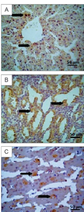
Immunohistochemical staining of viral antigen (Fig. 4A) in the livers of hamsters infected with ROCV was intense between day 1-15 p.i. and decreased be-tween day 16-30, with low but persistent levels being observed thereafter.
The lungs of hamsters infected with ROCV were characterised by thickening of the alveolar walls due to oedema, congestion and mononuclear infiltration (Fig. 3H, I). In addition, areas of overt pneumonitis and ne-crotic foci were observed. However, there were no overt signs of parenchymal condensation that would histo-pathologically characterise pneumonia or areas or foci of haemorrhage. The histopathological changes in the lung peaked on day 7 p.i., then remained stable until day 15
and improved substantially between days 15-30. Mini-mal alterations were observed 30 days p.i.
Viral antigen was detected in the pulmonary paren-chyma of animals infected with ROCV. The immunos-taining was similar to that observed in the liver, with the highest intensity observed until day 15 p.i. The staining intensity declined thereafter and persisted at a low level after day 30.
Congested glomeruli associated with oedema and foci of necrosis were observed in the kidneys of infected hamsters (Fig. 3E). A mild infiltrate, consisting of lym-phocytes and plasma cells, was primarily observed in the interstitium of the renal parenchyma (Fig. 3F). Cellular swelling was observed at the tubular level, as were dis-crete foci of tubular necrosis. These alterations peaked on day 7 p.i., continued until day 15 and improved after day 17 p.i. The inflammatory process and tubular cell swelling continued after day 30 p.i.
Immunostaining for viral antigens in the kidneys (Fig. 4B) of hamsters infected with ROCV was positive in the renal parenchyma, with peak intensity on day 7 p.i. The immunostaining intensity remained high until day 15 p.i., then decreased thereafter and persisted at discrete levels after day 30.
Congestion and mononuclear infiltration of the me-ninges were observed in the CNS of animals infected with ROCV (Fig. 3J). This infiltration was associated with intraparenchymatous oedema characterised by the presence of vacuolisation around neuronal bodies and cerebral gliosis (Fig. 3K). No alterations were apparent in the Virchow-Robin spaces and no cerebral gliosis or glial nodules were observed. These alterations peaked between day 7-8 p.i. and remained at a similar level until day 15, followed by attenuation between day 16-30 and low-level persistence thereafter.
The analysis of the brains of hamsters infected with ROCV showed positive immunostaining for viral anti-gen (Fig. 4C) in the cerebral parenchyma. The staining intensity was more prominent between day 1-7 p.i. and persisted at low levels after day 15.
DISCUSSION
The results of this study demonstrate that young ham-sters are an appropriate experimental model for the study of ROCV infection. In addition to acute infection, these animals were also susceptible to persistent infection.
Infected hamsters developed viraemia, which was detected by an IFA of the supernatants of cell cultures in-oculated with blood, for a maximum period of four days p.i. Antibodies detectable by HI appeared in the serum of infected animals on day 5 p.i., which coincided with the disappearance of the virus from the blood. This phenom-enon was also observed by Tesh et al. (2005) in a study of the persistent infection of hamsters with West Nile virus. No viraemia was detected in the animals in this previous study after the appearance of HI antibodies, most likely due to the presence of neutralising antibodies that bind to the virus to form immune complexes that prevent the entry of the virus into uninfected cells and, consequently, viral replication. These complexes are then eliminated from the organism via immune defence mechanisms.
Viable viral particles were detected in the superna-tants of all of the cells infected with material from or-gans, blood, serum and urine, thus confirming infec-tion. Viral replication was demonstrated by a positive IFA, but persistence of the virus in Vero cells was not observed. However, direct culture of infected organs is most likely not the best strategy for the evaluation of per-sistent viral infection, as suggested by Xiao et al. (2001) and Siirin et al. (2007) in studies of West Nile virus and Saint Louis encephalitis virus, respectively. Intracellu-lar virus particles released during the maceration of the organ fragments may come into contact with antibodies present in the blood and interstitial fluids, thus reducing the sensitivity of this method for the demonstration of viral persistence. The authors suggested that co-culture is more appropriate for the demonstration of persistent infection with flaviviruses because washing the tissue with trypsin-EDTA during preparation of the specimen presumably eliminates almost all neutralising antibod-ies without destroying the intracellular virus, which is subsequently released and amplified by co-culture with the Vero cell monolayer (Tesh et al. 2005).
Urine has been shown to be a good clinical specimen for the isolation of flaviviruses and to demonstrate viral persistence in direct cultures of Vero cells (Tesh et al. 2005, Tonry et al. 2005, Siirin et al. 2007). In the present experiment, the virus was isolated from urine,
strating that the urine of infected animals is infectious. However, in studies reporting good viral isolation from urine, the urine was diluted in PBS before inoculation into Vero cells. In the present study, the urine was di-rectly inoculated into Vero cells at a final dilution of 1:50 and the toxicity of the urine to the Vero cells may have influenced the results obtained. qRT-PCR permitted the detection of even small concentrations of the virus in urine. The final three urine samples collected during the ROCV kinetic analysis (30, 60 and 75 days p.i.) were selected for analysis by qRT-PCR and the results were positive at 30 and 60 days, with titres of 1.1 and 0.67 PFU/mL, respectively (data not shown).
ROCV was detected in all samples tested by q RT-PCR (blood, liver and brain), indicating the persistence of the viral infection. Viral RNA was detected until the end of the experiment (3 months). The sensitivity of qRT-PCR for viral detection is as high as 99.09% (Yong et al. 2007, Nunes et al. 2011). qRT-PCR is even able to detect the genome of inactive viruses. In fact, qRT-PCR was much more sensitive than IFA, which only detected viral antigens in the supernatants of cells infected with brain samples collected prior to six days p.i. (Table).
ROCV caused pathological alterations in the ham-sters and viral antigens were expressed in the liver, kid-ney, lung and brain samples during the four months of the experiment. The pantropic ROCV infection caused inflammatory lesions in the CNS, liver, kidney, spleen, lung and heart. The intensity and extent of tissue dam-age varied among the different organs. The damdam-age was more intense damage in the CNS, which was expected because ROCV has previously been demonstrated to be neurotropic (Lopes et al. 1978, Iversson 1988), followed by the liver. These results were confirmed by immuno-histochemistry and qRT-PCR. The effect on the CNS is in agreement with the severity of the ROCV-induced hu-man encephalitis during an epidemic that occurred in SP (Tiriba 1975, Iversson 1988).
The severe CNS injuries, including neuronal damage to the cerebral parenchyma or meninges, which caused meningitis associated with an inflammatory infiltrate that persisted for four months, further demonstrates the neurotropism and capacity of ROCV to cause encepha-litis in hamsters. According to some authors, neuronal infection may facilitate the persistence of the virus in the brain. Mature neurons are more resistant to the in-duction of apoptosis. This resistance is possibly medi-ated by the neuronal expression of apoptosis inhibitors such as bcl-2, bcl-x and mcl-1, which are activated as a mechanism of neuronal preservation. These cells are also deficient in cellular components necessary for the presentation of antigens to cytotoxic T cells, i.e., they are deficient in the expression of major histocompatibility complex class I molecules (Griffin 1995).
No viral antigens could be detected by immunohis-tochemistry in spleen or heart fragments infected with ROCV, even though the virus replicated in Vero cells inoculated with these fragments, and there was evidence of alterations in these tissues. This suggests that the vi-rus that grew in the Vero cells was present in the blood passing through these organs. The presence of the virus
in the blood may have been responsible for the tissue damage and co-culture with Vero cells or qRT-PCR is necessary to demonstrate the persistence of viral infec-tion observed in the brain and liver.
Although the criteria used for the definition of per-sistence are arbitrary (Chambers & Diamond 2003), ac-cording to the classification of persistent infection pro-posed by Santos (2008), the present results suggest that ROCV causes a chronic persistent infection in which the virus is continuously replicated and excreted.
Taken together, the results obtained for ROCV here and those reported in other studies for different flavi-viruses (Tesh et al. 2005, Tonry et al. 2005, Siirin et al. 2007) suggest that a variety of flaviviruses cause per-sistent infections in vertebrates and that the persistence of these viruses is not a rare phenomenon. However, further studies are needed to determine exactly how a persistent ROCV infection is established, including the route by which the virus enters the CNS, the cells that permit infection of the CNS and the cell groups involved in the production of virus and in the protective immune response, as well as other aspects of pathogenesis.
REFERENCES
Beaty BJ, Calisher CH, Shope RE 1989. Arboviruses. In NJ Schmidt, EW Emmons, Diagnostic procedures for viral, rickettsial and chlamydial infections, 1st ed.. American Public Health Associa-tion Press, Washington, p. 797-855.
Carvalho VL, Nunes MRT, Silva EVP, Vieira CMA, Gomes M, Casseb SM, Rodrigues SG, Nunes-Neto JP, Quaresma JAO, Vas-concelos PFC 2009. Genetic characterization of orthobunyavirus Melao, strains BE AR633512 and BE AR8033, and experimen-tal infection in golden hamsters (Mesocricetus auratus). J Gen Virol 90: 223-233.
Chambers TJ, Diamond MS 2003. Pathogenesis of Flavivirus En-cephalitis. In TJ Chambers, TP Monath, The Flaviviruses: pathogenesis and immunity, Vol. 60, Elsevier Academic Press, San Diego, p. 273-316.
Clark DH, Casals J 1958. Technique for haemagglutination and hae-magglutination inhibition with arthropod-borne viruses. Am J Trop Med Hyg7: 561-573.
Fauquet CM, Mayo MA, Manillof J, Desselberger U, Ball LA 2005. Family Flaviviridae. CM In Fauquet, MA Mayo, J Manillof, U Desselberger, LA Ball, Virus taxonomy, Elsevier Academic Press, London,p. 981-988.
Griffin DE 1995. Arboviruses and the central nervous system. Spring-er Sem Immunopath17: 121-132.
Iversson LB 1988. Rocio encephalitis. In TP Monath, The arbovirus-es: epidemiology and ecology, CRC Press, Florida, p. 77-93.
Iversson LB, Coimbra TLM, Travassos da Rosa APA, Monath TP 1992. Use of immunoglobulin M antibody capture enzyme-linked immunosorbent assay in the surveillance of Rocio encephalitis.
J Braz Assoc Adv Sci44: 164-166.
Karabatsos N 1985. International catalogue of arboviruses, including certain other viruses of vertebrates, 3rd ed., American Society of Tropical Medicine and Hygiene, San Antonio, 1141 pp.
Kuno G 1998. A manual of cell culture techniques for small arbovirus diagnostic laboratories, Center for Disease Control, NCID/DV-BID/ADB 970-221-6400, Fort Collins, 87 pp.
vi-ral, rickettsial and chlamydial infections, 7 ed., American Public Health Association Press, Washington, p. 3-25.
Lopes OS, Coimbra TLM, Saccheta LA, Calisher CH 1978. Emer-gence of a new arbovirus disease in Brazil. I. Isolation and char-acterization of the etiologic agent, Rocio virus. Am J Epidemiol 107: 444.
Mitchell CJ, Forattini OP, Miller BR 1986. Vector competence ex-periments with Rocio virus and three mosquito species from the epidemic zone in Brazil. Rev Saude Publica20: 171-177.
Nunes MRT, Palacios G, Nunes KNB, Casseb SMM, Savji N, Lipkin WI, Vasconcelos PFC 2011. Evaluation of two molecular meth-ods for the detection of yellow fevervirus genome. J Virol Meth 174: 29-34.
Oldstone MBA 2005. Viral persistence: parameters, mechanisms and future predictions. Virology341: 111-118.
Pinheiro FP, Travassos da Rosa APA, Vasconcelos PFC 1997. Ar-boviroses. In R Veronesi, R Focaccia, Tratado de infectologia, Atheneu, São Paulo, p. 169-180.
Prophet EB, Millis B, Arrington IB, Sobin LM 1992. Laboratory methods in histotechnology, American Registry of Pathology, Washington, p. 3-80.
Santos NOS 2008. Patogenese das Infecções Virais. In NSO Santos, VTM Romanos, DM Wigg, Introdução à virologia humana, 2nd ed., Guanabara Koogan Press, Rio de Janeiro, p. 42-58.
Shope RE 1963. The use of a microhaemagglutination-inhibition test to follow antibody response after arthropod-borne virus infection in a community of forest animals. Ann Microbiol 11: 167-171.
Siirin M, Duan T, Lei H, Guzman H, Travassos da Rosa APA, Watts MD, Xiao SY, Tesh RB 2007. Chronic St. Louis encephalitis vi-rus infection in the golden hamster (Mesocricetus auratus). Am J Trop Med Hyg 76: 299-306.
Straatmann A, Torres SS, Vasconcelos PFC, Travassos da Rosa APA, Rodrigues SG, Tavares-Neto J 1997. Evidências sorológicas da circulação do arbovírus Rocio (Flaviviridae) na Bahia. Rev Soc Bras Med Trop30: 512-525.
Tesh RB 1979. A method for the isolation and identification of den-gue viruses using mosquito cell cultures. Am J Trop Med Hyg 28: 1053-1059.
Tesh RB, Siirin M, Guzman H, Travassos da Rosa APA, Xiaoyan W, Duan T, Lei H, Nunes MR, Xiao SY 2005. Persistent West Nile infection in the golden hamster: studies on its mechanism and possible implications for other flavivirus infection. J Infect Dis 192: 287-295.
Tiriba AC 1975. Epidemiologia de encefalite atribuída a arbovírus, ocorrida no litoral sul do estado de São Paulo em 1975: con-tribuição para o estudo clínico, PhD Thesis, Escola Paulista de Medicina, São Paulo, 202 pp.
Tiriba AC, Miziara AM, Lourenço R, Costa CRB, Costa CS, Pinto GH 1976. Encefalite humana primária epidêmica por arbovírus observada no litoral sul do estado de São Paulo. Rev Assoc Med Bras22: 415.
Travassos da Rosa APA, Pinheiro FP, Travassos da Rosa JFS, Vas-concelos PFC 1997. Arboviroses. In RNQ Leão, Doenças infec-ciosas e parasitárias, CEJUP-UEPA/Instituto Evandro Chagas, Belém, p. 207-226.
Tonry JH, Xiao SY, Siirin M, Chen H, Travassos da Rosa APA, Tesh RB 2005. Persistent shedding of West Nile virus in urine of experimentally infected hamsters. Am J Trop Med Hyg72: 320-324.
Vasconcelos PFC, Travassos da Rosa APA, Pinheiro FP, Shope RE, Travassos da Rosa JFS, Rodrigues SG, Dégallier N, Travassos da Rosa ES 1998. Arboviruses pathogenic for man in Brazil. In APA Travassos da Rosa, PFC Vasconcelos, JFS Travassos da Rosa, An overview of arbovirology in Brazil and neighboring countries, Instituto Evandro Chagas, Belém, p. 72-94.
Yong YK, Thayan R, Chong HT, Tan CT, Sekaran SD 2007. Rapid detection and serotyping of dengue virus by multiplex RT-PCR and real-time SYBR green RT-PCR. Singapore Med J48: 662.