online | memorias.ioc.fiocruz.br
Characterisation of
iunH
gene knockout strain
from
Mycobacterium tuberculosis
Anne Drumond Villela1, Valnês da Silva Rodrigues-Junior1,2,3, Antônio Frederico Michel Pinto1, Priscila Lamb Wink1,4, Zilpa Adriana Sánchez-Quitian1,4, Guilherme Oliveira Petersen1, Maria Martha Campos2,3,4, Luiz Augusto Basso1,2,4, Diógenes Santiago Santos1,4/+
1Pontifícia Universidade Católica do Rio Grande do Sul, Centro de Pesquisas em Biologia Molecular e Funcional, Instituto Nacional de Ciência e Tecnologia em Tuberculose, Porto Alegre, RS, Brasil
2Pontifícia Universidade Católica do Rio Grande do Sul,
Programa de Pós-Graduação em Medicina e Ciências da Saúde, Porto Alegre, RS, Brasil
3Pontifícia Universidade Católica do Rio Grande do Sul, Instituto de Toxicologia e Farmacologia, Porto Alegre, RS, Brasil
4Pontifícia Universidade Católica do Rio Grande do Sul, Programa de Pós-Graduação em Biologia Celular e Molecular, Porto Alegre, RS, Brasil
BACKGROUND Tuberculosis (TB) is an infectious disease caused mainly by the bacillus Mycobacterium tuberculosis. The better understanding of important metabolic pathways from M. tuberculosis can contribute to the development of novel therapeutic and prophylactic strategies to combat TB. Nucleoside hydrolase (MtIAGU-NH), encoded by iunH gene (Rv3393), is an enzyme from purine salvage pathway in M. tuberculosis. MtIAGU-NH accepts inosine, adenosine, guanosine, and uridine as substrates, which may point to a pivotal metabolic role.
OBJECTIVES Our aim was to construct a M. tuberculosis knockout strain for iunH gene, to evaluate in vitrogrowth and the effect of iunH deletion in M. tuberculosis in non-activated and activated macrophages models of infection.
METHODS A M. tuberculosis knockout strain for iunH gene was obtained by allelic replacement, using pPR27xylE plasmid. The complemented strain was constructed by the transformation of the knockout strain with pNIP40::iunH. MtIAGU-NH expression was analysed by Western blot and LC-MS/MS. In vitrogrowth was evaluated in Sauton’s medium. Bacterial load of non-activated and interferon-γ activated RAW 264.7 cells infected with knockout strain was compared with wild-type and complemented strains.
FINDINGS Western blot and LC-MS/MS validated iunH deletion at protein level. The iunH knockout led to a delay in M. tuberculosis growth kinetics in Sauton’s medium during log phase, but did not affect bases and nucleosides pool in vitro. No significant difference in bacterial load of knockout strain was observed when compared with both wild-type and complemented strains after infection of non-activated and interferon-γ activated RAW 264.7 cells.
MAIN CONCLUSION The disruption of iunH gene does not influence M. tuberculosis growth in both non-activated and activated RAW 264.7 cells, which show that iunH gene is not important for macrophage invasion and virulence. Our results indicated that MtIAGU-NH is not a target for drug development.
Key words: iunH gene - nucleoside hydrolase - gene knockout - Mycobacterium tuberculosis
doi: 10.1590/0074-02760160462
Financial support: CNPq (304051/1975-06, 520182/99-5, 163507/2014-7, 304156/2014-0), BNDES (14.2.0914.1), FAPERGS-CAPES (DOCFIX, 05/2013), Decit/SCTIE/MS-MCT-CNPq-FNDCT-CAPES to INCT-TB, CNPq (441720/2014-5), FAPERGS-CNPq-PRONEX-2009.
+ Corresponding author: [email protected] Received 18 October 2016
Accepted 2 December 2016
Tuberculosis (TB) is an infectious disease caused mainly by the bacillus Mycobacterium tuberculosis, and remains one of the world’s deadliest contagious diseases. TB incidence is slowly declining each year, however, novel drugs and vaccines are urgently needed to stop global transmission and to prevent the
develop-ment of drug-resistant strains (WHO 2016). The better
understanding of important metabolic pathways from M. tuberculosis can contribute to the development of novel therapeutic and prophylactic strategies to combat TB.
ef-fect of iunH deletion in M. tuberculosis in non-activated and activated macrophage model of infection, compar-ing with M. tuberculosis H37Rv wild-type (WT) and complemented (CP) strains.
MATERIALS AND METHODS
Plasmid construction for generation of the knock-out strain - A fragment of 1782 bp containing the iunH gene (927 bp) with its flanking region (Fig. 1A) was am-plified by polymerase chain reaction (PCR) from M. tu-berculosis H37Rv genomic DNA, using primers forward
(5’-tttttctagagcagcaggcgatgcgccagg-3’) and reverse (5’- tttttctagagacccgtcgccggcggtgc-3’), both containing XbaI restriction sites (underlined). The 1782 bp fragment was subsequently cloned into pUC19 using the XbaI restric-tion site. The iunH gene was disrupted by the inserrestric-tion of a kanamycin cassette from pUC4K into unique inter-nal enzyme restriction site XcmI (New England Biolabs, USA) (Fig. 1B). Insert was released from pUC19 deriva-tive vector by digestion with XbaI (New England Biolabs, USA), and subcloned into XbaI linearised pPR27xylE vec-tor (pPR27xylE::iunH kan) (Fig. 1B) (Pelicic et al. 1997).
Plasmid construction for generation of the comple-mented strain - The iunH gene flanked by about 200 bp upstream and 100 bp downstream, was amplified by PCR from M. tuberculosis H37Rv genomic DNA using
primers forward (5’-ttttctagacagcgcgagatcgatcttg-3’)
and reverse (5’-tttttctagacggtggtatctggagggaa-3’), both containing XbaI restriction sites (underlined), and was cloned into XbaI linearised pNIP40/b (pNIP40::iunH),
a mycobacteriophage Ms6-derived integrative vector
(Freitas-Vieira et al. 1998).
Construction of the M. tuberculosis knockout strain - Electrocompetent cells were prepared as described (Par-ish & Stocker 1998) with some modifications. M. tuber-culosis H37Rv strain was grown in 50 mL of Middle -brook 7H9 (Becton Dickinson, BD, USA) 10% OADC
(oleic acid, albumin, dextrose, and catalase) (BD, USA)
0.05% tween-80 (Sigma-Aldrich, USA) (liquid medium)
to an OD600 of 0.6. Cells were washed two times in 0.05%
tween-80, one time in 10% glycerol containing 0.05% tween-80, and were suspended in 500 µL of 10% glyc
-erol containing 0.05% tween-80. Aliquots (200 µL) of
fresh prepared competent cells were electroporated with
approximately 2 µg of pPR27xylE::iunH kan plasmid in
0.2 cm cuvettes with a single pulse (2.5 kV; 25 mF; 1000
ohms). The pPR27xylE plasmid contains a thermosensi-tive origin of replication, the xylE reporter gene, and the sacB counterselectable marker. Bacteria were plated on Middlebrook 7H10 (BD, USA) 10% OADC (solid
me-dium) containing 25 µg/mL kanamycin (Gibco, USA),
and incubated at 32ºC. After six weeks, 1% pyrocatechol (Sigma-Aldrich, USA) was dropped on colonies to se-lect those containing the plasmid. Three different yellow colonies were picked up from the transformant, and
culti-vated in liquid medium containing kanamycin 25 µg/mL
at 32ºC. Individual cultures were plated on solid medium
containing 25 µg/mL kanamycin, 2% sucrose (Fisher Sci -entific, USA), and cultivated at 39ºC. After four weeks, 1% pyrocatechol was dropped on colonies to select those that might be double crossover (DCO) strains. Nine white
colonies were inoculated in liquid medium containing 25 µg/mL kanamycin, and cultivated at 37ºC for three weeks.
Genomic DNA was isolated and PCRs were carried out
using gene-specific screening primers forward (5’- ttcag
-gaaacgagcgaaggt-3’) and reverse (5’-gtgctatccggcgga -cac-3’) to determine whether the WT or the KO strain was present in the targeted chromosomal region (Fig. 1B).
Construction of the M. tuberculosis complemented strain - In order to obtain the CP strain with the iunH gene, the KO strain was transformed by electroporation
with about 2 µg of the complementing plasmid construc -tion, pNIP40::iunH. Electrocompetent cells were pre-pared as described above. Bacteria were plated on solid
Fig. 1: genomic environment of iunH gene in Mycobacterium tuberculosis (A), regions cloned into pPR27xylE vector (B), and agarose gel electrophoresis of polymerase chain reaction (PCR) products from knockout strains (C). (A) Genomic region of iunH gene (927 bp) containing unique internal XcmI site and flanking genes; (B) the iunH gene and flanking regions (1782 bp) were amplified by PCR from M. tuberculo-sis H37Rv genomic DNA, and the iunH gene was disrupted by the insertion of a kanamycin cassette (kanR) into XcmI site (iunH::kanR). The
iunH::kanR fragment was cloned into pPR27xylE vector using XbaI restriction site. Annealing regions of gene-specific screening primers
forward (Primer F) and reverse (Primer R) for the possible knockout strains of iunH gene are indicated; (C) agarose gel electrophoresis of PCR
medium containing 50 µg/mL hygromycin (Invitrogen, USA) and 25 µg/mL kanamycin, and incubated at 37ºC. After three weeks, a single colony was cultivated in 5 mL of liquid medium with 50 µg/mL hygromycin 25 µg/
mL kanamycin at 37ºC.
Protein extraction - WT, KO and CP strains were
grown in 50 mL liquid medium containing the proper antibiotics until an optical density at 600 nm (OD600)
of 0.5-0.7. Cellular pellets were washed twice using 10
mM Tris HCl pH 8.0. Cells were resuspended in 1 mL of the same buffer containing protease inhibitor cock-tail (Promega, USA), and then transferred to 2 mL lys-ing matrix B tubes containlys-ing 0.1 mm diameter silica beads (MP Biomedicals, USA). Cells were disrupted using a L-Beader 3 (Loccus, Brazil) at a speed setting of 4000 rpm, 10 cycles of 30 s each, cooling between cycles. After lysis, the cell free supernatants were col-lected by centrifugation at 2300 x g for 10 min at room temperature. The supernatants were filtered through 0.22 mM Millex Durapore (Millipore, USA), and triton X-114 (Sigma-Aldrich, USA) extraction was carried out to obtain detergent and aqueous fractions as described previously (Malen et al. 2010).
Western blot - Anti-MtIAGU-NH mouse polyclonal
antibody was produced by immunising a mouse with 50
mg of MtIAGU-NH purified protein (Wink et al. 2013) containing Freund’s incomplete adjuvant (Sigma-Aldrich,
USA) (total volume of 100 µL) by subcutaneous route, fol -lowed by a booster injection after one month. After one more month, mouse was euthanised by deep isoflurane inhalation, and blood was collected by the descendant aorta. Serum was separated by centrifugation at 10,000 x g, for 10 min, aliquoted, and storage at -80ºC.
Myco-bacterial proteins from detergent fraction (50 mg) were loaded on sodium dodecyl sulphate 12% polyacrylamide gels (SDS-PAGE), and transferred to nitrocellulose mem-branes (iBlot Invitrogen, USA). Blots were blocked with
5% non-fat dried milk (Santa Cruz Biotechnology, USA) 0.05% tween-20 (Sigma-Aldrich, USA) in TBS (T-TBS),
and probed with anti-MtIAGU-NH polyclonal mouse
an-tibody in a 1:500 dilution. Membranes were washed three
times with T-TBS, and alkaline phosphatase-conjugated anti-mouse secondary antibody (Invitrogen, USA) was
used at a dilution of 1:5000. Chemiluninescent substrate
(Novex by Life Technologies, USA) was used for detec-tion with ChemiDoc (Bio-Rad, USA).
LC-MS/MS - The presence of MtIAGU-NH in the WT, KO and CP strains was also investigated by liquid chromatography coupled to mass spectrometry (LC-MS/MS) of SDS-PAGE slices. Sections from 28-40 kDa of each lane/strain were excised and submitted to
in-gel digestion (Shevchenko et al. 2006). LC-MS/MS of
peptides was performed on an Eksigent nanoLC Ultra 1D plus with AS-2 autosampler, coupled to a LTQ-XL Orbitrap Discovery (Thermo Scientific, USA). Peptide mixtures from SDS-PAGE slices were separated in
re-versed phase gradients in C18 (5 µM ODS-AQ C18 Ya -mamura Chemical Lab) column. Samples were analysed in technical triplicates. Mass spectra were acquired in
a top 8 data-dependent manner with dynamic exclusion applied, and were searched against a non-redundant M. tuberculosis database for candidate peptides with the software Comet (Eng et al. 2012) in the platform
Pat-ternLab for Proteomics (Carvalho et al. 2016). The va -lidity of the peptide spectra matches was assessed using the module Search Engine Processor from Patternlab for Proteomics, with a false discovery rate of 1%. Identified peptides from MtIAGU-NH were manually validated and peak areas of selected peptides were calculated with Skyline (MacLean et al. 2010).
In vitro characterisation - Growth curves were
deter-mined in Sauton’s medium containing 0.025% tyloxapol
(Sigma-Aldrich, USA), in duplicates, at 37ºC, 80 rpm until reach the stationary phase. Aliquots were removed from each culture at different time points and the OD600 was determined. The data were evaluated with the two-way ANOVA analysis, followed by Bonferroni’s post-test,
using GraphPad Prism 5.0. Differences were considered significant at the 95% level of confidence. In order to
quantify the intracellular concentration of the bases (ura-cil, guanine, adenine, and hypoxanthine) and nucleosides (uridine, guanosine, adenosine, and inosine) in the WT, KO and CP strains, bacteria were grown in Sauton’s me-dium until an OD600 of 0.6 - 1.0. Cells were washed twice with 1x PBS, and autoclaved at 121ºC for 30 min, followed by sonication. An HPLC method (Dionex Ultimate 3000, Thermo Scientific, USA) to identify and quantify bases and nucleosides was modified from Wink et al. (2013). Solution A (1% acetic acid) was maintained at 100% for 20 min, followed by a linear gradient of 2 min up to 80% B (20 mM ammonium acetate) and 20% C (methanol and acetonitrile 1:1, v/v) for 10 min. The absorbance was
mea-sured at 262 nm, injection volume was 50 µL, 0.5 mL/
min, and measurements were performed in triplicates.
Macrophage infection - Macrophage infection ex-periments are often used to determine mycobacterial strains virulence (Copenhaver et al. 2004, Katti et al.
2008). RAW 264.7 macrophage cell line was cultured
in DMEM (Gibco, USA), supplemented with 10% heat inactivated fetal bovine serum (FBS) and 1%
penicillin-streptomycin at 37ºC with 5% CO2. Before infection procedures, macrophages were seeded in 24-well culture plates at a density of 105 cells per well in DMEM me-dium with 10% FBS and incubated for 24 h at 37ºC with
5% CO2. Two independent experiments were performed,
one with non-activated and the other with activated
mac-rophages, which were activated by the addition of 5 ng/
mL of interferon-g (IFN-g) (R&D systems, USA) during
24 h. Infection of RAW 264.7 cells with WT, KO and CP
strains was performed at a multiplicity of infection of
2:1 (bacteria/macrophage) at 37ºC with 5% CO2. After 18 h, infection was terminated by removing the overlay-ing medium, and each well was washed twice with ster-ile 0.9% NaCl solution to remove extracellular bacteria. At 18 h after infection, and two, three, six, seven and 10 days of incubation, wells were washed with sterile 0.9% NaCl solution, and the infected macrophages were
then lysed with 0.025% SDS (Rodrigues Jr et al. 2014).
incubation. Lysates were serially diluted and plated on solid medium. Bacterial colony formation was evaluated after incubation of plates for three weeks at 37ºC. These experiments were performed in triplicates of each time point. The results were expressed as mean numbers of the logarithms of CFU per well, and were evaluated with the two-way ANOVA analysis, followed by Bonferroni’s
post-test, using GraphPad Prism 5.0. Differences were considered significant at the 95% level of confidence.
RESULTS AND DISCUSSION
Construction of the M. tuberculosis knockout and complemented strains - Among the nine clones screened for the KO of iunH gene, all suffered a double-crossover gene replacement event (Fig. 1C). To construct the CP strain, the pNIP40::iunH construction was transformed in the KO strain. The stability of the mutation introduced by gene replacement in M. tuberculosis was evaluated by plating KO and CP strains on media with and with-out antibiotics. The difference between the colonies ob-tained on plates containing antibiotics was not signifi-cant when compared with the ones obtained on plates without antibiotic, which indicates that the introduced mutation is stable (data not shown).
Evaluation of iunH knockout at protein level - As shown in Fig. 2A, a 32.9 kDa band was detected by West-ern blot assay in extracts from WT and CP strains but was absent in the KO strain. The identity of the protein was further confirmed by LC-MS/MS from SDS-PAGE slices (28-40 kDa). Spectra matching MtIAGU-NH pep-tides were identified in WT and CP but not in KO extracts (Fig. 2B), confirming that the protein observed in WT and CP extracts by western blot is in fact MtIAGU-NH. As shown in Fig. 2B, the areas of the peptides identified on
the WT sample are approximately 3-fold higher than the areas identified on CP sample. No peptides were
identi-fied on the KO sample; consequently peak areas could
not be calculated. Fig. 2C shows a representative MS/MS spectrum of the peptide LASVCGSSPVMR, identified in both WT and CP samples. These results indicated that the disruption of iunH gene abolishes the expression of MtIAGU-NH protein in KO strain, and the MtIAGU-NH protein expression was restored in CP strain.
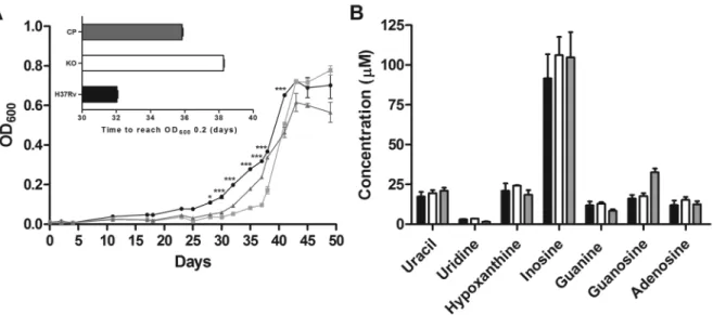
In vitro characterisation - The growth rate of the WT, KO, and CP strains were compared to determine whether iunH disruption lead to alterations during in vi-tro cultivation. Sauton’s is a defined medium that was used for in vitro characterisation experiments to avoid interferences with traces of bases and nucleosides from 7H9 medium. As shown in Fig. 3A, the deletion of iunH gene led to a delay of growth in Sauton’s medium dur-ing log phase. The iunH KO strain entered the early log phase of growth (OD600 of 0.2) six days later than WT strain (inset in Fig. 3A). No significant differences were found during lag and stationary phases of growth be-tween WT and KO strains (Fig. 3A). Two independent experiments were performed, and similar results were obtained. The intracellular concentration of the bases and nucleosides was quantified in the WT, KO and CP strains. Uracil, guanine, adenine, hypoxanthine, uridine, inosine, guanosine and adenosine presented retention
times of 10.53, 13.35, 14.34, 16.98, 19.89, 29.51, 29.64, and 30.36 minutes respectively. The calibration curves for all analyses were made from 0.48 to 62.5 µM and all
presented a correlation coefficient above 0.99. No sig-nificant difference was observed in intracellular concen-trations of either bases or nucleosides among the three strains when grown in Sauton’s medium (Fig. 3B). The
Fig. 2: evaluation of MtIAGU-NH expression. (A) Western blot analysis of protein extracts (50 mg) from detergent fractions of wild-type H37Rv
(H37Rv), knockout (KO), and complemented (CP) strains. Bands were detected by incubation with anti-MtIAGU-NH polyclonal antibody (1:500) fol
-lowed by alkaline phosphatase-conjugated anti-mouse secondary antibody (1:5000); (B) peak areas of peptides LASVCGSSPVMR (m/z 632.3128+2), GIGYAELPASNR (m/z 624.3226+2) and IGMSVDPAVFFDR (m/z 543.8024+2) were calculated using Skyline. LC-MS/MS analyses were performed 28-40 kDa section of SDS-PAGE samples. MS/MS spectra of identified peptides were manually validated; (C) representative MS/MS spectrum of the
peptide LASVCGSSPVMR. The peptide was identified in both WT and CP samples. Doubly charged parent ion with neutral loses ([MH-H2O] +2 and
absence of accumulation of purine bases and nucleosides in iunH KO strain could be explained by the redundan-cy found in nucleotide metabolism of M. tuberculosis. There are at least five enzymes in nucleotide salvage pathway that use MtIAGU-NH substrates or products: purine nucleoside phosphorylase (Rv3307),
hypoxan-thine-guanine phosphoribosyltransferase (Rv3624c), adenine phosphoribosyltransferase (Rv2584c), uracil
phosphoribosyltransferase (Rv3309c), and pyrimidine nucleoside phosphorylase (Rv3314c) (Ducati et al. 2011,
Villela et al. 2011); which might compensate for the ab -sence of iunH gene in KO strain.
Macrophage infection - To examine whether the iunH gene was important for invasion and growth in
Fig. 3: in vitro characterisation of iunH knockout strain. Growth curve of WT (circle), KO for iunH gene (square), and CP (triangle) Mycobacte-rium tuberculosis strains grown in Sauton’s medium (A); to start growth curves the different strains were diluted to reach an OD600 of 0.01. Inset shows the time required for each strain to reach an OD600 of 0.2. Asterisks represent significant differences between WT and KO strains by the Bonferroni post-test, *p < 0.05, ***p < 0.001. Intracellular concentrations of bases (uracil, guanine, adenine, and hypoxanthine) and nucleosides (uridine, guanosine, adenosine, and inosine) in WT (black bar), KO (white bar), and CP (grey bar) M. tuberculosis strains grown in Sauton’s
medium (B); adenine was not detected, and it was not included in graph; all measurements were performed in triplicates.
phagocytic cells, we determined the bacterial loads of the WT, KO and CP strains by using the macrophage model of infection. Non-activated macrophages were in-fected with 1.4 x 10⁵, 2.2 x 10⁵, and 2.0 x 10⁵ CFU, while
activated cells were infected with 2.6 x 10⁵, 2.5 x 10⁵,
and 2.6 x 10⁵ CFU of WT, KO and CP strains,
respec-tively, as determined at the day of infection. As shown in Fig. 4A, no significant difference was observed in intracellular growth among WT, KO, and CP strains in non-activated macrophages after 18 h, two, three, seven and 10 days after infection. Similar results were obtained with IFN-g activated macrophages, where no significant difference in bacterial load of KO strain was observed when compared with both WT and CP strains after 18 h, two, three and six days after infection (Fig.
4B). The disruption of iunH gene does not affect the M. tuberculosis growth in both non-activated and IFN-g
ac-tivated RAW 264.7 cells. The concentration of 5 ng/mL
of IFN-g or lower was shown to cause the activation of the endocytic pathway during the immune activation of
RAW 264.7 (Pei et al. 2015), to induce the upregulation of the pro-inflammatory cytokine IL-18 in RAW 264.7
mouse macrophages (Kim et al. 2000), and to lead to its own production in mouse peritoneal macrophages (Di Marzio et al. 1994). In order to evaluate cytokine expression by non-activated macrophages infected with WT, KO and CP strains, the supernatants from 18 h, and three days post infection were collected and interleukin (IL)-1b, tumor necrosis factor (TNF)-a, and IFN-g were quantified by ELISA using a commercial kit (data not shown). TNF-a and IL-1b levels of expression were not significantly different among groups, while IFN-g ex-pression was not detected in culture medium of cells in-fected with all strains (data not shown).
In this work, we constructed a M. tuberculosis KO strain for iunH gene, validated the gene deletion at pro-tein level, characterised the KO strain in vitro, and eval-uated its ability to invade and grow in non-activated and IFN-g activated macrophages. The absence of accumu-lation of purine bases and nucleosides in M. tuberculosis iunH KO strain in Sauton’s medium, together with the fact that iunH gene is not important for M. tuberculo-sis virulence in macrophages, could be explained by the redundancy found in nucleoside/nucleotide metabolism of M. tuberculosis. As mentioned previously, there are at least five enzymes in nucleotide salvage pathway from M. tuberculosis that use MtIAGU-NH substrates or products (Ducati et al. 2011, Villela et al. 2011). These enzymes might compensate for the absence of iunH gene in KO strain, consequently maintaining the nucleotide pool within the cell. Our results indicated that MtIAGU-NH is not a target for drug development.
ACKNOWLEDGEMENTS
To Dr Mary Jackson and Dr Brigitte Gicquel, for provid -ing the pNIP40/b and pPR27xylE plasmids.
AUTHORS’ CONTRIBUTION
ADV constructed and validated the Mycobacterium tu-berculosis knockout and complemented strains and wrote the manuscript; ADV and VSRJ performed macrophage infection experiments; AFMP carried out LC-MS/MS experiments; PLW and ZASQ constructed the plasmids; ADV and GOP performed in vitro characterisation; MMC participated in the design of the study and statistical analysis; LAB contributed in the analysis of results and revised the manuscript; and DSS conceived the study and participated in its design and coordination.
REFERENCES
Carvalho PC, Lima DB, Leprevost FV, Santos MD, Fischer JS, Aqui -no PF, et al. Integrated analysis of shotgun proteomic data with
PatternLab for proteomics 4.0. Nat Protoc. 2016; 11(1): 102-17. Copenhaver RH, Sepulveda E, Armitige LY, Actor JK, Wanger A, Nor
-ris SJ, et al. A mutant of Mycobacterium tuberculosis H37Rv that
lacks expression of antigen 85A is attenuated in mice but retains vaccinogenic potential. Infect Immun. 2004; 72(12): 7084-95.
Di Marzio P, Puddu P, Conti L, Belardelli F, Gessani S. Interferon gamma upregulates its own gene expression in mouse peritoneal
macrophages. J Exp Med. 1994; 179(5): 1731-6.
Ducati RG, Breda A, Basso LA, Santos DS. Purine salvage pathway in
Mycobacterium tuberculosis. Curr Med Chem. 2011; 18(9): 1258-75.
Eng JK, Jahan TA, Hoopmann MR. Comet: an open source tandem
mass spectrometry sequence database search tool. Proteomics.
2012; 13(1): 22-4.
Freitas-Vieira A, Anes E, Moniz-Pereira J. The site-specific recom
-bination locus of mycobacteriophage Ms6 determines DNA inte -gration at the tRNA(Ala) gene of Mycobacterium spp.
Microbiol-ogy. 1998; 144(12): 3397-406.
Katti MK, Dai G, Armitige LY, Marrero CR, Daniel S, Singh CR, et
al. The Delta f bpA mutant derived from Mycobacterium tubercu-losis H37Rv has an enhanced susceptibility to intracellular anti-microbial oxidative mechanisms, undergoes limited phagosome maturation and activates macrophages and dendritic cells. Cell
Microbiol. 2008; 10(6): 1286-303.
Kim YM, Im JY, Han SH, Kang HS, Choi I. IFN-gamma up-regulates
IL-18 gene expression via IFN consensus sequence-binding
pro-tein and activator propro-tein-1 elements in macrophages. J Immunol. 2000; 165(6): 3198-205.
MacLean B, Tomazela DM, Shulman N, Chambers M, Finney GL, Frewen B, et al. Skyline: an open source document editor for cre-ating and analyzing targeted proteomics experiments.
Bioinfor-matics. 2010; 26(7): 966-8.
Malen H, Pathak S, Softeland T, de Souza GA, Wiker HG.Definition of novel cell envelope associated proteins in Triton X-114 extracts of
Mycobacterium tuberculosis H37Rv. BMC Microbiol. 2010; 10: 132.
Parish T, Stocker NG. Electroporation of mycobacteria. In: Parish T, Stocker NG, editors. Methods in molecular biology mycobacteria
protocols. Totowa: Humana Press Inc; 1998. 129-44.
Pei G, Schnettger L, Bronietzki M, Repnik U, Griffiths G, Gutierrez
MG. Interferon-γ-inducible Rab20 regulates endosomal morphol
-ogy and EGFR degradation in macrophages. Mol Biol Cell. 2015; 26(17): 3061-70.
Pelicic V, Jackson M, Reyrat JM, Jacobs Jr WR, Gicquel B, Guilhot
C. Efficient allelic exchange and transposon mutagenesis in My-cobacterium tuberculosis. Proc Natl Acad Sci USA. 1997; 94(20):
10955-60.
Rodrigues Jr V, dos Santos A, Villela AD, Belardinelli JM, Morbidoni HR, Basso LA, et al. IQG-607 abrogates the synthesis of mycolic
acids and displays intracellular activity against Mycobacterium tuberculosis in infected macrophages. Int J Antimicrob Agents.
2014; 43(1): 82-5.
Shevchenko A, Tomas H, Havlis J, Olsen JV, Mann M. In-gel diges -tion for mass spectrometric characteriza-tion of proteins and
pro-teomes. Nature Protocols. 2006; 1(6): 2856-60.
Villela AD, Sánchez-Quitian ZA, Ducati RG, Santos DS, Basso LA. Pyrimidine salvage pathway in Mycobacterium tuberculosis.
Curr Med Chem. 2011; 18(9): 1286-98.
WHO - World Health Organization. Global tuberculosis report 2016. Geneva: WHO; 2016.